CN1803193A - Use of stroma stem cell derived from bone marrow in preparing formulation for treating hypoxic ischemic cerebral palsy - Google Patents
Use of stroma stem cell derived from bone marrow in preparing formulation for treating hypoxic ischemic cerebral palsy Download PDFInfo
- Publication number
- CN1803193A CN1803193A CN 200510032737 CN200510032737A CN1803193A CN 1803193 A CN1803193 A CN 1803193A CN 200510032737 CN200510032737 CN 200510032737 CN 200510032737 A CN200510032737 A CN 200510032737A CN 1803193 A CN1803193 A CN 1803193A
- Authority
- CN
- China
- Prior art keywords
- cells
- bone marrow
- stem cells
- mesenchymal stem
- derived mesenchymal
- Prior art date
- Legal status (The legal status is an assumption and is not a legal conclusion. Google has not performed a legal analysis and makes no representation as to the accuracy of the status listed.)
- Pending
Links
Landscapes
- Micro-Organisms Or Cultivation Processes Thereof (AREA)
- Medicines Containing Material From Animals Or Micro-Organisms (AREA)
Abstract
本发明属于干细胞与组织工程领域,涉及骨髓来源的间质干细胞用于制备治疗缺氧缺血性脑性瘫痪的制剂的用途。所述的制剂由骨髓来源的间质干细胞与生理盐水配制而成,用于缺氧缺血性脑病的新生儿大鼠模型的有效剂量为5×10⁵细胞/次,用于新生儿脑瘫患者病人的有效剂量为1.0~5.0×106细胞/次。本发明为临床治疗缺氧缺血性脑性瘫痪开辟细胞治疗的新途径。
The invention belongs to the field of stem cells and tissue engineering, and relates to the use of bone marrow-derived mesenchymal stem cells for preparing preparations for treating hypoxic-ischemic cerebral palsy. The preparation is formulated from bone marrow-derived mesenchymal stem cells and normal saline, and the effective dose for the neonatal rat model of hypoxic-ischemic encephalopathy is 5×10 5 cells/time, and it is used for neonatal cerebral palsy patients The effective dosage for patients is 1.0-5.0×10 6 cells/time. The invention opens up a new way of cell therapy for the clinical treatment of hypoxic-ischemic cerebral palsy.
Description
技术领域technical field
本发明属于干细胞与组织工程领域,涉及间质干细胞的新用途,尤其是涉及骨髓来源的间质干细胞用于制备治疗缺氧缺血性脑性瘫痪的制剂的用途。The invention belongs to the field of stem cells and tissue engineering, and relates to new uses of mesenchymal stem cells, in particular to the use of bone marrow-derived mesenchymal stem cells for preparing preparations for treating hypoxic-ischemic cerebral palsy.
背景技术Background technique
围产儿窒息所致的缺血缺氧性脑损伤(HIE)是引起新生儿脑性瘫痪(cerebral palsy)的主要原因。近20年来,尽管产科和新生儿医疗保健发展快速,但脑性瘫痪在新生儿中发病仍无明显降低,据1998年北京医科大学等单位抽样调查发现,我国0-6岁儿童中约有脑瘫患儿31万,并且每年以4.6万的速度递增,美国围生协作项目的一项前瞻性调查发现,小儿脑瘫发病率达4‰。患儿主要表现为非进行性中枢性运动功能障碍,或伴有智力低下等。目前的治疗方法主要有:(1)以运动康复为主的综合康复医疗;(2)以营养神经、松弛肌肉、活血等为主的药物疗法;(3)选择性脊神经后根切断术疗法;(4)以按摩、针刺等为主的中医疗法。但由于脑瘫是由固定的脑部病变所引起,以上治疗方法不能去除或矫正病因,疗效不肯定。然而对于脑瘫患者尤其是严重病人,患儿不仅承受巨大的肉体痛苦,还会因脑瘫不能自理而导致的窒息,使脑缺氧而使其智力呈进行性下降。因此,如果能研究出新的有效的脑瘫治疗方法,将给无数家庭带来幸福。Hypoxic-ischemic brain injury (HIE) caused by perinatal asphyxia is the main cause of neonatal cerebral palsy. In the past 20 years, despite the rapid development of obstetrics and newborn medical care, the incidence of cerebral palsy in newborns has not been significantly reduced. According to a sample survey conducted by Beijing Medical University and other units in 1998, it was found that children aged 0-6 in my country had cerebral palsy. There are 310,000 children, and the rate is increasing by 46,000 every year. A prospective survey by the American Perinatal Cooperation Project found that the incidence of cerebral palsy in children is as high as 4‰. The main manifestations of children are non-progressive central motor dysfunction, or accompanied by mental retardation. The current treatment methods mainly include: (1) comprehensive rehabilitation medicine based on sports rehabilitation; (2) drug therapy based on nourishing nerves, relaxing muscles, and promoting blood circulation; (3) selective posterior rhizotomy therapy; (4) Chinese medicine therapy mainly including massage and acupuncture. However, since cerebral palsy is caused by fixed brain lesions, the above treatment methods cannot remove or correct the cause, and the curative effect is uncertain. However, for patients with cerebral palsy, especially severe ones, the children not only suffer great physical pain, but also suffer from suffocation caused by cerebral palsy, which leads to brain hypoxia and progressive decline in intelligence. Therefore, if a new and effective treatment method for cerebral palsy can be developed, it will bring happiness to countless families.
过去的几十年来,采用干细胞移植治疗脑瘫患者尚未见报道,但在治疗中枢神经系统其它疾病中取得了一定的进展。In the past few decades, the use of stem cell transplantation to treat patients with cerebral palsy has not been reported, but some progress has been made in the treatment of other diseases of the central nervous system.
干细胞来源主要有:1、胚胎神经干细胞:有研究报道,采用6-9W流产胎儿脑细胞治疗Parkinson`s病能明显改善部份患者功能,甚至在有些患者体内还能合成多巴胺。但是,由于细胞来源、伦理道德等诸多因素局限了胚胎神经干细胞在移植治疗神经系统疾病的临床应用。2、骨髓来源的间质干细胞:70年代,Friedenstein等从骨髓中分离培养出一种间质干细胞,深入的研究发现间质干细胞不仅可以在脑组织成活、迁移,而且表达神经元、神经胶质细胞等多种神经细胞特异性标志,明显改善中风等模型大鼠功能。目前,间质干细胞主要来源于成人骨髓,但成人骨髓细胞来源有限、病原体污染不可避免。尚无采用胚胎骨髓作为干细胞来源的报道。The main sources of stem cells are: 1. Embryonic neural stem cells: According to research reports, using 6-9W aborted fetal brain cells to treat Parkinson's disease can significantly improve the function of some patients, and even synthesize dopamine in some patients. However, due to many factors such as cell source and ethics, the clinical application of embryonic neural stem cells in the treatment of nervous system diseases is limited. 2. Bone marrow-derived mesenchymal stem cells: In the 1970s, Friedenstein et al. isolated and cultured a kind of mesenchymal stem cells from bone marrow. In-depth research found that mesenchymal stem cells can not only survive and migrate in brain tissue, but also express neurons and glia. Cells and other specific markers of nerve cells, significantly improving the function of stroke and other model rats. At present, mesenchymal stem cells are mainly derived from adult bone marrow, but the source of adult bone marrow cells is limited, and pathogen contamination is inevitable. There is no report on the use of embryonic bone marrow as a source of stem cells.
显然,在现有条件下,对缺氧缺血性脑性瘫痪治疗作用的有关技术和方法均存在种种缺陷和不足。Obviously, under the existing conditions, there are various defects and deficiencies in the relevant technologies and methods for the treatment of hypoxic-ischemic cerebral palsy.
发明内容Contents of the invention
本发明的目的在于提供一种骨髓来源的间质干细胞用于制备治疗缺氧缺血性脑性瘫痪的制剂的用途。The purpose of the present invention is to provide a use of bone marrow-derived mesenchymal stem cells for preparing preparations for treating hypoxic-ischemic cerebral palsy.
所述的制剂由骨髓来源的间质干细胞与生理盐水配制而成,用于缺氧缺血性脑病的新生儿大鼠模型的有效剂量为5×10⁵细胞/次。该有效剂量是通过动物模型实验取得治疗效果而确定的。The preparation is formulated from bone marrow-derived mesenchymal stem cells and physiological saline, and the effective dosage for the neonatal rat model of hypoxic-ischemic encephalopathy is 5×10 5 cells/time. The effective dose is determined by obtaining therapeutic effects through animal model experiments.
所述的骨髓来源的间质干细胞是按以下方法制备的:利用无菌术分离大鼠孕中期胚胎骨髓有核细胞,接种在含胎牛血清的低糖DMEM培养基的培养瓶内,细胞贴壁增殖接近融合时,进行消化、传代,依此法反复传代,达到纯化和扩增间质干细胞的目的,收集细胞冻存、复苏备用。The bone marrow-derived mesenchymal stem cells are prepared according to the following method: use aseptic technique to isolate the bone marrow nucleated cells of rat second-trimester embryos, inoculate them in a culture bottle containing low-sugar DMEM medium containing fetal bovine serum, and adhere the cells to the wall. When the proliferation is close to fusion, digestion and passage are carried out, and this method is repeatedly passaged to achieve the purpose of purifying and expanding mesenchymal stem cells.
所述的骨髓来源的间质干细胞制备的制剂,用于对新生儿大鼠进行脑内定位注射,剂量为5.0×10⁵细胞/次,在缺氧缺血性脑病模型的制作后24小时内移植。The preparation prepared from bone marrow-derived mesenchymal stem cells is used for intracerebral injection of neonatal rats at a dose of 5.0×10 5 cells/time, within 24 hours after the establishment of the hypoxic-ischemic encephalopathy model transplant.
所述的制剂由骨髓来源的间质干细胞与生理盐水配制而成,用于新生儿脑瘫患者病人的有效剂量为1.0~5.0×10⁶细胞/次。该有效剂量是通过动物模型实验取得治疗效果以及人体用药常识而确定的。The preparation is prepared from bone marrow-derived mesenchymal stem cells and physiological saline, and the effective dose for neonatal cerebral palsy patients is 1.0-5.0×10 6 cells/time. The effective dose is determined by obtaining therapeutic effects through animal model experiments and common knowledge of human medicine.
所述的骨髓来源的间质干细胞是按以下方法制备的:利用无菌术从人胚流产材料中分离胚胎骨髓有核细胞,接种在含胎牛血清的低糖DMEM培养基的培养瓶内,细胞贴壁增殖接近融合时,进行消化、传代,依此法反复传代,达到纯化和扩增间质干细胞的目的,收集细胞冻存、复苏备用。The bone marrow-derived mesenchymal stem cells are prepared according to the following method: use aseptic technique to separate embryonic bone marrow nucleated cells from human embryo abortion materials, inoculate them in a culture bottle containing low-sugar DMEM medium containing fetal bovine serum, and the cells When adherent proliferation is close to confluence, digestion and passage are carried out, and this method is repeatedly passaged to achieve the purpose of purifying and expanding mesenchymal stem cells. The cells are collected, frozen and recovered for later use.
所述的骨髓来源的间质干细胞制备的制剂,用于对新生儿脑瘫患者进行脑内定位注射,剂量为1.0~5.0×10⁶/次,在缺氧缺血性脑病发生后24-48小时内移植。The preparation prepared from bone marrow-derived mesenchymal stem cells is used for localized injection in the brain of neonatal patients with cerebral palsy, with a dose of 1.0-5.0×10 6 /time, 24-48 hours after the occurrence of hypoxic-ischemic encephalopathy Intragraft.
本发明利用胚胎骨髓来源的间质干细胞通过脑定位输注方法移植给缺氧缺性脑瘫患儿,促进其脑组织神经细胞修复,改善其平衡协调、运动功能,提高学习、记忆能力,达到提高患儿生活质量、减轻家庭和社会负担的目的,为临床治疗脑瘫开辟细胞治疗的新途径。In the present invention, mesenchymal stem cells derived from embryonic bone marrow are transplanted to children with hypoxia-deficiency cerebral palsy through brain positioning and infusion, so as to promote the repair of nerve cells in their brain tissue, improve their balance and coordination, motor function, and improve their learning and memory abilities. The purpose of improving the quality of life of children and reducing family and social burdens is to open up a new way of cell therapy for the clinical treatment of cerebral palsy.
所述的骨髓来源的间质干细胞制备的制剂用于治疗缺氧缺血性脑性瘫痪的效果评价:Evaluation of the effect of the preparation prepared from the bone marrow-derived mesenchymal stem cells for the treatment of hypoxic-ischemic cerebral palsy:
1、生存能力:包括1. Viability: including
a)1周内模型大鼠存活率;a) Survival rate of model rats within 1 week;
b)1月内模型大鼠体重增长情况;b) Body weight gain of model rats within 1 month;
2、移植后第42天的学习、记忆功能;2. Learning and memory functions on the 42nd day after transplantation;
3、第10周解剖,脑组织病理特征;3. Anatomy and pathological features of brain tissue at the 10th week;
4、免疫组化和间接免疫荧光分析外源细胞在脑组织内存活、迁移、神经细胞特异性抗原表达。4. Immunohistochemistry and indirect immunofluorescence were used to analyze the survival, migration, and expression of nerve cell-specific antigens of exogenous cells in brain tissue.
本发明的优点在于:The advantages of the present invention are:
(1)将胚胎骨髓来源的间充质干细胞移植到新生缺氧缺血性模型大鼠脑内,能明显提高模型大鼠生存能力;改善其运动、学习、记忆能力;调理受损脑组织微环境、促进神经细胞再生和/或功能的修复。(1) Transplantation of embryonic bone marrow-derived mesenchymal stem cells into the brain of neonatal hypoxic-ischemic model rats can significantly improve the viability of the model rats; improve their motor, learning, and memory abilities; environment, promoting nerve cell regeneration and/or restoration of function.
(2)为临床治疗缺氧缺血性脑病开辟细胞疗法的新途径。本发明利用胚胎骨髓来源的间充质干细胞细胞原始、增殖潜能大、制备简便,连续培养及冻存复苏后仍具有多向分化潜能,细胞同质性高,具有正常的核型和端粒酶活性。(2) Open up a new way of cell therapy for the clinical treatment of hypoxic-ischemic encephalopathy. The invention utilizes embryonic bone marrow-derived mesenchymal stem cells to be original, has high proliferation potential, is easy to prepare, has multi-directional differentiation potential after continuous culture and cryopreservation and recovery, has high cell homogeneity, and has normal karyotype and telomerase active.
(3)本发明利用骨髓来源的间质干细胞脑内定位注射治疗大鼠缺氧缺血性脑性瘫痪模型证明,骨髓来源的间质干细胞能够阻滞和逆转缺氧缺血性脑性瘫痪的发展,为临床治疗缺氧缺血性脑性瘫痪开辟细胞治疗的新途径。(3) In the present invention, bone marrow-derived mesenchymal stem cells are injected into the brain to treat hypoxic-ischemic cerebral palsy model. It is proved that bone marrow-derived mesenchymal stem cells can block and reverse the progression of hypoxic-ischemic cerebral palsy. Development, opening up a new way of cell therapy for the clinical treatment of hypoxic-ischemic cerebral palsy.
附图说明Description of drawings
图1为SD大鼠脑组织病理切片检查结果(H&E染色,×200)图。其中,A图显示正常对照组皮质层细胞分布;B图显示HIE模型中神经元变性坏死(↑);C图显示HIE模型中小脑蒲肯野细胞核固缩(↑);D图显示HIE/hFBMMSCs移植组皮质层神经细胞广泛增多。Figure 1 is the results of pathological examination of SD rat brain tissue sections (H&E staining, ×200). Among them, Figure A shows the distribution of cortical cells in the normal control group; Figure B shows the degeneration and necrosis of neurons in the HIE model (↑); Figure C shows the pyknosis of the cerebellar Purkinje cells in the HIE model (↑); Figure D shows HIE/hFBMMSCs In the transplantation group, cortical nerve cells increased extensively.
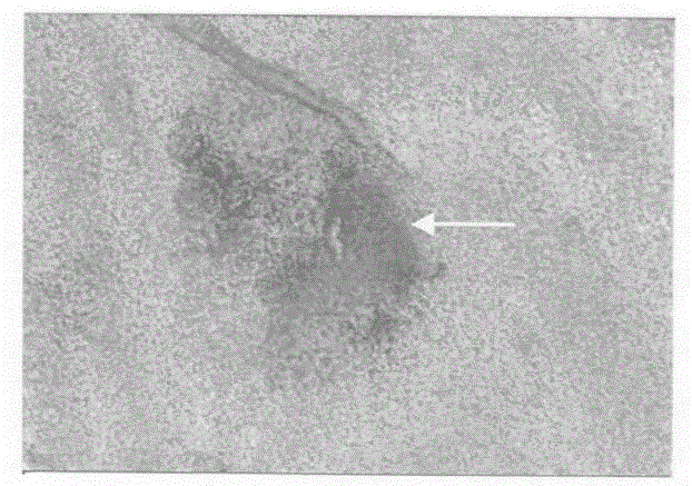
图2为hFBMMSCs移植组脑组织免疫组化染色检测结果(DAB,×100)图,A图显示抗人NSE阳性(↑);B图显示抗人GFAP阳性(↑)。Figure 2 is the results of immunohistochemical staining of brain tissue in the hFBMMSCs transplantation group (DAB, ×100). Panel A shows positive anti-human NSE (↑); panel B shows positive anti-human GFAP (↑).
图3为HIE/hFBMMSCs间接免疫荧光检测结果(×100)图。其中,1A和1B显示:同一张切片同一部位,A中绿色荧光者为抗人RNP阳性;B中桔红色荧光者为抗GFAP阳性;2A和2B显示:同一张切片同一部位,A中绿色荧光者为抗人RNP阳性;B中桔红色荧光者为抗NSE阳性。Fig. 3 is the results (×100) of indirect immunofluorescence detection of HIE/hFBMMSCs. Among them, 1A and 1B show: the same part of the same slice, the one with green fluorescence in A is positive for anti-human RNP; the one with orange-red fluorescence in B is positive for anti-GFAP; 2A and 2B show: the same part of the same slice, the one with green fluorescence in A Those with anti-human RNP are positive; those with orange-red fluorescence in B are anti-NSE positive.
具体实施方式Detailed ways
实施例一:骨髓来源的间质干细胞(MSC)的制备:Example 1: Preparation of bone marrow-derived mesenchymal stem cells (MSCs):
利用无菌术分离孕中期胚胎骨髓有核细胞,接种在含8%胎牛血清的低糖DMEM培养基的培养瓶内,视生长情况隔天换液1次,细胞贴壁增殖接近融合至细胞成90%融合态时,进行消化、传代,依此法反复传代,达到纯化和扩增间质干细胞的目的,收集细胞冻存、复苏备用。The bone marrow nucleated cells of the second-trimester embryo were isolated by aseptic technique, inoculated in the culture flask of low-sugar DMEM medium containing 8% fetal bovine serum, and the medium was changed once every other day depending on the growth situation, and the cells adhered to the wall and proliferated close to the fusion of the cells. When 90% of the fusion state is reached, digestion and passage are carried out, and repeated passages are carried out in this way to achieve the purpose of purifying and expanding mesenchymal stem cells.
所述的低糖DMEM增养基选用HYCLONE公司的CAT NO.SH30002.01产品,或者GIBCO公司的同类产品。Described low-sugar DMEM growth base selects the CAT NO.SH30002.01 product of HYCLONE Company, or the similar product of GIBCO Company.
实施例二:骨髓来源的间质干细胞(MSC)的制备:Example 2: Preparation of bone marrow-derived mesenchymal stem cells (MSCs):
利用无菌术分离孕中期胚胎骨髓有核细胞,接种在含10%胎牛血清的低糖DMEM培养基的培养瓶内,视生长情况3天换液1次,细胞贴壁增殖接近融合至细胞成80%融合态时,进行消化、传代,依此法反复传代,达到纯化和扩增间质干细胞的目的,收集细胞冻存、复苏备用。Isolate the nucleated cells from the bone marrow of the second-trimester embryo by aseptic technique, inoculate them in a culture bottle containing 10% fetal bovine serum low-sugar DMEM medium, change the medium once every 3 days depending on the growth situation, and the cells adhere to the wall and proliferate close to the fusion of the cells. When the fusion state is 80%, carry out digestion and passaging, and repeat passaging according to this method to achieve the purpose of purifying and expanding mesenchymal stem cells.
所述的低糖DMEM培养基选用HYCLONE公司的CAT NO.SH30002.01产品,或者GIBCO公司的同类产品。Described low-sugar DMEM culture medium selects the CAT NO.SH30002.01 product of HYCLONE Company, or the similar product of GIBCO Company.
实施例三:骨髓来源的间质干细胞(MSC)的制备:Example 3: Preparation of bone marrow-derived mesenchymal stem cells (MSCs):
利用无菌术分离孕中期胚胎骨髓有核细胞,接种在含15%胎牛血清的低糖DMEM培养基的培养瓶内,视生长情况4天换液1次,细胞贴壁增殖接近融合至细胞成70%融合态,进行消化、传代,依此法反复传代,达到纯化和扩增间质干细胞的目的,收集细胞冻存、复苏备用。The bone marrow nucleated cells of the second-trimester embryo were isolated by aseptic technique, inoculated in the culture flask of low-sugar DMEM medium containing 15% fetal bovine serum, and the medium was changed once every 4 days depending on the growth situation, and the cells adhered to the wall and proliferated close to the fusion of the cells. 70% confluent state, digested, passaged, repeated passages according to this method, to achieve the purpose of purification and expansion of mesenchymal stem cells, collected cells cryopreserved, resuscitated for later use.
所述的低糖DMEM培养基选用HYCLONE公司的CAT NO.SH30002.01产品,或者GIBCO公司的同类产品。Described low-sugar DMEM culture medium selects the CAT NO.SH30002.01 product of HYCLONE Company, or the similar product of GIBCO Company.
实施例四:缺氧缺血性脑瘫新生大鼠造模:Example 4: Modeling of newborn rats with hypoxic-ischemic cerebral palsy:
1天龄新生F344大鼠(体重5.85±0.39g),乙醚吸入麻醉后仰卧位固定,颈正中切开皮肤,游离左侧颈总动脉,5号线结扎后缝合切口,2h后置入37℃恒温密闭容器中,以1L/min的速度输入8%O2、92%N2的混合气体维持3h,取出后置于原饲养环境中接受母鼠喂养。One-day-old newborn F344 rats (body weight 5.85±0.39g) were fixed in the supine position after ether inhalation anesthesia, the skin was incised in the middle of the neck, the left common carotid artery was freed, the incision was ligated with No. In a constant-temperature airtight container, a mixed gas of 8% O2 and 92% N2 was input at a rate of 1 L/min for 3 hours, and after taking it out, it was placed in the original feeding environment to accept feeding of female mice.
实施例五:胚胎骨髓来源的间质干细胞输注实验:Example 5: Infusion experiment of embryonic bone marrow-derived mesenchymal stem cells:
1、治疗时机:模型制作后24小时内。1. Timing of treatment: within 24 hours after model making.
2、剂量:5.0×10⁵/g。2. Dose: 5.0×10⁵/g.
3、途径:脑定位注射。3. Approach: brain localized injection.
4、试验对照组:采用成组设计,同窝大鼠分为模型/干细胞移植组、模型/生理盐水组、模型/未治疗、正常对照4组。4. Experimental control group: group design was adopted, and littermate rats were divided into 4 groups: model/stem cell transplantation group, model/normal saline group, model/untreated group, and normal control group.
效果评价:Evaluation:
1、生存能力1. Viability
(1)1周内大鼠存活情况:干细胞移植组,存活率与正常对照组比较不存在显著性差异;高于生理盐水组和未治疗组。详见表1(1) Survival of rats within one week: the survival rate of the stem cell transplantation group had no significant difference compared with the normal control group; it was higher than that of the normal saline group and the untreated group. See Table 1 for details
(2)1月内新生大鼠体重增长情况:移植后第5天起,受移植组大鼠体重增长加快,第30天与正常对照组不存在显著性差异,重于生理盐水组和未治疗组。详见表2(2) Body weight gain of newborn rats within 1 month: From the 5th day after transplantation, the weight gain of the rats in the transplanted group accelerated, and there was no significant difference from the normal control group on the 30th day, which was heavier than that of the normal saline group and the untreated group. Group. See Table 2 for details
2、功能改变:细胞移植后第42天,采用Morris水迷宫实验测试移植组大鼠学习和记忆能力,具备与正常组大鼠相近的学习、记忆能力,强于生理盐水注射和未治疗组。详见表32. Functional changes: On the 42nd day after cell transplantation, the Morris water maze test was used to test the learning and memory abilities of the rats in the transplanted group. The learning and memory abilities of the rats in the transplanted group were similar to those of the normal group, and stronger than those of the normal saline injection and untreated groups. See Table 3 for details
3、解剖结果:第10W对实验大鼠进行脑组织结构分析。干细胞移植组脑组织出现萎缩和水肿比率明显低于生理盐水注射组和未治疗组,与正常对照组不存在显著性差异。3. Anatomical results: In the 10th week, the brain tissue structure of the experimental rats was analyzed. The rate of brain atrophy and edema in the stem cell transplantation group was significantly lower than that in the normal saline injection group and the untreated group, and there was no significant difference with the normal control group.
4、脑组织病理切片检查结果:H&E染色,光学显微镜下,干细胞移植组可见大脑神经细胞增生,尤其在大脑皮质层、海马等部位;部份大脑血管壁,可见大量细胞迁移环绕;干细胞移植进针部位可见大量细胞成团或散在分布,呈方向性向周围迁移。而未治疗组和生理盐水组可见小脑、皮质层、海马等区域神经细胞明显减少,神经元核固缩、坏死、胶质细胞吞噬以及胶质细胞反应性增生形成胶质细胞结节等病变现象。详见图1。图1为SD大鼠脑组织病理切片检查结果(H&E染色,×200)图。其中,A图显示正常对照组皮质层细胞分布;B图显示HIE模型中神经元变性坏死(↑);C图显示HIE模型中小脑蒲肯野细胞核固缩(↑);D图显示HIE/hFBMMSCs移植组皮质层神经细胞广泛增多。4. Pathological examination results of brain tissue: H&E staining, under the light microscope, the stem cell transplantation group can see the proliferation of brain nerve cells, especially in the cerebral cortex, hippocampus and other parts; part of the cerebral blood vessel wall, a large number of cells can be seen migrating around; stem cell transplantation A large number of cells can be seen in agglomerates or scattered distribution at the needle site, migrating to the surrounding in a directional manner. In the untreated group and the normal saline group, neurons in the cerebellum, cortex, and hippocampus were significantly reduced, neuronal nucleus shrinkage, necrosis, glial cell phagocytosis, and glial cell reactive hyperplasia to form glial cell nodules and other lesions . See Figure 1 for details. Figure 1 is the results of pathological examination of SD rat brain tissue sections (H&E staining, ×200). Among them, Figure A shows the distribution of cortical cells in the normal control group; Figure B shows the degeneration and necrosis of neurons in the HIE model (↑); Figure C shows the pyknosis of the cerebellar Purkinje cells in the HIE model (↑); Figure D shows HIE/hFBMMSCs In the transplantation group, cortical nerve cells increased extensively.
5、免疫组化分析结果:采用免疫组化技术分析,移植组大鼠脑组织中存在抗人NSE、GFAP反应细胞,以皮质层和海马等区域较明显;其它3组实验动物均未见NSE或GFAP阳性细胞。详见图2。图2为hFBMMSCs移植组脑组织免疫组化染色检测结果(DAB,×100)图,A图显示抗人NSE阳性(↑),B图显示抗人GFAP阳性(↑)。5. Immunohistochemical analysis results: Immunohistochemical analysis showed that there were anti-human NSE and GFAP reactive cells in the brain tissue of the rats in the transplantation group, especially in the cortex and hippocampus; no NSE was found in the other three groups of experimental animals or GFAP positive cells. See Figure 2 for details. Figure 2 is the results of immunohistochemical staining of brain tissue in the hFBMMSCs transplantation group (DAB, ×100). Figure A shows positive anti-human NSE (↑), and picture B shows positive anti-human GFAP (↑).
6、间接免疫荧光检测结果:移植组两侧大脑皮质层、海马等部位检测到抗人RNP/CD45、抗人RNP/NSE或抗人RNP/GFAP双阳性完整细胞,尤其是在进针部位,大量双阳性细胞成团或散在存在,向周围迁移并且密度呈梯度下降,而其它3组动物脑组织切片均未出现抗人RNP、CD45、NSE、GFAP阳性细胞。详见图3。图3为HIE/hFBMMSCs间接免疫荧光检测结果(×100)图。其中,1A和1B显示:同一张切片同一部位,A中绿色荧光者为抗人RNP阳性;B中桔红色荧光者为抗GFAP阳性;2A和2B显示:同一张切片同一部位,A中绿色荧光者为抗人RNP阳性;B中桔红色荧光者为抗NSE阳性。6. Indirect immunofluorescence test results: anti-human RNP/CD45, anti-human RNP/NSE or anti-human RNP/GFAP double-positive intact cells were detected in the cerebral cortex and hippocampus on both sides of the transplantation group, especially at the needle insertion site. A large number of double-positive cells existed in agglomerates or scattered, and migrated to the surroundings with a gradient decrease in density, while no anti-human RNP, CD45, NSE, and GFAP positive cells appeared in the brain tissue sections of animals in the other three groups. See Figure 3 for details. Fig. 3 is the results (×100) of indirect immunofluorescence detection of HIE/hFBMMSCs. Among them, 1A and 1B show: the same part of the same slice, the one with green fluorescence in A is positive for anti-human RNP; the one with orange-red fluorescence in B is positive for anti-GFAP; 2A and 2B show: the same part of the same slice, the one with green fluorescence in A Those with anti-human RNP are positive; those with orange-red fluorescence in B are anti-NSE positive.
表1 新生大鼠1周内成活率
表2 4组新生大鼠1M内体重增长情况(
x±SD)
表3 4组实验大鼠Morris水迷宫训练和测试结果(
X±SD)sec
*:2只鼠的实验结果;#:3只鼠的实验结果 * : Experimental results of 2 mice; #: Experimental results of 3 mice
Claims (7)
Priority Applications (1)
| Application Number | Priority Date | Filing Date | Title |
|---|---|---|---|
| CN 200510032737 CN1803193A (en) | 2005-01-13 | 2005-01-13 | Use of stroma stem cell derived from bone marrow in preparing formulation for treating hypoxic ischemic cerebral palsy |
Applications Claiming Priority (1)
| Application Number | Priority Date | Filing Date | Title |
|---|---|---|---|
| CN 200510032737 CN1803193A (en) | 2005-01-13 | 2005-01-13 | Use of stroma stem cell derived from bone marrow in preparing formulation for treating hypoxic ischemic cerebral palsy |
Publications (1)
| Publication Number | Publication Date |
|---|---|
| CN1803193A true CN1803193A (en) | 2006-07-19 |
Family
ID=36865424
Family Applications (1)
| Application Number | Title | Priority Date | Filing Date |
|---|---|---|---|
| CN 200510032737 Pending CN1803193A (en) | 2005-01-13 | 2005-01-13 | Use of stroma stem cell derived from bone marrow in preparing formulation for treating hypoxic ischemic cerebral palsy |
Country Status (1)
| Country | Link |
|---|---|
| CN (1) | CN1803193A (en) |
Cited By (3)
| Publication number | Priority date | Publication date | Assignee | Title |
|---|---|---|---|---|
| CN110461344A (en) * | 2017-01-16 | 2019-11-15 | 美迪因诺医疗创新科技有限公司 | Composition for the treatment of neonatal HIE |
| CN112236152A (en) * | 2018-04-04 | 2021-01-15 | 杜克大学 | Methods of treating cerebral palsy and hypoxic-ischemic encephalopathy using human umbilical cord tissue-derived mesenchymal stromal cells |
| CN114886921A (en) * | 2022-05-13 | 2022-08-12 | 深圳中检联新药检测有限责任公司 | Novel stem cell preparation and application thereof in treating cerebral hemorrhage |
-
2005
- 2005-01-13 CN CN 200510032737 patent/CN1803193A/en active Pending
Cited By (4)
| Publication number | Priority date | Publication date | Assignee | Title |
|---|---|---|---|---|
| CN110461344A (en) * | 2017-01-16 | 2019-11-15 | 美迪因诺医疗创新科技有限公司 | Composition for the treatment of neonatal HIE |
| CN110461344B (en) * | 2017-01-16 | 2024-01-09 | 美迪因诺医疗创新科技有限公司 | Composition for treating neonatal HIE |
| CN112236152A (en) * | 2018-04-04 | 2021-01-15 | 杜克大学 | Methods of treating cerebral palsy and hypoxic-ischemic encephalopathy using human umbilical cord tissue-derived mesenchymal stromal cells |
| CN114886921A (en) * | 2022-05-13 | 2022-08-12 | 深圳中检联新药检测有限责任公司 | Novel stem cell preparation and application thereof in treating cerebral hemorrhage |
Similar Documents
| Publication | Publication Date | Title |
|---|---|---|
| Burnstein et al. | Differentiation and migration of long term expanded human neural progenitors in a partial lesion model of Parkinson’s disease | |
| JP5871865B2 (en) | Method for inducing differentiation and proliferation of neural progenitor cells or neural stem cells into neurons, composition for inducing differentiation and proliferation, and pharmaceutical preparation | |
| Yang et al. | Repeated injections of human umbilical cord blood-derived mesenchymal stem cells significantly promotes functional recovery in rabbits with spinal cord injury of two noncontinuous segments | |
| KR101511184B1 (en) | Lithium stimulation of cord blood stem cell proliferation and growth factor production | |
| Li et al. | Transplantation of placenta-derived mesenchymal stem cell-induced neural stem cells to treat spinal cord injury | |
| RU2347579C1 (en) | Method for production of cell culture for treatment of vascular demyelinating diseases of nervous system and cell culture produced by this method (versions) | |
| ES2550456T3 (en) | Use of a composition containing mesenchymal stem cells derived from human umbilical cord blood to induce differentiation and proliferation of neural precursor cells or neural stem cells to neural cells | |
| Feng et al. | Bone marrow stromal cells promote neuromotor functional recovery, via upregulation of neurotrophic factors and synapse proteins following traumatic brain injury in rats | |
| CN109715175A (en) | For treating oligodendrocyte progenitor cells derived from the pluripotent stem cell of spinal cord injury | |
| Zhao et al. | Effect of neural stem cell transplantation combined with erythropoietin injection on axon regeneration in adult rats with transected spinal cord injury | |
| CN1803193A (en) | Use of stroma stem cell derived from bone marrow in preparing formulation for treating hypoxic ischemic cerebral palsy | |
| CN117778310B (en) | A preparation method and application of umbilical cord mesenchymal stem cells | |
| Sumarwotoa et al. | Enhancement of Schwann-like cells differentiation from adipose-derived mesenchymal stem cells by administration of platelet-rich plasma: An in vitro study | |
| CN1966080A (en) | Neural stem cell injection for treating senile dementia and Parkinson's disease | |
| JP2008531695A5 (en) | ||
| KR20170021015A (en) | A composition containing primo stem cell for treating damaged neural tissue | |
| CN1624113A (en) | Biological differentiation revellent, its preparation process and application differenting inducing in from stem cells to cardiacy muscular tissue cells | |
| Ye et al. | Rhesus monkey neural stem cell transplantation promotes neural regeneration in rats with hippocampal lesions | |
| JPWO2019216380A1 (en) | Treatment of spinal cord injury | |
| KR101423659B1 (en) | Composition Comprising Pre-differentiated Amniotic Fluid Stem Cells for Treating Urinary Incontinence | |
| Gol'dberg et al. | Modern views on the problem of stem cells and potentialities of their use in medicine | |
| KR101590722B1 (en) | Pharmaceutical composition for treating nervous system disease comprising human blood derived hematosphere | |
| AU2013203149B2 (en) | Lithium stimulation of cord blood stem cell proliferation and growth factor production | |
| RU2567671C1 (en) | Method of pre-differentiation of stromal stem cells of bone marrow in tyrosine hydroxylase-positive cells | |
| CN119799639A (en) | Application of neural stem cells derived from mesenchymal umbilical cord stem cells in the treatment of nervous system damage |
Legal Events
| Date | Code | Title | Description |
|---|---|---|---|
| C06 | Publication | ||
| PB01 | Publication | ||
| C10 | Entry into substantive examination | ||
| SE01 | Entry into force of request for substantive examination | ||
| C12 | Rejection of a patent application after its publication | ||
| RJ01 | Rejection of invention patent application after publication |