CN1903365A - Drug-carried nanometer particles, and its preparing process for preparing medicien prepn. for anti-restenosis of blood-vessel - Google Patents
Drug-carried nanometer particles, and its preparing process for preparing medicien prepn. for anti-restenosis of blood-vessel Download PDFInfo
- Publication number
- CN1903365A CN1903365A CNA2005100146432A CN200510014643A CN1903365A CN 1903365 A CN1903365 A CN 1903365A CN A2005100146432 A CNA2005100146432 A CN A2005100146432A CN 200510014643 A CN200510014643 A CN 200510014643A CN 1903365 A CN1903365 A CN 1903365A
- Authority
- CN
- China
- Prior art keywords
- drug
- nanoparticles
- drugs
- restenosis
- loaded
- Prior art date
- Legal status (The legal status is an assumption and is not a legal conclusion. Google has not performed a legal analysis and makes no representation as to the accuracy of the status listed.)
- Pending
Links
Images


Landscapes
- Pharmaceuticals Containing Other Organic And Inorganic Compounds (AREA)
Abstract
本发明公开了一种载药纳米微粒及其制备方法和该微粒在制备抗血管再狭窄制剂中的应用。载药纳米微粒由可生物降解高分子材料与药物组成,可生物降解高分子材料包括聚己内酯(PCL)、聚乳酸(PLA)和聚乳酸-聚羟基乙酸共聚物(PLGA)中的一种;其药物分别是抑制血管内皮增生药物、抗细胞增殖药物、溶血栓药物、抗凝血药物及抗炎药;纳米微粒含有70-95%的生物可降解高分子,5-30%的药物。微粒直径范围为50-500纳米;纳米微粒表面经过修饰物修饰,为了加强载药微粒抗血管再狭窄的作用,可以将不同药物作用的载药微粒根据病情按用药量的比例混合,配成混合悬液制剂。
The invention discloses a drug-loaded nano particle, a preparation method thereof and an application of the particle in preparing an anti-restenosis preparation. Drug-loaded nanoparticles are composed of biodegradable polymer materials and drugs, and biodegradable polymer materials include polycaprolactone (PCL), polylactic acid (PLA) and polylactic acid-polyglycolic acid copolymer (PLGA). The drugs are anti-vascular endothelial hyperplasia drugs, anti-cell proliferation drugs, thrombolytic drugs, anticoagulant drugs and anti-inflammatory drugs; nanoparticles contain 70-95% of biodegradable polymers, 5-30% of drugs . The particle diameter ranges from 50 to 500 nanometers; the surface of the nanoparticles is modified with modifiers. In order to enhance the anti-restenosis effect of the drug-loaded particles, the drug-loaded particles with different drug effects can be mixed according to the proportion of the dosage according to the disease condition to form a mixed drug. Suspension formulations.
Description
技术领域technical field
本发明属于一种专用于把药品制成特殊的物理形状的方法和制剂的新应用,具体涉及一种载药纳米微粒及其制备方法和该微粒在制备抗血管再狭窄制剂中的应用The present invention belongs to a method and a new application of a preparation dedicated to making medicine into a special physical shape, and specifically relates to a drug-loaded nanoparticle and its preparation method and the application of the particle in the preparation of an anti-restenosis preparation
背景技术Background technique
纳米微粒包括纳米微球和纳米微囊,它们是直径在1-1000nm之间的固态胶体粒子,活性成分(药物、生物活性材料等)可以包裹于粒子内部,也可附着于粒子表面。纳米微粒是极具前途的一种新剂型,作为药物载体,纳米微粒除了可改变药物的体内分布、具有靶向性外,还具有调节释药速度、提高生物利用度等优点,因而成为人们近年来研究的热点。现在已有用多种材料制备了各具特点的纳米控释系统。制备纳米控释系统的方法有聚合反应法和聚合材料分散法,前者如使用不同单体通过聚合反应进行制备的乳液聚合和界面聚合技术;后者如利用高分子聚合物采用乳化-溶剂挥发法进行制备的方法。纳米控释系统中的药物可通过表面扩散而释放出来,也可通过基质本身逐渐溶蚀降解而使其中的药物释放出来。纳米微粒的高分子基质可控制药物的缓慢释放,从而达到一次用药长期有效的目的。Nanoparticles include nanospheres and nanocapsules, which are solid colloidal particles with a diameter of 1-1000 nm. Active ingredients (drugs, bioactive materials, etc.) can be wrapped inside the particles or attached to the surface of the particles. Nanoparticles are a promising new dosage form. As a drug carrier, nanoparticles not only can change the distribution of drugs in vivo and have targeting properties, but also have the advantages of adjusting drug release speed and improving bioavailability. to research hotspots. Now a variety of materials have been used to prepare nano-controlled release systems with different characteristics. The method of preparing nano-controlled release system includes polymerization reaction method and polymer material dispersion method. The former is emulsion polymerization and interfacial polymerization technology prepared by polymerization reaction using different monomers; the latter is such as emulsification-solvent evaporation method using high molecular polymer The method of preparation. The drug in the nano-controlled release system can be released through surface diffusion, or the drug can be released through the gradual erosion and degradation of the matrix itself. The polymer matrix of nanoparticles can control the slow release of drugs, so as to achieve the purpose of a long-term effective drug.
血管再狭窄是血管在介入性治疗(如血管成形术、动脉旋切术、植入血管内支架等)之后,再次发生狭窄、阻塞的过程。药物防治再狭窄的途径通常集中在干预其早期过程,如血小板沉积、血栓形成等,或者阻断其中、后期过程,即平滑肌细胞增生或基质形成过程。这些药包括阻断早期过程的溶血栓药、抗炎药和抗血小板药。而抗增生药具有生长因子抑制剂和细胞抑制剂的作用,可抑制再狭窄的后期过程。因此将不同作用的药物释放于血管病变部位,作用于引起再狭窄过程的不同阶段,来更加有效地防治再狭窄。Vascular restenosis is a process in which blood vessels become narrowed and blocked again after interventional treatment (such as angioplasty, atherectomy, implantation of intravascular stents, etc.). Drugs to prevent restenosis usually focus on intervening in its early processes, such as platelet deposition and thrombus formation, or blocking the middle and late processes, that is, smooth muscle cell proliferation or matrix formation. These drugs include thrombolytics, anti-inflammatory drugs, and antiplatelet drugs that block the early process. Antiproliferative drugs have the effect of growth factor inhibitors and cytostatics, which can inhibit the later process of restenosis. Therefore, drugs with different effects are released on the vascular lesion site, acting on different stages of the process of causing restenosis, so as to prevent and treat restenosis more effectively.
中国专利1561987A公开了载有紫杉醇的可生物降解高分子纳米微球在治疗癌症方面的应用及制备方法,其纳米微球中含有80-90%的聚内酯-聚乙二醇嵌段共聚物,10-20%的紫杉醇。以聚乙烯醇或明胶的水溶液为分散介质,把含有聚内酯-聚乙二醇共聚物与紫杉醇的有机溶剂加入其中,进行超声乳化,然后减压挥发有机溶剂,得到粒径为300-800纳米的纳米微球。而用人工合成可生物降解高分子材料制备、并经过表面修饰的纳米微粒在治疗血管再狭窄药物制剂中的应用尚未见报道。Chinese patent 1561987A discloses the application and preparation method of biodegradable polymer nanospheres loaded with paclitaxel in the treatment of cancer. The nanospheres contain 80-90% polylactone-polyethylene glycol block copolymer , 10-20% Paclitaxel. Use the aqueous solution of polyvinyl alcohol or gelatin as the dispersion medium, add the organic solvent containing polylactone-polyethylene glycol copolymer and paclitaxel to it, perform ultrasonic emulsification, and then evaporate the organic solvent under reduced pressure to obtain a particle size of 300-800 Nano nanospheres. However, the application of surface-modified nanoparticles prepared from artificially synthesized biodegradable polymer materials in pharmaceutical preparations for the treatment of vascular restenosis has not been reported yet.
中国专利1507928A公开了一种具有药物涂层的可用于预防/治疗血管再狭窄的支架,它具有抗血管再狭窄的作用。其通过将具有多种作用机制的药物涂层涂在支架上,来有效地防止/治疗术后再狭窄。但是在一些特殊部位如血管交汇处、比较细的血管等不能放置支架的地方,就无法利用支架防止血管发生再狭窄。Chinese patent 1507928A discloses a drug-coated stent for preventing/treating vascular restenosis, which has an anti-restenosis effect. It effectively prevents/treats postoperative restenosis by coating drug coatings with multiple mechanisms of action on the stent. However, in places where stents cannot be placed in some special parts such as the intersection of blood vessels and relatively thin blood vessels, stents cannot be used to prevent restenosis of blood vessels.
发明内容Contents of the invention
本发明的目的是为了解决现有技术中,在支架不能到达的地方也能预治血管再狭窄,而提供一种载药纳米微粒及其制备方法和该微粒在制备抗血管再狭窄制剂中的应用The purpose of the present invention is to solve the problem of preventing vascular restenosis in places where stents cannot reach in the prior art, and to provide a drug-loaded nanoparticle, a preparation method thereof, and the use of the particle in preparing an anti-restenosis preparation. application
发明技术构思:Invention technical idea:
载药纳米微粒由于其超微小体积使其非常容易进入组织间隙,并可被细胞摄取,它还可以安全地通过人体最小的毛细血管(4μm)而进入靶器官,因此,可被用作靶向给药的载体。本发明采用乳化-溶剂挥发法制备纳米微粒。纳米微粒经修饰能够增加其与血管组织粘附性的物质,可选择适当的物理或化学方法如物理共混、表面吸附和化学偶联等方法将其结合于纳米微粒表面,从而提高纳米微粒在血管组织中的含量。使用时根据不同病情,可以将不同药物作用的载药微粒按用药量的比例混合,配成混合悬液制剂使用,采用本领域的技术人员公知的介入疗法用球囊导管把载有不同作用药物的纳米微粒,释放到血管病变部位,作用于引起再狭窄过程的不同阶段,来更加有效地防治再狭窄。纳米控释技术与介入治疗相结合可实现血管内局部给药防治在支架不能到达的血管发生再狭窄。Due to its ultra-small size, drug-loaded nanoparticles can easily enter the tissue space and be taken up by cells. It can also safely enter the target organ through the smallest capillary (4 μm) in the human body. Therefore, it can be used as a target organ. Carriers for administration. The invention adopts emulsification-solvent volatilization method to prepare nano particles. Nanoparticles are modified to increase their adhesion to vascular tissue. Appropriate physical or chemical methods such as physical blending, surface adsorption, and chemical coupling can be selected to bind them to the surface of nanoparticles, thereby improving the adhesion of nanoparticles. content in blood vessels. During use, according to different conditions, drug-loaded microparticles with different drug effects can be mixed according to the dosage ratio to form a mixed suspension preparation, and a balloon catheter for interventional therapy known to those skilled in the art can be used to transfer drugs with different effects The nano-particles released into the vascular lesion site act on different stages of the restenosis process to prevent restenosis more effectively. The combination of nano-controlled release technology and interventional therapy can realize local drug delivery in blood vessels to prevent restenosis in blood vessels that cannot be reached by stents.
发明技术方案:Invention technical solution:
一种载药纳米微粒,其由可生物降解高分子材料与药物组成,其可生物降解高分子材料包括聚己内酯(PCL)、聚乳酸(PLA)和聚乳酸-聚羟基乙酸共聚物(PLGA)中的一种,其药物分别是抑制血管内皮增生药物、抗细胞增殖药物、溶血栓药物、抗凝血药物及抗炎药,其高分子材料重量百分比为70-95%,药物总重量百分数比为5-30%,微粒直径范围为50-500纳米。纳米微粒表面经过修饰物修饰。A drug-loaded nanoparticle, which is composed of a biodegradable polymer material and a drug, and its biodegradable polymer material includes polycaprolactone (PCL), polylactic acid (PLA) and polylactic acid-polyglycolic acid copolymer ( PLGA), the drugs are anti-vascular endothelial hyperplasia drugs, anti-cell proliferation drugs, thrombolytic drugs, anticoagulant drugs and anti-inflammatory drugs, the weight percentage of the polymer material is 70-95%, the total weight of the drug The percentage is 5-30%, and the particle diameter ranges from 50-500 nanometers. The surface of the nanoparticles is modified with modifiers.
所述的纳米微粒,其抑制血管内皮增生药物是紫杉醇;抗细胞增殖药物是细胞松弛素B;溶血栓药物包括尿激酶、链激酶或组织性纤溶酶原激活剂等;抗凝血药物是肝素;抗炎药是地塞米松。Described nanoparticle, its anti-vascular endothelial hyperplasia drug is paclitaxel; anti-cell proliferation drug is cytochalasin B; thrombolytic drug includes urokinase, streptokinase or tissue plasminogen activator etc.; anticoagulant drug is Heparin; anti-inflammatory drug is dexamethasone.
所述的纳米微粒,其促进纳米微粒进入血管壁组织的修饰物包括:溴化双十二烷基二甲基(DMAB)、多聚左旋赖氨酸和硫酸鱼精蛋白中的一种,修饰物的重量百分比为2-10%,纳米微粒的重量百分比为90-98%。The nanoparticles, the modifiers that promote the entry of the nanoparticles into the blood vessel wall tissue include: one of didodecanyldimethyl bromide (DMAB), poly-L-lysine and protamine sulfate, modified The weight percentage of the substance is 2-10%, and the weight percentage of the nanoparticles is 90-98%.
一种载药纳米微粒的制备方法,本发明采用乳化-溶剂挥发法制备纳米微粒,其将制备好的纳米微粒进行修饰,包括下列步骤:A method for preparing drug-loaded nanoparticles, the present invention adopts an emulsification-solvent volatilization method to prepare nanoparticles, which modifies the prepared nanoparticles, comprising the following steps:
(1)将溴化双十二烷基二甲基铵(DMAB)、多聚左旋赖氨酸和硫酸鱼精蛋白中的一种,溶解在水里,配制浓度为0.5-4mg/ml的修饰物水溶液;(1) Dissolve one of didodecyldimethylammonium bromide (DMAB), poly-L-lysine and protamine sulfate in water, and prepare a modification with a concentration of 0.5-4mg/ml aqueous solution;
(2)将纳米微粒加入上述修饰物水溶液,配成浓度9.5-76mg/ml的悬浮液,高速搅拌或超声处理10-120s,使纳米微粒均匀悬浮;(2) Adding the nanoparticles to the aqueous solution of the modification to prepare a suspension with a concentration of 9.5-76 mg/ml, stirring at a high speed or ultrasonic treatment for 10-120 s, to uniformly suspend the nanoparticles;
(3)冷冻干燥。(3) freeze-drying.
纳米微粒在制备抗血管再狭窄制剂中的应用。Application of nanoparticle in preparation of anti-restenosis preparation.
为了更好地理解本发明的实质,以下用血管中纳米微粒的吸收量及骼动脉病理组织学形态试验及其结果来说明载药纳米微粒在防治血管再狭窄中的有效性。In order to better understand the essence of the present invention, the effectiveness of drug-loaded nanoparticles in preventing and treating vascular restenosis will be described below by using the absorption amount of nanoparticles in blood vessels and the histopathological morphology test of iliac artery and the results thereof.
1.血管中纳米微粒吸收量的试验1. Test of nanoparticle absorption in blood vessels
为了增加疗效,血管壁上需要驻留更多的纳米微粒,增加悬浮液中纳米微粒的浓度,可以增加血管中纳米微粒的含量。紫杉醇纳米微粒动物实验显示,用不同浓度的纳米微粒悬浮液灌注后,离体猪颈动脉中的紫杉醇含量测定(用氯仿提取,然后用高效液相法检测)结果见表1。随着浓度的增大,血管中紫杉醇的含量也随之增加。表明浓度对纳米微粒的吸收量有非常重要的影响。从理论上讲,动脉壁对纳米微粒的吸收增加到一定程度会出现饱和现象。但在本实验的浓度范围内(5mg/ml-50mg/ml)尚未显示饱和迹象。考虑到采用导管系统给药时,纳米微粒需穿过导管的微孔,浓度越高,阻塞微孔的可能性越大,故建议体内实验时,采用30mg/ml左右浓度的纳米微粒悬浮液较好,即能顺利通过球囊微孔,又能达到较高的血管吸收量。这种高浓度方法消耗较多的药物,而纳米微粒的表面经过修饰,相同浓度的悬浮液就可增加血管中微粒的吸收量。修饰前纳米微粒表面电荷的电位(Zeta电位)为-3.27mv,修饰后纳米微粒表面平均电荷Zeta电位为20.10mv,因为DMAB为阳离子表面活性剂,其改变了纳米微粒的表面电荷,血管壁组织中富含带负电荷的糖胺多糖,因而通过正负电荷的相互作用,促进了纳米微粒在血管壁中的吸收和驻留。紫杉醇纳米微粒经DMAB修饰后,可使血管中纳米微粒的吸收量提高近3倍。按照同样方法,用同样比例的三种修饰物修饰过的紫杉醇纳米微粒,都可提高血管组织对其的吸收量,效果见表2。In order to increase the curative effect, more nanoparticles need to reside on the blood vessel wall, and increasing the concentration of nanoparticles in the suspension can increase the content of nanoparticles in the blood vessel. The paclitaxel nanoparticle animal experiment showed that after perfusion with different concentrations of nanoparticle suspension, the paclitaxel content determination (extracted with chloroform, and then detected by high performance liquid chromatography) in the isolated porcine carotid artery was shown in Table 1. As the concentration increases, the content of paclitaxel in blood vessels also increases. It shows that the concentration has a very important influence on the absorption of nanoparticles. Theoretically speaking, the absorption of nanoparticles by the arterial wall increases to a certain extent and saturation occurs. But in the concentration range of this experiment (5mg/ml-50mg/ml) there is no sign of saturation. Considering that when the catheter system is used for drug administration, the nanoparticles need to pass through the micropores of the catheter. The higher the concentration, the greater the possibility of blocking the micropores. Therefore, it is recommended that the nanoparticle suspension with a concentration of about 30 mg/ml is more effective for in vivo experiments. Well, it can pass through the micropores of the balloon smoothly and achieve high vascular absorption. This high-concentration method consumes more drugs, and the surface of nanoparticles is modified, and the same concentration of suspension can increase the absorption of particles in blood vessels. The potential (Zeta potential) of the surface charge of the nanoparticles before modification is -3.27mv, and the Zeta potential of the average charge on the surface of the nanoparticles after modification is 20.10mv, because DMAB is a cationic surfactant, which changes the surface charge of the nanoparticles, and the blood vessel wall tissue It is rich in negatively charged glycosaminoglycans, so through the interaction of positive and negative charges, it promotes the absorption and residence of nanoparticles in the blood vessel wall. Paclitaxel nanoparticles modified by DMAB can increase the absorption of nanoparticles in blood vessels by nearly 3 times. According to the same method, paclitaxel nanoparticles modified with the same proportion of the three kinds of modifiers can increase the absorption of vascular tissue, and the effect is shown in Table 2.
表1 纳米粒子悬浮液浓度对其血管吸收的影响
注:各组间相比,P<0.001Note: P<0.001 compared between each group
表2 纳米微粒表面修饰对其血管组织吸收的影响
通过表2可看出三种修饰物中以DMAB效果最好,可使血管中纳米微粒的吸收量提高近3倍。It can be seen from Table 2 that among the three modifiers, DMAB has the best effect, which can increase the absorption of nanoparticles in blood vessels by nearly 3 times.
2.骼动脉病理组织学形态试验2. Histopathological morphology test of iliac artery
预防和降低再狭窄的机理是抑制内膜增生,本发明对此有明显的效果。此实验是在兔的过度拉伤骼动脉的模型上进行的,共10例。与各个对比组相比,在灌注纳米微粒28天后,显示血管内局部灌注地塞米松纳米微粒对血管再狭窄的抑制作用明显,结果如表3所示。表3中后三组的结果可用图1、图2更直观地表示。结果表明,局部血管内灌注地塞米松纳米微粒(DEX-NP)组与静脉注射DEX-NP组和局部灌注空白纳米微粒组相比,中膜面积相近似(P>0.05),而管腔面积增大(P<0.05),内膜面积减小(P<0.05)。说明局部血管内灌注地塞米松纳米微粒后,内膜增生受到明显抑制,从而得以维持较大的管腔面积。静脉注射DEX-NP组与局部灌注空白纳米微粒组相比,对新生内膜的形成无明显影响,说明全身应用DEX-NP对损伤血管的内膜增生无抑制作用。与静脉注射DEX-NP组和局部灌注空白纳米微粒组相比,局部血管内灌注DEX-NP组的内膜/中膜面积比值分别减小了46.9%和45.9%。The mechanism of preventing and reducing restenosis is to inhibit intimal hyperplasia, and the present invention has obvious effects on this. This experiment was carried out on the model of excessive straining of the iliac artery in rabbits, with a total of 10 cases. Compared with each control group, after infusion of nanoparticles for 28 days, it was shown that intravascular local infusion of dexamethasone nanoparticles had a significant inhibitory effect on vascular restenosis, and the results are shown in Table 3. The results of the latter three groups in Table 3 can be shown more intuitively in Figure 1 and Figure 2. The results showed that the media area of the local intravascular perfusion of dexamethasone nanoparticles (DEX-NP) group was similar to that of the intravenous injection of DEX-NP group and the local perfusion of blank nanoparticles group (P>0.05), while the lumen area increased (P<0.05), and decreased intima area (P<0.05). It shows that after local intravascular perfusion of dexamethasone nanoparticles, intimal hyperplasia is significantly inhibited, thereby maintaining a larger lumen area. Compared with the local perfusion blank nanoparticle group, intravenous injection of DEX-NP group had no significant effect on the formation of neointima, indicating that systemic application of DEX-NP had no inhibitory effect on intimal hyperplasia of injured vessels. Compared with the intravenous DEX-NP group and the local perfusion blank nanoparticle group, the intima/media area ratio of the local intravascular perfusion DEX-NP group was reduced by 46.9% and 45.9%, respectively.
表3.髂动脉病理组织学形态测量分析结果(
X±SD)
注:MIT,最大内膜增生厚度;LA,管腔面积;IEL,内弹力膜内面积;EEL,外弹力膜内面积;IA,内膜面积;MA,中膜面积;Intima/Media,内膜与中膜面积比值,即增生指数。*与其左侧各组相比,P>0.05;**与其左侧各组相比,P<0.05Note: MIT, maximum thickness of intimal hyperplasia; LA, luminal area; IEL, internal area of internal elastic membrane; EEL, internal area of external elastic membrane; IA, intima area; MA, media area; Intima/Media, intima The ratio to the media area is the proliferation index. *Compared with the groups on the left side, P>0.05; **Compared with the groups on the left side, P<0.05
本发明通过修饰的载药纳米微粒,能增加血管中纳米微粒的含量,产生剂量小疗效好的效果。并通过球囊导管将载药纳米微粒输送到不能放支架的血管受损处,不同作用药物的纳米微粒,作用于引起再狭窄过程的不同阶段,来更加有效地防治再狭窄。纳米控释技术与介入治疗相结合可实现血管内局部给药防治血管再狭窄。从而有效地防止支架不能到达的血管发生再狭窄。The invention can increase the content of nano particles in blood vessels through the modified drug-loaded nano particles, and produce the effect of small dose and good curative effect. And through the balloon catheter, the drug-loaded nanoparticles are delivered to the damaged blood vessel where the stent cannot be placed. The nanoparticles of different drugs act on different stages of the process of causing restenosis, so as to prevent and treat restenosis more effectively. The combination of nano-controlled release technology and interventional therapy can realize intravascular local drug delivery to prevent and treat vascular restenosis. Thereby effectively preventing restenosis of blood vessels that cannot be reached by the stent.
附图说明Description of drawings
图1:不同给药方式对于血管再狭窄的抑制作用Figure 1: Inhibitory effects of different administration methods on vascular restenosis
图2:不同给药方式对于内膜增生的影响Figure 2: Effects of different administration methods on intimal hyperplasia
图3:脂溶性药物的载药纳米微粒的制备流程Figure 3: Preparation process of drug-loaded nanoparticles of fat-soluble drugs
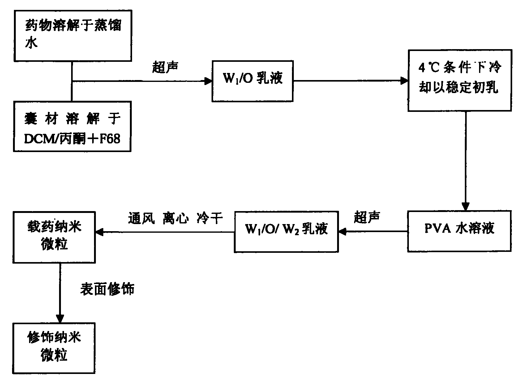
图4:水溶性药物的载药纳米微粒的制备流程Figure 4: Preparation process of drug-loaded nanoparticles for water-soluble drugs
具体实施方式Detailed ways
本发明的载有抗血管再狭窄药物的可生物降解高分子纳米微粒的制备方法如下:The preparation method of the biodegradable polymer nanoparticles loaded with anti-restenosis drugs of the present invention is as follows:
一、脂溶性药物的载药纳米微粒的制备方法1. Preparation method of drug-loaded nanoparticles of fat-soluble drugs
紫杉醇和地塞米松等脂溶性药物采用水包油(O/W)乳化-溶剂挥发法制备(见图3),方法如下:Fat-soluble drugs such as paclitaxel and dexamethasone are prepared by the oil-in-water (O/W) emulsification-solvent evaporation method (see Figure 3), and the method is as follows:
1.一定体积二氯甲烷溶解适量可生物降解高分子浓度0.5-4%,一定体积丙酮溶解适量药物浓度为0.02-4%,然后混合二氯甲烷/丙酮(比例为9∶1或8∶2)有机溶液。1. A certain volume of dichloromethane dissolves an appropriate amount of biodegradable polymer with a concentration of 0.5-4%, a certain volume of acetone dissolves an appropriate amount of a drug concentration of 0.02-4%, and then mixes dichloromethane/acetone (the ratio is 9:1 or 8:2 ) organic solution.
2.混合有机溶液加入到一定体积的(聚乙烯醇)PVA水溶液(0.5-2.5%)中(有机溶液/水溶液=1∶3至1∶4),冰浴条件下超声处理30s-10min。2. Add the mixed organic solution into a certain volume of (polyvinyl alcohol) PVA aqueous solution (0.5-2.5%) (organic solution/water solution = 1:3 to 1:4), and sonicate for 30s-10min under ice bath conditions.
3.常压下搅拌挥发2-18h,再减压挥发1-6h,以除去有机溶剂。3. Stir and volatilize under normal pressure for 2-18h, then volatilize under reduced pressure for 1-6h to remove the organic solvent.
4.低温(4℃-15℃)、15000-25000rpm下超速离心30min,收集固化的纳米微粒,再用蒸馏水洗涤、离心1-3次除去纳米微粒悬液中的聚乙烯醇。4. Ultracentrifuge at low temperature (4°C-15°C) at 15000-25000 rpm for 30 minutes to collect solidified nanoparticles, wash with distilled water and centrifuge 1-3 times to remove polyvinyl alcohol in the nanoparticle suspension.
5.冷冻干燥,完成纳米微粒制备。5. Freeze drying to complete the preparation of nanoparticles.
6.配制0.5-4mg/ml浓度的修饰物水溶液。6. Prepare an aqueous solution of the modifier with a concentration of 0.5-4mg/ml.
7.将纳米微粒加入上述修饰物水溶液,配制浓度为9.5-76mg/ml的悬浮液,高速搅拌或超声处理10-120s,使纳米微粒均匀悬浮。7. Add the nanoparticles to the aqueous solution of the modifier, prepare a suspension with a concentration of 9.5-76 mg/ml, stir at a high speed or ultrasonically treat for 10-120 s, and suspend the nanoparticles evenly.
8.冷冻干燥,完成抗血管再狭窄载药纳米微粒制备。8. Freeze-drying to complete the preparation of anti-restenosis drug-loaded nanoparticles.
9.采用15-30kGy之间的钴60发射的γ射线对纳米微粒照射进行灭菌。9. Sterilize the nanoparticles by irradiation with gamma rays emitted by cobalt 60 between 15-30 kGy.
二、水溶性药物的载药纳米微粒的制备方法Two, the preparation method of the drug-loaded nanoparticles of water-soluble drug
尿激酶和肝素等水溶性药物采用复乳(W/O/W)化-溶剂挥发法制备(见图4),方法如下:Water-soluble drugs such as urokinase and heparin are prepared by double emulsion (W/O/W)-solvent evaporation method (see Figure 4), and the method is as follows:
1.一定质量的水溶性药物溶解于蒸馏水中,浓度为20-30%,一定体积的二氯甲烷(DCM)或二氯甲烷/丙酮(8∶2或9∶1)有机溶液溶解适量可生物降解高分子囊材(浓度为0.5-10%),有机溶液中含有一定比例(0.01%-0.1%)的普罗尼克F68(Pluronic F68)作为乳化剂1. A certain quality of water-soluble drugs is dissolved in distilled water at a concentration of 20-30%, and a certain volume of dichloromethane (DCM) or dichloromethane/acetone (8:2 or 9:1) organic solution is dissolved in an appropriate amount to be biologically Degradable polymer capsule material (concentration: 0.5-10%), the organic solution contains a certain proportion (0.01%-0.1%) of Pluronic F68 (Pluronic F68) as an emulsifier
2.将药物水溶液加入到有机溶液中(药物水溶液∶有机溶剂体积比=1∶4-20),立即在冰浴条件下超声或高速搅拌30s-10min,形成W1/O溶液。2. Add the aqueous drug solution to the organic solution (aqueous drug solution: organic solvent volume ratio = 1:4-20), and immediately ultrasonically or high-speed stir for 30s-10min under ice bath conditions to form a W 1 /O solution.
3.在4℃条件下冷却以稳定初乳3. Cool at 4°C to stabilize colostrum
4.初乳加入到浓度为0.5-2.5%的PVA水溶液中,同样在冰浴条件下进行超声或高速搅拌乳化30s-10min,即形成W1/O/W2复乳。4. Add the colostrum into the PVA aqueous solution with a concentration of 0.5-2.5%, and carry out ultrasonic or high-speed stirring emulsification under ice bath conditions for 30s-10min to form W 1 /O/W 2 double emulsion.
5.常压下搅拌挥发2-18h,再减压挥发1-6h,以除去有机溶剂。5. Stir and volatilize under normal pressure for 2-18h, then volatilize under reduced pressure for 1-6h to remove the organic solvent.
6.低温(4℃-15℃),15000-25000rpm下超速离心30min,收集固化的纳米微粒,再用蒸馏水洗涤、离心1-3次除去纳米微粒悬浮液中的聚乙烯醇。6. Low temperature (4°C-15°C), ultracentrifugation at 15000-25000rpm for 30min, collect solidified nanoparticles, wash with distilled water, and centrifuge 1-3 times to remove polyvinyl alcohol in the nanoparticle suspension.
7.冷冻干燥,完成纳米微粒制备。7. Freeze drying to complete the preparation of nanoparticles.
8.配制0.5-4mg/ml浓度的修饰物水溶液。8. Prepare an aqueous solution of the modifier with a concentration of 0.5-4mg/ml.
9.将纳米微粒加入上述修饰物水溶液,配制浓度9.5-76mg/ml的悬浮液,高速搅拌或超声处理10-120s,使纳米微粒均匀悬浮。9. Add the nanoparticles into the above aqueous solution of the modification, prepare a suspension with a concentration of 9.5-76 mg/ml, stir at high speed or ultrasonically treat for 10-120 s, to suspend the nanoparticles uniformly.
10.冷冻干燥,完成抗血管再狭窄纳米微粒制备10. Freeze-drying to complete the preparation of anti-restenosis nanoparticles
11.采用15-30kGy之间的钴60发射的γ射线对纳米微粒照射进行灭菌。11. Sterilize the nanoparticles by irradiation with gamma rays emitted by cobalt 60 between 15-30 kGy.
具体实施例specific embodiment
实施例1.紫杉醇纳米微粒Example 1. Paclitaxel Nanoparticles
将9毫升2%浓度的PLGA二氯甲烷溶液与1毫升含85.96毫克紫杉醇丙酮溶液混合,再加入到40毫升1%浓度的PVA水浓液中,冰浴条件下超声(40W)乳化2min。然后室温电磁搅拌3h,减压挥发1h。低温、23000rpm离心30min,收集固化的纳米微粒。用蒸馏水洗涤、离心1次,冷冻干燥36h,得到载药量30.54%的紫杉醇纳米微粒,其包封率为90.75%,平均粒径为385nm。将得到的上述纳米微粒加入5毫升浓度为1mg/ml的DMAB水溶液,超声处理10-30s。冷冻干燥得到表面修饰的纳米微粒。修饰后的纳米微粒平均粒径为462.2nm。Mix 9 ml of 2% PLGA dichloromethane solution with 1 ml of acetone solution containing 85.96 mg of paclitaxel, then add to 40 ml of 1% PVA water concentrate, and emulsify by ultrasonic (40W) for 2 min in an ice bath. Then electromagnetically stirred at room temperature for 3 h, and evaporated under reduced pressure for 1 h. Centrifuge at 23000 rpm for 30 min at low temperature to collect solidified nanoparticles. It was washed with distilled water, centrifuged once, and freeze-dried for 36 hours to obtain paclitaxel nanoparticles with a drug loading capacity of 30.54%, an encapsulation efficiency of 90.75%, and an average particle diameter of 385 nm. Add the obtained above-mentioned nanoparticles to 5 ml of DMAB aqueous solution with a concentration of 1 mg/ml, and sonicate for 10-30 s. Freeze-drying yields surface-modified nanoparticles. The average particle size of the modified nanoparticles is 462.2nm.
实施例2.地塞米松纳米微粒
用10ml二氯甲烷溶解200mg PLGA,1ml丙酮溶解42.5mg醋酸地塞米松药粉,然后混合两种有机溶液,将此混合有机溶液加入到40ml 2%浓度的PVA水溶液中,冰浴条件下超声(40W)乳化10min,室温电磁搅拌12h,减压挥发1.5h,除去有机溶剂,在低温、23000rpm下超速离心30min,收集固化的纳米微粒,再用蒸馏水洗涤、离心三次除去纳米微粒表面的PVA,冷冻干燥36h,得到载药量为17.37%的地塞米松纳米微粒,其包封率为98.97%,平均粒径为260nm。将得到的上述纳米微粒加入5毫升浓度为1mg/ml的DMAB水溶液,超声处理10-30s。冷冻干燥得到表面修饰的纳米微粒。Dissolve 200mg PLGA with 10ml dichloromethane, 1ml acetone dissolves 42.5mg dexamethasone acetate powder, then mix two kinds of organic solutions, this mixed organic solution joins in the PVA aqueous solution of 40ml 2% concentration, under ice-bath condition ultrasonic (40W ) emulsified for 10 min, stirred electromagnetically at room temperature for 12 h, volatilized under reduced pressure for 1.5 h, removed the organic solvent, ultracentrifuged at low temperature and 23,000 rpm for 30 min, collected solidified nanoparticles, washed with distilled water, centrifuged three times to remove PVA on the surface of the nanoparticles, and freeze-dried After 36 hours, dexamethasone nanoparticles with a drug load of 17.37% were obtained, the encapsulation efficiency was 98.97%, and the average particle diameter was 260 nm. Add the obtained above-mentioned nanoparticles to 5 ml of DMAB aqueous solution with a concentration of 1 mg/ml, and sonicate for 10-30 s. Freeze-drying yields surface-modified nanoparticles.
实施例3.肝素纳米微粒Example 3. Heparin Nanoparticles
将6毫升PLGA二氯甲烷溶液(浓度为6%)加入到含0.6毫升肝素的水溶液(浓度为26%)的小瓶中,立即在冰浴条件下超声乳化3min(输出功率为40W),得到初乳,将此初乳加入到24毫升0.5%浓度的PVA水溶液,同样在冰浴条件下进行超声乳化3min,即形成W1/O/W2复乳。再将此复乳进行磁力搅拌,常压下挥发有机溶剂2h,高速离心(23000rpm×20min)收集固化的纳米微粒,用蒸馏水洗涤三次,冷冻干燥36h。得到载药量为5.35%的肝素纳米微粒,纳米微粒平均粒径297nm。6 milliliters of PLGA dichloromethane solution (concentration is 6%) is added in the vial containing the aqueous solution (concentration is 26%) of 0.6 milliliter heparin, ultrasonic emulsification 3min (output power is 40W) under ice-bath condition immediately, obtain initial Milk, the colostrum was added to 24 ml of 0.5% PVA aqueous solution, and ultrasonic emulsification was also carried out for 3 minutes under ice bath conditions to form W 1 /O/W 2 double emulsion. Then the double emulsion was magnetically stirred, and the organic solvent was volatilized under normal pressure for 2 hours, and the solidified nanoparticles were collected by high-speed centrifugation (23000rpm×20min), washed three times with distilled water, and freeze-dried for 36 hours. The heparin nanoparticles with a drug load of 5.35% were obtained, and the average particle size of the nanoparticles was 297nm.
实施例4Example 4
为了加强载药微粒抗血管再狭窄的作用,可以将不同药物作用的载药微粒根据病情按用药量的比例混合,配成30mg/ml浓度的盐水混合悬液制剂使用,采用本领域的技术人员公知的介入疗法将纳米微粒送到受损的血管部位。In order to strengthen the anti-restenosis effect of drug-loaded particles, drug-loaded particles with different drug effects can be mixed according to the proportion of the dosage according to the disease condition, and prepared into a saline mixed suspension preparation with a concentration of 30mg/ml for use. Known interventional therapies deliver nanoparticles to damaged vascular sites.
Claims (5)
Priority Applications (1)
| Application Number | Priority Date | Filing Date | Title |
|---|---|---|---|
| CNA2005100146432A CN1903365A (en) | 2005-07-28 | 2005-07-28 | Drug-carried nanometer particles, and its preparing process for preparing medicien prepn. for anti-restenosis of blood-vessel |
Applications Claiming Priority (1)
| Application Number | Priority Date | Filing Date | Title |
|---|---|---|---|
| CNA2005100146432A CN1903365A (en) | 2005-07-28 | 2005-07-28 | Drug-carried nanometer particles, and its preparing process for preparing medicien prepn. for anti-restenosis of blood-vessel |
Publications (1)
| Publication Number | Publication Date |
|---|---|
| CN1903365A true CN1903365A (en) | 2007-01-31 |
Family
ID=37672827
Family Applications (1)
| Application Number | Title | Priority Date | Filing Date |
|---|---|---|---|
| CNA2005100146432A Pending CN1903365A (en) | 2005-07-28 | 2005-07-28 | Drug-carried nanometer particles, and its preparing process for preparing medicien prepn. for anti-restenosis of blood-vessel |
Country Status (1)
| Country | Link |
|---|---|
| CN (1) | CN1903365A (en) |
Cited By (10)
| Publication number | Priority date | Publication date | Assignee | Title |
|---|---|---|---|---|
| CN101953776A (en) * | 2010-09-26 | 2011-01-26 | 中国医学科学院生物医学工程研究所 | Non-spherical drug-loaded particles and controlled release preparation of lactyl polymer and preparation methods thereof |
| CN102018996A (en) * | 2010-12-14 | 2011-04-20 | 中国医学科学院生物医学工程研究所 | Manufacturing method of drug vessel support with antibody immobilized on surface of support |
| US8962032B2 (en) | 2009-12-17 | 2015-02-24 | The Queen's University Of Belfast | Modulator |
| CN104840429A (en) * | 2015-04-23 | 2015-08-19 | 丽珠医药集团股份有限公司 | Preparation method of acetic acid goserelin microspheres |
| CN105617389A (en) * | 2016-03-23 | 2016-06-01 | 首都医科大学附属北京朝阳医院 | Positively charged nano microsphere medicine carrier for eye and preparation method of positively charged nano microsphere medicine |
| CN107441502A (en) * | 2017-07-25 | 2017-12-08 | 首都医科大学附属北京安贞医院 | A kind of nano-particle for carrying combination drug composition |
| WO2019162951A1 (en) * | 2018-02-26 | 2019-08-29 | Yissum Research Development Company Of The Hebrew University Of Jerusalem Ltd | Drug delivery systems |
| CN111467319A (en) * | 2020-03-20 | 2020-07-31 | 苏州医本生命科技有限公司 | Drug-loaded microparticles, storage tube for storing the microparticles, and implantation system |
| CN115006605A (en) * | 2022-07-20 | 2022-09-06 | 苏州中天医疗器械科技有限公司 | Drug coating balloon and preparation method and application thereof |
| CN115252913A (en) * | 2022-08-02 | 2022-11-01 | 赛诺神畅医疗科技有限公司 | Vascular drug delivery coating and preparation method and application thereof |
-
2005
- 2005-07-28 CN CNA2005100146432A patent/CN1903365A/en active Pending
Cited By (13)
| Publication number | Priority date | Publication date | Assignee | Title |
|---|---|---|---|---|
| US8962032B2 (en) | 2009-12-17 | 2015-02-24 | The Queen's University Of Belfast | Modulator |
| CN101953776B (en) * | 2010-09-26 | 2012-07-04 | 中国医学科学院生物医学工程研究所 | Non-spherical drug-loaded particles and controlled release preparation of lactyl polymer and preparation methods thereof |
| CN101953776A (en) * | 2010-09-26 | 2011-01-26 | 中国医学科学院生物医学工程研究所 | Non-spherical drug-loaded particles and controlled release preparation of lactyl polymer and preparation methods thereof |
| CN102018996A (en) * | 2010-12-14 | 2011-04-20 | 中国医学科学院生物医学工程研究所 | Manufacturing method of drug vessel support with antibody immobilized on surface of support |
| CN102018996B (en) * | 2010-12-14 | 2013-03-06 | 中国医学科学院生物医学工程研究所 | Manufacturing method of drug vessel support with antibody immobilized on surface of support |
| CN104840429B (en) * | 2015-04-23 | 2017-12-01 | 丽珠医药集团股份有限公司 | The preparation method of goserelin acetate microballoon |
| CN104840429A (en) * | 2015-04-23 | 2015-08-19 | 丽珠医药集团股份有限公司 | Preparation method of acetic acid goserelin microspheres |
| CN105617389A (en) * | 2016-03-23 | 2016-06-01 | 首都医科大学附属北京朝阳医院 | Positively charged nano microsphere medicine carrier for eye and preparation method of positively charged nano microsphere medicine |
| CN107441502A (en) * | 2017-07-25 | 2017-12-08 | 首都医科大学附属北京安贞医院 | A kind of nano-particle for carrying combination drug composition |
| WO2019162951A1 (en) * | 2018-02-26 | 2019-08-29 | Yissum Research Development Company Of The Hebrew University Of Jerusalem Ltd | Drug delivery systems |
| CN111467319A (en) * | 2020-03-20 | 2020-07-31 | 苏州医本生命科技有限公司 | Drug-loaded microparticles, storage tube for storing the microparticles, and implantation system |
| CN115006605A (en) * | 2022-07-20 | 2022-09-06 | 苏州中天医疗器械科技有限公司 | Drug coating balloon and preparation method and application thereof |
| CN115252913A (en) * | 2022-08-02 | 2022-11-01 | 赛诺神畅医疗科技有限公司 | Vascular drug delivery coating and preparation method and application thereof |
Similar Documents
| Publication | Publication Date | Title |
|---|---|---|
| Iyer et al. | Nanoparticle eluting-angioplasty balloons to treat cardiovascular diseases | |
| CN105833272B (en) | A kind of multifunctional nano pharmaceutical composition and its preparation method and application | |
| CN101889985B (en) | A drug-loaded nano-microsphere and its preparation method | |
| CN103768605B (en) | A kind of preparation method of hybrid nanofiber drug bearing microsphere | |
| Yang et al. | Intravitreal administration of dexamethasone-loaded PLGA-TPGS nanoparticles for the treatment of posterior segment diseases | |
| CN110302395A (en) | A nanoparticle capable of promoting tumor coagulation and enzyme/pH dual-responsive drug release, its preparation method and application | |
| Lim et al. | A novel technique for loading of paclitaxel‐PLGA nanoparticles onto ePTFE vascular grafts | |
| WO2021142884A1 (en) | Use of aspirin in preparation of platelet-targeted drug delivery system | |
| CN101653420A (en) | Ultrasonically controlled-release target medicinal preparation and production method thereof | |
| CN110051652B (en) | PLGA/FK506 drug-loaded nano-microsphere as well as preparation method and application thereof | |
| CN105079885A (en) | Injectable PLGA (poly(lactic-co-glycolic acid)) porous composite microsphere preparation embedded with BMP-2 and TGF-beta1 containing microspheres as well as preparation method and application of composite microsphere preparation | |
| CN103990185A (en) | Carrageenan and gelatin microsphere embolization agent and preparation method thereof | |
| CN103655484B (en) | A kind ofly utilize self-assembling technique method preparing taxol slow release microballoons and products thereof | |
| Lee et al. | Soft extrudable dendritic particles with nanostructured tendrils for local adhesion and drug release to bladder cancers | |
| CN114288262B (en) | Drug-loaded microsphere and preparation method and application thereof | |
| CN116270493A (en) | A kind of rapamycin microsphere for targeted drug delivery and its preparation method and application | |
| CN100381183C (en) | Preparation method of drug-loaded sustained-release micro/nanosphere digestive tract stent | |
| CN101040848B (en) | Preparation method of amphiphilic polymer drug-loaded nanoparticles | |
| CN1470289A (en) | Polymeric nano medicine carrier and preparation preparing method | |
| Alvi et al. | De novo drug delivery modalities for treating damaged hearts: Current challenges and emerging solutions | |
| CN103520728B (en) | The preparation method of arsenic trioxide invisible immunity targeting anti-tumor preparation | |
| Jin et al. | Cellular uptake and radiosensitization of SR-2508 loaded PLGA nanoparticles | |
| Zhao et al. | Study on the role of paclitaxel nano-drug delivery system in inhibiting intimal hyperplasia and improving vascular remodeling in abdominal aortic injury model | |
| Sengel | Delivery of nanoparticles for the treatment of cardiovascular diseases | |
| CN114831966A (en) | Photothermal conversion nano composite material without toxic and side effects and preparation method and application thereof |
Legal Events
| Date | Code | Title | Description |
|---|---|---|---|
| C06 | Publication | ||
| PB01 | Publication | ||
| C02 | Deemed withdrawal of patent application after publication (patent law 2001) | ||
| WD01 | Invention patent application deemed withdrawn after publication |