CN1965760A - synchrotron radiation X-ray phase contrasting computed tomography and experimental method thereof - Google Patents
synchrotron radiation X-ray phase contrasting computed tomography and experimental method thereof Download PDFInfo
- Publication number
- CN1965760A CN1965760A CN 200510086904 CN200510086904A CN1965760A CN 1965760 A CN1965760 A CN 1965760A CN 200510086904 CN200510086904 CN 200510086904 CN 200510086904 A CN200510086904 A CN 200510086904A CN 1965760 A CN1965760 A CN 1965760A
- Authority
- CN
- China
- Prior art keywords
- sample
- phase contrast
- imaging
- turntable
- image
- Prior art date
- Legal status (The legal status is an assumption and is not a legal conclusion. Google has not performed a legal analysis and makes no representation as to the accuracy of the status listed.)
- Granted
Links
- 238000002474 experimental method Methods 0.000 title claims description 38
- 230000005469 synchrotron radiation Effects 0.000 title claims description 17
- 238000002591 computed tomography Methods 0.000 title abstract description 16
- 239000013078 crystal Substances 0.000 claims abstract description 89
- 238000003384 imaging method Methods 0.000 claims abstract description 78
- 238000000034 method Methods 0.000 claims abstract description 20
- 238000013519 translation Methods 0.000 claims abstract description 9
- 230000014616 translation Effects 0.000 claims abstract description 9
- 238000013170 computed tomography imaging Methods 0.000 claims description 31
- 238000013480 data collection Methods 0.000 claims description 8
- 230000000694 effects Effects 0.000 claims description 6
- 238000012545 processing Methods 0.000 claims description 6
- 238000002247 constant time method Methods 0.000 claims description 5
- 238000012544 monitoring process Methods 0.000 claims 1
- 238000004458 analytical method Methods 0.000 abstract description 19
- 230000005855 radiation Effects 0.000 abstract 1
- 230000001360 synchronised effect Effects 0.000 abstract 1
- 238000010998 test method Methods 0.000 abstract 1
- 239000000523 sample Substances 0.000 description 59
- 238000010521 absorption reaction Methods 0.000 description 8
- 238000010586 diagram Methods 0.000 description 8
- 230000003287 optical effect Effects 0.000 description 8
- 210000004872 soft tissue Anatomy 0.000 description 4
- 239000000463 material Substances 0.000 description 3
- 238000011160 research Methods 0.000 description 3
- 238000013500 data storage Methods 0.000 description 2
- 238000003745 diagnosis Methods 0.000 description 2
- 206010028980 Neoplasm Diseases 0.000 description 1
- 239000012472 biological sample Substances 0.000 description 1
- 230000005540 biological transmission Effects 0.000 description 1
- 238000001514 detection method Methods 0.000 description 1
- 201000010099 disease Diseases 0.000 description 1
- 208000037265 diseases, disorders, signs and symptoms Diseases 0.000 description 1
- 239000003814 drug Substances 0.000 description 1
- 238000013399 early diagnosis Methods 0.000 description 1
- 238000002594 fluoroscopy Methods 0.000 description 1
- 229910052732 germanium Inorganic materials 0.000 description 1
- GNPVGFCGXDBREM-UHFFFAOYSA-N germanium atom Chemical compound [Ge] GNPVGFCGXDBREM-UHFFFAOYSA-N 0.000 description 1
- 230000031700 light absorption Effects 0.000 description 1
- 239000002184 metal Substances 0.000 description 1
- 210000004088 microvessel Anatomy 0.000 description 1
- 238000012806 monitoring device Methods 0.000 description 1
- 238000010606 normalization Methods 0.000 description 1
- 230000035945 sensitivity Effects 0.000 description 1
- 229910052710 silicon Inorganic materials 0.000 description 1
- 239000010703 silicon Substances 0.000 description 1
- 239000000126 substance Substances 0.000 description 1
Images

Landscapes
- Analysing Materials By The Use Of Radiation (AREA)
Abstract
Description
技术领域technical field
本发明涉及计算机断层成像技术领域,特别是一种同步辐射X射线相位衬度CT成像装置及实验方法。The invention relates to the technical field of computer tomography, in particular to a synchrotron radiation X-ray phase contrast CT imaging device and an experimental method.
背景技术Background technique
自1895年伦琴发现X射线至今的一百多年里,基于吸收衬度的X射线透视成像,因为能无损地得到样品的内部结构信息,在医学诊断和材料研究等领域已经成为重要的检测手段。特别是20世纪中、后期A.M.Cormack和G.N.Hounsfield等人发明X射线CT(计算机断层成像术)后,CT目前已成为医学诊断的必备手段。但无论是X射线透视成像还是传统CT,成像衬度都是基于样品对X射线的吸收差异。对于以轻元素为主要成分的生物软组织而言,由于轻元素材料对硬X射线的吸收非常小,而不同轻元素材料之间的吸收差别就更小,所以只能得到衬度较低的吸收像。因此,基于吸收差异的常规CT,即使提高了空间分辨率,仍然不能对生物软组织的内部细微结构产生衬度,还无法看到生物软组织内部的结构细节(如早期肿瘤、微血管等)。这极大限制了X射线吸收成像方法在生物软组织研究方面的应用。Since Roentgen discovered X-rays in 1895, X-ray fluoroscopy imaging based on absorption contrast has become an important detection method in the fields of medical diagnosis and material research because it can obtain the internal structure information of samples non-destructively. . Especially after the invention of X-ray CT (computed tomography) by A.M.Cormack and G.N.Hounsfield and others in the middle and late 20th century, CT has become an essential means of medical diagnosis. But whether it is X-ray fluoroscopic imaging or traditional CT, the imaging contrast is based on the difference in the absorption of X-rays by the sample. For biological soft tissues with light elements as the main components, since the absorption of light element materials on hard X-rays is very small, and the absorption difference between different light element materials is even smaller, only low-contrast absorption can be obtained. picture. Therefore, conventional CT based on absorption difference, even if the spatial resolution is improved, still cannot generate contrast to the internal fine structure of biological soft tissue, and cannot see the internal structural details of biological soft tissue (such as early tumors, microvessels, etc.). This greatly limits the application of X-ray absorption imaging methods in the study of biological soft tissues.
近年来,随着X射线相位衬度成像技术的出现,人们对X射线的成像机制有了进一步的认识。相位衬度成像是通过记录X射线穿过物体后相位的变化而成像的技术,在硬X射线波段(λ≤0.2nm),轻元素的相位项δ比吸收项β大3~4个数量级。因此,相位衬度成像比传统X射线吸收成像具有更高的灵敏度和分辨率,更有利于在轻元素范围内对物质内部结构信息的研究,并有可能在生物样品的内部结构细节以及某些疾病的早期诊断方面发挥一定的作用,这对生物、医学方面的相关研究具有重要的意义。In recent years, with the emergence of X-ray phase contrast imaging technology, people have a further understanding of the imaging mechanism of X-rays. Phase contrast imaging is an imaging technique that records the phase change after X-rays pass through an object. In the hard X-ray band (λ≤0.2nm), the phase term δ of light elements is 3 to 4 orders of magnitude larger than the absorption term β. Therefore, phase contrast imaging has higher sensitivity and resolution than traditional X-ray absorption imaging, and is more conducive to the study of the internal structure information of substances in the range of light elements, and it is possible to reveal the internal structure details of biological samples and some It plays a certain role in the early diagnosis of diseases, which is of great significance to related research in biology and medicine.
虽然理论和实验都表明,X射线相位衬度成像能达到较高的衬度和分辨率,但是它仍然是二维成像,是样品在垂直于光束方向各个层面投影像的重叠,还不能获得样品的三维结构。因此,将具有高衬度和分辨率的相位衬度成像技术应用于三维成像、开展同步辐射X射线相位衬度CT成像方法及应用研究是一个迫切需要解决的问题。Although both theory and experiments have shown that X-ray phase contrast imaging can achieve high contrast and resolution, it is still a two-dimensional imaging, which is the superimposition of the projection images of the sample at various levels perpendicular to the beam direction, and the sample cannot be obtained yet. three-dimensional structure. Therefore, it is an urgent problem to be solved to apply phase contrast imaging technology with high contrast and resolution to three-dimensional imaging, and to carry out synchrotron radiation X-ray phase contrast CT imaging method and application research.
图1是相位衬度成像中广泛应用的两种成像模式光路示意图。Figure 1 is a schematic diagram of the optical paths of two imaging modes widely used in phase contrast imaging.
同步辐射X射线相位衬度成像装置一般由单色器晶体、样品台、分析晶体、电离室和探测器组成。The synchrotron radiation X-ray phase contrast imaging device generally consists of a monochromator crystal, a sample stage, an analysis crystal, an ionization chamber and a detector.
单色器晶体是将入射的“白光”X射线进行单色化、获得带宽很窄的单色X射线装置,单色器晶体一般选用硅或锗单晶体,通过调整单色器晶体衍射面与入射“白光”X射线的角度(布喇格角),可以选择出射X射线的波长(能量)。分析晶体一般是与单色器晶体相同类型的晶体。在衍射增强相位衬度成像模式中,分析晶体是精度很高的角度滤波器;而在同轴相衬成像模式中,分析晶体是单色X射线的偏转器。在本发明的装置中,单色器晶体和分析晶体都固定在相应的转轴上,转轴的转台由步进电机通过相应的减速传动装置实现。转轴的转角分辨远远小于双晶摇摆曲线的半高宽(FWHM),以满足在摇摆曲线的不同部位进行成像的要求。以能量为10KeV的X射线为例,Si(111)晶体的双晶摇摆曲线的半高宽约为8角秒,在本发明的装置中,单色器晶体和分析晶体转轴的转动精度可以达到0.05角秒,足以满足在摇摆曲线不同部位成像的需要。The monochromator crystal is a monochromatic X-ray device that monochromatizes the incident "white light" X-ray and obtains a very narrow bandwidth. The monochromator crystal is generally made of silicon or germanium single crystal. By adjusting the diffraction surface and the incident The angle (Bragg angle) of the "white light" X-rays allows the selection of the wavelength (energy) of the emitted X-rays. The analytical crystal is generally the same type of crystal as the monochromator crystal. In the diffraction-enhanced phase-contrast imaging mode, the analytical crystal is a high-precision angular filter; while in the coaxial phase-contrast imaging mode, the analytical crystal is a deflector for monochromatic X-rays. In the device of the present invention, both the monochromator crystal and the analysis crystal are fixed on the corresponding rotating shaft, and the turntable of the rotating shaft is realized by a stepping motor through a corresponding reduction transmission device. The rotational angle resolution of the rotating shaft is much smaller than the full width at half maximum (FWHM) of the twin crystal rocking curve, so as to meet the requirement of imaging at different parts of the rocking curve. Be that the X-ray of 10KeV is example with energy, the FWHM of the twin crystal rocking curve of Si (111) crystal is about 8 arc seconds, and in the device of the present invention, the rotation precision of monochromator crystal and analytical crystal rotating shaft can reach 0.05 arc seconds is enough to meet the needs of imaging at different parts of the rocking curve.
电离室的主要作用是实时监测分析晶体出射的X射线的强度。通过微静电计可将电离室测得的X射线产生的光电流显示出来。电离室的具体作用体现在两个方面:1)不放置样品,当旋转分析晶体时,通过电离室记录的从分析晶体出射X射线的强度可以测定双晶摇摆曲线,并确定分析晶体衍射面相对于单色器晶体衍射面的相对角度位置,以满足不同相位衬度成像模式的要求。2)放置样品进行实验时,通过电离室记录的从分析晶体出射X射线的强度变化,可以确定曝光时间和实验过程中X射线强度下降带来的影响,为后续图像数据处理和归一化提供依据。The main function of the ionization chamber is to monitor and analyze the intensity of X-rays emitted by the crystal in real time. The photocurrent generated by the X-rays measured in the ionization chamber can be displayed by the micro-electrometer. The specific role of the ionization chamber is reflected in two aspects: 1) When the sample is not placed, when the analytical crystal is rotated, the intensity of the X-rays emitted from the analytical crystal recorded by the ionization chamber can be used to determine the rocking curve of the twin crystal, and determine that the diffraction surface of the analytical crystal is relative to The relative angular position of the crystal diffraction surface of the monochromator to meet the requirements of different phase contrast imaging modes. 2) When the sample is placed for the experiment, the intensity change of the X-ray emitted from the analysis crystal recorded by the ionization chamber can determine the exposure time and the influence of the X-ray intensity drop during the experiment, and provide information for subsequent image data processing and normalization. in accordance with.
成像探测器的一般为二维面探测器,要求是便于图像的数字化以及具有一定的空间分辨率。成像探测器便于图像数字化可以方便地对图像进行同步或后续处理。同时,为了满足成像分辨率的要求,探测器必须有足够的空间分辨率。Imaging detectors are generally two-dimensional surface detectors, which are required to facilitate image digitization and have a certain spatial resolution. Imaging detectors facilitate image digitization for easy synchronization or subsequent processing of images. At the same time, in order to meet the requirements of imaging resolution, the detector must have sufficient spatial resolution.
发明内容Contents of the invention
本发明的目的在于提供一种同步辐射X射线相位衬度CT成像装置及实验方法。The object of the present invention is to provide a synchrotron radiation X-ray phase contrast CT imaging device and an experimental method.
在同步辐射相位衬度CT成像实验中,由于光源是固定的,后续的实验装置必须根据光源位置及方向调整,所以在开展相位衬度CT实验时,只有通过实验样品的旋转来完成。In synchrotron radiation phase-contrast CT imaging experiments, since the light source is fixed, subsequent experimental devices must be adjusted according to the position and direction of the light source. Therefore, the phase-contrast CT experiment can only be completed by rotating the experimental sample.
单色器晶体、样品转台、分析晶体、电离室和成像探测器组成组成了本发明的X射线相位衬度CT实验装置。A monochromator crystal, a sample turntable, an analysis crystal, an ionization chamber and an imaging detector constitute the X-ray phase contrast CT experimental device of the present invention.
在上述的X射线相位衬度成像CT实验装置中,结合计算机可以实现单色器晶体、分析晶体和样品台的转台、电离室数据读出和记录、成像的拍摄及记录等过程的完全自动化进行。In the above-mentioned X-ray phase contrast imaging CT experimental device, the combination of computer can realize the complete automation of monochromator crystal, analysis crystal and sample stage turntable, ionization chamber data readout and recording, imaging shooting and recording, etc. .
在衍射增强相位衬度CT成像实验中,样品转台放置在单色器晶体和分析晶体之间,根据电离室测得的双晶摇摆曲线确定分析晶体和单色器晶体之间的夹角。固定调好的分析晶体和单色器晶体,样品转台(转台1)每转台一个特定的角度后,利用成像探测器获得一张样品的衍射增强投影像,样品转台的范围可为180°、360°或其它任意角度,最终获得相应张数的衍射增强二维投影像(二维投影像张数=样品转台度数/转台步长)。根据衍射增强成像的相关理论和相应CT算法,对所获得的二维投影像进行处理,即可得到样品的衍射增强相位衬度CT断层像。In the diffraction-enhanced phase contrast CT imaging experiment, the sample turret is placed between the monochromator crystal and the analysis crystal, and the angle between the analysis crystal and the monochromator crystal is determined according to the double crystal rocking curve measured in the ionization chamber. Fixed and adjusted analytical crystal and monochromator crystal, sample turret (turntable 1) after a specific angle for each turntable, using imaging detector to obtain a diffraction enhanced projection image of the sample, the range of sample turret can be 180°, 360° ° or other arbitrary angles, and finally obtain the corresponding number of diffraction-enhanced two-dimensional projection images (number of two-dimensional projection images = sample turret degree/turntable step size). According to the relevant theory of diffraction-enhanced imaging and the corresponding CT algorithm, the obtained two-dimensional projection image is processed to obtain the diffraction-enhanced phase contrast CT tomographic image of the sample.
在同轴相位衬度CT成像实验中,样品台放置在两块晶体的后面,两块晶体组成双晶单色器,探测器放置在可沿X射线光路方向自动滑动的直线滑台上。根据晶体衍射的布喇格角,可以选择照射到样品上的X射线波长;根据电离室测得的双晶摇摆曲线,可将两块晶体的衍射面调节平行。根据同轴相位衬度成像理论,选择合适的样品-探测器距离,可在成像探测器上获得同轴相位衬度二维投影像。按照特定步长旋转样品同时在每一个角度位置采集二维投影像,样品旋转整个预定转台范围后,可采集到完整的投影像数据。根据同轴相位衬度成像的相关理论和相应CT算法,对所获得的二维投影像进行处理,即可得到样品的同轴相位衬度CT断层像。In the coaxial phase contrast CT imaging experiment, the sample stage is placed behind two crystals, which form a double-crystal monochromator, and the detector is placed on a linear slide that can automatically slide along the direction of the X-ray optical path. According to the Bragg angle of crystal diffraction, the X-ray wavelength irradiated on the sample can be selected; according to the double crystal rocking curve measured in the ionization chamber, the diffraction planes of the two crystals can be adjusted to be parallel. According to the coaxial phase contrast imaging theory, the coaxial phase contrast two-dimensional projection image can be obtained on the imaging detector by selecting an appropriate sample-detector distance. Rotate the sample according to a specific step length and collect two-dimensional projection images at each angular position. After the sample rotates the entire predetermined turntable range, complete projection image data can be collected. According to the relevant theory of coaxial phase contrast imaging and corresponding CT algorithm, the obtained two-dimensional projection image is processed to obtain the coaxial phase contrast CT tomographic image of the sample.
在本发明所述的装置中,确定好不同成像条件下的曝光时间后,在程序中输入样品需转台的总角度(样品扫描范围)和转台步长,确定好数据在计算机上存放的路径后,整个数据采集过程即可自动运行直至数据采集过程结束。程序的运动过程是:采集二维投影像并存盘→样品旋转一个步长→再次采集二维投影像并存盘→样品继续旋转一个步长,如此反复,直到样品旋转完成整个扫描范围。图3是本发明的相位衬度CT成像实验工作过程示意图。In the device of the present invention, after determining the exposure time under different imaging conditions, input the total angle of the turntable (sample scanning range) and the step length of the turntable for the sample in the program, and determine the path where the data is stored on the computer , the entire data collection process can run automatically until the end of the data collection process. The movement process of the program is: collect the two-dimensional projection image and save it to disk → rotate the sample by one step → collect the two-dimensional projection image again and save it on disk → continue to rotate the sample by one step, and so on, until the sample rotates to complete the entire scanning range. Fig. 3 is a schematic diagram of the working process of the phase contrast CT imaging experiment of the present invention.
以本发明所述装置为基础,同步辐射X射线相位衬度CT成像实验工作模式主要有:Based on the device of the present invention, the synchrotron radiation X-ray phase contrast CT imaging experiment mode mainly includes:
衍射增强相位衬度CT成像实验的主要工作模式有如下四种:There are four main working modes of diffraction-enhanced phase-contrast CT imaging experiments:
1)样品转轴平行于单色器晶体转轴和分析晶体转轴,此种模式通过调整转台2得以实现,1) The sample rotation axis is parallel to the monochromator crystal rotation axis and the analysis crystal rotation axis. This mode can be realized by adjusting the turntable 2,
2)样品转轴垂直于单色器晶体和分析晶体转轴,此种模式通过在上一模式基础上使转台2旋转90°得以实现,2) The rotation axis of the sample is perpendicular to the rotation axis of the monochromator crystal and the analysis crystal. This mode is realized by rotating the turntable 2 by 90° on the basis of the previous mode.
3)单色器晶体和分析晶体调谐模式,单色器晶体和分析晶体衍射面完全平行,3) Monochromator crystal and analysis crystal tuning mode, monochromator crystal and analysis crystal diffraction planes are completely parallel,
4)单色器晶体和分析晶体失谐模式,单色器晶体和分析晶体的衍射面存在一非常小的夹角;4) There is a very small angle between the monochromator crystal and the analysis crystal in the detuning mode of the monochromator crystal and the diffraction plane of the analysis crystal;
同轴相位衬度CT成像实验的主要工作模式有如下两种:The main working modes of the coaxial phase contrast CT imaging experiment are as follows:
1)此种模式需要预先确定好样品和探测器之间的最佳距离,然后在样品-探测器距离不变的条件下进行CT成像实验,1) This mode needs to pre-determine the optimal distance between the sample and the detector, and then conduct CT imaging experiments under the condition that the sample-detector distance remains unchanged.
2)选取几个不同的样品-探测器距离,在不同距离处分别进行CT成像实验数据采集,然后利用所获的几套数据在数据处理中获得最优相位衬度。2) Select several different sample-detector distances, collect CT imaging experiment data at different distances, and then use the obtained several sets of data to obtain the optimal phase contrast in data processing.
在不同相位衬度CT成像实验工作模式下,本发明所述的相位衬度CT成像实验流程。其具体步骤为:Under different phase contrast CT imaging experiment working modes, the phase contrast CT imaging experiment process described in the present invention. The specific steps are:
步骤S1,不同成像模式下相位衬度成像条件的确定:Step S1, determination of phase contrast imaging conditions in different imaging modes:
(1)在衍射增强相位衬度CT成像实验中,根据样品所需的成像条件和双晶摇摆曲线上的相应位置的关系,确定在单色器晶体和分析晶体之间的夹角,(1) In the diffraction-enhanced phase contrast CT imaging experiment, according to the relationship between the required imaging conditions of the sample and the corresponding positions on the twin crystal rocking curve, determine the angle between the monochromator crystal and the analysis crystal,
(2)在同轴相位衬度CT成像实验中,根据不同样品确定合适的样品和探测器的距离以获得最佳的成像效果或得到所需的信息,(2) In the coaxial phase contrast CT imaging experiment, determine the appropriate distance between the sample and the detector according to different samples to obtain the best imaging effect or obtain the required information,
(3)根据X射线光强、探测器等的不同参数确定不同条件下相应的成像曝光时间;(3) Determine the corresponding imaging exposure time under different conditions according to different parameters such as X-ray light intensity and detectors;
步骤S2,相位衬度CT成像实验数据的获取,在步骤S1所确定的成像条件下,样品转台(1)每转动一个特定的角度后,利用成像探测器获得一张样品的相位衬度投影像,样品转台的范围可为180°、360°或其它任意角度,最终获得相应张数的相位衬度二维投影像,在所述的装置中,确定好不同成像条件下的曝光时间后,在程序中输入样品需转动的总角度和转动步长,确定好数据在计算机上存放的路径后,整个数据采集过程即可自动运行直至数据采集过程结束;Step S2, the acquisition of phase contrast CT imaging experiment data, under the imaging conditions determined in step S1, after the sample turntable (1) rotates a specific angle, use the imaging detector to obtain a phase contrast projection image of the sample , the range of the sample turntable can be 180°, 360° or other arbitrary angles, and finally obtain a corresponding number of phase contrast two-dimensional projection images. In the device, after determining the exposure time under different imaging conditions, the Input the total angle and rotation step of the sample to be rotated in the program, and after determining the data storage path on the computer, the entire data collection process can run automatically until the end of the data collection process;
步骤S3,相位衬度成像实验数据的重建,以衍射增强相位衬度成像或同轴相位衬度成像的相关理论作为算法基础,对所获得的二维投影像进行处理即可重建样品的相位衬度CT断层像。Step S3, the reconstruction of the experimental data of phase contrast imaging, based on the relevant theory of diffraction-enhanced phase contrast imaging or coaxial phase contrast imaging, the phase contrast of the sample can be reconstructed by processing the obtained two-dimensional projection image CT tomogram.
附图说明Description of drawings
图1是相位衬度成像中广泛应用的两种成像模式光路示意图。Figure 1 is a schematic diagram of the optical paths of two imaging modes widely used in phase contrast imaging.
图2是本发明中的样品转台示意图。Fig. 2 is a schematic diagram of the sample turntable in the present invention.
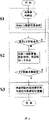
图3是本发明的同步辐射X射线相位衬度CT成像方法流程图。Fig. 3 is a flowchart of the synchrotron radiation X-ray phase contrast CT imaging method of the present invention.
具体实施方式Detailed ways
图1是相位衬度成像中广泛应用的两种成像模式光路示意图。其中,图1(a)是同轴相位衬度CT成像实验光路图,图1(b)是衍射增强相位衬度CT成像实验光路图。Figure 1 is a schematic diagram of the optical paths of two imaging modes widely used in phase contrast imaging. Among them, Fig. 1(a) is the optical path diagram of the coaxial phase contrast CT imaging experiment, and Fig. 1(b) is the optical path diagram of the diffraction-enhanced phase contrast CT imaging experiment.
在同步辐射相位衬度成像实验中,由于光源是固定的,后续的实验装置必须根据光源位置及方向调整,所以在相位衬度CT成像CT实验中,投影数据的采集只有通过实验样品的旋转而不是光源的旋转来完成。In synchrotron radiation phase-contrast imaging experiments, since the light source is fixed, subsequent experimental devices must be adjusted according to the position and direction of the light source. Therefore, in phase-contrast CT imaging CT experiments, the acquisition of projection data can only be achieved through the rotation of the experimental sample. Not the rotation of the light source to do it.
本发明的同步辐射X射线相位衬度CT成像装置,所述的装置包括由单色器晶体、样品转台、分析晶体、电离室和成像探测器组成,在衍射增强相位衬度CT成像中,单色器晶体、样品转台、分析晶体、电离室和成像探测器顺序排列在光路上;在同轴相位衬度CT成像中,单色器晶体、分析晶体、样品转台、电离室和成像探测器依次排列在光路上;电离室的作用是作为X射线强度的监测设备,电离室测得的X射线光强以光电流的形式通过微静电计实时显示。In the synchrotron radiation X-ray phase contrast CT imaging device of the present invention, the device includes a monochromator crystal, a sample turntable, an analysis crystal, an ionization chamber, and an imaging detector. In diffraction-enhanced phase contrast CT imaging, a single Chromochromator crystal, sample turret, analysis crystal, ionization chamber and imaging detector are sequentially arranged on the optical path; in coaxial phase contrast CT imaging, monochromator crystal, analysis crystal, sample turret, ionization chamber and imaging detector are sequentially arranged Arranged on the optical path; the role of the ionization chamber is as a monitoring device for X-ray intensity, and the X-ray intensity measured by the ionization chamber is displayed in real time by a micro-electrometer in the form of photocurrent.
图2是本发明中的样品转台示意图。转台1、转台2、转台3分别对应于ω1、ω2和ω3,平动1、平动2分别对应图中P1和P2。Fig. 2 is a schematic diagram of the sample turntable in the present invention.
本发明的样品转台由三个转台和两个平动组成,三个转台中,转台1(ω1)的作用是转动固定在转台1上样品,是CT实验过程中的主要转台;转台2(ω1)用来调整样品相对于从单色器晶体出射的X射线的方位以满足不同成像条件的需要,因为转台1固定在转台2上,所以转台2转台时,固定于转台1上的样品相对于入射X射线的方位将随转台2变动(例如,通过转台2可实现样品转轴由水平转台变为竖直转台);转台3(ω3)用来调整样品台相对入射X射线的方位以保证X射线对样品的正入射,转台3转台时,固定于转台3上的转台2随转,固定在转台2上的转台1同时随转以实现X射线对样品的正入射,两个平动中,平动P1用以实现样品台相对于X射线高度位置的调节,平动P2用以实现样品台相对于X射线水平位置的调节。The sample turntable of the present invention is made up of three turntables and two translations, among the three turntables, the effect of turntable 1 (ω1) is to rotate and fix the sample on
此外,为避免同步辐射X射线入射到金属部件上带来的强散射的影响,转台2的中央通过X射线的部位设计为一个足够大的X射线通道;样品转台(转台1)的转动轴要有足够高的同心稳定性以满足CT成像分辨率的要求。In addition, in order to avoid the impact of strong scattering of synchrotron radiation X-rays incident on the metal parts, the central part of the turntable 2 where the X-rays pass through is designed as a sufficiently large X-ray channel; the rotation axis of the sample turntable (turntable 1) must be It has high enough concentric stability to meet the requirements of CT imaging resolution.
图3是同步辐射X射线相位衬度成像CT方法流程,其具体步骤如下:Fig. 3 is the process flow of the synchrotron radiation X-ray phase contrast imaging CT method, and its specific steps are as follows:
步骤S1,不同成像模式下相位衬度成像条件的确定:Step S1, determination of phase contrast imaging conditions in different imaging modes:
(1)在衍射增强相位衬度CT成像实验中,根据样品所需的成像条件和双晶摇摆曲线上的相应位置的关系,确定在单色器晶体和分析晶体之间的夹角,(1) In the diffraction-enhanced phase contrast CT imaging experiment, according to the relationship between the required imaging conditions of the sample and the corresponding positions on the twin crystal rocking curve, determine the angle between the monochromator crystal and the analysis crystal,
(2)在同轴相位衬度CT成像实验中,根据不同样品确定合适的样品和探测器的距离以获得最佳的成像效果或得到所需的信息,(2) In the coaxial phase contrast CT imaging experiment, determine the appropriate distance between the sample and the detector according to different samples to obtain the best imaging effect or obtain the required information,
(3)根据X射线光强、探测器等的不同参数确定不同条件下相应的成像曝光时间;(3) Determine the corresponding imaging exposure time under different conditions according to different parameters such as X-ray light intensity and detectors;
步骤S2,相位衬度CT成像实验数据的获取,在步骤S1所确定的成像条件下,样品转台(1)每转动一个特定的角度后,利用成像探测器获得一张样品的相位衬度投影像,样品转台的范围可为180°、360°或其它任意角度,最终获得相应张数的相位衬度二维投影像,在所述的装置中,确定好不同成像条件下的曝光时间后,在程序中输入样品需转动的总角度和转动步长,确定好数据在计算机上存放的路径后,整个数据采集过程即可自动运行直至数据采集过程结束;Step S2, the acquisition of phase contrast CT imaging experiment data, under the imaging conditions determined in step S1, after the sample turntable (1) rotates a specific angle, use the imaging detector to obtain a phase contrast projection image of the sample , the range of the sample turntable can be 180°, 360° or other arbitrary angles, and finally obtain a corresponding number of phase contrast two-dimensional projection images. In the device, after determining the exposure time under different imaging conditions, the Input the total angle and rotation step of the sample to be rotated in the program, and after determining the data storage path on the computer, the entire data collection process can run automatically until the end of the data collection process;
步骤S3,相位衬度成像实验数据的重建,以衍射增强相位衬度成像或同轴相位衬度成像的相关理论作为算法基础,对所获得的二维投影像进行处理即可重建样品的相位衬度CT断层像。Step S3, the reconstruction of the experimental data of phase contrast imaging, based on the relevant theory of diffraction-enhanced phase contrast imaging or coaxial phase contrast imaging, the phase contrast of the sample can be reconstructed by processing the obtained two-dimensional projection image CT tomogram.
Claims (5)
Priority Applications (1)
| Application Number | Priority Date | Filing Date | Title |
|---|---|---|---|
| CNB2005100869041A CN100457040C (en) | 2005-11-17 | 2005-11-17 | synchrotron radiation X-ray phase contrasting computed tomography and experimental method thereof |
Applications Claiming Priority (1)
| Application Number | Priority Date | Filing Date | Title |
|---|---|---|---|
| CNB2005100869041A CN100457040C (en) | 2005-11-17 | 2005-11-17 | synchrotron radiation X-ray phase contrasting computed tomography and experimental method thereof |
Publications (2)
| Publication Number | Publication Date |
|---|---|
| CN1965760A true CN1965760A (en) | 2007-05-23 |
| CN100457040C CN100457040C (en) | 2009-02-04 |
Family
ID=38074863
Family Applications (1)
| Application Number | Title | Priority Date | Filing Date |
|---|---|---|---|
| CNB2005100869041A Expired - Fee Related CN100457040C (en) | 2005-11-17 | 2005-11-17 | synchrotron radiation X-ray phase contrasting computed tomography and experimental method thereof |
Country Status (1)
| Country | Link |
|---|---|
| CN (1) | CN100457040C (en) |
Cited By (11)
| Publication number | Priority date | Publication date | Assignee | Title |
|---|---|---|---|---|
| CN102413767A (en) * | 2009-03-02 | 2012-04-11 | 罗切斯特大学 | Methods and apparatus for differential phase contrast fan-beam CT, cone-beam CT, and hybrid cone-beam CT |
| CN101952900B (en) * | 2008-02-14 | 2013-10-23 | 皇家飞利浦电子股份有限公司 | X-ray detectors for phase contrast imaging |
| CN103356208A (en) * | 2012-04-01 | 2013-10-23 | 中国科学院高能物理研究所 | Two-dimensional imaging system and method for medical test of human body |
| US8971488B2 (en) | 2008-12-01 | 2015-03-03 | The University Of North Carolina At Chapel Hill | Systems and methods for detecting an image of an object using multi-beam imaging from an X-ray beam having a polychromatic distribution |
| US8976925B2 (en) | 2006-02-27 | 2015-03-10 | University Of Rochester | Method and apparatus of cone beam CT dynamic imaging |
| CN105513057A (en) * | 2015-11-30 | 2016-04-20 | 首都医科大学 | Tumor auxiliary diagnosis method |
| US9364191B2 (en) | 2013-02-11 | 2016-06-14 | University Of Rochester | Method and apparatus of spectral differential phase-contrast cone-beam CT and hybrid cone-beam CT |
| CN105675638A (en) * | 2016-03-24 | 2016-06-15 | 西安交通大学 | Universality efficient synchrotron radiation visual representation method of crystal microstructure |
| CN109215094A (en) * | 2017-09-22 | 2019-01-15 | 上海联影医疗科技有限公司 | phase contrast image generating method and system |
| CN111896566A (en) * | 2020-07-20 | 2020-11-06 | 上海交通大学 | A device and method for increasing the imaging range of a synchrotron radiation light source |
| CN112179926A (en) * | 2020-09-24 | 2021-01-05 | 首都师范大学 | A Phase-Absorption Inversion and Material Quantitative Imaging Method Based on Coaxial CT |
Family Cites Families (5)
| Publication number | Priority date | Publication date | Assignee | Title |
|---|---|---|---|---|
| US5259013A (en) * | 1991-12-17 | 1993-11-02 | The United States Of America As Represented By The Secretary Of Commerce | Hard x-ray magnification apparatus and method with submicrometer spatial resolution of images in more than one dimension |
| JPH09187455A (en) * | 1996-01-10 | 1997-07-22 | Hitachi Ltd | Phase type X-ray CT system |
| DE69730550T2 (en) * | 1996-03-29 | 2005-11-10 | Hitachi, Ltd. | Phase contrast X-ray imaging system |
| JP2000193609A (en) * | 1998-12-24 | 2000-07-14 | Hamamatsu Photonics Kk | Radiation tomograph |
| US7286628B2 (en) * | 2001-11-05 | 2007-10-23 | Vanderbilt University | Phase-contrast enhanced computed tomography |
-
2005
- 2005-11-17 CN CNB2005100869041A patent/CN100457040C/en not_active Expired - Fee Related
Cited By (15)
| Publication number | Priority date | Publication date | Assignee | Title |
|---|---|---|---|---|
| US8976925B2 (en) | 2006-02-27 | 2015-03-10 | University Of Rochester | Method and apparatus of cone beam CT dynamic imaging |
| US8576983B2 (en) | 2008-02-14 | 2013-11-05 | Koninklijke Philips N.V. | X-ray detector for phase contrast imaging |
| CN101952900B (en) * | 2008-02-14 | 2013-10-23 | 皇家飞利浦电子股份有限公司 | X-ray detectors for phase contrast imaging |
| US8971488B2 (en) | 2008-12-01 | 2015-03-03 | The University Of North Carolina At Chapel Hill | Systems and methods for detecting an image of an object using multi-beam imaging from an X-ray beam having a polychromatic distribution |
| CN102413767A (en) * | 2009-03-02 | 2012-04-11 | 罗切斯特大学 | Methods and apparatus for differential phase contrast fan-beam CT, cone-beam CT, and hybrid cone-beam CT |
| CN102413767B (en) * | 2009-03-02 | 2015-07-22 | 罗切斯特大学 | Methods and apparatus for differential phase contrast fan-beam CT, cone-beam CT, and hybrid cone-beam CT |
| CN103356208B (en) * | 2012-04-01 | 2015-01-14 | 中国科学院高能物理研究所 | Two-dimensional imaging system and method for medical test of human body |
| CN103356208A (en) * | 2012-04-01 | 2013-10-23 | 中国科学院高能物理研究所 | Two-dimensional imaging system and method for medical test of human body |
| US9364191B2 (en) | 2013-02-11 | 2016-06-14 | University Of Rochester | Method and apparatus of spectral differential phase-contrast cone-beam CT and hybrid cone-beam CT |
| CN105513057A (en) * | 2015-11-30 | 2016-04-20 | 首都医科大学 | Tumor auxiliary diagnosis method |
| CN105675638A (en) * | 2016-03-24 | 2016-06-15 | 西安交通大学 | Universality efficient synchrotron radiation visual representation method of crystal microstructure |
| CN105675638B (en) * | 2016-03-24 | 2017-04-05 | 西安交通大学 | A kind of synchrotron radiation visual representation method of crystal microscopic structure |
| CN109215094A (en) * | 2017-09-22 | 2019-01-15 | 上海联影医疗科技有限公司 | phase contrast image generating method and system |
| CN111896566A (en) * | 2020-07-20 | 2020-11-06 | 上海交通大学 | A device and method for increasing the imaging range of a synchrotron radiation light source |
| CN112179926A (en) * | 2020-09-24 | 2021-01-05 | 首都师范大学 | A Phase-Absorption Inversion and Material Quantitative Imaging Method Based on Coaxial CT |
Also Published As
| Publication number | Publication date |
|---|---|
| CN100457040C (en) | 2009-02-04 |
Similar Documents
| Publication | Publication Date | Title |
|---|---|---|
| Stampanoni et al. | TOMCAT: A beamline for TOmographic Microscopy and Coherent rAdiology experimenTs | |
| FI123402B (en) | Method and apparatus for generating a digital tomatosynthetic three-dimensional X-ray image of a subject | |
| JP6529968B2 (en) | X-ray grating phase contrast imaging apparatus and method | |
| CN105628718A (en) | Multi-energy-spectrum X-ray grating imaging system and imaging method | |
| CN108449980A (en) | Radiation image generating means | |
| US9155510B2 (en) | Systems and methods for generating x-ray phase contrast images using a conventional x-ray imaging system | |
| CN105606633A (en) | X-ray phase contrast imaging system and X-ray imaging method | |
| CN1965760A (en) | synchrotron radiation X-ray phase contrasting computed tomography and experimental method thereof | |
| WO2013063577A1 (en) | Color x-ray histology for multi-stained biologic sample | |
| CN109709127A (en) | Low scattered x-ray fluorescence CT imaging method | |
| CN104254786B (en) | Computerized axial tomography imaging method and system | |
| CN105842267A (en) | Non-synchrotron radiation micro-X-ray fluorescence CT imaging system and method | |
| EP2293721B1 (en) | Medical x-ray examination apparatus and method for k-edge imaging | |
| CN109975334A (en) | A single-exposure X-ray two-dimensional phase contrast imaging method | |
| WO2022247234A1 (en) | Cone-beam x-ray fluorescence imaging method and system, and terminal and storage medium | |
| WO2020246220A1 (en) | Radiography system and enlarged absorption contrast image generation method | |
| CN1584566A (en) | Device for capturing structural data of an object | |
| KR101680602B1 (en) | System, apparatus and method for reconstructing three dimensional internal image and non-transitory computer-readable recording medium | |
| CN1176370C (en) | X-ray fluorescence holographic tomography device | |
| Rivers et al. | Recent developments in computed tomography at GSECARS | |
| JP2012167985A (en) | Method and apparatus of x-ray and neutron beam imaging | |
| Guo | Methodological development and implementation of fast quantitative hard X-ray fluorescence multi-length-scale tomography for next-generation X-ray imaging at the NANOSCOPIUM beamline of SOLEIL synchrotron and its application to study physico-chemical processes involved in the pathologic calcifications | |
| WO2012056992A1 (en) | Radiograph detection device, radiography device, radiography system | |
| Cai et al. | X-ray Imaging | |
| JP2006071472A (en) | Ct method and ct apparatus |
Legal Events
| Date | Code | Title | Description |
|---|---|---|---|
| C06 | Publication | ||
| PB01 | Publication | ||
| C10 | Entry into substantive examination | ||
| SE01 | Entry into force of request for substantive examination | ||
| C14 | Grant of patent or utility model | ||
| GR01 | Patent grant | ||
| CF01 | Termination of patent right due to non-payment of annual fee |
Granted publication date: 20090204 Termination date: 20141117 |
|
| EXPY | Termination of patent right or utility model |