EP0031151A2 - Device for fitting an attachment to an endoscope ocular section - Google Patents
Device for fitting an attachment to an endoscope ocular section Download PDFInfo
- Publication number
- EP0031151A2 EP0031151A2 EP80108034A EP80108034A EP0031151A2 EP 0031151 A2 EP0031151 A2 EP 0031151A2 EP 80108034 A EP80108034 A EP 80108034A EP 80108034 A EP80108034 A EP 80108034A EP 0031151 A2 EP0031151 A2 EP 0031151A2
- Authority
- EP
- European Patent Office
- Prior art keywords
- face
- attachment
- cylindrical
- ocular section
- coupling ring
- Prior art date
- Legal status (The legal status is an assumption and is not a legal conclusion. Google has not performed a legal analysis and makes no representation as to the accuracy of the status listed.)
- Granted
Links
Images

Classifications
-
- A—HUMAN NECESSITIES
- A61—MEDICAL OR VETERINARY SCIENCE; HYGIENE
- A61B—DIAGNOSIS; SURGERY; IDENTIFICATION
- A61B1/00—Instruments for performing medical examinations of the interior of cavities or tubes of the body by visual or photographical inspection, e.g. endoscopes; Illuminating arrangements therefor
- A61B1/00112—Connection or coupling means
- A61B1/00121—Connectors, fasteners and adapters, e.g. on the endoscope handle
- A61B1/00126—Connectors, fasteners and adapters, e.g. on the endoscope handle optical, e.g. for light supply cables
-
- A—HUMAN NECESSITIES
- A61—MEDICAL OR VETERINARY SCIENCE; HYGIENE
- A61B—DIAGNOSIS; SURGERY; IDENTIFICATION
- A61B1/00—Instruments for performing medical examinations of the interior of cavities or tubes of the body by visual or photographical inspection, e.g. endoscopes; Illuminating arrangements therefor
- A61B1/00112—Connection or coupling means
- A61B1/00121—Connectors, fasteners and adapters, e.g. on the endoscope handle
- A61B1/00128—Connectors, fasteners and adapters, e.g. on the endoscope handle mechanical, e.g. for tubes or pipes
-
- A—HUMAN NECESSITIES
- A61—MEDICAL OR VETERINARY SCIENCE; HYGIENE
- A61B—DIAGNOSIS; SURGERY; IDENTIFICATION
- A61B1/00—Instruments for performing medical examinations of the interior of cavities or tubes of the body by visual or photographical inspection, e.g. endoscopes; Illuminating arrangements therefor
- A61B1/00163—Optical arrangements
- A61B1/00195—Optical arrangements with eyepieces
-
- A—HUMAN NECESSITIES
- A61—MEDICAL OR VETERINARY SCIENCE; HYGIENE
- A61B—DIAGNOSIS; SURGERY; IDENTIFICATION
- A61B1/00—Instruments for performing medical examinations of the interior of cavities or tubes of the body by visual or photographical inspection, e.g. endoscopes; Illuminating arrangements therefor
- A61B1/04—Instruments for performing medical examinations of the interior of cavities or tubes of the body by visual or photographical inspection, e.g. endoscopes; Illuminating arrangements therefor combined with photographic or television appliances
-
- G—PHYSICS
- G02—OPTICS
- G02B—OPTICAL ELEMENTS, SYSTEMS OR APPARATUS
- G02B23/00—Telescopes, e.g. binoculars; Periscopes; Instruments for viewing the inside of hollow bodies; Viewfinders; Optical aiming or sighting devices
- G02B23/24—Instruments or systems for viewing the inside of hollow bodies, e.g. fibrescopes
- G02B23/2476—Non-optical details, e.g. housings, mountings, supports
- G02B23/2484—Arrangements in relation to a camera or imaging device
-
- G—PHYSICS
- G02—OPTICS
- G02B—OPTICAL ELEMENTS, SYSTEMS OR APPARATUS
- G02B7/00—Mountings, adjusting means, or light-tight connections, for optical elements
- G02B7/02—Mountings, adjusting means, or light-tight connections, for optical elements for lenses
Definitions
- This invention relates to a device for fitting an endoscope ocular section with an attachment such as a photographic camera.
- a photographic camera or any other attachment is generally fitted to an endoscope ocular section by a bayonet mechanism.
- the ocular section and attachment are relatively rotated for mutual engagement.
- the end face of the attachment slidably contacting the end face of the ocular section is undesirably rubbed to present a light reflective or glossy surface.
- An endoscope attachment-fitting device embodying this invention comprises a cylindrical receptacle which is formed in an endoscope ocular section and has an end face, a mount which is set on an ocular attachment and fitted around the cylindrical receptacle by relative rotation between the cylindrical receptacle and the mount, a coupling ring which is rotatably provided in the mount and whose end face is made to abut against the end face of the cylindrical receptacle, and means for connecting the coupling ring and cylindrical receptacle together in a relatively immovable state.
- Provision of the coupling means prevents the end face of the cylindrical receptacle and that of the coupling ring from sliding over each other, thereby eliminating the appearance of a gloss on the end face of the cylindrical receptacle due to its rubbing against that of the coupling ring and the resultant obstruction of a coeliac observation.
- an endoscope 1 comprises a substantially rectangular box-shaped control section 2, a tubular insertion section 3 projecting from one end of the control section 2, an umbilical cord or connection cord 4 which extends from one lateral wall of the control section 2 and whose free end is fitted with a connector 4a coupled to an external light source (not shown), and a cylindrical ocular section 5 connected to the other end of the control section 2.
- the distal end of the tubular insertion section 3 is provided with an. observation window and an illumination window.
- the observation window is optically connected to the ocular section 5 by means of an image-transmitting optical system extending through the control section 2 and tubular insertion section 3.
- an illumination light supplied from the external light source by means of an illumination optical guide extending through the control section 2, tubular insertion section 3 and umbilical cord 4.
- the other parts of the endoscope of this invention are constructed in the same way as those of the conventional endoscope, description thereof being omitted.
- a photographic camera 6 one of the ocular attachments, is detachably fitted to the ocular section 5 of the endoscope 1 by a bayonet mechanism.
- a hollow cylindrical adapter 7 is formed on the front wall of the photographic camera 6.
- a plurality of bayonet pawls 9 radially project from the inner peripheral wall of the foremost edge portion of the distal end opening 8 equidistantly in the circumferential direction.
- a bayonet ring 10 having an outer diameter substantially equal to the inner diameter of the adapter 7.
- Bayonet grooves 11 engageable with the bayonet pawls 9 are formed in the outer peripheral wall of the bayonet ring 10 equidistantly in the circumferential direction.
- the bayonet grooves 11 receive the corresponding bayonet pawls 9.
- the bayonet pawls 9 act as engagement members, and the bayonet ring 10 is used as a receptacle of the engagement members.
- an ocular 12 is set so as to face the proximal end of an image guide 13 formed of an optical fiber bundle constituting part of the image-transmitting optical system.
- An operation ring 14 is rotatably set on the outer periphery of the ocular section 5 close to the bayonet ring 10.
- Formed in the outer peripheral wall of the operation ring 14 are axially extending notches 15 corresponding to the bayonet grooves 11. Where the notches 15 are aligned with the corresponding grooves 11 by rotating the operation ring 14 in inserting the ocular section 5 into the adapter 7 of the photographic camera 6, the bayonet pawls 9 pass through the bayonet grooves 11 to be engaged with the corresponding notches 15.
- the bayonet ring 10 is securely fitted into the opening 8 of the adapter 7. Later when the adapter 7 is rotated, the operation ring 14 is turned in the same direction, causing the bayonet pawls 9 to be caught by the notches 15 and the inner end of the bayonet ring 10, and enabling the photographic camera 6 to be securely connected to the ocular section 5.
- a smaller diameter cylindrical hole 7a is formed close to the forward end of the adapter 7.
- a larger diameter cylindrical hole 7b is formed close to the rear end of the adapter 7 concentrically with said smaller diameter cylindrical hole 7a.
- the cylindrical holes 7a, 7b are rendered coaxial with the optical axis X-X of the ocular 12.
- a coupling ring 16 comprises a hollow cylindrical body 16a slidably contacting the inner wall of the smaller diameter cylindrical hole 7a and a flange 18 formed concentrically with the hollow cylindrical body 16a at one end thereof and set in a larger diameter cylindrical hole 7b.
- a notch 24 is cut out in the front end portion of a hollow cylindrical body 16a.
- An engagement pin 25 axially projecting from the front end face or distal end face 17 of the bayonet ring 10 in the proximity of the outer periphery of the distal end face 17 of the bayonet ring 10 of the ocular section 5 is carried into the notch 24 when the bayonet ring 10 is inserted into the adapter 7.
- the notch 24 and engagement pin 25 jointly constitute means for preventing the relative rotation of the bayonet ring 10 and coupling ring 16.
- a hollow cylindrical lens mount 22 fixed to the front wall of the photographic camera 6 by set screws 22a.
- a space is provided between the front end face 22b of the lens mount 22 and the front end face 16b of the coupling ring 16, thereby allowing the coupling ring 16 to reciprocate axially of the adapter 7.
- Set in the lens mount 22 is an objective 21 which is rendered coaxial with an optical axis X-X of the ocular 12 when the photographic camera 6 is fitted to the ocular section 5.
- elastically urging means is provided between the front side 22b of the lens mount 22 and the corresponding front end face 16b of the coupling ring 16 normally to urge the coupling ring 16 toward the front end of the adapter 7.
- the rear end face 16c of the coupling ring 16 elastically abuts against the end face 17 of the ocular section 5 by the urging means, and clamps the bayonet ring 10 of the ocular section 5 between the coupling ring 16 and bayonet pawls 9.
- the above-mentioned urging means comprises, for example, a plurality of plate springs 20 shown in Figs. 3 and 4.
- Each plate spring 20 comprises a base portion 20a securely set on the front end face 22b of the lens mount 22 in parallel therewith by a set screw 23 and an arm 20b which extends from the base portion 20a toward the front end face 16b of the coupling ring 16 in an outward convexed form and whose free end portion contacts the front end face 16b in parallel therewith.
- three plate springs 20 are provided (only two are indicated in Fig. 3).
- the rear end face 16c of the coupling ring 16 abuts against the distal end face 17 of the ocular section 5.
- the engagement pin 25 of the ocular section 5 is engaged with the notch 24 of the coupling ring 16. Therefore, the coupling ring 16 and ocular section 5 are assembled as if into a single body, thereby preventing the bayonet ring 10 and coupling ring 16 from being rotated relative to each other even when the adapter 7 is later rotated. In other words, when sliding on the side of the adapter 7, the coupling ring 16 does not rub the distal end face 17 of the ocular section 5. Thus, the distal end face 17 is not mirror polished.
- the coupling ring 16 is pressed by the distal end face 17 of the ocular section 5, and is retracted against the urging force of the plate spring 20, thus remaining in a pressed state in tight contact with the end face 17 of the ocular section 5.
- the bayonet pawls 9 are stopped at the rear end of the bayonet ring 10 and the coupling ring 16 is pressed against the bayonet ring 10 when connection is effected between the ocular section 5 and coupling ring 16, the photographic camera 6 is securely supported by the ocular section 5.
- an endoscope attachment is detachably fitted to the ocular section of the endoscope, it is possible to suppress the appearance of an unnecessary gloss on the end face of the ocular section due to its rubbing.
- an endoscope is applied, for example, in a room where an illumination source is set, the front end face of the ocular section fitted with a photographic camera does not shine at all, thereby eliminating the obstruction of a coeliac observation.
- a photographic camera was used as an attachment to the ocular section of the endoscope.
- this invention is obviously applicable to the case when any other attachment (for example, a teaching scope) is fitted to the ocular section of the endoscope.
Landscapes
- Health & Medical Sciences (AREA)
- Life Sciences & Earth Sciences (AREA)
- Physics & Mathematics (AREA)
- Surgery (AREA)
- Optics & Photonics (AREA)
- Engineering & Computer Science (AREA)
- Heart & Thoracic Surgery (AREA)
- Animal Behavior & Ethology (AREA)
- Radiology & Medical Imaging (AREA)
- Nuclear Medicine, Radiotherapy & Molecular Imaging (AREA)
- Biophysics (AREA)
- Biomedical Technology (AREA)
- Veterinary Medicine (AREA)
- Medical Informatics (AREA)
- Molecular Biology (AREA)
- Pathology (AREA)
- General Health & Medical Sciences (AREA)
- Public Health (AREA)
- General Physics & Mathematics (AREA)
- Mechanical Engineering (AREA)
- Multimedia (AREA)
- Astronomy & Astrophysics (AREA)
- Instruments For Viewing The Inside Of Hollow Bodies (AREA)
- Endoscopes (AREA)
Abstract
Description
- This invention relates to a device for fitting an endoscope ocular section with an attachment such as a photographic camera.
- A photographic camera or any other attachment is generally fitted to an endoscope ocular section by a bayonet mechanism. In this case, the ocular section and attachment are relatively rotated for mutual engagement. The end face of the attachment slidably contacting the end face of the ocular section is undesirably rubbed to present a light reflective or glossy surface. Where, therefore, a coeliac cavity is observed by an endoscope in a room where an illumination source is set, the rubbed end face of the attachment shines, and light reflections therefrom enter the observer's eye, obstructing the proper observation of the coeliac cavity.
- It is accordingly the object of this invention to provide an endoscope attachment-fitting device which suppresses the appearance of a gloss on the end face of the ocular section resulting from its rubbing when the endoscope attachment is fitted to the ocular section while being rotated, thereby ensuring a good coeliac observation.
- An endoscope attachment-fitting device embodying this invention comprises a cylindrical receptacle which is formed in an endoscope ocular section and has an end face, a mount which is set on an ocular attachment and fitted around the cylindrical receptacle by relative rotation between the cylindrical receptacle and the mount, a coupling ring which is rotatably provided in the mount and whose end face is made to abut against the end face of the cylindrical receptacle, and means for connecting the coupling ring and cylindrical receptacle together in a relatively immovable state.
- Provision of the coupling means prevents the end face of the cylindrical receptacle and that of the coupling ring from sliding over each other, thereby eliminating the appearance of a gloss on the end face of the cylindrical receptacle due to its rubbing against that of the coupling ring and the resultant obstruction of a coeliac observation.
- This invention can be fully understood from the following detailed description with reference to the accompanying drawings, in which:
- Fig. 1 shows an aligned arrangement of a front view of an endoscope and a lateral view of a photographic camera to be connected to the ocular section of the endoscope, the ocular section and the adapter of the camera being provided with an attachment-fitting device embodying this invention;
- Fig. 2 is an exploded oblique view of an attachment-fitting device embodying this invention;
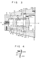
- Fig. 3 is a longitudinal cross sectional view of Fig. 2; and
- Fig. 4 is an oblique view of a plate spring used with the embodiment of Figs. 1 and 2.
- Referring to Fig. 1, an endoscope 1 comprises a substantially rectangular box-
shaped control section 2, atubular insertion section 3 projecting from one end of thecontrol section 2, an umbilical cord orconnection cord 4 which extends from one lateral wall of thecontrol section 2 and whose free end is fitted with a connector 4a coupled to an external light source (not shown), and a cylindricalocular section 5 connected to the other end of thecontrol section 2. The distal end of thetubular insertion section 3 is provided with an. observation window and an illumination window. The observation window is optically connected to theocular section 5 by means of an image-transmitting optical system extending through thecontrol section 2 andtubular insertion section 3. Through the illumination window is emitted an illumination light supplied from the external light source by means of an illumination optical guide extending through thecontrol section 2,tubular insertion section 3 andumbilical cord 4. The other parts of the endoscope of this invention are constructed in the same way as those of the conventional endoscope, description thereof being omitted. - With the endoscope of the invention, a
photographic camera 6, one of the ocular attachments, is detachably fitted to theocular section 5 of the endoscope 1 by a bayonet mechanism. This arrangement is detailed below. A hollowcylindrical adapter 7 is formed on the front wall of thephotographic camera 6. A plurality ofbayonet pawls 9 radially project from the inner peripheral wall of the foremost edge portion of the distal end opening 8 equidistantly in the circumferential direction. - Provided at the distal end of the
ocular section 5 is abayonet ring 10 having an outer diameter substantially equal to the inner diameter of theadapter 7. Bayonet grooves 11 engageable with thebayonet pawls 9 are formed in the outer peripheral wall of thebayonet ring 10 equidistantly in the circumferential direction. Where thephotographic camera 6 is fitted to theocular section 5, thebayonet grooves 11 receive thecorresponding bayonet pawls 9. In other words, thebayonet pawls 9 act as engagement members, and thebayonet ring 10 is used as a receptacle of the engagement members. - As shown in Fig. 3, an ocular 12 is set so as to face the proximal end of an
image guide 13 formed of an optical fiber bundle constituting part of the image-transmitting optical system. Anoperation ring 14 is rotatably set on the outer periphery of theocular section 5 close to thebayonet ring 10. Formed in the outer peripheral wall of theoperation ring 14 are axially extendingnotches 15 corresponding to thebayonet grooves 11. Where thenotches 15 are aligned with thecorresponding grooves 11 by rotating theoperation ring 14 in inserting theocular section 5 into theadapter 7 of thephotographic camera 6, thebayonet pawls 9 pass through thebayonet grooves 11 to be engaged with thecorresponding notches 15. As a result, thebayonet ring 10 is securely fitted into the opening 8 of theadapter 7. Later when theadapter 7 is rotated, theoperation ring 14 is turned in the same direction, causing thebayonet pawls 9 to be caught by thenotches 15 and the inner end of thebayonet ring 10, and enabling thephotographic camera 6 to be securely connected to theocular section 5. - Reverting to Fig. 3, a smaller diameter cylindrical hole 7a is formed close to the forward end of the
adapter 7. A larger diametercylindrical hole 7b is formed close to the rear end of theadapter 7 concentrically with said smaller diameter cylindrical hole 7a. Thecylindrical holes 7a, 7b are rendered coaxial with the optical axis X-X of the ocular 12. - A
coupling ring 16 comprises a hollow cylindrical body 16a slidably contacting the inner wall of the smaller diameter cylindrical hole 7a and aflange 18 formed concentrically with the hollow cylindrical body 16a at one end thereof and set in a larger diametercylindrical hole 7b. Anotch 24 is cut out in the front end portion of a hollow cylindrical body 16a. Anengagement pin 25 axially projecting from the front end face ordistal end face 17 of thebayonet ring 10 in the proximity of the outer periphery of thedistal end face 17 of thebayonet ring 10 of theocular section 5 is carried into thenotch 24 when thebayonet ring 10 is inserted into theadapter 7. Insertion of theengagement pin 25 into thenotch 24 prevents thecoupling ring 16 from being rotated relative to thebayonet ring 10. Thenotch 24 andengagement pin 25 jointly constitute means for preventing the relative rotation of thebayonet ring 10 andcoupling ring 16. - Securely inserted into the larger diameter
cylindrical hole 7b of theadapter 7 is a hollowcylindrical lens mount 22 fixed to the front wall of thephotographic camera 6 by set screws 22a. A space is provided between thefront end face 22b of thelens mount 22 and thefront end face 16b of thecoupling ring 16, thereby allowing thecoupling ring 16 to reciprocate axially of theadapter 7. Set in thelens mount 22 is an objective 21 which is rendered coaxial with an optical axis X-X of the ocular 12 when thephotographic camera 6 is fitted to theocular section 5. - In a space in the larger diameter
cylindrical hole 7b, elastically urging means is provided between thefront side 22b of thelens mount 22 and the correspondingfront end face 16b of thecoupling ring 16 normally to urge thecoupling ring 16 toward the front end of theadapter 7. Where theadapter 7 is fitted to theocular section 5, the rear end face 16c of thecoupling ring 16 elastically abuts against theend face 17 of theocular section 5 by the urging means, and clamps thebayonet ring 10 of theocular section 5 between thecoupling ring 16 andbayonet pawls 9. - The above-mentioned urging means comprises, for example, a plurality of
plate springs 20 shown in Figs. 3 and 4. Eachplate spring 20 comprises a base portion 20a securely set on thefront end face 22b of thelens mount 22 in parallel therewith by aset screw 23 and anarm 20b which extends from the base portion 20a toward thefront end face 16b of thecoupling ring 16 in an outward convexed form and whose free end portion contacts thefront end face 16b in parallel therewith. For example, threeplate springs 20 are provided (only two are indicated in Fig. 3). They are equidistantly set in the circumferential direction of thefront end face 22b of thelens mount 22 in such a manner that thearms 20b of therespective plate springs 20 are directed to the same circumferential sense, whereby the plate springs 20 axially urge thecoupling ring 16 with an equal force. - Description is now given of the process of fitting the
photographic camera 6 to theocular section 5 of the endoscope 1. First, thebayonet pawls 9 of theadapter 7 are aligned with thebayonet grooves 11, and thebayonet ring 10 is inserted into theadapter 7. As a result, thebayonet pawls 9 pass through thebayonet grooves 11 and are engaged with thenotches 15 of theoperation ring 14. Later, theadapter 7 is rotated to effect the joint rotation of thebayonet pawls 9 andoperation ring 14, thereby causing thebayonet pawls 9 to stop at the rear end of thebayonet ring 10. Where theocular section 5 is going to be connected to theadapter 7, the rear end face 16c of thecoupling ring 16 abuts against thedistal end face 17 of theocular section 5. Prior to said abutment, theengagement pin 25 of theocular section 5 is engaged with thenotch 24 of thecoupling ring 16. Therefore, thecoupling ring 16 andocular section 5 are assembled as if into a single body, thereby preventing thebayonet ring 10 and couplingring 16 from being rotated relative to each other even when theadapter 7 is later rotated. In other words, when sliding on the side of theadapter 7, thecoupling ring 16 does not rub thedistal end face 17 of theocular section 5. Thus, thedistal end face 17 is not mirror polished. - Further, where the
ocular section 5 is inserted into theadapter 7, thecoupling ring 16 is pressed by thedistal end face 17 of theocular section 5, and is retracted against the urging force of theplate spring 20, thus remaining in a pressed state in tight contact with theend face 17 of theocular section 5. Where thebayonet pawls 9 are stopped at the rear end of thebayonet ring 10 and thecoupling ring 16 is pressed against thebayonet ring 10 when connection is effected between theocular section 5 andcoupling ring 16, thephotographic camera 6 is securely supported by theocular section 5. - The opposite process to that of fitting the
photographic camera 6 to theocular section 5 enables theadapter 7 to be released from theocular section 5. In this case, too, thecoupling ring 16 andocular section 5 are engaged with each other, until thecoupling ring 16 is removed from theend face 17 of theocular section 5, thereby preventing thecoupling ring 16 andocular section 5 from being rotated relative to each other. The possibility is eliminated that thedistal end face 17 of theocular section 5 and the rear end face 16c of thecoupling ring 16 are rubbed against each other. - Where, with an endoscope embodying this invention, an endoscope attachment is detachably fitted to the ocular section of the endoscope, it is possible to suppress the appearance of an unnecessary gloss on the end face of the ocular section due to its rubbing. In other words, even where an endoscope is applied, for example, in a room where an illumination source is set, the front end face of the ocular section fitted with a photographic camera does not shine at all, thereby eliminating the obstruction of a coeliac observation.
- With the foregoing embodiment, a photographic camera was used as an attachment to the ocular section of the endoscope. However, this invention is obviously applicable to the case when any other attachment (for example, a teaching scope) is fitted to the ocular section of the endoscope.
Claims (6)
Priority Applications (1)
| Application Number | Priority Date | Filing Date | Title |
|---|---|---|---|
| AT80108034T ATE9615T1 (en) | 1979-12-24 | 1980-12-18 | DEVICE FOR ATTACHING AN ACCESSORIES TO A SECTION OF AN ENDOSCOPOCULAR. |
Applications Claiming Priority (2)
| Application Number | Priority Date | Filing Date | Title |
|---|---|---|---|
| JP16777579A JPS5689232A (en) | 1979-12-24 | 1979-12-24 | Endoscope apparatus |
| JP167775/79 | 1979-12-24 |
Publications (3)
| Publication Number | Publication Date |
|---|---|
| EP0031151A2 true EP0031151A2 (en) | 1981-07-01 |
| EP0031151A3 EP0031151A3 (en) | 1982-02-10 |
| EP0031151B1 EP0031151B1 (en) | 1984-09-26 |
Family
ID=15855871
Family Applications (1)
| Application Number | Title | Priority Date | Filing Date |
|---|---|---|---|
| EP80108034A Expired EP0031151B1 (en) | 1979-12-24 | 1980-12-18 | Device for fitting an attachment to an endoscope ocular section |
Country Status (5)
| Country | Link |
|---|---|
| US (1) | US4323304A (en) |
| EP (1) | EP0031151B1 (en) |
| JP (1) | JPS5689232A (en) |
| AT (1) | ATE9615T1 (en) |
| DE (1) | DE3069321D1 (en) |
Cited By (6)
| Publication number | Priority date | Publication date | Assignee | Title |
|---|---|---|---|---|
| US4600277A (en) * | 1984-02-13 | 1986-07-15 | Murray Jr Joseph E | Telescope with multiple image-viewing assemblies |
| WO1990013841A1 (en) * | 1989-05-01 | 1990-11-15 | MILLER, Patricia, Hermione (heir of MILLER, David, Dushko (deceased)) | Endoscope support system |
| US5249082A (en) * | 1991-05-08 | 1993-09-28 | Eastman Kodak Company | Exact constraint arrangement for and methods of mounting an element such as a lens |
| US5673143A (en) * | 1995-10-11 | 1997-09-30 | Hughes Electronics | Thermal imaging device with selectively replaceable telescopic lenses and automatic lens identification |
| EP2003482A1 (en) * | 2007-06-11 | 2008-12-17 | Swarovski-Optik KG | Adapter for connecting an optical recording device with an observation device |
| EP2674811A3 (en) * | 2012-06-11 | 2014-01-15 | Swarovski Optik Kg | Lens for an image recording apparatus |
Families Citing this family (29)
| Publication number | Priority date | Publication date | Assignee | Title |
|---|---|---|---|---|
| US4638792A (en) * | 1979-12-20 | 1987-01-27 | Burgin Kermit H | Adjustable speculum with incorporated lighting system |
| USD270643S (en) | 1981-03-17 | 1983-09-20 | Polaroid Corporation | Photographic camera or similar article |
| JPS5865133A (en) * | 1981-10-12 | 1983-04-18 | オリンパス光学工業株式会社 | Endoscope |
| GB2149932B (en) * | 1983-11-12 | 1986-11-19 | Warner Lambert Tech | Improved endoscope |
| JPH0514258Y2 (en) * | 1985-09-05 | 1993-04-16 | ||
| US4776668A (en) * | 1986-01-09 | 1988-10-11 | Machida, Inc. | Image focusing ocular piece for a viewing scope including mechanism for accommodating differential expansion |
| US4807594A (en) * | 1988-01-15 | 1989-02-28 | Medical Concepts, Incorporated | Adapter assembly for endoscopic video camera |
| US4844071A (en) * | 1988-03-31 | 1989-07-04 | Baxter Travenol Laboratories, Inc. | Endoscope coupler device |
| US5419313A (en) * | 1988-10-27 | 1995-05-30 | Rosemarie Lemke | Microwave sterilizable and reusable endoscope |
| US5205280A (en) * | 1990-12-21 | 1993-04-27 | Mp Video, Inc. | Quick-release endoscopic coupling assembly |
| US5846186A (en) * | 1996-09-24 | 1998-12-08 | Mercury Enterprises, Inc. | Endoscope system and coupling arrangement for use therewith |
| US6106457A (en) * | 1997-04-04 | 2000-08-22 | Welch Allyn, Inc. | Compact imaging instrument system |
| US6840903B2 (en) * | 2002-03-21 | 2005-01-11 | Nuvista Technology Corporation | Laryngoscope with image sensor |
| FR2857248B1 (en) * | 2003-07-07 | 2006-01-27 | Acep France | DEVICE FOR ENSURING THE EXAMINATION OF THE RETINA OF A PATIENT |
| US20060276690A1 (en) * | 2005-05-11 | 2006-12-07 | Farris Jesse L Iii | Visualization system |
| US20070179342A1 (en) * | 2006-01-12 | 2007-08-02 | Kb Port Llc | Wireless Laryngoscope with Internal Antennae and One Piece Construction Adapted for Laryngoscopy Training |
| US9622651B2 (en) | 2012-01-27 | 2017-04-18 | Kbport Llc | Wireless laryngoscope simulator with onboard event recording adapted for laryngoscopy training |
| DE102012008970B3 (en) * | 2012-05-03 | 2013-06-27 | Joimax Gmbh | Surgical tooling |
| JP5980559B2 (en) * | 2012-05-07 | 2016-08-31 | オリンパス株式会社 | camera |
| CN107260109B (en) * | 2013-08-08 | 2019-06-14 | 夏普株式会社 | In-vivo imaging device, support tube for in-vivo imaging device |
| JP2015052746A (en) * | 2013-09-09 | 2015-03-19 | ソニー株式会社 | Stereoscopic endoscope |
| US10264166B2 (en) * | 2013-10-31 | 2019-04-16 | Sharp Kabushiki Kaisha | Intracorporeal-monitoring camera system, support tube for intracorporeal-monitoring camera system, and cable holder for intracorporeal-monitoring camera system |
| DE102014222880A1 (en) * | 2014-11-10 | 2016-05-12 | Xion Gmbh | Adapter for endoscopy |
| JP6464104B2 (en) * | 2016-01-20 | 2019-02-06 | 富士フイルム株式会社 | Endoscope connector, endoscope and endoscope system |
| JP6527646B2 (en) * | 2017-05-26 | 2019-06-05 | オリンパス株式会社 | Optical unit |
| JP7086641B2 (en) * | 2018-02-28 | 2022-06-20 | ソニー・オリンパスメディカルソリューションズ株式会社 | Camera head and endoscope |
| DE102018130553A1 (en) | 2018-11-30 | 2020-06-04 | Henke-Sass, Wolf Gmbh | endoscope |
| US11766166B2 (en) * | 2021-06-21 | 2023-09-26 | Karl Storz Se & Co. Kg | Endoscope eyepiece grasping mechanism and endoscope camera head with endoscope eyepiece grasping mechanism |
| DE102021119204A1 (en) | 2021-07-23 | 2023-01-26 | Xion Gmbh | Camera head for endoscopes |
Family Cites Families (15)
| Publication number | Priority date | Publication date | Assignee | Title |
|---|---|---|---|---|
| GB598572A (en) * | 1945-07-07 | 1948-02-20 | Kershaw & Sons Ltd A | Improvements in or relating to securing means for mounting detachable components |
| US2500050A (en) * | 1948-01-07 | 1950-03-07 | Eastman Kodak Co | Camera lens attaching and clamping mechanism |
| DE861754C (en) * | 1948-03-12 | 1953-01-05 | Kern & Co Ag | Frame for photographic lenses with an aperture ring and a spacer ring |
| US2870674A (en) * | 1956-11-01 | 1959-01-27 | Cecil I Mize | Microscope |
| SU143572A1 (en) | 1961-04-25 | 1961-11-30 | А.П. Ломовцев | Photo set |
| US3680459A (en) * | 1970-11-06 | 1972-08-01 | Asahi Optical Co Ltd | Camera objective adjustable for flash and non-flash photography |
| GB1423836A (en) * | 1972-07-04 | 1976-02-04 | Makepeace A P W | Coupling together of an instrument for bodily examination and another instrument such as a camera |
| DE2257191A1 (en) * | 1972-11-22 | 1974-05-30 | Weber Josef | CAMERA LENS |
| JPS5127677A (en) * | 1974-08-30 | 1976-03-08 | Osaka Denki Co Ltd | INTAAROTSUKUSEIGYOSOCHI |
| JPS52144179A (en) * | 1976-05-27 | 1977-12-01 | Fuji Photo Optical Co Ltd | Device for operating endscope camera |
| JPS5922927B2 (en) * | 1977-02-04 | 1984-05-30 | キヤノン株式会社 | Camera accessory mounting device |
| US4188942A (en) * | 1978-02-27 | 1980-02-19 | American Hospital Supply Corporation | Endoscope and urological instrument lock |
| US4426135A (en) * | 1978-05-22 | 1984-01-17 | Shinano Kikaku Co., Ltd. | Lens attchment for forming wide-ranging images |
| US4157216A (en) * | 1978-06-26 | 1979-06-05 | Polaroid Corporation | Adapter for optically coupling a photographic camera with a viewing device |
| FR2453421A1 (en) * | 1979-04-06 | 1980-10-31 | Beaulieu Sa | Lens fixing for camera - in which dowel maintains correct angular alignment when screwing down captive collar nut on attachment |
-
1979
- 1979-12-24 JP JP16777579A patent/JPS5689232A/en active Pending
-
1980
- 1980-12-12 US US06/215,755 patent/US4323304A/en not_active Expired - Lifetime
- 1980-12-18 EP EP80108034A patent/EP0031151B1/en not_active Expired
- 1980-12-18 AT AT80108034T patent/ATE9615T1/en not_active IP Right Cessation
- 1980-12-18 DE DE8080108034T patent/DE3069321D1/en not_active Expired
Cited By (9)
| Publication number | Priority date | Publication date | Assignee | Title |
|---|---|---|---|---|
| US4600277A (en) * | 1984-02-13 | 1986-07-15 | Murray Jr Joseph E | Telescope with multiple image-viewing assemblies |
| WO1990013841A1 (en) * | 1989-05-01 | 1990-11-15 | MILLER, Patricia, Hermione (heir of MILLER, David, Dushko (deceased)) | Endoscope support system |
| US5249082A (en) * | 1991-05-08 | 1993-09-28 | Eastman Kodak Company | Exact constraint arrangement for and methods of mounting an element such as a lens |
| US5673143A (en) * | 1995-10-11 | 1997-09-30 | Hughes Electronics | Thermal imaging device with selectively replaceable telescopic lenses and automatic lens identification |
| EP2003482A1 (en) * | 2007-06-11 | 2008-12-17 | Swarovski-Optik KG | Adapter for connecting an optical recording device with an observation device |
| US7924515B2 (en) | 2007-06-11 | 2011-04-12 | Swarovski-Optik Kg. | Adapter for connecting an optical recording device to an observation device |
| CN101414042B (en) * | 2007-06-11 | 2014-05-07 | 施华洛世奇光学公司 | Adapter for connecting an optical recording device with an observation device |
| EP2674811A3 (en) * | 2012-06-11 | 2014-01-15 | Swarovski Optik Kg | Lens for an image recording apparatus |
| US9883087B2 (en) | 2012-06-11 | 2018-01-30 | Swarovski-Optik Kg. | Objective for an image recording device |
Also Published As
| Publication number | Publication date |
|---|---|
| JPS5689232A (en) | 1981-07-20 |
| EP0031151B1 (en) | 1984-09-26 |
| DE3069321D1 (en) | 1984-10-31 |
| EP0031151A3 (en) | 1982-02-10 |
| ATE9615T1 (en) | 1984-10-15 |
| US4323304A (en) | 1982-04-06 |
Similar Documents
| Publication | Publication Date | Title |
|---|---|---|
| EP0031151B1 (en) | Device for fitting an attachment to an endoscope ocular section | |
| US4643170A (en) | Endoscope apparatus | |
| JP3739592B2 (en) | Laparoscopic device | |
| US4727859A (en) | Right angle detachable prism assembly for borescope | |
| US5101807A (en) | Endoscope connecting apparatus | |
| US4534339A (en) | Endoscope | |
| JP5283722B2 (en) | Ultra-compact form factor single-core optical fiber interconnect system having push-push insertion / extraction mechanism, shutter modular connector and shutter adapter, and method of use thereof | |
| JP4328711B2 (en) | Optical connector plug and optical connector | |
| US6540668B1 (en) | Endoscope with a coupling device (video coupler) for connection of a video camera | |
| JP5000778B2 (en) | Optical connector plug | |
| US5264928A (en) | Universal adapter for mounting cameras to microscopes | |
| US5800343A (en) | Endoscope light guide connector allowing adjustment of the angle of incident light rays | |
| US4569334A (en) | Apparatus for restoring the light transmittance of an image-transmitting optical fiber bundle used in a fiber optic endoscope | |
| US6007255A (en) | Adapter assembly for light source | |
| EP0027651B1 (en) | Endoscope ocular section | |
| JPH0627393A (en) | Observation device | |
| US6522466B2 (en) | Apparatus for observing interior from an ultramicropore space | |
| JPH10178A (en) | Illumination light transmission system adapter for endoscope device | |
| JP4450912B2 (en) | Endoscope holding device | |
| CA1204973A (en) | Endoscope | |
| JPH05281439A (en) | Optical connector | |
| JPH10300985A (en) | Multi-core optical connector | |
| EP0392718B1 (en) | Optical connector for endoscope | |
| JPH0353831B2 (en) | ||
| JPH03224534A (en) | Connector device for endoscope |
Legal Events
| Date | Code | Title | Description |
|---|---|---|---|
| PUAI | Public reference made under article 153(3) epc to a published international application that has entered the european phase |
Free format text: ORIGINAL CODE: 0009012 |
|
| AK | Designated contracting states |
Designated state(s): AT BE CH DE FR GB IT NL SE |
|
| PUAL | Search report despatched |
Free format text: ORIGINAL CODE: 0009013 |
|
| RHK1 | Main classification (correction) |
Ipc: G02B 7/02 |
|
| AK | Designated contracting states |
Designated state(s): AT BE CH DE FR GB IT NL SE |
|
| 17P | Request for examination filed |
Effective date: 19820204 |
|
| GRAA | (expected) grant |
Free format text: ORIGINAL CODE: 0009210 |
|
| AK | Designated contracting states |
Designated state(s): AT BE CH DE FR GB IT LI NL SE |
|
| PG25 | Lapsed in a contracting state [announced via postgrant information from national office to epo] |
Ref country code: SE Effective date: 19840926 Ref country code: NL Effective date: 19840926 Ref country code: LI Effective date: 19840926 Ref country code: IT Free format text: LAPSE BECAUSE OF FAILURE TO SUBMIT A TRANSLATION OF THE DESCRIPTION OR TO PAY THE FEE WITHIN THE PRESCRIBED TIME-LIMIT;WARNING: LAPSES OF ITALIAN PATENTS WITH EFFECTIVE DATE BEFORE 2007 MAY HAVE OCCURRED AT ANY TIME BEFORE 2007. THE CORRECT EFFECTIVE DATE MAY BE DIFFERENT FROM THE ONE RECORDED. Effective date: 19840926 Ref country code: FR Free format text: THE PATENT HAS BEEN ANNULLED BY A DECISION OF A NATIONAL AUTHORITY Effective date: 19840926 Ref country code: CH Effective date: 19840926 Ref country code: BE Effective date: 19840926 Ref country code: AT Effective date: 19840926 |
|
| REF | Corresponds to: |
Ref document number: 9615 Country of ref document: AT Date of ref document: 19841015 Kind code of ref document: T |
|
| REF | Corresponds to: |
Ref document number: 3069321 Country of ref document: DE Date of ref document: 19841031 |
|
| REG | Reference to a national code |
Ref country code: CH Ref legal event code: PL |
|
| NLV1 | Nl: lapsed or annulled due to failure to fulfill the requirements of art. 29p and 29m of the patents act | ||
| GBPC | Gb: european patent ceased through non-payment of renewal fee | ||
| PLBE | No opposition filed within time limit |
Free format text: ORIGINAL CODE: 0009261 |
|
| STAA | Information on the status of an ep patent application or granted ep patent |
Free format text: STATUS: NO OPPOSITION FILED WITHIN TIME LIMIT |
|
| EN | Fr: translation not filed | ||
| 26N | No opposition filed | ||
| PG25 | Lapsed in a contracting state [announced via postgrant information from national office to epo] |
Ref country code: GB Effective date: 19881118 |
|
| PGFP | Annual fee paid to national office [announced via postgrant information from national office to epo] |
Ref country code: DE Payment date: 19951222 Year of fee payment: 16 |
|
| PG25 | Lapsed in a contracting state [announced via postgrant information from national office to epo] |
Ref country code: DE Effective date: 19970902 |