EP0062459A2 - Gerät zur Diagnose in der Orthopädie - Google Patents
Gerät zur Diagnose in der Orthopädie Download PDFInfo
- Publication number
- EP0062459A2 EP0062459A2 EP82301581A EP82301581A EP0062459A2 EP 0062459 A2 EP0062459 A2 EP 0062459A2 EP 82301581 A EP82301581 A EP 82301581A EP 82301581 A EP82301581 A EP 82301581A EP 0062459 A2 EP0062459 A2 EP 0062459A2
- Authority
- EP
- European Patent Office
- Prior art keywords
- frequency
- joint
- emission
- transducers
- emissions
- Prior art date
- Legal status (The legal status is an assumption and is not a legal conclusion. Google has not performed a legal analysis and makes no representation as to the accuracy of the status listed.)
- Granted
Links
Images


Classifications
-
- A—HUMAN NECESSITIES
- A61—MEDICAL OR VETERINARY SCIENCE; HYGIENE
- A61B—DIAGNOSIS; SURGERY; IDENTIFICATION
- A61B7/00—Instruments for auscultation
- A61B7/006—Detecting skeletal, cartilage or muscle noise
Definitions
- This invention concerns orthopaedic diagnostic procedures and apparatus therefor, and more particularly such procedures and apparatus for detecting and assessing pathological conditions in the joints.
- the present invention has been developed following a further, detailed and extensive investigation of the microphone-based acoustic techniques. The result of this investigation was to establish that such techniques could not be improved to the point where they formed the basis for a routine clinical procedure.
- an improved orthopeadic diagnostic procedure and apparatus provide for detecting, preferably with a piezoelectric accelerometer, from a moving joint vibration emission extending into the subsonic frequency range, and analysing the detected emission to determine whether it has a characteristic of predetermined form indicative of a pathological condition.
- the apparatus of Figure 1 comprises three piezoelectric accelerometers 10 having their outputs applied by way of individual preamplifiers 11 to respective tracks of a tape recorder 12.
- a further track of the recorder 12 can record the output of a microphone and/or goniometer 13 to provide a verbal commentary on the procedure and/or to denote the varying joint position.
- the tape recorder can apply its record outputs individually to a frequency analyser 14 having an auxiliary recorder 15.
- the tape recorder outputs can also, or the preamplifier outputs can directly, be applied in parallel to a multi-track printer 16.
- the form of apparatus of Figure 1 has been primarily suited to development and is not necessarily to be regarded as commercial clinical equipment. However, some of the features suiting development may also suit practical clinical requirements.
- the apparatus need not be used as a whole at any one time insofar as the components up to and including the tape recorder 12 can be transported to different locations in order to generate signal records for various subjects, and the remaining components used, with the tape recorder, or a duplicate thereof, thereafter for purposes of analysis at a central site.
- the accelerometers, and correspondingly the associated preamplifiers and multi-track tape recorder are employed in a multiple presence to allow simultaneous detection in mutually spaced positions adjacent to a joint and the consequent possibility of localisation of a condition within the joint. Such localisation can in fact be effected and this capability will be relevant to some clinical requirements.
- the accelerometers should be small and have suitable response characteristics in recognition of the findings (2) and (3) above. This presents no difficulty vis-a-vis available products. The same is true in respect of corresponding requirements in operational characteristic for the preamplifiers.
- the former mode was preferred because the cables connecting the accelerometers and preamplifiers will have a capacitive effect which can be matched with the accelerometers and allow the convenience of long cables without affecting the lower frequency sensitivity of the accelerometers. This choice also requires the corresponding use of a charge type preamplifier.
- tape recorder clearly involves a requirement for an appropriate multi-track facility and frequency response. A further requirement is the ability to replay at slow speeds to facilitate detailed analysis without loss of low frequency content. This is met by the use of a recorder which operates in the frequency modulation (FM) mode and in practice a B & K Type 7003 has been used.
- FM frequency modulation
- the frequency analyser desirably provides a range of capabilities including display of the vibration signals in original time-amplitude mode and in frequency spectrum form as by Fourier transform, and averaging.
- the analyser used was a B & K Type 2031 which provides these capabilities, with exponential or linear averaging being available, plus, among other things, an adjustable trigger mode whereby corresponding portions of a cyclic input of continuous or intermittent form can be automatically selected for processing, and an interface facility (IEC 625-1) allowing digital transfer with compatable peripheral equipment.
- the auxilliary recorder for the tape recorder was chosen as an X-Y printer, which in practice was of B & K Type 2308.
- the printer is required to respond satisfactorily to rapidly changing signals containing high frequency components in excess of 100 Hz and a review of possibly suitable multi-track types show ink-jet forms to be preferable in affording this capability while producing an instantly visible permanent record.
- An additional benefit is that such printers are in common clinical usage in association with electrocardiographs (ECG).
- ECG electrocardiographs
- Siemens-Elema Mingograf 34 has been employed.
- non-invasive application is found to provide results at least as good as invasive application, with signal components of interest being enhanced in the former case compared to the latter in many tests. Also, no material improvement was found to occur with use of a coupling medium. In the result non-invasive application directly to the skin with the accelerometers simply held in place by adhesive medical tape is satisfactory.
- results can be obtained which are consistent in a mutual sense, in comparison with traditional diagnostic procedures as deployed by experts, and in comparison with subsequent findings during surgery.
- results involve the provision of data which conforms to a consistent pattern for a given bone joint species, and which contains consistent irregularities identifying abnormalities when present in an individual joint of a species.
- the general pattern for a joint species is found to be one of slow waves on which faster impulses and transients of relatively low amplitude are superimposed. Irregularities occur as prominent impulses and transients and these coincide with conventional detection of palpable crepitus.
- the results can serve not only to detect and identify a pathological abnormality, but also to localise the same in the joint by comparison of the outputs from the accelerometers by spacing them around the joint.
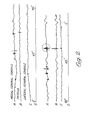
- Figure 2 illustrates such as just discussed in relation to the knee joint with a medial meniscal tear.
- This Figure is in four parts A to D of which the first three indicate the outputs from the accelerometers when located across the joint in the coronal plane adjacent the medial femoral condyle, the patella, and the tibial femoral condyle, respectively.
- the fourth part indicates movement of the joint from 0 to 90° flexion and back again. Irregularities of particular interest are the encircled transients which occur at about 30° flexion during the return movement, which transients indicate the meniscal tear and, by their relative magnitudes, localise the tear in the medial meniscus.
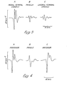
- Figure 3 illustrates similar results for a medial meniscal tear in another patient, only the transients, A, B and C being shown in this case.
- Figure 4 concerns the same patient as Figure 3 and shows transients A, B and C derived from the accelerometers when located to the posterior, middle and anterior of the medial tibial condyle in the transverse plane. These results localised the tear in the anterior portion of the meniscus by virtue of higher amplitude in transient C.
- the present invention is considered to be applicable to this last purpose.
- multiple accelerometers have been sited in various locations around the infant pelvis, in contact with the sacrum and sacro-iliac areas, and also the anterior superior iliac spines and symphysis pubis.
- the former locations can be accomodated by housing the accelerometers in a board on which the infant is placed.
- Signal outputs followed a consistent pattern with dislocation of the joint being indicated by a low frequency displacement signal on which a high frequency component was superimposed at completion of the actual dislocation. This was followed by slow wave components of very low frequency and amplitude representing the dislocated femoral head in the pseudo-acetabular area.
- FIG. 5 shows typical signal outputs during dislocation and reduction at A and B, respectively.
- Frequency analysis shows the peak frequency at the sacrum to be in the region of 5 Hz for both events, while that on the anterior superior iliac spines is about double.
- the higher frequency components are represented at lower power levels on the side of the lesion under observation. It is probable that, as with knee joint lesions, a driving frequency and a natural frequency are present with the latter predominating further from the lesion.
- the illustrated apparatus is constrained in usage by the fact that the frequency analyser must be used sequentially on different accelerometer signals, whereas a likely commercial development can involve simultaneous analysis by way of a microprocessor adapted to operate on the basis of comparison of results with predetermined patterns and parameters for a given joint species or condition.
Landscapes
- Health & Medical Sciences (AREA)
- Life Sciences & Earth Sciences (AREA)
- Medical Informatics (AREA)
- Engineering & Computer Science (AREA)
- Biomedical Technology (AREA)
- Heart & Thoracic Surgery (AREA)
- Rheumatology (AREA)
- Molecular Biology (AREA)
- Surgery (AREA)
- Animal Behavior & Ethology (AREA)
- General Health & Medical Sciences (AREA)
- Public Health (AREA)
- Veterinary Medicine (AREA)
- Measurement Of The Respiration, Hearing Ability, Form, And Blood Characteristics Of Living Organisms (AREA)
Applications Claiming Priority (2)
| Application Number | Priority Date | Filing Date | Title |
|---|---|---|---|
| GB8110528 | 1981-04-03 | ||
| GB8110528 | 1981-04-03 |
Publications (3)
| Publication Number | Publication Date |
|---|---|
| EP0062459A2 true EP0062459A2 (de) | 1982-10-13 |
| EP0062459A3 EP0062459A3 (en) | 1983-09-21 |
| EP0062459B1 EP0062459B1 (de) | 1986-12-30 |
Family
ID=10520909
Family Applications (1)
| Application Number | Title | Priority Date | Filing Date |
|---|---|---|---|
| EP82301581A Expired EP0062459B1 (de) | 1981-04-03 | 1982-03-25 | Gerät zur Diagnose in der Orthopädie |
Country Status (4)
| Country | Link |
|---|---|
| US (1) | US4437473A (de) |
| EP (1) | EP0062459B1 (de) |
| DE (1) | DE3274793D1 (de) |
| GB (1) | GB2096319B (de) |
Cited By (7)
| Publication number | Priority date | Publication date | Assignee | Title |
|---|---|---|---|---|
| GB2156983A (en) * | 1983-08-26 | 1985-10-16 | Mintowt Czyz Witek | Method and apparatus for assessing the structure and mechanical integrity of osseous systems |
| DE3830412A1 (de) * | 1988-09-07 | 1990-03-15 | Gw Elektronik Gmbh | Diagnosegeraet zum akustischen erfassen der lokalen geraeuschbildung im menschlichen oder tierischen koerper |
| WO1996029007A1 (en) * | 1995-03-21 | 1996-09-26 | David John Walker | Activity recording device |
| GB2313672A (en) * | 1995-03-21 | 1997-12-03 | David John Walker | Activity recording device |
| WO2010128423A1 (en) * | 2009-05-04 | 2010-11-11 | Koninklijke Philips Electronics N.V. | Method and system for analyzing motion data |
| US9849216B2 (en) | 2006-03-03 | 2017-12-26 | Smith & Nephew, Inc. | Systems and methods for delivering a medicament |
| US10357292B2 (en) | 2008-10-15 | 2019-07-23 | Smith & Nephew, Inc. | Composite internal fixators |
Families Citing this family (27)
| Publication number | Priority date | Publication date | Assignee | Title |
|---|---|---|---|---|
| US4748987A (en) * | 1984-02-21 | 1988-06-07 | The University Of Michigan | Acoustic myography |
| US4836218A (en) * | 1984-04-09 | 1989-06-06 | Arthrotek, Inc. | Method and apparatus for the acoustic detection and analysis of joint disorders |
| WO1985004563A1 (en) * | 1984-04-09 | 1985-10-24 | Thomas Gay | Method for the acoustic detection and analysis of joint disorders |
| JPS61501823A (ja) * | 1984-04-09 | 1986-08-28 | ゲイ,ト−マス | 関節異常の音響検出及び分析のための装置 |
| US4986281A (en) * | 1984-08-23 | 1991-01-22 | Starkey Laboratories, Inc. | Method for obtaining a signal for analyzing human and animal joint functions |
| US4817628A (en) * | 1985-10-18 | 1989-04-04 | David L. Zealear | System and method for evaluating neurological function controlling muscular movements |
| JPS62117553A (ja) * | 1985-11-16 | 1987-05-29 | 肥後 矢吉 | 機能評価装置 |
| US5154080A (en) * | 1986-10-29 | 1992-10-13 | Westinghouse Electric Corp. | Integrated check valve testing system |
| GB8724687D0 (en) * | 1987-10-21 | 1987-11-25 | Campbell F C | Surface vibration analysis |
| US4823807A (en) * | 1988-02-11 | 1989-04-25 | Board Of Regents, Univ. Of Texas System | Device for non-invasive diagnosis and monitoring of articular and periarticular pathology |
| US5031637A (en) * | 1990-08-17 | 1991-07-16 | Parra Jorge M | Non-invasive diagnostic method and apparatus |
| US5309922A (en) * | 1992-09-21 | 1994-05-10 | Center For Innovative Technology | Respiratory sound analyzer for use in high noise environments |
| US5396891A (en) * | 1993-04-07 | 1995-03-14 | Osteo Sciences Corporation | System and method for external acoustic bone velocity measurement |
| US5720290A (en) * | 1993-04-07 | 1998-02-24 | Metra Biosystems, Inc. | Apparatus and method for acoustic analysis of bone using optimized functions of spectral and temporal signal components |
| US5413116A (en) * | 1993-06-24 | 1995-05-09 | Bioresearch | Method and apparatus for diagnosing joints |
| DE4422451B4 (de) * | 1993-07-01 | 2007-05-31 | Schwalbe, Hans-Joachim, Prof. Dr. | Orthopädisches Untersuchungsverfahren |
| US5402800A (en) * | 1993-08-02 | 1995-04-04 | Hollis; J. Marcus | Ankle laxity measurement system |
| US6024711A (en) | 1998-03-12 | 2000-02-15 | The University Of British Columbia | Diagnosis of osteoporosis using acoustic emissions |
| DE10018654A1 (de) * | 2000-04-14 | 2001-12-13 | Jacek Czernicki | Verfahren und Vorrichtung zur Ermittlung des anatomischen Zustands eines Menschen oder eines Tieres, insbesondere zur Diagnose von Osteoporose |
| CA2325236A1 (en) | 2000-11-06 | 2002-05-06 | Cyril B. Frank | Auditory display of knee joint vibration signals |
| WO2005023093A2 (en) * | 2003-09-05 | 2005-03-17 | William Marsh Rice University | Noninvasive tissue assessment |
| AU2005304613A1 (en) * | 2004-11-12 | 2006-05-18 | Andrew H. Elser | Equine wireless physiological monitoring system |
| US8486070B2 (en) | 2005-08-23 | 2013-07-16 | Smith & Nephew, Inc. | Telemetric orthopaedic implant |
| US7879043B2 (en) * | 2006-11-28 | 2011-02-01 | Robert Michael Meneghini | System and method for preventing intraoperative fracture in cementless hip arthroplasty |
| US9445720B2 (en) | 2007-02-23 | 2016-09-20 | Smith & Nephew, Inc. | Processing sensed accelerometer data for determination of bone healing |
| WO2009032969A1 (en) | 2007-09-06 | 2009-03-12 | Smith & Nephew, Inc. | System and method for communicating with a telemetric implant |
| CN104019890B (zh) * | 2014-06-05 | 2016-12-07 | 无锡智感星际科技有限公司 | 一种次声波检测方法及其检测装置 |
Family Cites Families (10)
| Publication number | Priority date | Publication date | Assignee | Title |
|---|---|---|---|---|
| US3181528A (en) * | 1961-02-16 | 1965-05-04 | Roy E Brackin | Process and apparatus for analyzing joint disorders |
| AT276808B (de) * | 1966-07-01 | 1969-12-10 | Vibro Meter Ag | Anordnung zur Messung mechanischer Großen mit Hilfe von piezoelektrischen Meßwertwandlern |
| GB1319054A (en) | 1969-05-28 | 1973-05-31 | Ransome Hoffmann Pollard | Vibration measuring equipment |
| GB1274687A (en) | 1969-06-24 | 1972-05-17 | Secr Defence | Machinery vibration monitor |
| US3786285A (en) * | 1972-12-08 | 1974-01-15 | R Reibold | Moment actuated transducer |
| FR2224742A1 (en) * | 1973-04-03 | 1974-10-31 | Laguerre Rene | Human physiological difference tester - uses comparator for comparing physical data |
| US3985121A (en) | 1975-05-20 | 1976-10-12 | International Telephone And Telegraph Corporation | Heart detective |
| US4008711A (en) | 1975-06-30 | 1977-02-22 | Charles P. Olinger | Method and apparatus for non-invasive detection of intracranial aneurysms |
| US4144877A (en) | 1976-08-12 | 1979-03-20 | Yeda Research And Development Co. Ltd. | Instrument for viscoelastic measurement |
| US4444205A (en) * | 1980-05-31 | 1984-04-24 | University Of Strathclyde | Apparatus for assessing joint mobility |
-
1982
- 1982-03-25 DE DE8282301581T patent/DE3274793D1/de not_active Expired
- 1982-03-25 GB GB8208840A patent/GB2096319B/en not_active Expired
- 1982-03-25 EP EP82301581A patent/EP0062459B1/de not_active Expired
- 1982-03-26 US US06/362,516 patent/US4437473A/en not_active Expired - Lifetime
Cited By (9)
| Publication number | Priority date | Publication date | Assignee | Title |
|---|---|---|---|---|
| GB2156983A (en) * | 1983-08-26 | 1985-10-16 | Mintowt Czyz Witek | Method and apparatus for assessing the structure and mechanical integrity of osseous systems |
| DE3830412A1 (de) * | 1988-09-07 | 1990-03-15 | Gw Elektronik Gmbh | Diagnosegeraet zum akustischen erfassen der lokalen geraeuschbildung im menschlichen oder tierischen koerper |
| WO1996029007A1 (en) * | 1995-03-21 | 1996-09-26 | David John Walker | Activity recording device |
| GB2313672A (en) * | 1995-03-21 | 1997-12-03 | David John Walker | Activity recording device |
| GB2313672B (en) * | 1995-03-21 | 1999-09-15 | David John Walker | Activity recording device |
| US9849216B2 (en) | 2006-03-03 | 2017-12-26 | Smith & Nephew, Inc. | Systems and methods for delivering a medicament |
| US10357292B2 (en) | 2008-10-15 | 2019-07-23 | Smith & Nephew, Inc. | Composite internal fixators |
| US11096726B2 (en) | 2008-10-15 | 2021-08-24 | Smith & Nephew, Inc. | Composite internal fixators |
| WO2010128423A1 (en) * | 2009-05-04 | 2010-11-11 | Koninklijke Philips Electronics N.V. | Method and system for analyzing motion data |
Also Published As
| Publication number | Publication date |
|---|---|
| EP0062459A3 (en) | 1983-09-21 |
| US4437473A (en) | 1984-03-20 |
| GB2096319B (en) | 1986-02-19 |
| EP0062459B1 (de) | 1986-12-30 |
| DE3274793D1 (en) | 1987-02-05 |
| GB2096319A (en) | 1982-10-13 |
Similar Documents
| Publication | Publication Date | Title |
|---|---|---|
| US4437473A (en) | Orthopedic diagnostic procedures and apparatus therefor | |
| McCoy et al. | Vibration arthrography as a diagnostic aid in diseases of the knee. A preliminary report | |
| Chu et al. | Detection of knee joint diseases using acoustical pattern recognition technique | |
| Oster et al. | Characterization of temporomandibular joint sounds: a preliminary investigation with arthrographic correlation | |
| US4154231A (en) | System for non-invasive cardiac diagnosis | |
| Frank et al. | Analysis of knee joint sound signals for non-invasive diagnosis of cartilage pathology | |
| Maussavi et al. | Screening of vibroarthrographic signals via adaptive segmentation and linear prediction modeling | |
| EP0444594B1 (de) | Verfahren und Gerät zur Anzeige und Auswertung der Schluckbewegung eines Patienten | |
| Mollan et al. | A critical appraisal of auscultation of human joints. | |
| JPH05505954A (ja) | 心筋虚血検出システム | |
| Gay et al. | The acoustical characteristics of the normal temporomandibular joint | |
| KERNOHAN et al. | The diagnostic potential of vibration arthrography | |
| WO2009109917A1 (en) | Non invasive analysis of body sounds | |
| Zhang et al. | Adaptive cancellation of muscle contraction interference in vibroarthrographic signals | |
| Hermeling et al. | Confluence of incident and reflected waves interferes with systolic foot detection of the carotid artery distension waveform | |
| Shen et al. | Localization of knee joint cartilage pathology by multichannel vibroarthrography | |
| Murphy et al. | Chest auscultation in the diagnosis of pulmonary asbestosis | |
| Mollan et al. | Artefact encountered by the vibration detection system | |
| Cao et al. | Earsteth: Cardiac auscultation audio reconstruction using earbuds | |
| Vatolik et al. | Development of a multi-modal sensor network to detect and monitor knee joint condition | |
| White et al. | Analysing heart murmurs using time-frequency methods | |
| Sarkady et al. | Computer analysis techniques for phonocardiogram diagnosis | |
| Widmalm et al. | The frequency range of TMJ sounds | |
| Bohadana et al. | Contour maps of auscultatory percussion in healthy subjects and patients with large intrapulmonary lesions | |
| Hughes et al. | Continuous measurement of aortic radius change in vivo with an intra-aortic ultrasonic catheter |
Legal Events
| Date | Code | Title | Description |
|---|---|---|---|
| PUAI | Public reference made under article 153(3) epc to a published international application that has entered the european phase |
Free format text: ORIGINAL CODE: 0009012 |
|
| AK | Designated contracting states |
Designated state(s): CH DE FR LI |
|
| 17P | Request for examination filed |
Effective date: 19830329 |
|
| PUAL | Search report despatched |
Free format text: ORIGINAL CODE: 0009013 |
|
| AK | Designated contracting states |
Designated state(s): CH DE FR LI |
|
| GRAA | (expected) grant |
Free format text: ORIGINAL CODE: 0009210 |
|
| AK | Designated contracting states |
Kind code of ref document: B1 Designated state(s): CH DE FR LI |
|
| ET | Fr: translation filed | ||
| REF | Corresponds to: |
Ref document number: 3274793 Country of ref document: DE Date of ref document: 19870205 |
|
| PLBE | No opposition filed within time limit |
Free format text: ORIGINAL CODE: 0009261 |
|
| STAA | Information on the status of an ep patent application or granted ep patent |
Free format text: STATUS: NO OPPOSITION FILED WITHIN TIME LIMIT |
|
| 26N | No opposition filed | ||
| REG | Reference to a national code |
Ref country code: FR Ref legal event code: TP |
|
| REG | Reference to a national code |
Ref country code: CH Ref legal event code: PUE Owner name: BRITISH TECHNOLOGY GROUP LTD |
|
| PGFP | Annual fee paid to national office [announced via postgrant information from national office to epo] |
Ref country code: FR Payment date: 19960222 Year of fee payment: 15 |
|
| PGFP | Annual fee paid to national office [announced via postgrant information from national office to epo] |
Ref country code: DE Payment date: 19960530 Year of fee payment: 15 |
|
| PGFP | Annual fee paid to national office [announced via postgrant information from national office to epo] |
Ref country code: CH Payment date: 19970326 Year of fee payment: 16 |
|
| PG25 | Lapsed in a contracting state [announced via postgrant information from national office to epo] |
Ref country code: FR Free format text: LAPSE BECAUSE OF NON-PAYMENT OF DUE FEES Effective date: 19971128 |
|
| PG25 | Lapsed in a contracting state [announced via postgrant information from national office to epo] |
Ref country code: DE Effective date: 19971202 |
|
| REG | Reference to a national code |
Ref country code: FR Ref legal event code: ST |
|
| PG25 | Lapsed in a contracting state [announced via postgrant information from national office to epo] |
Ref country code: LI Free format text: LAPSE BECAUSE OF NON-PAYMENT OF DUE FEES Effective date: 19980331 Ref country code: CH Free format text: LAPSE BECAUSE OF NON-PAYMENT OF DUE FEES Effective date: 19980331 |
|
| REG | Reference to a national code |
Ref country code: CH Ref legal event code: PL |