EP0073247B1 - Colorants a l'argent pour les proteines contenues dans des gels - Google Patents
Colorants a l'argent pour les proteines contenues dans des gels Download PDFInfo
- Publication number
- EP0073247B1 EP0073247B1 EP82901207A EP82901207A EP0073247B1 EP 0073247 B1 EP0073247 B1 EP 0073247B1 EP 82901207 A EP82901207 A EP 82901207A EP 82901207 A EP82901207 A EP 82901207A EP 0073247 B1 EP0073247 B1 EP 0073247B1
- Authority
- EP
- European Patent Office
- Prior art keywords
- polypeptide
- gel
- stain
- polypeptides
- silver
- Prior art date
- Legal status (The legal status is an assumption and is not a legal conclusion. Google has not performed a legal analysis and makes no representation as to the accuracy of the status listed.)
- Expired
Links
Images






Classifications
-
- G—PHYSICS
- G01—MEASURING; TESTING
- G01N—INVESTIGATING OR ANALYSING MATERIALS BY DETERMINING THEIR CHEMICAL OR PHYSICAL PROPERTIES
- G01N33/00—Investigating or analysing materials by specific methods not covered by groups G01N1/00 - G01N31/00
- G01N33/48—Biological material, e.g. blood, urine; Haemocytometers
- G01N33/50—Chemical analysis of biological material, e.g. blood, urine; Testing involving biospecific ligand binding methods; Immunological testing
- G01N33/68—Chemical analysis of biological material, e.g. blood, urine; Testing involving biospecific ligand binding methods; Immunological testing involving proteins, peptides or amino acids
- G01N33/6803—General methods of protein analysis not limited to specific proteins or families of proteins
- G01N33/6827—Total protein determination, e.g. albumin in urine
- G01N33/683—Total protein determination, e.g. albumin in urine involving metal ions
- G01N33/6836—Silver staining
-
- G—PHYSICS
- G01—MEASURING; TESTING
- G01N—INVESTIGATING OR ANALYSING MATERIALS BY DETERMINING THEIR CHEMICAL OR PHYSICAL PROPERTIES
- G01N27/00—Investigating or analysing materials by the use of electric, electrochemical, or magnetic means
- G01N27/26—Investigating or analysing materials by the use of electric, electrochemical, or magnetic means by investigating electrochemical variables; by using electrolysis or electrophoresis
- G01N27/416—Systems
- G01N27/447—Systems using electrophoresis
- G01N27/44704—Details; Accessories
- G01N27/44717—Arrangements for investigating the separated zones, e.g. localising zones
- G01N27/44721—Arrangements for investigating the separated zones, e.g. localising zones by optical means
- G01N27/44726—Arrangements for investigating the separated zones, e.g. localising zones by optical means using specific dyes, markers or binding molecules
-
- Y—GENERAL TAGGING OF NEW TECHNOLOGICAL DEVELOPMENTS; GENERAL TAGGING OF CROSS-SECTIONAL TECHNOLOGIES SPANNING OVER SEVERAL SECTIONS OF THE IPC; TECHNICAL SUBJECTS COVERED BY FORMER USPC CROSS-REFERENCE ART COLLECTIONS [XRACs] AND DIGESTS
- Y10—TECHNICAL SUBJECTS COVERED BY FORMER USPC
- Y10S—TECHNICAL SUBJECTS COVERED BY FORMER USPC CROSS-REFERENCE ART COLLECTIONS [XRACs] AND DIGESTS
- Y10S436/00—Chemistry: analytical and immunological testing
- Y10S436/905—Photochemical activation of reactions
-
- Y—GENERAL TAGGING OF NEW TECHNOLOGICAL DEVELOPMENTS; GENERAL TAGGING OF CROSS-SECTIONAL TECHNOLOGIES SPANNING OVER SEVERAL SECTIONS OF THE IPC; TECHNICAL SUBJECTS COVERED BY FORMER USPC CROSS-REFERENCE ART COLLECTIONS [XRACs] AND DIGESTS
- Y10—TECHNICAL SUBJECTS COVERED BY FORMER USPC
- Y10T—TECHNICAL SUBJECTS COVERED BY FORMER US CLASSIFICATION
- Y10T436/00—Chemistry: analytical and immunological testing
- Y10T436/25—Chemistry: analytical and immunological testing including sample preparation
Definitions
- This invention relates to improved ultra-sensitive metallic silver stains for polypeptides, especially when fixed in synthetic gels, particularly polyacrylamide gels.
- polypeptides Detection and characterization of polypeptides is of fundamental importance to many areas of biology and clinical medicine. In some endeavors, such as genetic screening for mutational events, monitoring for pathophysiologic changes in disease states, and the diagnosis of genetic diseases, the efficiency of the search is directly proportional to the number of polypeptides that can be detected and characterized in cellular extracts of body fluids. Additionally polypeptides, hormones, etc., that are present in trace amounts are often of great importance for various medical reasons.
- Electrophoresis (defined generally as the movement of charged particles in solution under the influence of an electrical field), is a primary laboratory detection and characterization technique, especially useful for polypeptides and other macromolecules, such as nucleic acids. It is also useful in separating small particles such as viruses, cells, sub-cellular organelles and organic molecules such as steroids and amino acids.
- Coomassie Blue The most commonly used conventional polypeptide stain is Coomassie Blue, which may be considered as a prototype. Dyes of this type are mainly dependent upon the electrostatic attraction between dye and polypeptide, stabilized by Van der Wall's forces. In fact, Coomassie Blue and a variety of other dyes exhibit particular affinities for polypeptides of specific charge. Coomassie Blue, an acidic dye, stains basic polypeptides most intensely, while crystal violet is the most effective stain for acidic polypeptides. Other dyes for which quantitative aspects of staining have been investigated include Amido Black, Fast Green, and Fe2+-bathophenanthroline sulfonate. In contrast, the Remazol Brilliant Blue R method depends on a covalent bond between dye and polypeptide.
- MDPF (2-methoxy-2-4-diphenyl-3(2H)furanone) may be used to label polypeptides fluorescently prior to electrophoresis, with linearity from 10 ng to 10 pg of protein. However fluorescent staining of polypeptides prior to electrophoresis may alter their electrophoretic patterns.
- Radioactive detection techniques offer a higher degree of sensitivity than the stains but are often impractical to use. /n vivo radiolabelling may alter cellular metabolism and it may be impossible to label certain human polypeptides. /n vitro radiolabelling has the disadvantage that it might alter the electrophoretic mobility of polypeptides. Furthermore, radioactive reagents sometimes prove too expensive and long exposure to detect trace polypeptide may result irrthe problem of "autoradiographic spreading".
- Polypeptides labelled with a radioactive precursor may be detected by autoradiography and/or fluorography, which have been standardized and used quantitatively.
- a set of radiographic standards placed next to the gel during exposure of the film may facilitate quantification.
- Fluorography requires impregnation of the gel with a scintillation fluor and is of greatest use when a low energy beta emitter has been used for labelling or when an increase in sensitivity of detection is required.
- Quantitative use of fluorography requires prefogging of the film. Recently, in vitro methods for chemically radiolabelling polypeptides prior to electrophoresis have been described.
- Kerényi and Gallyas in "A Highly Sensitive Method for Demonstrating Proteins in Electrophoretic, Immuno-electrophoretic and Immuno-diffusion Preparations", Clin. Chim. Acta, 38, 465-467 (1972) discloses a silver stain for proteins in agar gel in which the gel is immersed in potassium ferrocyanide, and then in a two solution developer containing sodium carbonate and water in the first solution and ammonium nitrate, silver nitrate, tungsto-silicic acid, and formaldehyde in the second solution. The possibility of using polyacrylamide gel is mentioned.
- Veerheecke in "Agar gel Electrophoresis of Unconcentrated Cerebrospinal Fluid", J. Neurol., 209, 59 ⁇ 63 (1975) discloses silver staining in agar gel utilizing two solutions after immersion of the protein-containing gel in potassium ferrocyanide.
- the first solution contains sodium carbonate in water
- the second solution contains ammonium nitrate, water, formaldehyde, and tungsto-silicic acid as well as silver nitrate.
- the results reported are mixed, although generally favorable.
- Switzer, Merril and Shifrin in "A Highly Sensitive Silver Stain for Detecting Proteins and Peptides in Polyacrylamide Gels", Anal. Biochem, 98, 231-237 (1979), discloses a silver stain in which the proteins are fixed by soaking of the gel in various methanol/acetic acid mixtures for at least 2.5 hours, soaking the gel in a paraformaldelyde solution for .5 hours, placing the gel in a cupric nitrate/silver nitrate solution for at least .5 hours, placing the gel in a diammine solution (a mixture of silver nitrate, NaOH, NH 4 0H, and ethanol) for 10 min, and twice reducing the gel stain with formaldehyde and citric acid.
- the stain was found to be 100 times more sensitive than Coomassie Blue and comparable to autoradiography.
- the disclosed process utilizes (1) soaking the gel in glutaraldehyde for 30 minutes, (2) rinsing and soaking the gel in water for at least 2 hours, (3) adding ammoniacal silver solution (a mixture of NH 4 0H, NAOH, and AgN0 3 ), (4) transferring the gel to a mixture of citric acid and formaldehyde, and (6) washing in water for at least 1 hour.
- ammoniacal silver solution a mixture of NH 4 0H, NAOH, and AgN0 3
- (4) transferring the gel to a mixture of citric acid and formaldehyde and (6) washing in water for at least 1 hour.
- U.S. 3,873,433 does not disclose silver stains, but does disclose protein staining by the formation of complex organic salts with bivalent elements such as calcium and magnesium.
- U.S. 4,167,467 does not disclose silver stains, but does disclose the quantification of lipoprotein free cholesterols using a cholesterol oxidase substrate by enzymatic determination.
- This invention provides improved silver stain methods capable of detecting as little as .01 ng/mm 2 of polypeptide.
- the methods are analogous to photographic chemistry and comprise the sequential steps of: (1) fixing the polypeptide; (2) photo-reversal using an oxidant; (3) latent image formation, preferably with silver nitrate; and (4) image development to afford a visible stain.
- the improved silver stain methods of this invention find utility in: clinical and laboratory examination of blood-derived and other polypeptides separated by electrophoresis; prenatal diagnosis of fetal abnormalities by examination of amniotic fluid; diagnosis of central nervous system diseases by analysis of polypeptide patterns in spinal fluid; visualization and quantitation of polypeptides hormones, and analysis of cellular protein patterns for physiological and/or patho-physiological studies; and many other applications in which the detection and/or quantitation of proteins or polypeptides is desired.
- Typical gels are polyacrylamide, agarose, and cellulose acetate.
- Polyacrylamide is preferred, because both cellulose acetate and agarose gels when used with the method of this invention give brownish backgrounds (agarose being somewhat darker). If the brownish backgrounds can be avoided, all of these gels would be suitable.
- stain methods of this invention find their primary utility in connection with staining polypeptides on gels
- the same stain methods (sometimes without the fixing step) are also useful in conducting protein assays for polypeptides filtered from source materials and for histological-type stains of tissue slices such as in biopsies.
- the improved silver stain method of this invention comprises the four basic sequential steps of (1) washing and fixing, (2) photo-reversal, (3) latent image formation, and (4) image development.
- the invention method may be used in connection with gel electrophoresis, polypeptide assays, and tissue staining. Because of the sensitivity of the silver stains of this invention, the method is most useful in conjunction with electrophoresis, especially two-dimensional electrophoresis.
- the method of this invention is as follows.
- any of the conventional fixatives may be used, for example, glutaraldehyde, oxides of heavy metals such as mercury, lead, and osmium, formaldehyde, paraformaldehyde, trichloroacetic acid, and acetic acid.
- glutaraldehyde and heavy metal oxides pose health hazards and are therefore least preferred.
- Trichloroacetic acid (10%), acetic acid, formaldehyde, or paraformaldehyde are preferred fixatives, but all of these must be washed out of the gel prior to staining, usually with methanol or ethanol.
- fixative is an aqueous mixture of ethanol and/or methanol (about 10-50%) and acetic acid (about 5-20%), with about 10% ethanol and about 15% acetic acid being optimum.
- the effect of fixatives is not accurately known.
- the above fixing agents have been employed in both histology and protein electrophoresis for years. Without current quantitative methods utilizing external and internal standard proteins with the gels, it is at least known that the relative concentrations of proteins remain constant with the above fixatives. Typically, the gels were fixed by immersion in the fixative for a period of about 5-15 minutes, and immersion in fresh fixer was then repeated up to 3 times.
- rinsing When using a fixative other than the acetic acid-methanol and/or ethanol mixture, this was following by rinsing, preferably with methanol and/or ethanol. Where the most preferred fixative of an acetic acid-methanol and/or ethanol mixture is used, rinsing is not necessary.
- the polypeptide-gel is treated with an oxidizing agent to effect "photoreversal" and avoid silver staining of non-polypeptides.
- Suitable agents include dichromates, potassium permanganate, molecular oxygen, potassium ferricyanide, iodine, and quinone, of which acid dichromates, potassium dichromate and molecular oxygen are preferred. Potassium dichromate is particularly preferred. Potassium ferricyanide was originally tried in this invention, but it was discovered to lack sensitivity. Empirical testing indicated that .03 M potassium dichromate yielded the best results.
- this step is effected by soaking the fixed polypeptide-gel for about 3-10 minutes (preferably about 5 minutes), preferably in an aqueous solution of potassium dichromate and a small amount of nitric acid.
- a solution of about .03 M potassium dichromate and about .0015-.03 (preferably about .0016) N nitric acid is not preferred.
- this step may be followed by rapidly rinsing the gel in deionized water.
- the polypeptide-gel is immersed in a photosensitive salt solution, and optionally subjected to actuating irradiation.
- a photosensitive salt solution silver, gold, platinum, palladium and/or iridium salts may be used, although silver salts are preferred because they are more available and less costly.
- Silver nitrate is the most preferred salt.
- the concentration of the salt to be used is inversely (but not proportionally) dependent upon the thickness of the gel. Thus, an ultra-thin gel of .01 mm or less requires higher concentrations of the utilized metallic salt, while thicker gels require less.
- a polyacrylamide gel of 0.8 mm thickness a .01 M aqueous silver nitrate solution was empirically found to be most effective.
- step 3 With the proper concentration of oxidizing agent (such as dichromate) in above step 2, light or other irradiation is not required in step 3. In fact, light may be detrimental in that it increases the background. For this reason, it is preferred to prepare the stain in the dark or with a red safety light such as that used in a photographic darkroom.
- This procedure is analogous to photochemical reversal in photography, wherein the proper oxidizing reagent may permit photoreversal without exposure to light.
- most commercial photoreversal procedures (such as in the production of color slide film) is totally a chemical process and does not utilize light or other irradiation actuation.
- the use of light irradiation may be possible and even preferred under certain conditions, such as when the gel thickness is greater than about 1 mm, or when staining certain organic polymers such as DNA.
- the irradiation may be in any conventional manner and must be sufficient to effect the photochemical reaction. Light radiation from actinic through infrared and/or heat has been found to be effective. When used, the irradiation is preferably during the first minutes of the immersion in the photosensitive salt, to produce maximum sensitivity.
- a bright uniform light source such as a 160 watt fluorescent grid lamp which emits light equivalent to a 1500 watt tungsten source may be used. When a saturating light source of this intensity was used, a 5 minute exposure was found to be adequate.
- the sensitivity of the staining method was reduced by more than 50%.
- the immersion of the polypeptide-gel in the silver nitrate solution should be for about 10-30 minutes, preferably about 20 minutes. If a radiation source of sufficient intensity is used, the irradiation may be for only a part of the immersion time, although it may be just as convenient to arrange the silver nitrate immersion time and irradiation time so that they are equal and simultaneous.
- the polypeptide-gel is then removed from the photosensitive salt solution and optionally rinsed, after which it is subjected to a reducing agent or image developer.
- the photosensitive salt solution may be recycled by monitoring it and restoring it to the desired concentration.
- Many developers known in photography may be used, including metallic compounds of iron, tungsten, vanadium and molybdenum, and organic compounds including hydroquinone, pyrogallol, p-aminophenol, p-phenylenediamine, paraformaldehyde, and formaldehyde. Of these, the organic compounds are preferred, and formaldehyde and/or paraformaldehyde are most preferred.
- an alkalizing agent typically may be sodium carbonate or sodium metaborate.
- an alkalizing agent typically may be sodium carbonate or sodium metaborate.
- a typical reducing solution When using formaldehyde and/or paraformaldehyde, a typical reducing solution would be an aqueous mixture of 0.3 M sodium carbonate and 0.5 ml of formaldehyde per liter.
- the reducing (developer) solution is discarded and the polypeptide-gel is washed thoroughly with distilled water.
- the polypeptide-gel is immersed in an acid solution immediately it is removed from the reducing solution and prior to washing.
- the acid solution may be an aqueous solution of acetic, citric, or hydrochloric acids, acetic acid (1-3%) being preferred.
- the acid solution acts to stop the stain development and should be used for about 5 minutes.
- Example 1 (a silverstain according to this invention)
- Polypeptides were separated by the two-dimensional electrophoretic method developed by O'Farrell and disclosed in J. Biol. Chem. 250,4007-4021 (1975). The second dimension gels of 10% acrylamide were 16x12 cm, and 0.8 mm thick. Polypeptides were fixed and excess sodium dilauryl sulfate removed from the gels by three 200 ml, 10 minute rinses with an aqueous mixture of ethanol (10%) and acetic acid (5%).
- the polypeptide-gels were then soaked for 5 minutes in a 200 ml solution of 0.034 M potassium dichromate and 0.032 N nitric acid.
- the gels were washed 3 times, for two minutes each time, in 200 ml deionized water, and placed in 200 ml of 0.012 M silver nitrate for 20 minutes. This was followed by rinsing rapidly with two 300 ml aliquots of the image developer solution which contained 0.21 M sodium carbonate and 0.5 ml of commercial formaldehyde per liter.
- the gels were gently agitated in a third aliquot of this solution until the image had reached the desired intensity. Development was stopped by discarding the developer and adding 100 ml of 0.2 acetic acid.
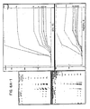
- the silver stain of this invention was demonstrated to be at least equal and perhaps somewhat more sensitive than the histologically derived silver stain (Fig. 1). Densities obtained with the two stains were proportional over a wide range of protein concentrations. Polypeptide "spot" counts made on two-dimensional gels established that the photochemical silver stain was at least as efficient as the histological stain in detecting the presence of polypeptides in gels. Both stains displayed the same specificity for polypeptides. Treatment of E. coli or human cell lysates with protease k (an enzyme which degrades polypeptides) resulted in the disappearance of all discrete spots on the gels.
- protease k an enzyme which degrades polypeptides
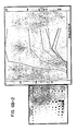
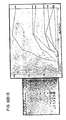
- FIGS 2 and 3 The utility of the stain is illustrated in Figures 2 and 3 in which two-dimensional electrophoretic patterns of primate cerebrospinal fluid (CSF) from various regions and of plasma are presented.
- CSF was obtained from 6 Rhesus macaque, from the lateral ventricle, cisterna magna, and lumbar space. Quantitative differences were noted in the distribution of a number of polypeptides, including albumin, al-antitrypsin, haptoglobin, immunoglobulin G (light and heavy chains), Gc globulin, a2 HS glycoprotein, al-antichymotrypsin, al-p-glycoprotein and transferrin, with lower concentrations in CSF from the lateral ventricle.
- polypeptides including albumin, al-antitrypsin, haptoglobin, immunoglobulin G (light and heavy chains), Gc globulin, a2 HS glycoprotein, al-antichymotrypsin, al-p-
- CSF polypeptides not seen in plasma were also noted to be present in lower concentration in lateral ventricle CSF.
- CSF polypeptides several of which are indicated in the Figures by arrows, were not distinguished in lateral ventricular CSF, demonstrating that individual polypeptide variations occur in the subregions of CSF.
- a lysis solution (100 pl/sample) containing 2% sodium dilauryl sulfate, 5% mercaptoethanol, 20% glycerol, 2% Biolyte 3/10 (Bio-Rad, Richmond, California, U.S.A.), and 2% Nonidet P-40 (BRL, Bethesda, Md., U.S.A.) was added and the sample heated at 95°C for 5 minutes. After heating, the sample was rapidly cooled in an ice bath, brought to room temperature and centrifuged at 12,OOOxG for 2 minutes. The supernatant was stored at -70°C. Plasma samples were prepared from heparinized venous blood by heating with six volumes of lysis solution to denature the proteins and were then stored at -70°C.
- Electrophoresis Two-dimensional electrophoresis of lymphocyte and E. coli proteins was accomplished according to the procedure of O'Farrell. A mixture of 1.6% (5-7) and 0.4% (3-10) ampholines was used for the isoelectric focusing which was carried out at 500 volts for 20 hours. A 10% uniform acrylamide gel (0.8 mm thick) and a 4.75% acrylamide stacking gel were used for the second dimension, which was run at 20 mA/gel. Plasma protein standards were prepared by mixing 5 ⁇ l of denatured plasma with 10 pl of 20% glycerol and placing this solution into a trough at the top of a PAGE gel in the presence of 0.1 % SDS.
- the gel was sliced into strips cut parallel to the direction of electric flow, fixed with methanol (50%) and acetic acid (12%) and stored for use. Purified, denatured, low and high molecular weight marker proteins were placed in preformed wells and polyacrylamide gel electrophoresis was carried out in a manner similar to the plasma proteins.
- a conventional histochemically-derived stain was performed as described by Switzer, Merril and Shifin (1979), above, to contrast and compare it with the more recently developed improved method of silver staining according to this invention.
- the histochemical stain is based on the copper-silver method of de Olmos as described at Brain Behav. Eval., 2, 213-237 (1969). It required 3 hours to perform and 915 g of silver per gel.
- Example 3 an improved silver stain according to this invention
- Densitometry The gels were photographed next to a stepped density standard (as an aid for normalization of gel densities) on 120 mm Tri-X film (Kodak). The negatives were scanned with a P-1000HS scanning densitometer (Optronics International, Inc., Chelmsford, Mass.) at 100 11m resolution using the "3D" optical density range. The data was processed with a PDP 11/60 computer equipped with a DeAnza IP5000 image processor. Identical gel regions were measured for particular polypeptide bands and spots. The density standard on the negative was used to normalize the computer images to correct for variations in photographic and scanning techniques.
- the light source for these experiments and for all staining was a 160 w fluorescent grid lamp (Aristo Grid Lamp Products Inc., Washington, N.Y., U.S.A.) which emits light equivalent to a 1500 w tungsten source.
- the staining of polypeptides according to the improved silver staining method of this invention is a process which is, in practice, greatly affected by three variables: light exposure, polypeptide concentration, and time of development.
- light exposure When intense exposure to light during the silver nitrate step was omitted, polypeptide spot densities were less than 50% of the densities obtained in an equivalent gel with intense light exposure. Using a saturating light exposure; it was found that no increase in sensitivity occurred after 5 min of exposure with the light source.
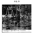
- Difficulties may occur in the quantitative or qualitative use of silver stains, two of which are shown in Figure 9.
- a region of lower density is occasionally noted in the middle of a dense polypeptide spot. Rarely, there may be a region of lower background adjacent to a dense spot.
- Similar artifacts were observed by early photographers who were forced to use thick emulsions which limited the efficiency of diffusion of fresh developer and of reaction products which might limit development. By increasing the time of development, such negative staining becomes less prominent and may disappear, but the background increases, the slope of polypeptide concentration versus density relationship increases, and the region of staining linearity is diminished.
- Saturation of staining limits the concentration of polypeptide that can be quantitated in a spot or band (usually any concentration greater than 2 ng/mm 2 ).
- a third problem may occur with gels thicker than those used (0.8 mm). A separate image may develop on each side of the slab gel. This could be due to an accentuation of the diffusion problem discussed above or to an actual difference in the distribution of polypeptides within these thick gels.
- Two dimensional electrophoresis is becoming a valuable tool in physiology and genetics because of its ability to resolve hundreds of polypeptides in a single array.
- Quantitative analysis of these polypeptides depends on accurate, sensitive microdensitometric measurements of polypeptide "spots" whose densities are regularly related to polypeptide concentration. Methods of polypeptide visualization possessing this regularity include autoradiography, some methods using organic dyes and, as described herein, the silver stains of this invention.
- Successful quantitative analysis of these polypeptides must also depend on choice of the appropriate analytic strategy for intersample comparisons. Advantage was taken for this invention of the large number of polypeptides which are synthesized constitutively and are present in nearly constant quantities within a particular cell type or preparation.
- Such polypeptides may be said to serve as "internal standards". These constitutive polypeptides are first used as internal markers of charge and molecular weight so that these parameters can be accurately estimated for polypeptides of interest and for precise pairing of polypeptide spots in different gels. Then, after densitometric measurements have been made, constitutive polypeptides densities are used to normalize the densities obtained from any particular gel to the densities found in the reference gel. Correlation coefficients between gels for similar samples and as many as 700 individual polypeptides are usually greater than 0.92. The average error between equivalent spots on separate gels is 15-30%. Improvements in image visualization and quantitative densitometry are adding a third dimension to the analytic technique of electrophoresis.
- Figure 4 shows identical subregions of lymphocyte lysate gels for the purpose of illustrating the effect of loading different amounts of polypeptide. Lysates were prepared and electrophoresced as described above. The following amounts of polypeptide were loaded onto each gel: 200 pg (20 pl), 100 pg (10 pl), 50 pg (5 pl), 25 pg (2.5 pl), and 1 pg (1 pl). The total density (average density-background density)xarea of 45 individual polypeptide spots in each gel was compared to the corresponding spot density in each of the other gels. Correlation coefficients and slopes were as follows:
- Polypeptides were precipitated with potassium dichromate and then filtered using cellulose nitrate filter paper under partial vacuum.
- the polypeptide source was purified bovine serum albumin, and the amounts tested ranged from 0.1 pg to 100 pg. After filtration, the polypeptides were successfully stained according to the preferred method of this invention.
- the fixing step was eliminated because no gel was used, but it is noted that the potassium dichromate used as an oxidant in the photoreversal step also acts as a fixative.
- the resultant stains were successful and sufficiently sensitive to indicate that this embodiment of the stain method of this invention could be used for quantitative polypeptide assays, by comparing the stain intensity to a standard.
- Thin tissue slices were successfully stained with the silver stain method of this invention.
- This embodiment shows that the method of this invention can readily be adapted to provide improved histological stains. This embodiment would be useful for biopsies.
- the Mean protein spot density desireably rises with repeats of steps 4-6, but that the Mean background undesireably also rises.
- steps 4-6 should preferably be conducted from one to three times, most preferably twice.
- the increased mean protein spot density permitted a number of proteins which originally were very faint to be easily identified.
Landscapes
- Life Sciences & Earth Sciences (AREA)
- Health & Medical Sciences (AREA)
- Molecular Biology (AREA)
- Physics & Mathematics (AREA)
- Engineering & Computer Science (AREA)
- Chemical & Material Sciences (AREA)
- Immunology (AREA)
- Urology & Nephrology (AREA)
- General Health & Medical Sciences (AREA)
- Analytical Chemistry (AREA)
- Biomedical Technology (AREA)
- Pathology (AREA)
- General Physics & Mathematics (AREA)
- Hematology (AREA)
- Biochemistry (AREA)
- Food Science & Technology (AREA)
- Bioinformatics & Computational Biology (AREA)
- Proteomics, Peptides & Aminoacids (AREA)
- Medicinal Chemistry (AREA)
- Microbiology (AREA)
- Biophysics (AREA)
- Bioinformatics & Cheminformatics (AREA)
- Cell Biology (AREA)
- Biotechnology (AREA)
- Spectroscopy & Molecular Physics (AREA)
- Chemical Kinetics & Catalysis (AREA)
- Electrochemistry (AREA)
- Investigating Or Analysing Biological Materials (AREA)
Claims (16)
Applications Claiming Priority (4)
| Application Number | Priority Date | Filing Date | Title |
|---|---|---|---|
| US24057781A | 1981-03-04 | 1981-03-04 | |
| US240577 | 1981-03-04 | ||
| US339886 | 1982-01-18 | ||
| US06/339,886 US4405720A (en) | 1981-03-04 | 1982-01-18 | Silver stains for protein in gels |
Publications (3)
| Publication Number | Publication Date |
|---|---|
| EP0073247A1 EP0073247A1 (fr) | 1983-03-09 |
| EP0073247A4 EP0073247A4 (fr) | 1983-04-18 |
| EP0073247B1 true EP0073247B1 (fr) | 1985-06-19 |
Family
ID=26933514
Family Applications (1)
| Application Number | Title | Priority Date | Filing Date |
|---|---|---|---|
| EP82901207A Expired EP0073247B1 (fr) | 1981-03-04 | 1982-03-03 | Colorants a l'argent pour les proteines contenues dans des gels |
Country Status (6)
| Country | Link |
|---|---|
| US (1) | US4405720A (fr) |
| EP (1) | EP0073247B1 (fr) |
| CA (1) | CA1182390A (fr) |
| DE (1) | DE3264186D1 (fr) |
| IT (1) | IT1148101B (fr) |
| WO (1) | WO1982003128A1 (fr) |
Families Citing this family (29)
| Publication number | Priority date | Publication date | Assignee | Title |
|---|---|---|---|---|
| US4468466A (en) * | 1982-02-16 | 1984-08-28 | The United States Of America As Represented By The Department Of Health And Human Services | Silver stains for protein in gels--a modified procedure |
| US4552848A (en) * | 1983-05-16 | 1985-11-12 | Eastman Kodak Company | Macromolecule determination by physical development |
| GB8321009D0 (en) * | 1983-08-04 | 1983-09-07 | New R R C | Immunological compositions |
| US4555490A (en) * | 1984-06-08 | 1985-11-26 | The United States Of America As Represented By The Department Of Health And Human Services | Rapid visualization system for gel electrophoresis |
| DE3503978A1 (de) * | 1984-08-01 | 1986-02-06 | Detlev Prof.Dr. 4000 Düsseldorf Riesner | Verfahren zur frueherkennung von pflanzenkrankheiten |
| US4575452A (en) * | 1984-09-21 | 1986-03-11 | E. I. Du Pont De Nemours And Company | Kit for silver staining proteins and nucleic acids |
| US4672043A (en) * | 1985-02-19 | 1987-06-09 | Eastman Kodak Company | Stabilization of developed electrophoregrams |
| GB8527687D0 (en) * | 1985-11-09 | 1985-12-11 | Wales University Of College Of | Silver intensification of diaminobenzidine |
| US4769334A (en) * | 1986-02-12 | 1988-09-06 | Linus Pauling Institute Of Science And Medicine | Method for detecting proteins in gels using radioactive dyes |
| US4703016A (en) * | 1986-05-05 | 1987-10-27 | The United States Of America As Represented By The Department Of Health And Human Services | Silver stain for rapid, quantitative detection of polypeptides and nucleic acids |
| US4946794A (en) * | 1986-12-18 | 1990-08-07 | Protein Databases, Inc. | Visualization of proteins on electrophoresis gels using planar dyes |
| US4782027A (en) * | 1987-01-22 | 1988-11-01 | President And Fellows Of Harvard College | Protein detection by negative staining |
| JP2804038B2 (ja) * | 1988-02-24 | 1998-09-24 | 株式会社日立製作所 | 塩基配列決定方法 |
| WO1992017611A1 (fr) * | 1991-03-28 | 1992-10-15 | The University Of Tennessee Research Corporation | Argentation d'adn |
| US5347139A (en) * | 1991-06-04 | 1994-09-13 | Molecular Dynamics | Dual energy tracer quantitative analysis |
| CA2071736A1 (fr) * | 1991-06-24 | 1992-12-25 | Naoyuki Kouno | Methode de teinture a l'acide |
| US5595878A (en) * | 1995-06-02 | 1997-01-21 | Boron Biologicals, Inc. | Detection of biopolymers and biooligomers with boron hydride labels |
| EP0963435B1 (fr) * | 1997-01-08 | 2008-05-28 | Invitrogen Corporation | Procedes de production de proteines |
| US5993627A (en) * | 1997-06-24 | 1999-11-30 | Large Scale Biology Corporation | Automated system for two-dimensional electrophoresis |
| ES2252263T3 (es) * | 2000-07-25 | 2006-05-16 | The Government Of The Usa, As Represented By The Secretary, Department Of Health And Human Services | Bacteriofago que tiene una gama de hospedadores multiple. |
| AU2002230438A1 (en) | 2000-11-20 | 2002-06-03 | Invitrogen Corporation | Materials for enhancing staining of biopolymers in matrices |
| WO2002079771A1 (fr) * | 2001-03-30 | 2002-10-10 | The Samuel Roberts Noble Foundation, Inc. | Procede de decoloration a l'argent |
| EP1331473B1 (fr) * | 2002-01-22 | 2005-04-06 | MPB MelTec Patent- und Beteiligungsgesellschaft mbH | Méthode et appareil pour la préparation d'échantillons pour analyser des échantillons biologiques |
| DE10243303A1 (de) * | 2002-09-13 | 2004-03-18 | Nordheim, Alfred, Prof. Dr.rer.nat. | Verfahren zur Detektion von Biomolekülen |
| EP2069386A4 (fr) | 2006-07-21 | 2009-10-28 | Life Technologies Corp | Étalons de masse moléculaire protéiques marqués à résolution très pointue |
| US9201063B2 (en) * | 2006-11-16 | 2015-12-01 | General Electric Company | Sequential analysis of biological samples |
| US8305579B2 (en) | 2006-11-16 | 2012-11-06 | Thomas Pirrie Treynor | Sequential analysis of biological samples |
| US7629125B2 (en) * | 2006-11-16 | 2009-12-08 | General Electric Company | Sequential analysis of biological samples |
| US9677125B2 (en) * | 2009-10-21 | 2017-06-13 | General Electric Company | Detection of plurality of targets in biological samples |
Family Cites Families (2)
| Publication number | Priority date | Publication date | Assignee | Title |
|---|---|---|---|---|
| AT320865B (de) * | 1973-02-12 | 1975-03-10 | Immuno Ag | Verfahren zum Bestimmen von normalen und pathologischen Plasma-Lipoproteinen in menschlichen Körperflüssigkeiten und Apparat zur Durchführung des Verfahrens |
| US4167467A (en) * | 1977-09-21 | 1979-09-11 | Helena Laboratories Corporation | Clinical procedure for measuring lipoprotein free cholesterols |
-
1982
- 1982-01-18 US US06/339,886 patent/US4405720A/en not_active Expired - Lifetime
- 1982-03-03 CA CA000397468A patent/CA1182390A/fr not_active Expired
- 1982-03-03 DE DE8282901207T patent/DE3264186D1/de not_active Expired
- 1982-03-03 EP EP82901207A patent/EP0073247B1/fr not_active Expired
- 1982-03-03 WO PCT/US1982/000267 patent/WO1982003128A1/fr not_active Ceased
- 1982-03-03 IT IT47901/82A patent/IT1148101B/it active
Non-Patent Citations (1)
| Title |
|---|
| SCIENCE, vol. 211, March 27, 1981 C.R. MERRIL: "Ultrasensitive Stain for Proteins in Polyacrylamide Gels Shows Regional Variation in Cerebrospinal Fluid Proteins", pages 1437-1438 * |
Also Published As
| Publication number | Publication date |
|---|---|
| IT1148101B (it) | 1986-11-26 |
| CA1182390A (fr) | 1985-02-12 |
| US4405720A (en) | 1983-09-20 |
| WO1982003128A1 (fr) | 1982-09-16 |
| IT8247901A0 (it) | 1982-03-03 |
| DE3264186D1 (en) | 1985-08-01 |
| EP0073247A1 (fr) | 1983-03-09 |
| EP0073247A4 (fr) | 1983-04-18 |
Similar Documents
| Publication | Publication Date | Title |
|---|---|---|
| EP0073247B1 (fr) | Colorants a l'argent pour les proteines contenues dans des gels | |
| Merril et al. | Simplified silver protein detection and image enhancement methods in polyacrylamide gels | |
| Merril et al. | A rapid sensitive silver stain for polypeptides in polyacrylamide gels | |
| US4555490A (en) | Rapid visualization system for gel electrophoresis | |
| Sammons et al. | Ultrasensitive silver‐based color staining of polypeptides in polyacrylamide gels | |
| Poehling et al. | Visualization of proteins with a silver “stain”: A critical analysis | |
| Switzer III et al. | A highly sensitive silver stain for detecting proteins and peptides in polyacrylamide gels | |
| Irie et al. | A faithful double stain of proteins in the polyacrylamide gels with Coomassie blue and silver | |
| Merril et al. | Ultrasensitive stain for proteins in polyacrylamide gels shows regional variation in cerebrospinal fluid proteins | |
| Görg et al. | The current state of two‐dimensional electrophoresis with immobilized pH gradients | |
| Syrový et al. | Staining and quantification of proteins separated by polyacrylamide gel electrophoresis | |
| Willoughby et al. | A sensitive silver stain for proteins in agarose gels | |
| US4468466A (en) | Silver stains for protein in gels--a modified procedure | |
| US4434234A (en) | Method and kit for silver staining substances supported in matrix | |
| Wijnen et al. | Capillary electrophoresis of serum proteins. Reproducibility, comparison with agarose gel electrophoresis and a review of the literature | |
| Talent et al. | A double stain for total and oxidized proteins from two-dimensional fingerprints | |
| Patton | Biologist's perspective on analytical imaging systems as applied to protein gel electrophoresis | |
| Harrington et al. | Two-dimensional electrophoresis and" ultrasensitive" silver staining of cerebrospinal fluid proteins in neurological diseases. | |
| Merril et al. | " Ultrasensitive" silver stains: their use exemplified in the study of normal human cerebrospinal fluid proteins separated by two-dimensional electrophoresis. | |
| Peats | Quantitation of protein and DNA in silver-stained agarose gels | |
| Glasson et al. | Development of mini‐gel technology in two‐dimensional electrophoresis for mass‐screening of samples: Application to tears | |
| US4946794A (en) | Visualization of proteins on electrophoresis gels using planar dyes | |
| US4508820A (en) | Method for simultaneously monitoring turnover rate in multiple proteins | |
| Rubin et al. | [12] Two-dimensional polyacrylamide gel electrophoresis of membrane proteins | |
| Chaudhuri et al. | A sensitive urea-silver stain method for detecting trace quantities of separated proteins in polyacrylamide gels |
Legal Events
| Date | Code | Title | Description |
|---|---|---|---|
| PUAI | Public reference made under article 153(3) epc to a published international application that has entered the european phase |
Free format text: ORIGINAL CODE: 0009012 |
|
| 17P | Request for examination filed |
Effective date: 19821209 |
|
| AK | Designated contracting states |
Designated state(s): CH DE FR GB LI NL SE |
|
| GRAA | (expected) grant |
Free format text: ORIGINAL CODE: 0009210 |
|
| AK | Designated contracting states |
Designated state(s): CH DE FR GB LI NL SE |
|
| REF | Corresponds to: |
Ref document number: 3264186 Country of ref document: DE Date of ref document: 19850801 |
|
| ET | Fr: translation filed | ||
| PLBE | No opposition filed within time limit |
Free format text: ORIGINAL CODE: 0009261 |
|
| STAA | Information on the status of an ep patent application or granted ep patent |
Free format text: STATUS: NO OPPOSITION FILED WITHIN TIME LIMIT |
|
| 26N | No opposition filed | ||
| PGFP | Annual fee paid to national office [announced via postgrant information from national office to epo] |
Ref country code: FR Payment date: 19940210 Year of fee payment: 13 |
|
| PGFP | Annual fee paid to national office [announced via postgrant information from national office to epo] |
Ref country code: CH Payment date: 19940214 Year of fee payment: 13 |
|
| PGFP | Annual fee paid to national office [announced via postgrant information from national office to epo] |
Ref country code: SE Payment date: 19940215 Year of fee payment: 13 |
|
| PGFP | Annual fee paid to national office [announced via postgrant information from national office to epo] |
Ref country code: DE Payment date: 19940224 Year of fee payment: 13 |
|
| PGFP | Annual fee paid to national office [announced via postgrant information from national office to epo] |
Ref country code: GB Payment date: 19940225 Year of fee payment: 13 |
|
| PGFP | Annual fee paid to national office [announced via postgrant information from national office to epo] |
Ref country code: NL Payment date: 19940331 Year of fee payment: 13 |
|
| EAL | Se: european patent in force in sweden |
Ref document number: 82901207.9 |
|
| PG25 | Lapsed in a contracting state [announced via postgrant information from national office to epo] |
Ref country code: GB Effective date: 19950303 |
|
| PG25 | Lapsed in a contracting state [announced via postgrant information from national office to epo] |
Ref country code: SE Effective date: 19950304 |
|
| PG25 | Lapsed in a contracting state [announced via postgrant information from national office to epo] |
Ref country code: LI Effective date: 19950331 Ref country code: CH Effective date: 19950331 |
|
| PG25 | Lapsed in a contracting state [announced via postgrant information from national office to epo] |
Ref country code: NL Effective date: 19951001 |
|
| GBPC | Gb: european patent ceased through non-payment of renewal fee |
Effective date: 19950303 |
|
| PG25 | Lapsed in a contracting state [announced via postgrant information from national office to epo] |
Ref country code: FR Free format text: LAPSE BECAUSE OF NON-PAYMENT OF DUE FEES Effective date: 19951130 |
|
| REG | Reference to a national code |
Ref country code: CH Ref legal event code: PL |
|
| NLV4 | Nl: lapsed or anulled due to non-payment of the annual fee |
Effective date: 19951001 |
|
| PG25 | Lapsed in a contracting state [announced via postgrant information from national office to epo] |
Ref country code: DE Effective date: 19951201 |
|
| EUG | Se: european patent has lapsed |
Ref document number: 82901207.9 |
|
| REG | Reference to a national code |
Ref country code: FR Ref legal event code: ST |