EP0096932A2 - Process for making a lyophilized product for use in skeletal imaging - Google Patents
Process for making a lyophilized product for use in skeletal imaging Download PDFInfo
- Publication number
- EP0096932A2 EP0096932A2 EP83200828A EP83200828A EP0096932A2 EP 0096932 A2 EP0096932 A2 EP 0096932A2 EP 83200828 A EP83200828 A EP 83200828A EP 83200828 A EP83200828 A EP 83200828A EP 0096932 A2 EP0096932 A2 EP 0096932A2
- Authority
- EP
- European Patent Office
- Prior art keywords
- pharmaceutically
- acid
- dry
- acceptable salts
- solution
- Prior art date
- Legal status (The legal status is an assumption and is not a legal conclusion. Google has not performed a legal analysis and makes no representation as to the accuracy of the status listed.)
- Granted
Links
Classifications
-
- A—HUMAN NECESSITIES
- A61—MEDICAL OR VETERINARY SCIENCE; HYGIENE
- A61K—PREPARATIONS FOR MEDICAL, DENTAL OR TOILETRY PURPOSES
- A61K51/00—Preparations containing radioactive substances for use in therapy or testing in vivo
- A61K51/12—Preparations containing radioactive substances for use in therapy or testing in vivo characterised by a special physical form, e.g. emulsion, microcapsules, liposomes, characterized by a special physical form, e.g. emulsions, dispersions, microcapsules
- A61K51/1241—Preparations containing radioactive substances for use in therapy or testing in vivo characterised by a special physical form, e.g. emulsion, microcapsules, liposomes, characterized by a special physical form, e.g. emulsions, dispersions, microcapsules particles, powders, lyophilizates, adsorbates, e.g. polymers or resins for adsorption or ion-exchange resins
-
- A—HUMAN NECESSITIES
- A61—MEDICAL OR VETERINARY SCIENCE; HYGIENE
- A61K—PREPARATIONS FOR MEDICAL, DENTAL OR TOILETRY PURPOSES
- A61K2123/00—Preparations for testing in vivo
Definitions
- This invention relates to radio-diagnostic agents for use in tissue imaging. More particularly, it relates to a process for preparing improved skeletal imaging products.
- Scintigraphic skeletal imaging and similar radiographic techniques for visualizing other tissues are finding ever-increasing application in biological and medical research and in diagnostic procedures.
- scintigraphic procedures involve the preparation of radioactive agents which, upon introduction into a biological subject, become localized in specific organs, tissue, or skeletal structures that are under study.
- traces, plots, or scintiphotos of the distribution of the radiographic materials can be made by various radiation detectors, e.g., traversing scanners and scintilation cameras.
- the distribution and corresponding relative intensity of the detected radioactive material not only indicates the position occupied by the tissue in which the radionuclide is localized, but also indicates the presence of aberrations, pathological conditions, and the like.
- a scintigraphic imaging agent as used in a hospital comprises a radionuclide, a carrier agent designed to target the specific organ, various auxiliary agents which affix the radionuclide to the carrier, water or other delivery vehicles suitable for injection into, or aspiration by, the patient, physiologic buffers and salts, and the like.
- the carrier attaches or complexes with the radionuclide, and localizes the material in the location where the carrier naturally concentrates in a biologic subject.
- Technetium-99m ( 99m Tc) is a radionuclide which is widely known for use in tissue imaging agents. This radionuclide is conveniently available commercially in the oxidized pertechnetate form ( 99 m TcO 4 - , hereinafter "pertechnetate-Tc99m"). However, the technetium in pertechnetate has a valance state of +7 and, thus, will not complex with the most commonly used carriers for radionuclide tissue imaging. This problem is easily overcome by reducing the technetium to what is believed to be the +3, +4, and/or +5 oxidation state.
- technetium-labeled imaging agents are generally prepared by admixing pertechnetate-Tc99m isotonic saline solution with a technetium reductant (reducing agent) such as the stannous, ferrous, or chromous salt of sulfuric or hydorchloric acid, and the desired carrier agent for targeting the organ of interest.
- a technetium reductant such as the stannous, ferrous, or chromous salt of sulfuric or hydorchloric acid
- organophos- phonates are known as suitable carrier agents which target technetium radionuclide to bone tissue.
- U.S. Patent 3,983,227, Tofe and Francis, issued September 28, 1976 discloses the use of reducing salts with radioactive pertechnetate-Tc99m solutions and organophosphonate bone-seeking carriers to prepare skeletal imaging agents.
- Technetium-containing scintigraphic imaging agents are known to be unstable in the presence of oxygen, primarily since oxidation of the reductant and/or the technetium destroys the reduced technetium/targeting carrier complex. Accordingly, such imaging agents are generally made oxygen-free by saturating the compositions with oxygen-free nitrogen gas or by preparing the agents in an oxygen-free atmosphere. Stabilization of imaging agents can also be achieved through chemical means.
- German Offenlegungsschrift 2,618,337, Tofe, published November 11, 1976 discloses the use of ascorbate stabilizers with technetium imaging agents.
- U.S. Patent 4,232,000, Fawzi, issued November 4, 1980 discloses the use of gentisyl alcohol as a stabilizer for technetium imaging agents.
- U.S. Patent 4,233,284, Fawzi, issued November 11, 1980 discloses the use of gentisic acid as a stabilizer.
- kits Commercial products for use in skeletal imaging are generally provided in liquid or dry powder mixture "kits" with vials containing phosphate or phosphonate bone seeking carriers. Skeletal imaging agents are formed by adding pertechnetate-Tc99m, in physiological saline, to such kits.
- Osteoscan-HDP R which comprises the disodium salt of methanehydroxydiphosphonic acid (HMDP), stannous chloride, and gentisic acid stabilizer, is one example of a freeze-dried (lyophilized) skeletal imaging kit.
- HMDP methanehydroxydiphosphonic acid
- stannous chloride stannous chloride
- gentisic acid stabilizer is one example of a freeze-dried (lyophilized) skeletal imaging kit.
- such kits are produced by a process which includes the steps of:
- the pH of the solution Prior to lyophilization in the manufacturing process, is 3.9. It has now been discovered that adjusting the pH of the bulk solution within a certain pH range produces a freeze-dried skeletal imaging kit that, when reconstituted with pertechnetate-Tc99m solution, forms a skeletal imaging agent with improved performance. Thus, it is an object of this invention to provide an improved process for preparing imaging kits.
- This invention encompasses a process for producing a dry-powder skeletal imaging kit comprising the steps of:
- This invention is based on the discovery that the adjustment of pH, within a particular range, during the process of manufacturing lyophilized diphosphonate-containing skeletal imaging kits yields a kit which produces a technetium skeletal imaging agent with superior imaging properties. This improved performance is manifested through faster blood clearance and higher skeletal uptake of the technetium imaging agent, i.e., the bone targeting carrier, complexed with technetium, is more quickly and more highly concentrated in bone tissue as compared to surrounding soft tissues in the body.
- the technetium imaging agent i.e., the bone targeting carrier, complexed with technetium
- Skeletal imaging kits produced in the process of this invention comprise:
- imaging refers to all radiographic tissue imaging processes for which the compositions made by the instant process may be used, including (but not limited to) skeletal imaging.
- imaging agent refers to compositions useful for tissue imaging, including (but not limited to) skeletal imaging, such compositions comprising the product of admixing pertechnetate-Tc99m, or other useful radioisotope, to an imaging kit comprising a tissue-seeking carrier, reductant, and, optionally, a stabilizer.
- imaging kit or “kit,” refers to the imaging agent before addition of a. solution of pertechnetate-Tc99m, or similar radionuclide.
- Kits for use commercially preferably contain sufficient material to form multiple doses of imaging agent. Obviously, the amount of material to be incorporated in such a kit will depend upon the number of doses of imaging agent desired. Further, specific quantities of carrier, reductant, and stabilizer (if any) may vary according to the particular compound used. Thus, the practitioner of this invention may determine appropriate quantities by refering to literature describing particular carriers, reductants, and stabilizers.
- R is hydrogen, alkyl containing from 1 to about 20 carbon atoms, amino alkyl, substituted aminoalkyl, alkenyl containing from 2 to about 20 carbon atoms, aryl (e.g., phenyl, naphthyl), phenylethenyl, benzyl, halogen (e.g., chlorine, bromine and fluorine), hydroxyl, amino, substituted amino (e.g., dimethylamino, diethylamino, N-hydroxy-N-ethylamino, acetylamino), -CH 2 COOH, -CH(COOH)CH 2 COOH, R' is hydrogen, lower alkyl (e.g., methyl, ethyl, propyl and butyl), amino, benzyl, halogen (e.g., chlorine, bromine, and fluorine), hydroxyl, or -CH 2
- Preferred carriers for use herein are the pharmaceutically-acceptable salts of methanediphosphonic acid (MDP), methanehydroxydiphosphonic acid (HMDP), and ethane-1-hydroxy-1 ,1-diphosphonic acid (EHDP).
- MDP methanediphosphonic acid
- HMDP methanehydroxydiphosphonic acid
- EHDP ethane-1-hydroxy-1 ,1-diphosphonic acid
- Oxidronate disodium, the disodium salt of HMDP is especially preferred.
- the aminodiphosphonate bone-seeking carriers of the above formula are also preferred for use in the instant invention.
- these aminodiphosphonate carriers are compounds and mixtures of compounds having the formulae: wherein n is an integer from 0 to 5; R is hydrogen, hydroxy, halogen, or amino; R' is hydrogen or alkyl containing from 1 to about 5 carbon atoms; R" is hydrogen, halogen, alkyl containing from 1 to about 8 carbon atoms, or aryl; X is hydrogen, alkyl containing from 1 to about 8 carbon atoms, aryl, alkylaryl, acetyl, or haloaryl; and the pharmaceutically-acceptable salts thereof.
- operable aminodiphosphonate carriers of formula (I) are: propane-1-hydroxy-3-amino-1,1-diphosphonic acid; ethane-t-hydroxy-2-amino-1,1-diphosphonic acid; butane-1-hydroxy-3-amino-1,1-diphosphonic acid; propane-1-hydroxy-2-amino-1,1-diphosphonic acid; propane-1-hydroxy-3-dimethytamino-1,1-diphosphonic acid; propane-1-hydroxy-3-diethylamino-1,1- diphosphonic acid; ethane-1-hydroxy-2-dimethylamino-1 ,1-diphosphonic acid; propane-1-hydroxy-1-dimethylamino-1 ,1-diphosphonic acid; and butane-1-hydroxy-3-dimethylamino-1,1-diphosphonic acid.
- operable aminodiphosphonate carriers of formula (II) are: methaneaminohydroxydiphosphonic acid; methaneaminodiphosphonic acid; methaneaminochlorodiphosphonic acid; methanediaminodiphosphonic acid; methane-N-methylamino- hydroxydiphosphonic acsid; methane-N-methylaminodiphosphonic acid; methane-N,N-dimethylaminohydroxydiphosphonic acid; methane-N,N-dimethylaminodiphosphonic acid; methaneaminochlorodiphosphonic acid; methane-N-butylaminodiphosphonic acid; methane-N-phenylaminodiphosphonic acid; methane-N-napthylaminodiphos- phonic acid; methane-N-acetylaminodiphosphonic acid; methane-N-(2-hydroxyethyl)-aminod
- Preferred aminodiphosphonate carriers include methaneaminodiphosphonic acid (AMDP), methaneaminohydroxydiphosphonic acid, methane-N-methylaminodiphosphonic acid, N,N-dimethylaminodiphosphonic acid, propane-l-hydroxy-3-amino-1,1-diphosphonic acid, and ethane-1-hydroxy-2-amino-1,1-diphosphonic acid.
- AMDP methaneaminodiphosphonic acid
- methaneaminohydroxydiphosphonic acid methane-N-methylaminodiphosphonic acid
- N,N-dimethylaminodiphosphonic acid propane-l-hydroxy-3-amino-1,1-diphosphonic acid
- ethane-1-hydroxy-2-amino-1,1-diphosphonic acid propane-l-hydroxy-3-amino-1,1-diphosphonic acid
- any pharmaceutically-acceptable, water-soluble salt or hydrolyzable ester of these diphosphonates are useful herein.
- the alkali metal and ammonium salts are preferred. Most preferred are the diphosphonate free acids themselves, and the sodium salts thereof.
- stannous ion Sn +2
- reductants for reducing the technetium in imaging compositions.
- the present invention incorporates one or more water-soluble, pharmaceutically-acceptable compounds which provide stannous ions when in solution e.g., stannous chloride, stannous fluoride, stannous sulfate, and stannous citrate, herein referred to as "reductant” or "stannous reductant.
- Stannous chloride (SnCl 2 ) is particularly preferred.
- a sufficient amount of stannous reductant must be included to ensure complete reduction of the technetium-99m added to the imaging kit.
- This amount herein "effective amount,” is greater than or equal to (not less than) the stoichiometric amount to reduce all of the technetium in the pertechnetate to be added to the imaging kit, i.e., when the reductant is dissolved, there must be enough stannous ion in solution to reduce technetium (+7) to a lower valence state, facilitating complexation with a diphosphonate carrier.
- stannous reductant incorporated into an imaging kit encompassed by this invention may vary according to such factors as the molecular weight of the salt, the amount of pertechnetate to be added to the kit, the desired storage time of the agent made from the kit, the presence of oxidants in the agent, and the presence of optional anti-oxidant stabilizers, as discussed below.
- Preferred skeletal imaging agents are formed from diphosphonate-containing kits wherein the molar ratio of diphosphonate to stannous tin in the reductant is about 65:1 or greater.
- stannous tin refers to elemental tin (Sn +2 ) contained in the reductant compound. See concurrently filed U.S. Patent Application Serial No. , "Radiographic Imaging Agents," Benedict and Van Duzee (incorporated by reference herein) and concurrently filed U. S. Patent Application Serial No. , “Radiographic Imaging Agents,” Van Duzee (incorporated by reference herein).
- the imaging kits formed in the process of this invention optionally contain an amount, herein "stabilizing amount,” of a stabilizer material sufficient to prevent or inhibit the oxidation of the reductant (e.g., oxidation of Sn +2 to Sn +4 ) during storage and/or to inhibit or reduce the reoxidation of reduced technetium-99m and/or to reduce the formation of technetium-labeled impurities which may form during use of the compositions.
- stabilizing amount of a stabilizer material sufficient to prevent or inhibit the oxidation of the reductant (e.g., oxidation of Sn +2 to Sn +4 ) during storage and/or to inhibit or reduce the reoxidation of reduced technetium-99m and/or to reduce the formation of technetium-labeled impurities which may form during use of the compositions.
- stabilizers is preferred in multi-dose kits.
- the stabilizers used herein are characterized by their toxicological acceptability under the conditions of use, their ability to stabilize the product for a reasonable period of storage and/or under usage conditions, and by their substantial non-interference with the delivery of the technetium radionuclide to bone mineral.
- gentisate stabilizers that meet the foregoing requirements and that are quite suitable for intravenous injection: U. S. Patent 4,232,200, Fawzi, issued November 4, 1980 (gentisyl alcohol); U.S. Patent 4,233,284, Fawzi, issued November 11, 1980 (gentisic acid); and U.S. Patent 4,229,427, Whitehouse, issued October 21, 1980 (hydroquinone).
- the sodium salt of gentisic acid is commercially available, quite water-soluble, and preferred for use herein.
- stabilizer materials can chelate/complex with technetium and cause it to be deposited in uncalcified soft tissue. Since the user of the kit formed in the process of the present invention will wish to avoid all unnecessary deposition of technetium in soft tissue, it will be appreciated that the amount of stabilizer material used, if any, in the present process should not be so great as to overshadow the bone-directing effect of the diphosphonate carrier thereby interfering with the skeletal imaging. Appropriate, non-interfering, stabilizing amounts of stabilizer materials for use in combination with the diphosphonates may vary according to the diphosphonate and/or stabilizer used.
- the weight ratio of stabilizer to reductant for the gentisate stabilizers useful herein is in the range of about 20:1 to about 1:20, preferably about 10:1 to about 1:1, more preferably about 5:1 to 1:1, most preferably about 3:1.
- the imaging agents made with the kit produced by this process are intended for intravenous injections into humans or lower animals. Accordingly, appropriate manufacturing and operating conditions are employed so as to provide suitably sterile, pyrogen-free compositions.
- a pharmaceutically-acceptable extender or filler to dilute the reducing and diphosphonate salts in order to simplify metering the requisite small quantities of such salts.
- Sodium chloride and glucose are preferred; sodium chloride is especially preferred inasmuch as its addition will assure that the resulting agent is at least isotonic even if the pertechnetate-Tc99m solution is hypo- tonic (as is the case when it must be diluted with sterile water to reduce its activity.)
- the preparation step in the process of this invention consists of co-dissolving the diphosphonate imaging carrier, the reductant, and the stabilizer and any other optional components, in an aqueous solution.
- a preferred method involves dissolving the carrier and optional components first, and then dissolving the reductant.
- sterile, deoxygenated water is used in processing and the product is stored under nitrogen.
- the quantity of these components incorporated into a preferred kit is enough to form multiple doses of imaging agent, as when reconstituted with a pertechnetate-Tc99m solution containing about 1 to 400 millicuries (mCi) of technetium-99m. (mCi) of technetium-99m. (mCi) of technetium-99m. (mCi) of technetium-99m. (mCi) of technetium-99m. (mCi) of technetium-99m. (mCi) of technetium-99m. (mCi) of technetium-99m. (
- a preferred kit formed in the process of this invention contains about 3.0 mg of the disodium salt of HMDP, about 0.24 mg stannous chloride, and about 0.84 mg gentisic acid.
- the pH of the bulk solution after the preparation step is typically below the pH encompassed by this invention, Therefore, the pH of the bulk solution is preferably adjusted through the addition of a pharmaceutically-acceptable base.
- Sodium hydroxide is a preferred base for this purpose.
- a pharmaceutically-acceptable acid such as hydrochloric acid, may be used to adjust the pH downward, if necessary.
- the pH is adjusted within the range of about 4.2 to about 4.8, preferably about 4.5.
- This pH adjustment step may be concurrent or subsequent to the solution preparation step of this invention; i.e., a predetermined amount of base may be added during the preparation step to provide a bulk solution with the proper pH.
- Lyophilization or freeze-drying, can be done through a variety of well-known methods.
- the pH-adjusted diphosphonate-containing solution can be lyophilized while in bulk solution or in smaller quantities.
- the bulk solution is preferably placed in sterile vials fitted with a rubber septum for ease of mixing with a pertechnetate-Tc99m solution by the user.
- the vials are preferably nitrogen-filled as an added protection against oxidation of the technetium reducing agent on storage.
- a preferred process of this invention executes the lyophilization step after the bulk solution has been metered into such mixing vials.
- compositions formed by the process of this invention are dissolved with a pertechnetate-Tc99m isotonic solution from a commercial technetium source to yield an imaging agent suitable for intravenous injection.
- the stability of such imaging agents is ample under ordinary hospital conditions.
- Administration is preferably done within about eight hours after addition of the pertechnetate-Tc99m solution.
- the concentration of reagents and technetium radionuclide is sufficient that about 1 milliliter of the solution is used in an adult of about 50-100 kg body weight.
- One milliliter of solution is preferably injected intravenously over a period of about 30 seconds.
- the total dosage of radionuclide for a sharp skeletal image ranges from about 5 mCi to about 30 mCi, preferably from about 10 mCi to about 20 .mCi. See also (incorporated by reference herein), U.S. Patent 4,234,562, Tofe et al., issued November 18, 1980; and U.S. Patent 4,247,534, Bevan, issued January 27, 1981.
- a kit was made, using the process of this invention, comprising the following components:
- the bulk solution was prepared by dissolving the HMDP, gentisic acid, and sodium chloride in sterile, nitrogen-purged (deoxygenated) water. After dissolution of these components, the stannous chloride was added and dissolved in the solution. The resulting bulk solution had a pH of about 3.0. A sodium hydroxide solution was added so as to adjust the bulk solution to pH 4.5. Sterile, nitrogen-purged water was added to bring the volume to 100 mi.
- One milliliter aliquots of the bulk solution were transferred to sterile vials which were kept under a nitrogen blanket in order to exclude oxygen.
- the vials were cooled and frozen under dry ice and dried under vacuum for 3 hours in a commercial lyophilization apparatus.
- the vials were then gradually heated to 25°C and lyophilized for an additional 16 hours.
- the vials, containing the lyophilized product, were stoppered and sealed under vacuum.
- An imaging agent is prepared using this kit by adding about 5 ml of a pertechnetate-Tc99m physiological saline with an activity of about 75 mCi, from a commercial technetium source. The vial is agitated until the kit components are dissolved. About 1 ml of the agent is slowly injected, over a period of about 30 seconds, into an adult human subject weighing about 75 kg. Excellent skeletal images are then obtained using a scintillation camera.
- HMDP HMDP
- MDP MDP
- EHDP disodium MDP HMDP
- EHDP disodium EHDP
- gentisyl alcohol hydroquinone, and pharmaceutically-acceptable salts thereof, respectively, are used instead of gentisic acid, with substantially similar results.
- a kit was made, using the process of this invention, comprising the following components:
- Vials containing a lyophilized product were prepared in the manner of Example I, above. Excellent skeletal images are obtained when this kit is used to prepare an imaging agent and the agent is injected, as in Example I.
- methaneaminohydroxydiphosphonic acid methane-N-methylaminodiphosphonic acid, methane-N,N-dimethylaminodiphosphonic acid, ethane-l-hydroxy-2-amino-1 ,1-diphosphonic acid, and monosodium salts thereof are, respectively, used instead of AMDP, with substantially similar results.
- stannous fluoride, stannous sulfate are, respectively, used with or instead of stannous chloride, with substantially similar results.
- a kit is made, using the process of this invention comprising the following components:
- Vials containing a lyophilized product are prepared as in Example I, above. Excellent skeletal images are obtained when this kit is used to prepare imaging agent and the agent is injected in the manner of Example I.
- a kit is made, using the process of this invention, comprising the following components:
- a bulk solution is prepared by admixing the HMDP, gentisic acid, and sodium chloride in sterile, deoxygenated water. Once these components are dissolved, the stannous chloride is added and dissolved. the pH of the resulting solution is adjusted to pH 4.2 by addition of a 1N NaOH solution. Sterile, deoxygenated water is then added to dilute the solution to a total volume of 28 liters.
- One milliliter aliquots of the bulk solution are placed in individual, sterile, deoxygenated vials.
- the vials are then frozen and placed under vacuum in a commercial lyophilization apparatus. Heat is applied and the product is dried for approximately 20 hours.
- the vials with the lyophilized product are stoppered and sealed.
- Kits are also made, using the process of this invention as described in the above example, by adjusting the bulk solution to any pH within the range of about 4.2 to about 4.8 through the use of any pharmaceutically-acceptable base and/or acid, with substantially similar or better results.
- Lyophilized kits were made including gentisic acid stabilizer according to the process of this invention described in Example 1, except that the bulk solution was adjusted to pH 3.0, 4.0, 5.0, and 6.0, respectively. Each kit vial was then reconstituted by addition of 5 ml of a pertechnetate-Tc99m physiological saline solution with an activity of approximately 22 mCi. A 50 microliter dose of each imaging agent thus formed was injected into fasted rats weighing approximately 200 g. One dose of an imaging agent was injected into each rat studied, with three rats injected for each kit made.
- Scintiscans were taken of rats that were injected with agents made from kits produced at each of the pH adjustments employed above. The rats were then sacrificed and blood, muscle, and bone tissue removed and placed in tared scintillation counting vials. The samples were weighed and radioassayed, along with a control sample of the original imaging agent, using a gamma-scintillation spectrometer.
- the following table summarizes the distribution of the technetium-99m imaging agent in the rat body, as a function of the pH employed in the production process of the imaging kit used. This distribution is indicated by relative bone to muscle retention ratio and the relative bone to blood retention ratio.
- kits were made without any stabilizer, according to the process of the invention described in Example II, except that the pH of the bulk solution was adjusted to pH 3.0, 4.0, 4.5, 5.0, and 6.0, respectively.
- Each kit vial was then reconstituted by addition of 5 ml of a pertechnetate-Tc99m physiological saline solution with an activity of approximately 27 mCi.
- a 50 microliter dose of each imaging agent thus formed was injected into a fasted rat weighing approximately 200 g.
- One dose of imaging agent was injected into each rat studied, with four rats injected for each kit made.
- a skeletal imaging kit produced according to the process of this invention wherein the pH of the bulk solution of components is adjusted between 4.2 and 4.8, preferably 4.5, yields an imaging agent with improved imaging qualities, i.e., increased bone/ soft tissue retention and enhanced blood clearance.
Landscapes
- Health & Medical Sciences (AREA)
- Chemical & Material Sciences (AREA)
- Pharmacology & Pharmacy (AREA)
- Physics & Mathematics (AREA)
- Medicinal Chemistry (AREA)
- Optics & Photonics (AREA)
- Dispersion Chemistry (AREA)
- Epidemiology (AREA)
- Life Sciences & Earth Sciences (AREA)
- Animal Behavior & Ethology (AREA)
- General Health & Medical Sciences (AREA)
- Public Health (AREA)
- Veterinary Medicine (AREA)
- Medicines Containing Antibodies Or Antigens For Use As Internal Diagnostic Agents (AREA)
Abstract
Description
- This invention relates to radio-diagnostic agents for use in tissue imaging. More particularly, it relates to a process for preparing improved skeletal imaging products.
- Scintigraphic skeletal imaging and similar radiographic techniques for visualizing other tissues are finding ever-increasing application in biological and medical research and in diagnostic procedures. Generally, scintigraphic procedures involve the preparation of radioactive agents which, upon introduction into a biological subject, become localized in specific organs, tissue, or skeletal structures that are under study. When so localized, traces, plots, or scintiphotos of the distribution of the radiographic materials can be made by various radiation detectors, e.g., traversing scanners and scintilation cameras. The distribution and corresponding relative intensity of the detected radioactive material not only indicates the position occupied by the tissue in which the radionuclide is localized, but also indicates the presence of aberrations, pathological conditions, and the like.
- In general, depending on the type of radionuclide used and the organ of interest, a scintigraphic imaging agent as used in a hospital comprises a radionuclide, a carrier agent designed to target the specific organ, various auxiliary agents which affix the radionuclide to the carrier, water or other delivery vehicles suitable for injection into, or aspiration by, the patient, physiologic buffers and salts, and the like. The carrier attaches or complexes with the radionuclide, and localizes the material in the location where the carrier naturally concentrates in a biologic subject.
- Technetium-99m (99mTc) is a radionuclide which is widely known for use in tissue imaging agents. This radionuclide is conveniently available commercially in the oxidized pertechnetate form (99 mTcO4 -, hereinafter "pertechnetate-Tc99m"). However, the technetium in pertechnetate has a valance state of +7 and, thus, will not complex with the most commonly used carriers for radionuclide tissue imaging. This problem is easily overcome by reducing the technetium to what is believed to be the +3, +4, and/or +5 oxidation state. Thus, technetium-labeled imaging agents are generally prepared by admixing pertechnetate-Tc99m isotonic saline solution with a technetium reductant (reducing agent) such as the stannous, ferrous, or chromous salt of sulfuric or hydorchloric acid, and the desired carrier agent for targeting the organ of interest. For example, organophos- phonates are known as suitable carrier agents which target technetium radionuclide to bone tissue. U.S. Patent 3,983,227, Tofe and Francis, issued September 28, 1976, discloses the use of reducing salts with radioactive pertechnetate-Tc99m solutions and organophosphonate bone-seeking carriers to prepare skeletal imaging agents.
- Technetium-containing scintigraphic imaging agents are known to be unstable in the presence of oxygen, primarily since oxidation of the reductant and/or the technetium destroys the reduced technetium/targeting carrier complex. Accordingly, such imaging agents are generally made oxygen-free by saturating the compositions with oxygen-free nitrogen gas or by preparing the agents in an oxygen-free atmosphere. Stabilization of imaging agents can also be achieved through chemical means. German Offenlegungsschrift 2,618,337, Tofe, published November 11, 1976, discloses the use of ascorbate stabilizers with technetium imaging agents. U.S. Patent 4,232,000, Fawzi, issued November 4, 1980, discloses the use of gentisyl alcohol as a stabilizer for technetium imaging agents. Similarly, U.S. Patent 4,233,284, Fawzi, issued November 11, 1980, discloses the use of gentisic acid as a stabilizer.
- Commercial products for use in skeletal imaging are generally provided in liquid or dry powder mixture "kits" with vials containing phosphate or phosphonate bone seeking carriers. Skeletal imaging agents are formed by adding pertechnetate-Tc99m, in physiological saline, to such kits. Osteoscan-HDPR, which comprises the disodium salt of methanehydroxydiphosphonic acid (HMDP), stannous chloride, and gentisic acid stabilizer, is one example of a freeze-dried (lyophilized) skeletal imaging kit. Generally, such kits are produced by a process which includes the steps of:
- (1) adding solid ingredients to sterile water,
- (2) metering of the resulting bulk solution into individual vials,
- (3) lyophilizing the solution in the vials, and
- (4) packaging.
- For Osteoscan-HDPR, the pH of the solution, prior to lyophilization in the manufacturing process, is 3.9. It has now been discovered that adjusting the pH of the bulk solution within a certain pH range produces a freeze-dried skeletal imaging kit that, when reconstituted with pertechnetate-Tc99m solution, forms a skeletal imaging agent with improved performance. Thus, it is an object of this invention to provide an improved process for preparing imaging kits.
- This invention encompasses a process for producing a dry-powder skeletal imaging kit comprising the steps of:
- (1) preparing an aqueous solution of a diphosphonate, a stannous reductant, and, optionally, a gentisate stabilizer;
- (2) adjusting the solution formed in step 1 to a pH within the range from about 4.2 to about 4.8; and
- (3) lyophilizing the pH-adjusted solution.
- This invention is based on the discovery that the adjustment of pH, within a particular range, during the process of manufacturing lyophilized diphosphonate-containing skeletal imaging kits yields a kit which produces a technetium skeletal imaging agent with superior imaging properties. This improved performance is manifested through faster blood clearance and higher skeletal uptake of the technetium imaging agent, i.e., the bone targeting carrier, complexed with technetium, is more quickly and more highly concentrated in bone tissue as compared to surrounding soft tissues in the body.
- Skeletal imaging kits produced in the process of this invention comprise:
- (1) a diphosphonate carrier;
- (2) a stannous reductant; and, optionally,
- (3) a gentisate stabilizer.
- These components, as well as the process of this invention for combining the components into skeletal imaging kits, are described below.
- As used herein, the term "imaging" refers to all radiographic tissue imaging processes for which the compositions made by the instant process may be used, including (but not limited to) skeletal imaging. The term "imaging agent" refers to compositions useful for tissue imaging, including (but not limited to) skeletal imaging, such compositions comprising the product of admixing pertechnetate-Tc99m, or other useful radioisotope, to an imaging kit comprising a tissue-seeking carrier, reductant, and, optionally, a stabilizer. Hence, the term "imaging kit," or "kit," refers to the imaging agent before addition of a. solution of pertechnetate-Tc99m, or similar radionuclide.
- Kits for use commercially preferably contain sufficient material to form multiple doses of imaging agent. Obviously, the amount of material to be incorporated in such a kit will depend upon the number of doses of imaging agent desired. Further, specific quantities of carrier, reductant, and stabilizer (if any) may vary according to the particular compound used. Thus, the practitioner of this invention may determine appropriate quantities by refering to literature describing particular carriers, reductants, and stabilizers.
- Particularly useful as carriers in the instant invention are compounds and mixtures of compounds of the formula:

- Preferred carriers for use herein are the pharmaceutically-acceptable salts of methanediphosphonic acid (MDP), methanehydroxydiphosphonic acid (HMDP), and ethane-1-hydroxy-1 ,1-diphosphonic acid (EHDP). Oxidronate disodium, the disodium salt of HMDP, is especially preferred. The aminodiphosphonate bone-seeking carriers of the above formula are also preferred for use in the instant invention. Specifically, these aminodiphosphonate carriers are compounds and mixtures of compounds having the formulae:

- Among the operable aminodiphosphonate carriers of formula (I) are: propane-1-hydroxy-3-amino-1,1-diphosphonic acid; ethane-t-hydroxy-2-amino-1,1-diphosphonic acid; butane-1-hydroxy-3-amino-1,1-diphosphonic acid; propane-1-hydroxy-2-amino-1,1-diphosphonic acid; propane-1-hydroxy-3-dimethytamino-1,1-diphosphonic acid; propane-1-hydroxy-3-diethylamino-1,1- diphosphonic acid; ethane-1-hydroxy-2-dimethylamino-1 ,1-diphosphonic acid; propane-1-hydroxy-1-dimethylamino-1 ,1-diphosphonic acid; and butane-1-hydroxy-3-dimethylamino-1,1-diphosphonic acid. Among the operable aminodiphosphonate carriers of formula (II) are: methaneaminohydroxydiphosphonic acid; methaneaminodiphosphonic acid; methaneaminochlorodiphosphonic acid; methanediaminodiphosphonic acid; methane-N-methylamino- hydroxydiphosphonic acsid; methane-N-methylaminodiphosphonic acid; methane-N,N-dimethylaminohydroxydiphosphonic acid; methane-N,N-dimethylaminodiphosphonic acid; methaneaminochlorodiphosphonic acid; methane-N-butylaminodiphosphonic acid; methane-N-phenylaminodiphosphonic acid; methane-N-napthylaminodiphos- phonic acid; methane-N-acetylaminodiphosphonic acid; methane-N-(2-hydroxyethyl)-aminodiphosphonic acid; methane-N-(chlorophenyl)aminodiphosphonic acid; methane-N-(2-phenylethyl)-aminodi- phosphonic acid; ethane-1-amino-1,1-diphosphonic acid; propane-1-amino-1,1-diphosphonic acid; butane-1-amino-1,1-diphosphonic acid; and pentane-1-amino-1,1-diphosphonic acid.
- Aminodiphosphonate compounds of these formulae are disclosed in the following documents incorporated by reference herein: U.S. Patent 3,983,227, Tofe, issued September 28, 1976; U.S. Patent 4,054,598, Blum, et aI., issued October 18,. 1977; and concurrently filed EPC Patent Application Serial No."Radiographic Imaging Agents," Benedict and Van Duzee.

- Preferred aminodiphosphonate carriers include methaneaminodiphosphonic acid (AMDP), methaneaminohydroxydiphosphonic acid, methane-N-methylaminodiphosphonic acid, N,N-dimethylaminodiphosphonic acid, propane-l-hydroxy-3-amino-1,1-diphosphonic acid, and ethane-1-hydroxy-2-amino-1,1-diphosphonic acid.
- Any pharmaceutically-acceptable, water-soluble salt or hydrolyzable ester of these diphosphonates. are useful herein. The alkali metal and ammonium salts are preferred. Most preferred are the diphosphonate free acids themselves, and the sodium salts thereof.
- In order for these targeting agents to be useful with technetium, commercially available technetium (pertechnetate) must be reduced to form trivalent, tetravalent, and/or pentavalent technetium, which is then available to attach or complex with the targeting carrier. Reducing metal cations, such as stannous ion (Sn+2) are known reductants for reducing the technetium in imaging compositions. The present invention incorporates one or more water-soluble, pharmaceutically-acceptable compounds which provide stannous ions when in solution e.g., stannous chloride, stannous fluoride, stannous sulfate, and stannous citrate, herein referred to as "reductant" or "stannous reductant. Stannous chloride (SnCl2) is particularly preferred.
- A sufficient amount of stannous reductant must be included to ensure complete reduction of the technetium-99m added to the imaging kit. This amount, herein "effective amount," is greater than or equal to (not less than) the stoichiometric amount to reduce all of the technetium in the pertechnetate to be added to the imaging kit, i.e., when the reductant is dissolved, there must be enough stannous ion in solution to reduce technetium (+7) to a lower valence state, facilitating complexation with a diphosphonate carrier. The specific quantity of stannous reductant incorporated into an imaging kit encompassed by this invention may vary according to such factors as the molecular weight of the salt, the amount of pertechnetate to be added to the kit, the desired storage time of the agent made from the kit, the presence of oxidants in the agent, and the presence of optional anti-oxidant stabilizers, as discussed below. Preferred skeletal imaging agents, with improved performance, are formed from diphosphonate-containing kits wherein the molar ratio of diphosphonate to stannous tin in the reductant is about 65:1 or greater. As used herein, "stannous tin" refers to elemental tin (Sn+2) contained in the reductant compound. See concurrently filed U.S. Patent Application Serial No. , "Radiographic Imaging Agents," Benedict and Van Duzee (incorporated by reference herein) and concurrently filed U. S. Patent Application Serial No. , "Radiographic Imaging Agents," Van Duzee (incorporated by reference herein).
- The imaging kits formed in the process of this invention optionally contain an amount, herein "stabilizing amount," of a stabilizer material sufficient to prevent or inhibit the oxidation of the reductant (e.g., oxidation of Sn+2 to Sn+4) during storage and/or to inhibit or reduce the reoxidation of reduced technetium-99m and/or to reduce the formation of technetium-labeled impurities which may form during use of the compositions. Use of such stabilizers is preferred in multi-dose kits. The stabilizers used herein are characterized by their toxicological acceptability under the conditions of use, their ability to stabilize the product for a reasonable period of storage and/or under usage conditions, and by their substantial non-interference with the delivery of the technetium radionuclide to bone mineral.
- The following documents, all incorporated by reference herein, describe useful stabilizers, herein referred to as "gentisate" stabilizers, that meet the foregoing requirements and that are quite suitable for intravenous injection: U. S. Patent 4,232,200, Fawzi, issued November 4, 1980 (gentisyl alcohol); U.S. Patent 4,233,284, Fawzi, issued November 11, 1980 (gentisic acid); and U.S. Patent 4,229,427, Whitehouse, issued October 21, 1980 (hydroquinone). The sodium salt of gentisic acid is commercially available, quite water-soluble, and preferred for use herein.
- As is known in the literature, stabilizer materials can chelate/complex with technetium and cause it to be deposited in uncalcified soft tissue. Since the user of the kit formed in the process of the present invention will wish to avoid all unnecessary deposition of technetium in soft tissue, it will be appreciated that the amount of stabilizer material used, if any, in the present process should not be so great as to overshadow the bone-directing effect of the diphosphonate carrier thereby interfering with the skeletal imaging. Appropriate, non-interfering, stabilizing amounts of stabilizer materials for use in combination with the diphosphonates may vary according to the diphosphonate and/or stabilizer used. In general, the weight ratio of stabilizer to reductant for the gentisate stabilizers useful herein is in the range of about 20:1 to about 1:20, preferably about 10:1 to about 1:1, more preferably about 5:1 to 1:1, most preferably about 3:1.
- The imaging agents made with the kit produced by this process are intended for intravenous injections into humans or lower animals. Accordingly, appropriate manufacturing and operating conditions are employed so as to provide suitably sterile, pyrogen-free compositions. Although not necessary to the practice of the present invention, it is preferable to use a pharmaceutically-acceptable extender or filler to dilute the reducing and diphosphonate salts in order to simplify metering the requisite small quantities of such salts. Sodium chloride and glucose are preferred; sodium chloride is especially preferred inasmuch as its addition will assure that the resulting agent is at least isotonic even if the pertechnetate-Tc99m solution is hypo- tonic (as is the case when it must be diluted with sterile water to reduce its activity.)
- The preparation step in the process of this invention consists of co-dissolving the diphosphonate imaging carrier, the reductant, and the stabilizer and any other optional components, in an aqueous solution. A preferred method involves dissolving the carrier and optional components first, and then dissolving the reductant. Preferably, sterile, deoxygenated water is used in processing and the product is stored under nitrogen. The quantity of these components incorporated into a preferred kit is enough to form multiple doses of imaging agent, as when reconstituted with a pertechnetate-Tc99m solution containing about 1 to 400 millicuries (mCi) of technetium-99m. (The number of doses ultimately obtained from such a kit depends upon such factors as the weight of the dosed subject and the type of tissue to be imaged.) Generally, then, a preferred kit comprises:
- (a) an amount of diphosphonate carrier sufficient to target the technetium in a pertechnetate solution containing from about 1 to 400 mCi of technetium-99m;
- (b) an amount of stannous reductant sufficient to reduce the technetium in a pertechnetate solution containing from about 1 to 400 mCi technetium-99m, and
- (c) an amount of gentisate stabilizer sufficient to prevent oxidation of the reductant and the reduced technetium-99m.
- A preferred kit formed in the process of this invention contains about 3.0 mg of the disodium salt of HMDP, about 0.24 mg stannous chloride, and about 0.84 mg gentisic acid.
- The pH of the bulk solution after the preparation step is typically below the pH encompassed by this invention, Therefore, the pH of the bulk solution is preferably adjusted through the addition of a pharmaceutically-acceptable base. Sodium hydroxide is a preferred base for this purpose. A pharmaceutically-acceptable acid, such as hydrochloric acid, may be used to adjust the pH downward, if necessary. The pH is adjusted within the range of about 4.2 to about 4.8, preferably about 4.5. This pH adjustment step may be concurrent or subsequent to the solution preparation step of this invention; i.e., a predetermined amount of base may be added during the preparation step to provide a bulk solution with the proper pH.
- Lyophilization, or freeze-drying, can be done through a variety of well-known methods. The pH-adjusted diphosphonate-containing solution can be lyophilized while in bulk solution or in smaller quantities. The bulk solution is preferably placed in sterile vials fitted with a rubber septum for ease of mixing with a pertechnetate-Tc99m solution by the user. The vials are preferably nitrogen-filled as an added protection against oxidation of the technetium reducing agent on storage. A preferred process of this invention executes the lyophilization step after the bulk solution has been metered into such mixing vials.
- The compositions formed by the process of this invention are dissolved with a pertechnetate-Tc99m isotonic solution from a commercial technetium source to yield an imaging agent suitable for intravenous injection. The stability of such imaging agents is ample under ordinary hospital conditions. Administration is preferably done within about eight hours after addition of the pertechnetate-Tc99m solution. Preferably, the concentration of reagents and technetium radionuclide is sufficient that about 1 milliliter of the solution is used in an adult of about 50-100 kg body weight. One milliliter of solution is preferably injected intravenously over a period of about 30 seconds. The total dosage of radionuclide for a sharp skeletal image ranges from about 5 mCi to about 30 mCi, preferably from about 10 mCi to about 20 .mCi. See also (incorporated by reference herein), U.S. Patent 4,234,562, Tofe et al., issued November 18, 1980; and U.S. Patent 4,247,534, Bevan, issued January 27, 1981.
- The process of this invention is illustrated in the following non-limiting examples.
- A kit was made, using the process of this invention, comprising the following components:

- The bulk solution was prepared by dissolving the HMDP, gentisic acid, and sodium chloride in sterile, nitrogen-purged (deoxygenated) water. After dissolution of these components, the stannous chloride was added and dissolved in the solution. The resulting bulk solution had a pH of about 3.0. A sodium hydroxide solution was added so as to adjust the bulk solution to pH 4.5. Sterile, nitrogen-purged water was added to bring the volume to 100 mi.
- One milliliter aliquots of the bulk solution were transferred to sterile vials which were kept under a nitrogen blanket in order to exclude oxygen. The vials were cooled and frozen under dry ice and dried under vacuum for 3 hours in a commercial lyophilization apparatus. The vials were then gradually heated to 25°C and lyophilized for an additional 16 hours. The vials, containing the lyophilized product, were stoppered and sealed under vacuum.
- An imaging agent is prepared using this kit by adding about 5 ml of a pertechnetate-Tc99m physiological saline with an activity of about 75 mCi, from a commercial technetium source. The vial is agitated until the kit components are dissolved. About 1 ml of the agent is slowly injected, over a period of about 30 seconds, into an adult human subject weighing about 75 kg. Excellent skeletal images are then obtained using a scintillation camera.
- In the process described above, HMDP, MDP, EHDP disodium MDP, and disodium EHDP, respectively are used instead of the disodium salt of HMDP, with substantially similar results. Also, in the foregoing kit, gentisyl alcohol, hydroquinone, and pharmaceutically-acceptable salts thereof, respectively, are used instead of gentisic acid, with substantially similar results.
- A kit was made, using the process of this invention, comprising the following components:

- Vials containing a lyophilized product were prepared in the manner of Example I, above. Excellent skeletal images are obtained when this kit is used to prepare an imaging agent and the agent is injected, as in Example I.
- In the kit prepared above, methaneaminohydroxydiphosphonic acid, methane-N-methylaminodiphosphonic acid, methane-N,N-dimethylaminodiphosphonic acid, ethane-l-hydroxy-2-amino-1 ,1-diphosphonic acid, and monosodium salts thereof are, respectively, used instead of AMDP, with substantially similar results.
- Also, in the foregoing kit, stannous fluoride, stannous sulfate, are, respectively, used with or instead of stannous chloride, with substantially similar results.
- A kit is made, using the process of this invention comprising the following components:

- Vials containing a lyophilized product are prepared as in Example I, above. Excellent skeletal images are obtained when this kit is used to prepare imaging agent and the agent is injected in the manner of Example I.
- A kit is made, using the process of this invention, comprising the following components:

- A bulk solution is prepared by admixing the HMDP, gentisic acid, and sodium chloride in sterile, deoxygenated water. Once these components are dissolved, the stannous chloride is added and dissolved. the pH of the resulting solution is adjusted to pH 4.2 by addition of a 1N NaOH solution. Sterile, deoxygenated water is then added to dilute the solution to a total volume of 28 liters.
- One milliliter aliquots of the bulk solution are placed in individual, sterile, deoxygenated vials. The vials are then frozen and placed under vacuum in a commercial lyophilization apparatus. Heat is applied and the product is dried for approximately 20 hours. The vials with the lyophilized product are stoppered and sealed.
- Excellent skeletal images are obtained when this kit is used to prepare an imaging agent and the agent is injected, as in Example I. Kits are also made, using the process of this invention as described in the above example, by adjusting the bulk solution to any pH within the range of about 4.2 to about 4.8 through the use of any pharmaceutically-acceptable base and/or acid, with substantially similar or better results.
- Lyophilized kits were made including gentisic acid stabilizer according to the process of this invention described in Example 1, except that the bulk solution was adjusted to pH 3.0, 4.0, 5.0, and 6.0, respectively. Each kit vial was then reconstituted by addition of 5 ml of a pertechnetate-Tc99m physiological saline solution with an activity of approximately 22 mCi. A 50 microliter dose of each imaging agent thus formed was injected into fasted rats weighing approximately 200 g. One dose of an imaging agent was injected into each rat studied, with three rats injected for each kit made.
- Scintiscans were taken of rats that were injected with agents made from kits produced at each of the pH adjustments employed above. The rats were then sacrificed and blood, muscle, and bone tissue removed and placed in tared scintillation counting vials. The samples were weighed and radioassayed, along with a control sample of the original imaging agent, using a gamma-scintillation spectrometer.
- The following table summarizes the distribution of the technetium-99m imaging agent in the rat body, as a function of the pH employed in the production process of the imaging kit used. This distribution is indicated by relative bone to muscle retention ratio and the relative bone to blood retention ratio.

- This data clearly demonstrates the effect on the distribution of a technetium-99m imaging agent as a function of the pH used in the process for producing an imaging kit. This data indicates maximum bone/muscle and bone/blood retention at a kit production pH between 4.2 and 4.8.
- In a second experiment, lyophilized kits were made without any stabilizer, according to the process of the invention described in Example II, except that the pH of the bulk solution was adjusted to pH 3.0, 4.0, 4.5, 5.0, and 6.0, respectively. Each kit vial was then reconstituted by addition of 5 ml of a pertechnetate-Tc99m physiological saline solution with an activity of approximately 27 mCi. A 50 microliter dose of each imaging agent thus formed was injected into a fasted rat weighing approximately 200 g. One dose of imaging agent was injected into each rat studied, with four rats injected for each kit made.
- Scintiscans and assays were made of the injected rats, as in Experiment I, described above. Similarly, data is presented in Table II, below.
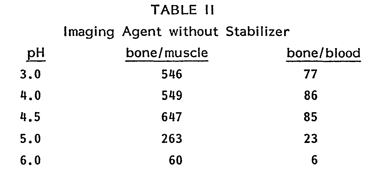
- This data indicates maximum bone/muscle and bone/ blood retention at a kit production pH between 4.2 and 4.8. Thus, a skeletal imaging kit produced according to the process of this invention, wherein the pH of the bulk solution of components is adjusted between 4.2 and 4.8, preferably 4.5, yields an imaging agent with improved imaging qualities, i.e., increased bone/ soft tissue retention and enhanced blood clearance.
Claims (10)


Priority Applications (1)
| Application Number | Priority Date | Filing Date | Title |
|---|---|---|---|
| AT83200828T ATE23799T1 (en) | 1982-06-10 | 1983-06-07 | PROCESS FOR MANUFACTURING A LYOPHILIZED PRODUCT FOR SKELETON VISIBLE. |
Applications Claiming Priority (2)
| Application Number | Priority Date | Filing Date | Title |
|---|---|---|---|
| US06/387,139 US4504463A (en) | 1982-06-10 | 1982-06-10 | Process for making a lyophilized product for use in skeletal imaging |
| US387139 | 1982-06-10 |
Publications (3)
| Publication Number | Publication Date |
|---|---|
| EP0096932A2 true EP0096932A2 (en) | 1983-12-28 |
| EP0096932A3 EP0096932A3 (en) | 1984-02-22 |
| EP0096932B1 EP0096932B1 (en) | 1986-11-26 |
Family
ID=23528637
Family Applications (1)
| Application Number | Title | Priority Date | Filing Date |
|---|---|---|---|
| EP83200828A Expired EP0096932B1 (en) | 1982-06-10 | 1983-06-07 | Process for making a lyophilized product for use in skeletal imaging |
Country Status (7)
| Country | Link |
|---|---|
| US (1) | US4504463A (en) |
| EP (1) | EP0096932B1 (en) |
| JP (1) | JPS5946229A (en) |
| AT (1) | ATE23799T1 (en) |
| AU (1) | AU554517B2 (en) |
| CA (1) | CA1210692A (en) |
| DE (1) | DE3367852D1 (en) |
Cited By (3)
| Publication number | Priority date | Publication date | Assignee | Title |
|---|---|---|---|---|
| EP0111414A3 (en) * | 1982-12-08 | 1985-05-02 | Mallinckrodt, Inc. (a Delaware corporation) | Radiographic imaging agents |
| EP0111415A3 (en) * | 1982-12-08 | 1985-05-08 | Mallinckrodt, Inc. | Process for making lyophilized radiographic imaging kit |
| EP0096933B1 (en) * | 1982-06-10 | 1986-10-08 | Mallinckrodt, Inc. (a Delaware corporation) | Process for making a lyophilized product for use in skeletal imaging |
Families Citing this family (6)
| Publication number | Priority date | Publication date | Assignee | Title |
|---|---|---|---|---|
| US4707353A (en) * | 1982-12-08 | 1987-11-17 | Mallinckrodt, Inc. | Radiographic imaging agents |
| US4582698A (en) * | 1983-09-19 | 1986-04-15 | The Dow Chemical Company | Process for imaging cardiac infarcts |
| IL74902A (en) * | 1984-06-04 | 1990-12-23 | Dow Chemical Co | Organic amine phosphonic acid complexes with particle-emitting radionuclides for the treatment of calcific tumors |
| US5066478A (en) * | 1984-06-04 | 1991-11-19 | The Dow Chemical Company | Radio labeled organic amine phosphonic acid complexes for the treatment of calcific tumors |
| ES2150916T3 (en) * | 1991-08-29 | 2000-12-16 | Mallinckrodt Medical Inc | USE OF GENISTIC ACID OR GENTISILIC ALCOHOL TO STABILIZE RADIO-MARKED PEPTIDES AND PROTEINS. |
| DE10049735A1 (en) * | 2000-09-28 | 2002-04-18 | Jenapharm Gmbh | Method for the isolation of pharmaceutically acceptable etidronate disodium |
Family Cites Families (11)
| Publication number | Priority date | Publication date | Assignee | Title |
|---|---|---|---|---|
| ZA761944B (en) * | 1975-04-30 | 1977-03-30 | Procter & Gamble | Stable radiographic scanning agents |
| US4364920A (en) * | 1975-04-30 | 1982-12-21 | Medi-Physics, Inc. | Stable diagnostic reagents |
| DE2543349C2 (en) * | 1975-09-29 | 1984-05-10 | Henkel KGaA, 4000 Düsseldorf | Preparations for the production of 99m technetium-radiagnostica |
| US4054645A (en) * | 1976-08-24 | 1977-10-18 | Minnesota Mining And Manufacturing Company | Radiodiagnostic complexes employing fluorine-containing tin reducing agents |
| CA1078731A (en) * | 1976-12-16 | 1980-06-03 | Charles E. Frosst And Co. | Skeletal imaging kit utilizing triethylene tetramine hexa (methylene phosphonic acid) |
| US4233284A (en) * | 1978-03-31 | 1980-11-11 | The Procter & Gamble Company | Stabilized radiographic scanning agents |
| US4229427A (en) * | 1978-06-28 | 1980-10-21 | The Procter & Gamble Company | Radioactive scanning agents with hydroquinone stabilizer |
| US4247534A (en) * | 1978-07-31 | 1981-01-27 | The Procter & Gamble Company | Radiographic scanning agent |
| JPS5569517A (en) * | 1978-11-20 | 1980-05-26 | Nippon Mejifuijitsukusu Kk | Labelling preparation for labelling of erythrocytes with radio-active technetium |
| JPS5750928A (en) * | 1980-09-13 | 1982-03-25 | Nippon Mejifuijitsukusu Kk | Diagnostic agent for scanning bone |
| DE3047107A1 (en) * | 1980-12-13 | 1982-07-29 | Hoechst Ag, 6000 Frankfurt | METHOD FOR PRODUCING 1-AMINO-ALKANE-1,1-DIPHOSPHONIC ACIDS |
-
1982
- 1982-06-10 US US06/387,139 patent/US4504463A/en not_active Expired - Fee Related
-
1983
- 1983-06-07 AT AT83200828T patent/ATE23799T1/en active
- 1983-06-07 DE DE8383200828T patent/DE3367852D1/en not_active Expired
- 1983-06-07 EP EP83200828A patent/EP0096932B1/en not_active Expired
- 1983-06-09 CA CA000430065A patent/CA1210692A/en not_active Expired
- 1983-06-09 AU AU15673/83A patent/AU554517B2/en not_active Ceased
- 1983-06-10 JP JP58104084A patent/JPS5946229A/en active Granted
Cited By (3)
| Publication number | Priority date | Publication date | Assignee | Title |
|---|---|---|---|---|
| EP0096933B1 (en) * | 1982-06-10 | 1986-10-08 | Mallinckrodt, Inc. (a Delaware corporation) | Process for making a lyophilized product for use in skeletal imaging |
| EP0111414A3 (en) * | 1982-12-08 | 1985-05-02 | Mallinckrodt, Inc. (a Delaware corporation) | Radiographic imaging agents |
| EP0111415A3 (en) * | 1982-12-08 | 1985-05-08 | Mallinckrodt, Inc. | Process for making lyophilized radiographic imaging kit |
Also Published As
| Publication number | Publication date |
|---|---|
| CA1210692A (en) | 1986-09-02 |
| AU1567383A (en) | 1983-12-15 |
| AU554517B2 (en) | 1986-08-21 |
| EP0096932B1 (en) | 1986-11-26 |
| ATE23799T1 (en) | 1986-12-15 |
| EP0096932A3 (en) | 1984-02-22 |
| DE3367852D1 (en) | 1987-01-15 |
| US4504463A (en) | 1985-03-12 |
| JPS5946229A (en) | 1984-03-15 |
| JPH0543690B2 (en) | 1993-07-02 |
Similar Documents
| Publication | Publication Date | Title |
|---|---|---|
| EP0096930B1 (en) | Stable radiographic imaging agents | |
| US4247534A (en) | Radiographic scanning agent | |
| US4104366A (en) | Compositions for preparation of aqueous solutions of low valence 99 technitium salts | |
| US4510125A (en) | Process for making lyophilized radiographic imaging kit | |
| CA2044812C (en) | Radiopharmaceutical formulations, their method of administration and process of preparation | |
| EP0096933B1 (en) | Process for making a lyophilized product for use in skeletal imaging | |
| JPS6255627B2 (en) | ||
| EP0006659B1 (en) | Compositions useful in the preparation of stabilized radiographic scanning agents, a method of preparing them and the use of stannous gentisilate as stabilizer in these compositions | |
| EP0096932B1 (en) | Process for making a lyophilized product for use in skeletal imaging | |
| EP0096931B1 (en) | Radiographic imaging agents | |
| EP0096934B1 (en) | Radiographic imaging agents | |
| EP0111414A2 (en) | Radiographic imaging agents | |
| US4432963A (en) | Radiographic scanning agent | |
| Archimandritis et al. | Technetium-1, 2-dihydroxy-1, 2-bis (dihydroxyphospinyl) ethane (99mTc-DHPE). A promising new skeletal imaging agent |
Legal Events
| Date | Code | Title | Description |
|---|---|---|---|
| PUAI | Public reference made under article 153(3) epc to a published international application that has entered the european phase |
Free format text: ORIGINAL CODE: 0009012 |
|
| PUAL | Search report despatched |
Free format text: ORIGINAL CODE: 0009013 |
|
| AK | Designated contracting states |
Designated state(s): AT BE CH DE FR GB IT LI LU NL SE |
|
| AK | Designated contracting states |
Designated state(s): AT BE CH DE FR GB IT LI LU NL SE |
|
| RAP1 | Party data changed (applicant data changed or rights of an application transferred) |
Owner name: MALLINCKRODT, INC. |
|
| 17P | Request for examination filed |
Effective date: 19840803 |
|
| GRAA | (expected) grant |
Free format text: ORIGINAL CODE: 0009210 |
|
| AK | Designated contracting states |
Kind code of ref document: B1 Designated state(s): AT BE CH DE FR GB IT LI LU NL SE |
|
| REF | Corresponds to: |
Ref document number: 23799 Country of ref document: AT Date of ref document: 19861215 Kind code of ref document: T |
|
| BECN | Be: change of holder's name |
Effective date: 19861126 |
|
| REF | Corresponds to: |
Ref document number: 3367852 Country of ref document: DE Date of ref document: 19870115 |
|
| ITF | It: translation for a ep patent filed | ||
| RAP2 | Party data changed (patent owner data changed or rights of a patent transferred) |
Owner name: MALLINCKRODT, INC. (A DELAWARE CORPORATION) |
|
| REG | Reference to a national code |
Ref country code: CH Ref legal event code: PUE Owner name: MALLINCKRODT, INC. (A DELAWARE CORPORATION) |
|
| NLT2 | Nl: modifications (of names), taken from the european patent patent bulletin |
Owner name: MALLINCKRODT, INC. (DELAWARE CORPORATION) TE ST. L |
|
| PLBE | No opposition filed within time limit |
Free format text: ORIGINAL CODE: 0009261 |
|
| STAA | Information on the status of an ep patent application or granted ep patent |
Free format text: STATUS: NO OPPOSITION FILED WITHIN TIME LIMIT |
|
| 26N | No opposition filed | ||
| PGFP | Annual fee paid to national office [announced via postgrant information from national office to epo] |
Ref country code: FR Payment date: 19930517 Year of fee payment: 11 Ref country code: CH Payment date: 19930517 Year of fee payment: 11 |
|
| PGFP | Annual fee paid to national office [announced via postgrant information from national office to epo] |
Ref country code: AT Payment date: 19930518 Year of fee payment: 11 |
|
| PGFP | Annual fee paid to national office [announced via postgrant information from national office to epo] |
Ref country code: SE Payment date: 19930521 Year of fee payment: 11 |
|
| PGFP | Annual fee paid to national office [announced via postgrant information from national office to epo] |
Ref country code: DE Payment date: 19930525 Year of fee payment: 11 Ref country code: BE Payment date: 19930525 Year of fee payment: 11 |
|
| PGFP | Annual fee paid to national office [announced via postgrant information from national office to epo] |
Ref country code: GB Payment date: 19930527 Year of fee payment: 11 |
|
| PGFP | Annual fee paid to national office [announced via postgrant information from national office to epo] |
Ref country code: LU Payment date: 19930621 Year of fee payment: 11 |
|
| ITTA | It: last paid annual fee | ||
| PGFP | Annual fee paid to national office [announced via postgrant information from national office to epo] |
Ref country code: NL Payment date: 19930630 Year of fee payment: 11 |
|
| EPTA | Lu: last paid annual fee | ||
| PG25 | Lapsed in a contracting state [announced via postgrant information from national office to epo] |
Ref country code: LU Free format text: LAPSE BECAUSE OF NON-PAYMENT OF DUE FEES Effective date: 19940607 Ref country code: GB Effective date: 19940607 Ref country code: AT Effective date: 19940607 |
|
| PG25 | Lapsed in a contracting state [announced via postgrant information from national office to epo] |
Ref country code: SE Effective date: 19940608 |
|
| PG25 | Lapsed in a contracting state [announced via postgrant information from national office to epo] |
Ref country code: LI Effective date: 19940630 Ref country code: CH Effective date: 19940630 Ref country code: BE Effective date: 19940630 |
|
| BERE | Be: lapsed |
Owner name: MALLINCKRODT INC. Effective date: 19940630 |
|
| PG25 | Lapsed in a contracting state [announced via postgrant information from national office to epo] |
Ref country code: NL Free format text: LAPSE BECAUSE OF NON-PAYMENT OF DUE FEES Effective date: 19950101 |
|
| EUG | Se: european patent has lapsed |
Ref document number: 83200828.8 Effective date: 19950110 |
|
| GBPC | Gb: european patent ceased through non-payment of renewal fee |
Effective date: 19940607 |
|
| NLV4 | Nl: lapsed or anulled due to non-payment of the annual fee | ||
| PG25 | Lapsed in a contracting state [announced via postgrant information from national office to epo] |
Ref country code: FR Effective date: 19950228 |
|
| REG | Reference to a national code |
Ref country code: CH Ref legal event code: PL |
|
| PG25 | Lapsed in a contracting state [announced via postgrant information from national office to epo] |
Ref country code: DE Effective date: 19950301 |
|
| EUG | Se: european patent has lapsed |
Ref document number: 83200828.8 |
|
| REG | Reference to a national code |
Ref country code: FR Ref legal event code: ST |