EP0106285A2 - Antigène relatif à la liaison glycosidique, procédé de production et agent anticancéreux le contenant - Google Patents
Antigène relatif à la liaison glycosidique, procédé de production et agent anticancéreux le contenant Download PDFInfo
- Publication number
- EP0106285A2 EP0106285A2 EP83110040A EP83110040A EP0106285A2 EP 0106285 A2 EP0106285 A2 EP 0106285A2 EP 83110040 A EP83110040 A EP 83110040A EP 83110040 A EP83110040 A EP 83110040A EP 0106285 A2 EP0106285 A2 EP 0106285A2
- Authority
- EP
- European Patent Office
- Prior art keywords
- binding
- lectin
- gra
- cells
- antibody
- Prior art date
- Legal status (The legal status is an assumption and is not a legal conclusion. Google has not performed a legal analysis and makes no representation as to the accuracy of the status listed.)
- Withdrawn
Links
Images




Classifications
-
- A—HUMAN NECESSITIES
- A61—MEDICAL OR VETERINARY SCIENCE; HYGIENE
- A61K—PREPARATIONS FOR MEDICAL, DENTAL OR TOILETRY PURPOSES
- A61K39/00—Medicinal preparations containing antigens or antibodies
- A61K39/0005—Vertebrate antigens
- A61K39/0011—Cancer antigens
- A61K39/001102—Receptors, cell surface antigens or cell surface determinants
Definitions
- the present invention relates to a novel glycosidic linkage related antigen derived from cancer cells (hereinafter referred to as "GRA”) and, in greater detail, to GRA which specifically acts on cancerous cells having GRA to easily induce cancerous cell-cytotoxic lymphocytes which destroy the cancerous cells (hereinafter referred to as "killer cells”).
- GAA novel glycosidic linkage related antigen derived from cancer cells
- immune response effector cells particularly T lymphocytes playing a main role in cell- mediated immune response, cause rejection of grafts due to foreign cell antigens, but exhibit no appreciable or very limited immuno-inhibition against cancer cells.
- the cancer cells are not destroyed and multiply in vivo, finally putting the cancer-bearing host to death.
- the above described GRA which is a cancerous cell membrane component of GRA-containing cancerous cells, is a component which can bind a lectin capable of binding terminal galactose and/or terminal N-acetylgalactosamine (hereinafter referred to as "Lectin receptor").
- cancerous cell membrane components contain a component having a property of binding an antibody which in turn is capable of binding the above described lectin receptor (hereinafter referred to as "GRA antibody”), the component being hereinafter referred to as "A-GRA”. It has also been found that this A-GRA is novel GRA which has effects similar to those in the above described lectin receptor.
- thermoly denatured GRA a thermally denatured antigen obtained by heat treatment of GRA composed of the above described lectin receptor and/or A-GRA (hereinafter referred to as "thermally denatured GRA") satisfies the above described requirements and the thermally denatured GRA can. be used for medical treatment and prevention for cancers in a wide dosage range since it has a low dependence on the concentration.
- the A-GRA of the present invention can be obtained from GRA-containing cancerous cells such as cultured human or animal cancerous cells, transplanted cancerous cells, spontaneous cancerous cells, chemically or virally induced cancerous cells or cancerous cells originated in operation tissue, etc. as follows. Namely, a cell membrane component is firstly separated from the above described cancerous cells and thereafter treated with a GRA antibody, to bind A-GRA and isolate the A-GRA-GRA antibody complex. Further, the A-GRA can be separated by treating with a lectin capable of binding terminal galactose and/or N-acetylgalactosamine to bind it to the lectin.
- GRA-containing cancerous cells such as cultured human or animal cancerous cells, transplanted cancerous cells, spontaneous cancerous cells, chemically or virally induced cancerous cells or cancerous cells originated in operation tissue, etc.
- a cell membrane component is firstly separated from the above described cancerous cells and thereafter treated with a GRA antibody, to bind A-GRA and isolate the
- Examples of the lectin which can specifically bind terminal galactose include peanut lectin (PNA), castor bean (Ricinus Communis) lectin and soybean lectin (SBA), etc. (J.B.C. 250, 8518-8523 (1975); Biochem. Biophvs. Res. Comm. 62, 144 (1975); Z. Immunitaetsforch. 138, 423-433 (1969); Br. J. Exp. Pathol, 27, 228-236 (1946); Proc. Nat. Acad. Sci. U.S.A., 75, No. 5, 2215-2219 (1978); Biochemistry 13, 196-204 (1974); and Carbohydrate Research, 51, 107-118 (1976).
- PNA peanut lectin
- Ricinus Communis castor bean
- SBA soybean lectin
- Examples of the lectin which can specifically bind terminal N-acetylgalactosamine include Dolichos bean lectin (DBA), braid orange lectin, Hilix pomatia lectin, lima bean lectin, SBA, Bauhinia bean lectin, etc.
- the autoantibodies of patients with cancers can be obtained as an antiserum which is collected according to the conventional method from GRA antibody-positive patients and can be used as is or as an immunoglobulin fraction obtained by purifying it as by salting-out, etc.
- the lectin receptor it is possible to use those which are obtained by separating a component having a property of binding a lectin which is capable of binding terminal galactose and/or terminal N-acetylgalactosamine from the cancerous cell membrane component according to the process for producing A-GRA of the present invention.
- Mammals to be subjected to immunization are not especially limited. Generally, rabbits, mice, rats and the like are used, and the immunization can be carried out by a conventional method.
- the above described lectin receptor is diluted with physiological saline, etc. so as to have a suitable concentration and it is administered into the animal by intradermal injection as a suspension with a Ferund's adjuvant. It is preferred that the administration be carried out several times every I to 2 weeks so that the whole dosage becomes I ⁇ g to 20 mg or so in case of, for example, rabbits, and the GRA antibody is collected as a serum.
- the monoclonal antibody it is possible to use those which are obtained by a method comprising screening a hybridoma cell line producing the desired antibody from hybridomas obtained by fusing spleen cells of an animal immunized as described above with known myeloma cells such as NS-1, P3, P3-UI, MPC-11, SP2, X63.6.5.3, etc., and separating as a supernatant of cultured medium in which the above-described hybridoma cells have been cultured, or multiplying the hybridoma cells by administering them into an animal which has compatibility therewith and separating from ascites.
- a method comprising screening a hybridoma cell line producing the desired antibody from hybridomas obtained by fusing spleen cells of an animal immunized as described above with known myeloma cells such as NS-1, P3, P3-UI, MPC-11, SP2, X63.6.5.3, etc.
- Separation of the cancerous cell membrane component can be carried out by known methods, such as a homogenizing method, or a solubilizing method using a solubilizing agent.
- a homogenizing method or a solubilizing method using a solubilizing agent.
- it can be carried out by a method which comprises homogenizing cancerous cells in physiological saline or in a suitable buffer solution, separating precipitate by centrifugal separation, etc., dissolving it in physiological saline or in a buffer solution in the presence of a solubilizing agent, and separating the supernatant by centrifugal separation, etc.
- solubilizing agent there are various surface active agents which are known to solubilize cell membranes, for example, nonionic surface active agents such as "Triton X-100” (produced by Wako Pure Chemical Industries, Ltd.), “NP-40” (produced by Shell Co.), digitonin or urea, etc. and anionic surface active agents such as sodium dodecylsulfate (SDS), etc.
- nonionic surface active agents such as "Triton X-100” (produced by Wako Pure Chemical Industries, Ltd.), "NP-40” (produced by Shell Co.), digitonin or urea, etc.
- anionic surface active agents such as sodium dodecylsulfate (SDS), etc.
- Separation of the A-GRA which can bind GRA antibody from the cell membrane component can be carried out by conventional physicochemical or biochemical means utilizing properties of the A-GRA.
- Such means include affinity.chromatography utilizing a column carrier contain- ing the above-described antibody, an immuno-precipitation process, a gel filtration process, an electrophoretic process, a physical precipitation process utilizing a glycoprotein precipitant such as polyethylene glycol or acetone, etc. and processes of suitable combinations of them.
- affinity chromatography utilizing a column carrier containing the antibody.
- the column carrier can be easily obtained by fixing the above antibody on an insoluble support.
- the method using a polysaccharide activated with cyanogen bromide comprises treating an insoluble support with cyanogen bromide and coupling the resulted activated material with a GRA antibody under mild conditions to fix the GRA antibody to the support.
- the support may be treated in water or acetonitrile at room temperature with keeping the pH at 7.5 to 12 with a basic compound such as sodium hydroxide or sodium hydrogen carbonate, etc. or in a buffer solution having a pH of 7.5 to 12 such as a O.lM sodium hydrogen carbonate buffer solution having a pH of about 8.7 or a 0.01M phosphoric acid buffer solution having a pH of about 7.7, etc. for about 1 to 12 minutes.
- the amount of the cyanogen bromide used is generally nearly the same weight as that of the insoluble support.
- the insoluble support it is possible to use any known insoluble supports that show a low non-specific adsorption to living matters in general and have a high porosity, which have a functional group capable of fixing the living matters under mild conditions and are sufficiently stabilized chemically and physically.
- the insoluble support include cellulose supports such as aminoethyl cellulose, carboxymethyl cellulose, bromoacetyl cellulose or p-anilino cellulose, etc., cross-linked dextran supports such as Sephadex or CM-Sephadex (produced by Pharmacia Co.), etc. and agarose supports such as Sepharose 2B, Sepharose 4B or Sepharose 6B (produced by Pharmacia Co.), etc.
- the. support activated with cyanogen bromide is used in an amount of 30 to 80 times by weight that of the GRA antibody, and the reaction is carried out generally at 0 to 40°C, preferably 2 to 8°C for about 10 to 20 hours in a suitable solvent, for example, a O.lM aqueous solution of sodium hydrogen carbonate (containing 0.5M of sodium chloride, pH 8.4).
- a suitable solvent for example, a O.lM aqueous solution of sodium hydrogen carbonate (containing 0.5M of sodium chloride, pH 8.4).
- substances capable of binding GRA antibody used in -the above described exchange reaction there are substances which bind to lectin capable of binding to galactose, such as galactose, disacchrides having galactose in the terminal or oligosaccharides having galactose in the terminal, etc. and substances which bind to lectin capable of binding to N-acetylgalactosamine, such as N-acetylgalactosamine, disaccharides having N-acetylgalactosamine in the terminal or oligosaccharides having N-acetylgalactosamine in the terminal.
- the resulting A-GRA of the present invention can be separated by treating with lectin capable of binding to terminal galactose and/or terminal N-acetylgalactosamine to bind to said lectin.
- the treatment with lectin can be carried out according to the above described affinity chromatography utilizing the column carrier containing lectin.
- the column carrier there are those available in the market and those obtained by the same method as the above described method of fixing the GRA antibody on the insoluble support.
- chromatography utilizing a carrier for affinity chromatography containing the lectin can be carried out similarly to the above described affinity chromatography of the GRA antibody.
- the A-GRA allowed to adsorb on the carrier is dissociated by an exchange reaction or with an adsorptive separator.
- the exchange reaction is carried out using the above described substances which can bind to.. a lectin capable of binding galactose when using the lectin capable of binding galactose as a carrier, or carried out using the above described substances which can bind a lectin capable of binding N-acetylgalactosamine when using the lectin capable of binding N-acetylgalactosamine as a carrier.
- a component which can bind a lectin is obtained from cancerous cell membranes by the above described treatment with lectin and, thereafter, subjected to the above described treatment with GRA antibody, by_ which the desired A-GRA can be obtained.
- the A-GRA of the present invention obtained as described above contains glycoproteins, glycolipids and/or saccharides each having galactose and/or N-acetylgalactosamine in the terminal.
- This A-GRA if necessary, can be purified or lyophilized by a conventional method.
- the lectin receptor can be obtained by separating the above described component having a property of being bound to a lectin capable of binding terminal galactose and/ or terminal N-acetylgalactosamine from the above described cancerous ; cell membrane component according to the above described process for producing A-GRA, preferably by affinity chromatography utilizing the above described column carrier containing such lectin.
- This lectin receptor contains glycoproteins, glycolipids and/or saccharides each having galactose and/or N-acetylgalactosamine in the terminal, which can be, if necessary, lyophilized or further purified by a conventional separating means.
- a process which comprises separating the lectin receptor separated with a lectin capable of binding galactose with a lectin capable of binding N-acetylgalactosamine or a process which comprises separating the lectin receptor separated with a lectin capable of binding N-acetylgalactosamine with a lectin capable of binding galactose.
- the GRA antibody of the present invention it-has been found for the first time that the above-described antibody is present in the body fluid of patients with cancers as an autoantibody. This antibody is confirmed (measured) by the method described in the following and is collected from antibody-positive patients as described above.
- the present invention provides a method of measurement for the above described GRA antibody.
- the method of measurement is useful for diagnosis of cancer, for medical treatment resistance, and for judgment of prognosis.
- the above-described method of measurement can be carried out by observing an antibody which can be bound to lectin receptor-positive cancerous cells by an indirect rosette method using the lectin receptor-positive cancerous cells, preferably, the same cancerous cells which do not express major histocompatibility complex (M.H.C.).
- the above described cancerous cells are suspended in a medium which is used for conventional cultivation of cells, such as RPMI-1640 medium, etc. so as to have a suitable population generally 3 x 10 6 cells/ml medium, and serum of a patient with cancer is added to the resulting cell suspension and a reaction is carried out at 4 to 37°C for 30 minutes to 4 hours.
- red blood cells such as bovine red blood cells (ORBC), etc.
- protein A bound to polystyrene beads or latex, or anti-human immunoglobulin antibody bound to polystyrene beads or latex are added thereto and reacted at 4 to 37°C for several minutes to 30 minutes by centrifugation (about 1000 rpm).
- the cells are fixed and dyed by a conventional method.and rosette forming cells are examined by visual observation, by which existence of GRA antibody in the serum of the patient can be confirmed. Namely, when using a serum of GRA antibody-positive patients, rosette formation is observed in the cancerous cells an the rate of formation is evaluated as a GRA antibody value.
- Thermal denaturation of the resulting GRA composed of A-GRA and/or the lectin receptor is carried out under a conventional condition for denaturing proteins. For example, it.is carried out by heating GRA in a solvent such as water, physiological saline or a phosphoric acid buffer solution at about 60 to 120°C, preferably about 90 to 110°C for 5 to 60 minutes, preferably 10 to 20 minutes.
- a solvent such as water, physiological saline or a phosphoric acid buffer solution
- the thermally denatured GRA of the present invention is a glycoprotein composed of a saccharide terminal structure of galactose and/or N-acetylgalactosamine and a thermally denatured protein.
- lymphocytes are sensitized with the A-GRA or the thermally denatured GRA of the present invention, killer cells are produced.
- lymphocytes used here are not especially restricted, and any lymphocytes of normal or cancer-carrying humans or animals can be used. Examples of them include those derived from peripheral blood, bone marrow, lymph node, spleen, tonsil and thymus gland, etc. These lymphocytes can be isolated by, for example, a physical or chemical process or a surface membrane process, and they can be used for the process for producing killer cells.
- Sensitization of, lymphocytes with thermally denatured GRA can be similarly carried out by incubating them for several hours to 10 days, preferably 1 to 5 days.
- the medium various kinds of medium conventionally used for incubating this kind of cells can be used, but it is preferred to use, for example, RPMI 1640 medium and Eagle MEM medium to which human serum, fetus calf serum (FCS), calf serum or horse serum, etc. is added.
- the A-GRA or the thermally denatured GRA to be added to the medium is preferred to be in an amount of 0.1 to 1000 ng/ml, preferably 1 to 500 ng/ml as a protein based on the 1 x 10 6 lympho- cytes/ml, when using A-GRA; and 1 to 2000 ng/ml, preferably 50 to 500 ng/ml as saccharide, when using thermally denatured GRA.
- Incubation is carried out, for example, at a temperature of 37°C and at a pH of 7.2 or so according to the conventional method.
- the thus obtained killer cells can be multiplied without any restriction in the above described medium containing T-cell growth factor (TCGF IL-2).
- selective incubation for cloning killer cells may be carried out by a conventional limiting dilution method.
- the killer cells can be stably preserved for a long period of time, if they are preserved in, for example, liquid nitrogen.
- the resulting killer cells are substantially normal lymphocytes, which are characterized by having a cytotoxicity specific to GRA. These killer cells are held in a state available from the applicant.
- the A-GRA and the thermally denatured GRA of the present invention obtained as described above are useful as an anticancer agent.
- the A-GRA and the thermally denatured GRA may be used alone as an effective ingredient. They may be used together with other antimicrobial agents and/or anticancer agents.
- the anticancer agent containing A-GRA or thermally denatured GRA of the present invention as an effective ingredient may have any form, if it is in a state of effectively containing A-GRA or thermally denatured * GRA- as a major active ingredient. However, it is generally administered by intravenous injection, subcutaneous injection or intramusclar injection in a state of a solution, a suspension or an emulsion, etc.
- Such liquid agents may contain suspending agents such as methyl cellulose, emulsifiers such as lecithin, antiseptics such as methyl-p-hydroxybenzoate and stabilizers or buffers which do not have an adverse influence upon an immune function of humans and animals, etc. It is possible to use physiological saline as an aqueous medium and vegetable oils such as sesame oil, etc., mineral oils such as paraffin, etc., vegetable and animal oils such as squalene, etc., and propylene glycol, etc. as a non-aqueous medium. Further, such a liquid agent may contain suitable adjuvants for promoting immunity. For example, there are Freund's complete adjuvant, saponin for animals and aluminium hydroxide for humans, etc.
- the above described anticancer agent can be administered for patients with cancers at a time or several times over a long period of time in order to remedy the disease, and it can be administered for persons who are in danger of taking a cancer in order to prevent the development of the cancer.
- killer cells obtained as described above are useful as an anticancer agent, too.
- Such an anticancer agent is preferred to be used as an injection preparation together with a vehicle used for.this kind of blood agents.
- the vehicle is not particularly restricted, it is preferred to use those which have the same toxicity as blood, preferably physiological saline.
- the killer cells are washed sufficiently with physiological saline to remove the above described medium, they are suspended in a vehicle.
- the concentration of killer cells in the above described agent is not particularly restricted, but it is generally preferred in a range of 10 5 to 10 9 cells/ml. Further; toxicity of killer cells is not observed when they are administered in an amount of 10 8 cells/mouse (intraperitoneally). The dosage depends upon the state of disease, age and sexuality, but it is preferred to administer at a time to several times in an amount of 10 5 to 10 12 cells/kg/day.
- DBA-FITC was obtained using DBA (produced by EY Co.) by the same manner as in (1) above.
- the ratio F/P 0.9.
- Results obtained are 'shown in Table 1.
- the specimens of cancer cells were all known and could be available in the First Pathology Section, Medical Department, Niigata University.
- GRA is not expressed on the surface of cells.
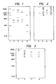
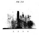
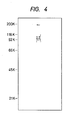
- Figures 1 and 5 are drawings representing electrophoretic diagram detected by a dyeing reaction of protein by means of a C.B.B. method (Biochemistry, Vol. 10, p. 2606 (1971)) and Figures 2 to 4 are representing electrophoretic diagram drawings detected by a dyeing reaction of saccharide by means of a Pas method (Anal. Biochem., Vol. 30, 148 (1962)).
- Receptor 1 obtained in Reference Example 2 in an amount of 5 g as protein together with a Freund's complete adjuvant was subcutaneously administered to a rabbit (2 to 3 kg) once every two weeks to immunize the animal. After immunizing three times, the blood was collected, and an antiserum was collected by centrifugal separation to obtain a GRA antibody (antibody-I).
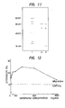
- the Figure 11 shows dyeing patterns by a C.B.B. method, wherein each number is as follows.
- a physiological saline containing 100 ⁇ g as protein of Receptor M-3 obtained in Reference Example 2 was heated to 100°C for 10 minutes in a boiling water bath to obtain a thermally denatured antigen.
- GRA-M-3-H a physiological saline containing 100 ⁇ g as protein of Receptor M-3 obtained in Reference Example 2
- the spleen of C57BL/6 mouse male, 6 weeks was picked out and washed twice with the medium RP M I-1640. After it was loosened by an injector needle, it was filtered by a stainless steel mesh (No. 100) to remove fragments having a .large size. After the cells filtered off was washed twice with the above described medium, they were centrifuged at 1,200 X g for 10 minutes to obtain 4 x 10 7 spleen lymphocytes.
- the thermally denatured antigen of the present invention is effective in a wide range, because it has a low dependency on the concentration when forming cell mediated immunity (induction of killer cells), and it is understood that it has a great effect.
- thermally denatured antigen of the present invention has a very high induction activity for killer cells.
- GRA-6-H was administered into a monkey of Macaca fuscata (Nippon Primates Co.) by intradermally administration at a dosage of 200 ng protein/monkey. After 3, 6, 24, 48 and 72 hours from administration, 5 ml of blood was collected from a femoral vein of the monkey using a heparin-treated syringe. Then lymphocytes were.separated by the difference of specific gravity using a specific gravity separating solution SMF (produced by JIMRO), and they were adjusted with the medium RPMI-1640 so as to be 2 x 10 6 cells/ml. Then, the cancerous cell cytotoxicity of the lymphocytes was measured.
- SMF specific gravity separating solution
- This activity test was carried out using target cells (QG90) obtained by the same manner as in Reference Example 13-(2). Likewise, 200 ⁇ l of each lymphocyte solution was added dropwise. After incubation at 37°C in a carbon dioxide gas incubator for 90 minutes, dyeing was carried out using Eosine, and the number of dead cells was measured. The results are shown in the following Table 9. In Table 9, the number of dead cells is shown as an average f SD per well. Further, the passage time: 0 hour means a control wherein lymphocytes which were collected just before administration of GRA-6-H were used.
- Peripheral blood lymphocytes obtained from a healthy adult by centrifugal separation by means of "Ficollpack" (Pharmacia Co.) were adjusted with the medium RPMI-1640 containing 10% of FCS so-as to be 1.5 x 10 6 cells/ml.
- the medium RPMI-1640 containing 10% of FCS so-as to be 1.5 x 10 6 cells/ml.
- GRA-3 was added so as to be 5, 25 or 50 ng protein/ml, and incubation was carried out at 37°C in a carbon dioxide gas incubator for 48 hours to obtain killer cells.
- Efector cells As target cells (T), those which were obtained by adjusting Daudi washed twice with the above described medium with the same medium containing 10% of FCS so as to be 1.5 x 10 6 cells/ml were used.
- Killer Activity ⁇ (Number of Cells When Using E of the Control) - (Number of Cells of Tested) ⁇ /(E' + T') wherein E' represents the number of survival cells after 100 ⁇ l of E is incubated at 37°C for 1 hour. T' represents the number of survival cells after 100 ⁇ l of T is incubated at 37°C for 1 hour. Results are shown in Table 11.
Landscapes
- Health & Medical Sciences (AREA)
- Life Sciences & Earth Sciences (AREA)
- Mycology (AREA)
- Pharmacology & Pharmacy (AREA)
- Chemical & Material Sciences (AREA)
- Immunology (AREA)
- Medicinal Chemistry (AREA)
- Microbiology (AREA)
- Cell Biology (AREA)
- Oncology (AREA)
- Epidemiology (AREA)
- Animal Behavior & Ethology (AREA)
- General Health & Medical Sciences (AREA)
- Public Health (AREA)
- Veterinary Medicine (AREA)
- Medicines Containing Antibodies Or Antigens For Use As Internal Diagnostic Agents (AREA)
- Medicines That Contain Protein Lipid Enzymes And Other Medicines (AREA)
- Peptides Or Proteins (AREA)
Applications Claiming Priority (4)
| Application Number | Priority Date | Filing Date | Title |
|---|---|---|---|
| JP57177411A JPS5967224A (ja) | 1982-10-08 | 1982-10-08 | 糖鎖関連抗原およびその製造法ならびにこれを有効成分とする抗癌剤 |
| JP177411/82 | 1982-10-08 | ||
| JP100528/83 | 1983-06-06 | ||
| JP58100528A JPS59225119A (ja) | 1983-06-06 | 1983-06-06 | 熱変性抗原の製造法 |
Publications (2)
| Publication Number | Publication Date |
|---|---|
| EP0106285A2 true EP0106285A2 (fr) | 1984-04-25 |
| EP0106285A3 EP0106285A3 (fr) | 1987-09-02 |
Family
ID=26441535
Family Applications (1)
| Application Number | Title | Priority Date | Filing Date |
|---|---|---|---|
| EP83110040A Withdrawn EP0106285A3 (fr) | 1982-10-08 | 1983-10-07 | Antigène relatif à la liaison glycosidique, procédé de production et agent anticancéreux le contenant |
Country Status (8)
| Country | Link |
|---|---|
| EP (1) | EP0106285A3 (fr) |
| CA (1) | CA1236016A (fr) |
| DK (1) | DK463283A (fr) |
| ES (2) | ES526622A0 (fr) |
| FI (1) | FI80711C (fr) |
| NO (1) | NO833655L (fr) |
| PH (1) | PH23701A (fr) |
| PT (1) | PT77472B (fr) |
Cited By (6)
| Publication number | Priority date | Publication date | Assignee | Title |
|---|---|---|---|---|
| EP0157427A3 (fr) * | 1984-04-06 | 1987-08-12 | Otsuka Pharmaceutical Co., Ltd. | Procédé de préparation d'un antigène apparenté à la liaison glycosidique |
| EP0162825A3 (en) * | 1984-05-18 | 1987-10-07 | Jan Roland Holmgren | The use of a specific carcinom associated antigen (hapten), fucosylsialosylgangliotetraose (fuc-gm1) in diagnostic or therapeutic procedures related to human lung cancer (small cell carcinomas) |
| US5132413A (en) * | 1990-03-13 | 1992-07-21 | Japan Immuno Research Laboratories Co., Ltd. | Sugar chain |
| WO1994012213A1 (fr) * | 1992-11-24 | 1994-06-09 | Ole Buchardt | Procede de production d'anticorps avec des conjugues de l'antigene de lymphocytes b et une molecule porteuse denaturee |
| US5376531A (en) * | 1992-09-03 | 1994-12-27 | Northwestern University | Method of detecting cancer |
| MD838G2 (ro) * | 1992-07-21 | 1998-09-30 | Alexandr Nicolaenco | Remediu biologic activ cu proprietate imunomodulatoare, procedeu de obţinere a lui, preparat pe baza lui şi procedeu de normalizare a stării fiziologice a omului şi animalelor |
Family Cites Families (3)
| Publication number | Priority date | Publication date | Assignee | Title |
|---|---|---|---|---|
| WO1982003988A1 (fr) * | 1981-05-19 | 1982-11-25 | Stephen Sugaar | Substances fortement antigeniques specifiques des tumeurs et procedes permettant de detecter le cancer en utilisant ces substances |
| GB2106935B (en) * | 1981-10-01 | 1985-01-30 | Otsuka Pharma Co Ltd | Cancer cell-combatting lymphocytes process for the production thereof and anticancer agents containing said lymphocytes |
| GB2121417A (en) * | 1982-02-22 | 1983-12-21 | Carlton Med Prod | Antigens and antibodies useful in the detection of cancer |
-
1983
- 1983-10-07 PH PH29673A patent/PH23701A/en unknown
- 1983-10-07 FI FI833652A patent/FI80711C/fi not_active IP Right Cessation
- 1983-10-07 PT PT77472A patent/PT77472B/pt not_active IP Right Cessation
- 1983-10-07 NO NO833655A patent/NO833655L/no unknown
- 1983-10-07 CA CA000438680A patent/CA1236016A/fr not_active Expired
- 1983-10-07 DK DK463283A patent/DK463283A/da not_active Application Discontinuation
- 1983-10-07 ES ES526622A patent/ES526622A0/es active Granted
- 1983-10-07 EP EP83110040A patent/EP0106285A3/fr not_active Withdrawn
-
1985
- 1985-05-30 ES ES544306A patent/ES8700056A1/es not_active Expired
Cited By (7)
| Publication number | Priority date | Publication date | Assignee | Title |
|---|---|---|---|---|
| EP0157427A3 (fr) * | 1984-04-06 | 1987-08-12 | Otsuka Pharmaceutical Co., Ltd. | Procédé de préparation d'un antigène apparenté à la liaison glycosidique |
| US4849510A (en) * | 1984-04-06 | 1989-07-18 | Otsuka Pharmaceutical Co., Ltd. | Process for preparing glycosidic linkage related antigen |
| EP0162825A3 (en) * | 1984-05-18 | 1987-10-07 | Jan Roland Holmgren | The use of a specific carcinom associated antigen (hapten), fucosylsialosylgangliotetraose (fuc-gm1) in diagnostic or therapeutic procedures related to human lung cancer (small cell carcinomas) |
| US5132413A (en) * | 1990-03-13 | 1992-07-21 | Japan Immuno Research Laboratories Co., Ltd. | Sugar chain |
| MD838G2 (ro) * | 1992-07-21 | 1998-09-30 | Alexandr Nicolaenco | Remediu biologic activ cu proprietate imunomodulatoare, procedeu de obţinere a lui, preparat pe baza lui şi procedeu de normalizare a stării fiziologice a omului şi animalelor |
| US5376531A (en) * | 1992-09-03 | 1994-12-27 | Northwestern University | Method of detecting cancer |
| WO1994012213A1 (fr) * | 1992-11-24 | 1994-06-09 | Ole Buchardt | Procede de production d'anticorps avec des conjugues de l'antigene de lymphocytes b et une molecule porteuse denaturee |
Also Published As
| Publication number | Publication date |
|---|---|
| CA1236016A (fr) | 1988-05-03 |
| FI833652A0 (fi) | 1983-10-07 |
| ES544306A0 (es) | 1986-10-16 |
| DK463283A (da) | 1984-04-09 |
| NO833655L (no) | 1984-04-09 |
| FI833652A7 (fi) | 1984-04-09 |
| ES8600692A1 (es) | 1985-11-01 |
| ES8700056A1 (es) | 1986-10-16 |
| PT77472A (en) | 1983-11-01 |
| ES526622A0 (es) | 1985-11-01 |
| EP0106285A3 (fr) | 1987-09-02 |
| FI80711B (fi) | 1990-03-30 |
| PT77472B (en) | 1986-02-12 |
| FI80711C (fi) | 1990-07-10 |
| DK463283D0 (da) | 1983-10-07 |
| PH23701A (en) | 1989-09-27 |
Similar Documents
| Publication | Publication Date | Title |
|---|---|---|
| Springer et al. | Blood group MN precursors as human breast carcinoma‐associated antigens and “naturally” occurring human cytotoxins against them | |
| Berg et al. | ATPase-associated antigen (M2): marker antigen for serological diagnosis of primary biliary cirrhosis | |
| Thomson et al. | A cross-reacting embryonic antigen in the membrane of rat sarcoma cells which is immunogenic in the syngeneic host | |
| US5389530A (en) | Methods for the production of antibodies and induction of immune responses to tumor-associated ganagliosides by immunization with gangloiside lactones | |
| Pilch et al. | Transfer of tumor immunity with ribonucleic acid | |
| Springer et al. | Blood group Tn-active macromolecules from human carcinomas and erythrocytes: characterization of and specific reactivity with mono-and poly-clonal anti-Tn antibodies induced by various immunogens | |
| EP0173663B1 (fr) | Application d'un antigène spécifique associé aux tumeurs, sialosyllactotétraose, dans des procédures de diagnostic ou de thérapie relatives aux maladies cancéreuses | |
| Claflin et al. | The Dunning R3327 prostate adenocarcinoma in the Fischer-Copenhagen F1 rat: a useful model for immunological studies | |
| CA1236016A (fr) | Antigene en relation avec la liaison glycosidique, procede de production et agent anticancereux le contenant a titre de principe actif | |
| Chism et al. | Evidence for common and distinct determinants of colon carcinoembryonic antigen, colon carcinoma antigen-III, and molecules with carcinoembryonic antigen activity isolated from breast and ovarian cancer | |
| KR870001375B1 (ko) | 글리코사이드결합 관련항원의 제조방법 | |
| Springer et al. | Tn epitopes, immunoreactive with ordinary anti-Tn antibodies, on normal, desialylated human erythrocytes and on Thomsen-Friedenreich antigen isolated therefrom | |
| EP0162825B1 (fr) | Utilisation d'un antigène spécifique (haptène) associé au carcinome, fucosylsialosylgangliotétrose (Fuc-GM1) dans des méthodes diagnostiques ou thérapeutiques en relation avec le cancer du poumon humain (carcinome de petite cellule) | |
| Gaini et al. | Ganglioside content and composition in human gliomas | |
| KR100206061B1 (ko) | 엠씨에이 28에이 32에 의해 인식되는 항원,씨티에이에이28에이32 | |
| US5418129A (en) | Blood treatment method | |
| CA1336404C (fr) | Facteur d'identification scm associe au cancer en general, sa preparation et sa methode d'utilisation | |
| James et al. | Waldenström's macroglobulinaemia in a bird breeder: a case history with pulmonary involvement and antibody activity of the monoclonal IgM to canary's droppings | |
| Huber et al. | Immune Response to a Mammary Adenocarcinoma: VI. Blocking of Tumor-Specific Cell-Mediated Cytotoxicity by Antibodies to the Cytotoxic Lymphocyte | |
| JPS591420A (ja) | 糖鎖関連抗原およびその製造法 | |
| CA1201988A (fr) | Lymphocytes destructeurs de cellules cancerigenes; methode pour les obtenir et agents anticancerigenes renfermant ces lymphocytes | |
| Umeda et al. | Agglutination of murine tumor cells by sera from various cancer patients | |
| Stavrou et al. | Chemical modification and antigenicity of glioma cells | |
| Easty | Cell surfaces in neoplasia | |
| Bird | Haematoserology of neoplasms |
Legal Events
| Date | Code | Title | Description |
|---|---|---|---|
| PUAI | Public reference made under article 153(3) epc to a published international application that has entered the european phase |
Free format text: ORIGINAL CODE: 0009012 |
|
| AK | Designated contracting states |
Designated state(s): AT BE CH DE FR GB IT LI NL SE |
|
| PUAL | Search report despatched |
Free format text: ORIGINAL CODE: 0009013 |
|
| AK | Designated contracting states |
Kind code of ref document: A3 Designated state(s): AT BE CH DE FR GB IT LI NL SE |
|
| 17P | Request for examination filed |
Effective date: 19871228 |
|
| 17Q | First examination report despatched |
Effective date: 19891017 |
|
| STAA | Information on the status of an ep patent application or granted ep patent |
Free format text: STATUS: THE APPLICATION IS DEEMED TO BE WITHDRAWN |
|
| 18D | Application deemed to be withdrawn |
Effective date: 19901023 |
|
| RIN1 | Information on inventor provided before grant (corrected) |
Inventor name: ADACHI, MASAKAZU |