EP0108400A2 - Determination of unsaturated thyroxine binding protein sites using fluorescence polarization techniques - Google Patents
Determination of unsaturated thyroxine binding protein sites using fluorescence polarization techniques Download PDFInfo
- Publication number
- EP0108400A2 EP0108400A2 EP83110998A EP83110998A EP0108400A2 EP 0108400 A2 EP0108400 A2 EP 0108400A2 EP 83110998 A EP83110998 A EP 83110998A EP 83110998 A EP83110998 A EP 83110998A EP 0108400 A2 EP0108400 A2 EP 0108400A2
- Authority
- EP
- European Patent Office
- Prior art keywords
- thyroxine
- thyroxine binding
- sample
- binding protein
- unsaturated
- Prior art date
- Legal status (The legal status is an assumption and is not a legal conclusion. Google has not performed a legal analysis and makes no representation as to the accuracy of the status listed.)
- Granted
Links
Classifications
-
- C—CHEMISTRY; METALLURGY
- C07—ORGANIC CHEMISTRY
- C07D—HETEROCYCLIC COMPOUNDS
- C07D493/00—Heterocyclic compounds containing oxygen atoms as the only ring hetero atoms in the condensed system
- C07D493/02—Heterocyclic compounds containing oxygen atoms as the only ring hetero atoms in the condensed system in which the condensed system contains two hetero rings
- C07D493/10—Spiro-condensed systems
-
- C—CHEMISTRY; METALLURGY
- C07—ORGANIC CHEMISTRY
- C07D—HETEROCYCLIC COMPOUNDS
- C07D311/00—Heterocyclic compounds containing six-membered rings having one oxygen atom as the only hetero atom, condensed with other rings
- C07D311/02—Heterocyclic compounds containing six-membered rings having one oxygen atom as the only hetero atom, condensed with other rings ortho- or peri-condensed with carbocyclic rings or ring systems
- C07D311/78—Ring systems having three or more relevant rings
- C07D311/80—Dibenzopyrans; Hydrogenated dibenzopyrans
- C07D311/82—Xanthenes
-
- G—PHYSICS
- G01—MEASURING; TESTING
- G01N—INVESTIGATING OR ANALYSING MATERIALS BY DETERMINING THEIR CHEMICAL OR PHYSICAL PROPERTIES
- G01N33/00—Investigating or analysing materials by specific methods not covered by groups G01N1/00 - G01N31/00
- G01N33/48—Biological material, e.g. blood, urine; Haemocytometers
- G01N33/50—Chemical analysis of biological material, e.g. blood, urine; Testing involving biospecific ligand binding methods; Immunological testing
- G01N33/74—Chemical analysis of biological material, e.g. blood, urine; Testing involving biospecific ligand binding methods; Immunological testing involving hormones or other non-cytokine intercellular protein regulatory factors such as growth factors, including receptors to hormones and growth factors
- G01N33/78—Thyroid gland hormones, e.g. T3, T4, TBH, TBG or their receptors
-
- Y—GENERAL TAGGING OF NEW TECHNOLOGICAL DEVELOPMENTS; GENERAL TAGGING OF CROSS-SECTIONAL TECHNOLOGIES SPANNING OVER SEVERAL SECTIONS OF THE IPC; TECHNICAL SUBJECTS COVERED BY FORMER USPC CROSS-REFERENCE ART COLLECTIONS [XRACs] AND DIGESTS
- Y10—TECHNICAL SUBJECTS COVERED BY FORMER USPC
- Y10S—TECHNICAL SUBJECTS COVERED BY FORMER USPC CROSS-REFERENCE ART COLLECTIONS [XRACs] AND DIGESTS
- Y10S436/00—Chemistry: analytical and immunological testing
- Y10S436/80—Fluorescent dyes, e.g. rhodamine
Definitions
- Free thyroxine is the generally accepted marker for the initial assessment of possible thyroid dysfunction: J. Clin, Invest., 45:133, 1966. Since approximately 0,05% of the total circulating thyroxine is physiologically active (i.e., free thyroxine), it is apparent that measurement of total thyroxine concentration only is not a completely satisfactory marker, Free thyroxine is however difficult to measure directly: Berger, S., and Quinn, J. L., Fundamentals of Clinical Chemistry, Tietz, N,W., Ed; W. B.
- T 3 uptake assays use 125 I labeled liothyronine (T 3 1 to competitively bind to the free TBG sites and a secondary solid support (viz: resin, charcoal, etc.), After an incubation period and suitable separation technique (yis : washing, centrifugation, etc, ) the remaining radioactivity on the .solid support is counted. This radioactivity is inversely proportional to the unsaturated thyroxine binding globulin concentration.
- a third methodology assesses the total thyroxine binding globulin capacity by equilibrating with a large excess of thyroxine to displace the endogenous thyroxine in the sample: U. S. Patent 3,960,492 (1976).
- Another methodology utilizes a thyroxine irreversible enzyme inhibitor conjugate, which binds by way of the thyroxine moiety of a site of unsaturation on the thyroxine binding globulin, thereby inactivating the irreversible enzyme inhibitor moiety.
- a thyroxine irreversible enzyme inhibitor conjugate which binds by way of the thyroxine moiety of a site of unsaturation on the thyroxine binding globulin, thereby inactivating the irreversible enzyme inhibitor moiety.
- thyroxine will bind to binding proteins other than thyroxine binding protein, in particular, albumin and pre-ablumin and therefore the presence of such proteins will influence free thyroxine levels.
- the present invention relates to a method for determining unsaturated thyroxine binding protein sites utilizing fluorescent polarization techniques.
- the present invention relates to a method for determining unsaturated thyroxine binding protein sites comprising contacting a sample containing thyroxine binding proteins, with a fluorescent labeled tracer wherein said fluorescent labeled tracer has a preferential binding affinity for thyroxine binding globulin, and the measuring the amount of fluorescent labeled tracer bound to thyroxine binding proteins utilizing fluorescent polarization techniques as a measure of unsaturated thyroxine binding protein sites.
- the measurement of unsaturated.thyroxine binding protein sites is used in conjunction with total thyroxine concentration to determine a free thyroxine index which is a generally accepted marker for the intitial assessment of thyroid dysfunction,
- Samples which can be assayed by the present invention for the determination of unsaturated thyroxine binding protein sites include biological fluids having either an unknown.T uptake (e.g,, a patient's sample) or biological fluids having a known T uptake (i.e., standards), Typical biological fluids include, but are not limited to plasma and serum. Serum is the preferred biological fluid employed in the analysis of the present invention.
- T uptake refers to the relative concentration of unsaturated thryoxine binding protein sites available for binding to thyroxine.
- the fluorescent labeled tracers which are utilized in the methods of the present invention preferentially bind thyroxine binding globulin rather than other specific binding proteins.
- the term "preferential binding affinity for thyroxine binding globulin” refers to the fact that the tracer has essentially the same relative binding affinity for thyroxine binding globulin, albumin and pre-albumin as does thyroxine. That is, the ratio of the relative assay response of a tracer to thryoxine binding globulin and the binding of the tracer to albumin and thyroxine binding pre-albumin should be greater than 1 and preferably greater than 3,
- the fluorescent labeled tracers comprise a thyroxine moiety or thyroxine analog, bound to a fluorescent label.
- Any suitable fluorescent label can be used to label the thyroxine or thyroxine analog, Fluorescent labels include, but are not limited to, fluorescein and rhodamine and derivatives thereof, It is preferred to employ a fluorescein derivative as the fluorescent label.
- the fluorescent labeled tracers wherein a thyroxine moiety or thyroxine analog are bound to fluorescein or fluorescein derivatives are hereinafter referred to as thyroxine-fluorescein conjugates,
- the following structures illustrate preferred thyroxine-fluorescein conjugates useful as tracers in conjunction with the methods of the present invention: O OH OH and biologically acceptable salts thereof, wherein the bonded carbonyl group is bonded to the 5 or 6 position on the fluorescein moiety; and OH wherein n is an integer of from 1 to 8 and biologically acceptable salts thereof, and wherein the carbonyl group is bonded to the 5 or 6 position on the fluorescein moietv; and wherein m is an integer of from 1 to 8, p is an integer 0 or 1,
- A is an acid salt having a pH less than 3, such as, for example, hydrochloride, trifluoroacetate, trichloroacetate, acetate, and the like, and biologically acceptable salts thereofm and wherein the-carbonyl group is bonded to the 5 or 6 position on the fluorescein moiety,
- a sample containing thyroxine binding protein is contacted with a fluorescent labeled tracer thus permitting the fluorescent labeled tracer to bind the unsaturated thyroxine binding protein sites in the sample and form a complex.
- the production of the complex increases the fluorescence polarization of the solution from that containing only free tracer.
- the increase in polarization of fluorescence caused by contacting the thyroxine bindina protein with the fluorescent labeled tracer can be measured by known polarized fluorometric techniques via either an endpoint or kinetic methodology.
- an endpoint fluorometric technique is employd in the assay of this invention,
- the sample must be treated with a composition comprising a nonfluorescent surfactant in a buffered medium having a pH greater than 7.0.
- Nonfluorescent surfactants utilized in such compositions are readily ascertained by one of ordinary skill in the art and include for example, sodium dodecyl sulfate.
- the buffered media utilized in such compositions include buffers sufficient to maintain the pH of the solution greater than pH 7 and preferably in the range of 7.5 - 9.5, and most preferably in the range of 8.0 - 9.0.
- Respective buffered media include bicarbonate buffers, tris(hydroxymethyl)aminomethane and the like.
- a buffer having a low pK such as, a phosphate buffer
- organic bases such as triethylamine and triethylenediamine may be employed in conjunction with a buffer to provide a buffered medium in the desired pH range.
- the methods of the present invention provide a direct means for determining a T uptake value for a sample in a homogeneous assay system without the need for additional reagents, such an antibodies to thyroxine,
- the thyroxine-fluorescein conjugates employed in the present invention are relatively easy to-produce and purify and are more stable than reagents employed in a T uptake assay employing enzyme inhibitor techniques.
- this is a fluorescence assay, the sample size is generally significantly lower than that required in T uptake assays using conventional techniques.
- thyroxine-fluorescein conjugates useful as tracers in conjunction with the methods of the present invention may generally be prepared in accordance with conventional procedures.
- the following examples serve to illustrate the preparation of thyroxine-fluorescein conjugates that may be employed in the methods of the present invention.
- the symbol [CF] appearing in the structural formulas illustrating the compounds prepared in the following examples, represents a moiety of the formula:
- D-thyroxine sodium salt 100 mg, 0.00012 mole
- di-t-butyl dicarbonate 27 mg, 0.00012 mole
- dimethylformamide 1 ml
- N-t-butoxycarbonyl D-thyroxine 51 mg, 0.00006 mole
- N-hydroxysuccinimide 7 mg, 0.00006 molel
- N,N'- dicyclohexylcarbodiimide 15 mg, 0.00006 mole
- the reaction mixture is stirred at room temperature for three hours after which time the reaction mixture is filtered into a solution of 1,3-diaminopropane (50 ⁇ l, 0.0003 mole) in 1 ml dimethylformamide with constant stirring.
- N-t-butoxycarbonyl D-thyroxine propylenediamine 10 mg, 0.00001 mole
- N-hydroxysuccinimide active ester of a mixture of 5- and 6-carboxyfluorescein 5 mq, 0.00001 mle
- the reaction mixture is stirred for thirty minutes at room temperature and the solvent is removed under vacuum.
- the residue is purified using a silica gel prep TLC plate eluting with a 9:1 mixture of methylene chloride:methanol to yield a mixture of N-t-butoxycarbonyl D-thyroxine propylenediamine carboxyfluorescein having the general formula:
- reaction was allowed to proceed for 18 hours after which time the reaction mixture was passed through an ion exchange resin column (Bio-Rad AGo 50W- X 8 (H + form) using methanol as the eluent.
Landscapes
- Chemical & Material Sciences (AREA)
- Health & Medical Sciences (AREA)
- Organic Chemistry (AREA)
- Life Sciences & Earth Sciences (AREA)
- Molecular Biology (AREA)
- Engineering & Computer Science (AREA)
- Urology & Nephrology (AREA)
- Endocrinology (AREA)
- Biomedical Technology (AREA)
- Hematology (AREA)
- Immunology (AREA)
- Biotechnology (AREA)
- General Health & Medical Sciences (AREA)
- Food Science & Technology (AREA)
- Medicinal Chemistry (AREA)
- Physics & Mathematics (AREA)
- Analytical Chemistry (AREA)
- Biochemistry (AREA)
- Microbiology (AREA)
- General Physics & Mathematics (AREA)
- Pathology (AREA)
- Cell Biology (AREA)
- Investigating Or Analysing Biological Materials (AREA)
- Investigating, Analyzing Materials By Fluorescence Or Luminescence (AREA)
- Heterocyclic Carbon Compounds Containing A Hetero Ring Having Oxygen Or Sulfur (AREA)
Abstract
Description
- Free thyroxine is the generally accepted marker for the initial assessment of possible thyroid dysfunction: J. Clin, Invest., 45:133, 1966. Since approximately 0,05% of the total circulating thyroxine is physiologically active (i.e., free thyroxine), it is apparent that measurement of total thyroxine concentration only is not a completely satisfactory marker, Free thyroxine is however difficult to measure directly: Berger, S., and Quinn, J. L., Fundamentals of Clinical Chemistry, Tietz, N,W., Ed; W. B. Saunders, Co,: Philadelphia, 1976, Chapter 14 and Robbins, J., Thyroid Hormone Metabolism, Harlem and Orr, Ed., 1975, Chapter 1, The remaining circulating thyroxine is bound to protein, primarily thyroxine binding globulin (TBG) : Robbins, J., and Rall, J. G., Proteins Associated with Thyroid Hormones, Physiol. Rev., 40, 415, 1960, Thus, the measurement of total circulating thyroxine concentration and of thyroxine binding globulin concentration provides a good assessment of the relative concentration of free thyroxine.
- Various methods have been used to assess the level of thyroxine binding globulin concentrations, The oldest method is the T3 uptake study which measures unsaturated thyroxine binding globulin capacity; J, Clin, Endocrinol, 17:33, January, 1957 and J. Clin, EndocrinoZ., 25:39-54, 1965. T3 uptake assays use 125I labeled liothyronine (T31 to competitively bind to the free TBG sites and a secondary solid support (viz: resin, charcoal, etc.), After an incubation period and suitable separation technique (yis : washing, centrifugation, etc, ) the remaining radioactivity on the .solid support is counted. This radioactivity is inversely proportional to the unsaturated thyroxine binding globulin concentration.
- Another methodology is a radioimmunoassay which utilizes antibody specific for thyroxine binding globulin, However methodology does not measure the binding ability of the thyroxine binding globulins: Clin, Chem, Acta., 87, (1978), 373-381.
- A third methodology assesses the total thyroxine binding globulin capacity by equilibrating with a large excess of thyroxine to displace the endogenous thyroxine in the sample: U. S. Patent 3,960,492 (1976).
- Another methodology utilizes a thyroxine irreversible enzyme inhibitor conjugate, which binds by way of the thyroxine moiety of a site of unsaturation on the thyroxine binding globulin, thereby inactivating the irreversible enzyme inhibitor moiety. Thus, the greater the amount of unsaturated thyroxine binding globulin, the more conjugate that will be bound and the amount of irreversible enzyme inhibitor which will be available to inhibit the enzyme will be reduced. That is: increased amounts of thyroxine binding globulin will result in greater enzyme activity. Thus, intermixing enzyme and substrate to the enzyme and the above-described thyroxine irreversible enzyme inhibitor with serum permits determination of unsaturated thyroxine binding globulin in serum: U. S. Patent 4,341,865.
- Another methodology involves the use of fluorescent polarization immunoassay techniques to measure T3 uptake. However, this procedure involves separation steps as well as the use of antibody against T3: U, S. Patent 4,347,059,
- It is known that thyroxine will bind to binding proteins other than thyroxine binding protein, in particular, albumin and pre-ablumin and therefore the presence of such proteins will influence free thyroxine levels. Fesco, et al, Clin. Chem., 28/6, p. 1325-1329 (1982).
- The present invention relates to a method for determining unsaturated thyroxine binding protein sites utilizing fluorescent polarization techniques. In particular the present invention relates to a method for determining unsaturated thyroxine binding protein sites comprising contacting a sample containing thyroxine binding proteins, with a fluorescent labeled tracer wherein said fluorescent labeled tracer has a preferential binding affinity for thyroxine binding globulin, and the measuring the amount of fluorescent labeled tracer bound to thyroxine binding proteins utilizing fluorescent polarization techniques as a measure of unsaturated thyroxine binding protein sites.
- The measurement of unsaturated.thyroxine binding protein sites is used in conjunction with total thyroxine concentration to determine a free thyroxine index which is a generally accepted marker for the intitial assessment of thyroid dysfunction,
- Samples which can be assayed by the present invention for the determination of unsaturated thyroxine binding protein sites include biological fluids having either an unknown.T uptake (e.g,, a patient's sample) or biological fluids having a known T uptake (i.e., standards), Typical biological fluids include, but are not limited to plasma and serum. Serum is the preferred biological fluid employed in the analysis of the present invention.
- As used herein the term "T uptake" refers to the relative concentration of unsaturated thryoxine binding protein sites available for binding to thyroxine.
- The fluorescent labeled tracers which are utilized in the methods of the present invention preferentially bind thyroxine binding globulin rather than other specific binding proteins. The term "preferential binding affinity for thyroxine binding globulin" refers to the fact that the tracer has essentially the same relative binding affinity for thyroxine binding globulin, albumin and pre-albumin as does thyroxine. That is, the ratio of the relative assay response of a tracer to thryoxine binding globulin and the binding of the tracer to albumin and thyroxine binding pre-albumin should be greater than 1 and preferably greater than 3,
- The fluorescent labeled tracers comprise a thyroxine moiety or thyroxine analog, bound to a fluorescent label. Any suitable fluorescent label can be used to label the thyroxine or thyroxine analog, Fluorescent labels include, but are not limited to, fluorescein and rhodamine and derivatives thereof, It is preferred to employ a fluorescein derivative as the fluorescent label. The fluorescent labeled tracers wherein a thyroxine moiety or thyroxine analog are bound to fluorescein or fluorescein derivatives are hereinafter referred to as thyroxine-fluorescein conjugates,
- The following structures illustrate preferred thyroxine-fluorescein conjugates useful as tracers in conjunction with the methods of the present invention: O OH OH


- It is preferred to employ tracers of formula (II) and (III) wherein n or m is in a range of 2 to 4 and more preferred to employ tracers of formula (IIIl wherein n is 2 or 3. It is most preferred to employ a tracer of formula (III) wherein m is 3.
- In accordance with the methods of the present invention a sample containing thyroxine binding protein is contacted with a fluorescent labeled tracer thus permitting the fluorescent labeled tracer to bind the unsaturated thyroxine binding protein sites in the sample and form a complex. The production of the complex increases the fluorescence polarization of the solution from that containing only free tracer. The increase in polarization of fluorescence caused by contacting the thyroxine bindina protein with the fluorescent labeled tracer can be measured by known polarized fluorometric techniques via either an endpoint or kinetic methodology. Preferably, an endpoint fluorometric technique is employd in the assay of this invention,
- To overcome problems associated with nonspecific tracer binding and variable background fluorescence, the sample must be treated with a composition comprising a nonfluorescent surfactant in a buffered medium having a pH greater than 7.0. Nonfluorescent surfactants utilized in such compositions are readily ascertained by one of ordinary skill in the art and include for example, sodium dodecyl sulfate. The buffered media utilized in such compositions include buffers sufficient to maintain the pH of the solution greater than pH 7 and preferably in the range of 7.5 - 9.5, and most preferably in the range of 8.0 - 9.0. Respective buffered media include bicarbonate buffers, tris(hydroxymethyl)aminomethane and the like. In addition, if a buffer having a low pK is employed, such as, a phosphate buffer, organic bases such as triethylamine and triethylenediamine may be employed in conjunction with a buffer to provide a buffered medium in the desired pH range.
- The following examples serve to further illustrate the present invention,
- A. Reagents
- 1. Pretreatment Solution A solution containing 0.15% sodium dodecyl sulfate 0.564 M triethylenediamine (DABCO), and 0.1% sodium azide in O.1M sodium phosphate buffer (pH 7.25).
- 2. T4 Fluorescein Tracer - A tracer of formula (III) wherein m = 3 is employed at a concentration of 2.4 X 10⁻⁷ M in a buffered medium containing 005% sodium dodecyl sulfate, 0.1% bovine gamma globulin, and 0,1% sodium azide in O.1M sodium phosphate buffer.
- 3. T Uptake Calibrators - Sheep anti-T antisera in a 4% human serum matrix having the following uptake values: 0, 0.5, 1.0, 1.5, 2.0, and 2.5. An uptake value of 1.0 is equivalent to the T uptake of normal serum.
- 4. Diluent buffer: 4.1M sodium phosphate containing 0.1% bovine gamma globulin and 0.1% sodium azide.
- All polarized fluorescence measurements were made using a polarization spectrofluorimeter (Abbott TDx m Fluorescence Polarization Analyzer.
-
- 1, To 1 µℓ, aliquot of an unknown sample is added 25 µℓ, of the pretreatment solution and the resulting mixture is diluted to 1 ml with diluent buffer. The resultant assay solution is mixed and the polarized fluorescence background is measured.
- 2. To the assay solution in Step 1. is added a second 1 µℓ aliquot of the unknown sample, 25 pt of the pretreatment solution 25 µℓ of T4 fluorescein tracer, and the buffer to a final volume of 2 ml. The resultant solution is mixed and the polarized fluorescence is measured.
- 3. The fluorescence polarization due to tracer binding is obtained by subtracting the polarized fluorescence intensities of the background from the final polarized fluorescence intensities of-the mixture.
- 4. The polarization values obtained are proportional to the T uptake of each sample.
- 5. The fluorescence polarization for a sample is cmopared to a standard curve prepared using calibrators of known T uptake values to indicate the T uptake value.
- In order to evaluate the efficacy of the T uptake fluorescence polarization assay of the present invention, a correlation study was conducted employing (i) a radioassay T3 uptake procedure set forth in TRIO-BEAD T3 Uptake Diagnostic Kit, Abbott Laboratories, North Chicago, Illinois and (ii) an enzyme inhibitor T uptake procedure set forth in Thyrozyme Uptake Diagnostic Kit, Abbott Laboratories, North Chicago, Illinois.
- The data obtained from the fluorescence polarization assay of the present invention, the radioassay and enzyme inhibitor assay are set forth in Table I.

- As evidenced from the above specification and examples, the methods of the present invention provide a direct means for determining a T uptake value for a sample in a homogeneous assay system without the need for additional reagents, such an antibodies to thyroxine, In addition, the thyroxine-fluorescein conjugates employed in the present invention are relatively easy to-produce and purify and are more stable than reagents employed in a T uptake assay employing enzyme inhibitor techniques. Furthermore, because this is a fluorescence assay, the sample size is generally significantly lower than that required in T uptake assays using conventional techniques.
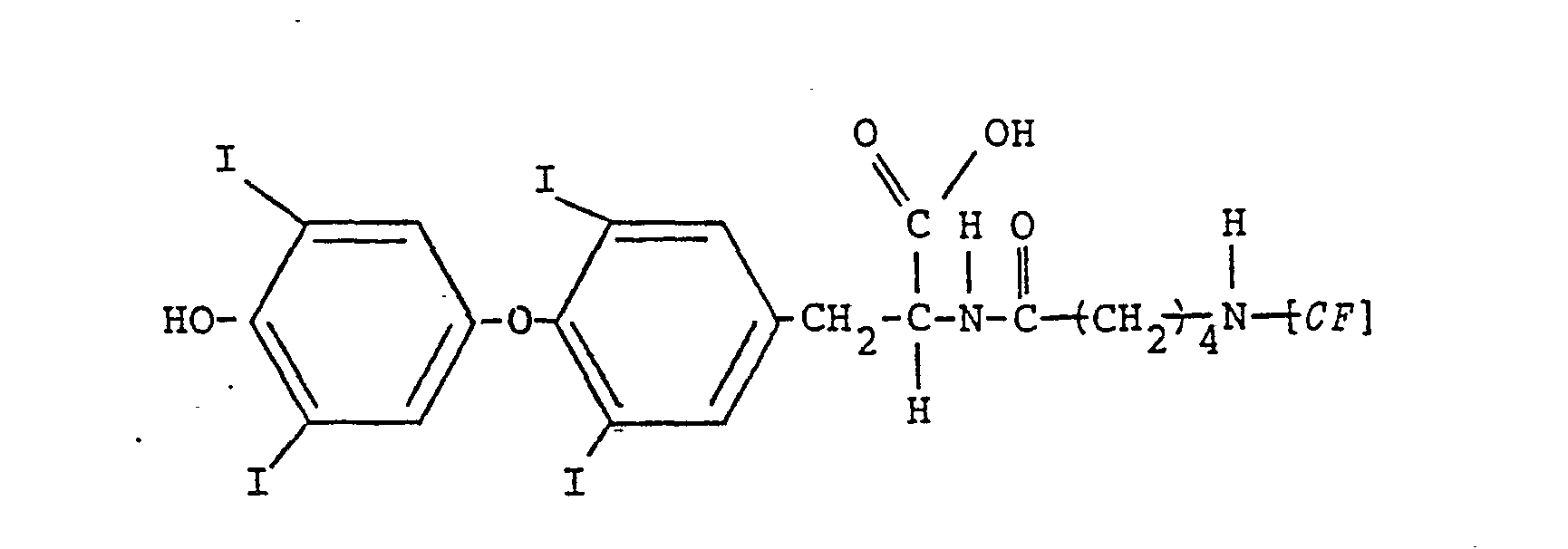
- The thyroxine-fluorescein conjugates useful as tracers in conjunction with the methods of the present invention may generally be prepared in accordance with conventional procedures. The following examples serve to illustrate the preparation of thyroxine-fluorescein conjugates that may be employed in the methods of the present invention. The symbol [CF] appearing in the structural formulas illustrating the compounds prepared in the following examples, represents a moiety of the formula:

- To 83 mg (0.22 mmoll of 6-carboxyfluorescein dissolved in 2 ml of dimethylformamide was added 28 mg (0.24 mmol) of N-hydroxysuccinimide and 55 mg (0.27 mmol) of N,N'- dicyclohexylcarbodiimide, The reaction mixture was stirred at 0° C under argon atmosphere for one hour and then maintained at 4° C for 16 hours to yield a N-hydroxysuccinimide active ester of carboxyfluorescein having the formula:

- D-thyroxine sodium salt (100 mg, 0.00012 mole) is added with constant stirring to a solution of di-t-butyl dicarbonate (27 mg, 0.00012 mole) in 1 ml of dimethylformamide. After stirring two hours at room temperature the solvent is removed under high vacuum, The residue is dissolved in
- methanol and 1N HC1 is added dropwise until a precipitate is formed. The solvent is decanted and the residue is dried under vacuum to yield 90 mg (82% yield) of N-t-butoxycarbonyl D-thyroxine having the formula:

- To a portion of the N-t-butoxycarbonyl D-thyroxine (51 mg, 0.00006 mole) dissolved in 1 ml of dimethylformamide is added N-hydroxysuccinimide (7 mg, 0.00006 molel and N,N'- dicyclohexylcarbodiimide (15 mg, 0.00006 mole), The reaction mixture is stirred at room temperature for three hours after which time the reaction mixture is filtered into a solution of 1,3-diaminopropane (50 µℓ, 0.0003 mole) in 1 ml dimethylformamide with constant stirring. After stirring for fifteen minutes at room temperature, the solvent is removed and the residue is purified on a Whatman C18 reversed phase prep TLC plate eluting with a 80/19/1 mixture of methanol-water-acetic acid to yield 33 mg, (61% yield) of N-t-butoxycarbonyl D-thyroxine propylene diamine having the formula;

- To a portion of the N-t-butoxycarbonyl D-thyroxine propylenediamine (10 mg, 0.00001 mole) dissolved in 1 ml of dimethylformamide is added N-hydroxysuccinimide active ester of a mixture of 5- and 6-carboxyfluorescein (5 mq, 0.00001 mle). The reaction mixture is stirred for thirty minutes at room temperature and the solvent is removed under vacuum. The residue is purified using a silica gel prep TLC plate eluting with a 9:1 mixture of methylene chloride:methanol to yield a mixture of N-t-butoxycarbonyl D-thyroxine propylenediamine carboxyfluorescein having the general formula:

- To a solution containing 5-aminovaleric acid (5.85 g, 0.05 mol) in 100 ml of 2% aqueous sodium hydroxide and 100 ml of dioxane was dropwise added a solution containing di-t-butyldicarbonate (10.9 g, 0.05 mol) in 40 ml of dioxane. The reaction mixture was stirred for 18 hours and then acidified to pH 3 using 1 N hydrochloric acid. The acidified mixture was extracted three times with dichloromethane. The organic layers were combined, washed with water, and dried over sodium sulfate to yield 10.1 g (93.5% yield) of 5-(t-butoxycarbonylamino)valeric acid as a white crystalline solid.
- To a portion of 5-(t-butoxycarbonylamino)valeric acid (0.434 g, .002 mol) was added N,N'-dicyclohexylcarbodiimide (0.412 g, .002 mol) and N-hydroxysuccinimide (0.25 g, .0022 mol) in 3 ml dichloromethane with constant stirring and the reaction was allowed to proceed for 18 hours to yield the N-hydroxysuccinimide active ester of 5-(t-butoxycarbonylamino)-valeric acid as an oily residue. To the oily residue was added L-thyroxine sodium salt pentahydrate (1.95 g, .0022 mol) in 30 ml of methanol.
- The reaction was allowed to proceed for 18 hours after which time the reaction mixture was passed through an ion exchange resin column (Bio-Rad AGo 50W-X8 (H+ form) using methanol as the eluent.
- The eluent was concentrated under vacuum to yield 1.96 g (90% yield) of intermediate of the formula:
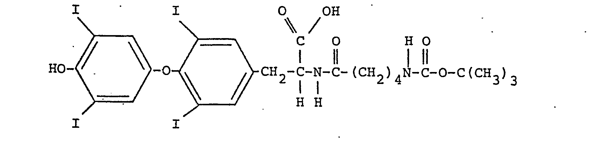
- A portion of the intermediate (.125 g, .00015 mol) was treated with trifluoroacetic acid (2.0 ml) for 30 minutes. The trifluoroacetic acid was removed via evaporation under reduced pressure and the resulting residue was dissolved in 1.5 ml of N,N'-dimethylformamide, The resulting solution was adjusted to a basic pH using triethylamine. To the resulting mixture was added N-hydroxysuccinimide active ester of carboxyfluorescein (75 mg, 0.000159 mol). The reaction was allowed to proceed for 18 hours. Diethylether was added to the reaction mixture to yield a precipitate which was purified via preparatory reverse phase TLC using a mixture of methanol: water:acetic acid (75:25:0.5) to yield .071 g of a thyroxine-6-carboxyfluorescein conjugate as an orange solid, having the formula:
- Although this invention has been described with respect to specific modifications, the details thereof are not to be construed as limitations, for it will be apparent that various equivalents, changes and modifications may be resorted to without departing from the spirit and scope thereof and it is understood that such equivalent embodiments are intended to be included therein.
Claims (10)


Applications Claiming Priority (2)
| Application Number | Priority Date | Filing Date | Title |
|---|---|---|---|
| US06/440,068 US4476228A (en) | 1982-11-08 | 1982-11-08 | Determination of unsaturated thyroxine binding protein sites using fluorescence polarization techniques |
| US440068 | 1989-11-22 |
Publications (3)
| Publication Number | Publication Date |
|---|---|
| EP0108400A2 true EP0108400A2 (en) | 1984-05-16 |
| EP0108400A3 EP0108400A3 (en) | 1984-06-13 |
| EP0108400B1 EP0108400B1 (en) | 1987-04-29 |
Family
ID=23747288
Family Applications (1)
| Application Number | Title | Priority Date | Filing Date |
|---|---|---|---|
| EP83110998A Expired EP0108400B1 (en) | 1982-11-08 | 1983-11-04 | Determination of unsaturated thyroxine binding protein sites using fluorescence polarization techniques |
Country Status (6)
| Country | Link |
|---|---|
| US (1) | US4476228A (en) |
| EP (1) | EP0108400B1 (en) |
| JP (1) | JPS59104554A (en) |
| CA (1) | CA1210690A (en) |
| DE (1) | DE3371251D1 (en) |
| ES (1) | ES8407209A1 (en) |
Cited By (5)
| Publication number | Priority date | Publication date | Assignee | Title |
|---|---|---|---|---|
| EP0257572A3 (en) * | 1986-08-25 | 1988-12-14 | Hoechst Aktiengesellschaft | Thyronin derivatives |
| WO1991001006A1 (en) * | 1989-07-11 | 1991-01-24 | Pb Diagnostic Systems, Inc. | Diagnostic assay system |
| US5055594A (en) * | 1990-07-19 | 1991-10-08 | Becton, Dickinson And Company | Fluorogenic trypotophanase substrates |
| EP0576095A1 (en) * | 1992-06-26 | 1993-12-29 | Johnson & Johnson Clinical Diagnostics, Inc. | Immunoassays with labeled thyronine hapten analogues |
| EP0597333A3 (en) * | 1992-11-10 | 1995-05-10 | Hoffmann La Roche | Compounds for fluorescence polarization immunoassays and immunoassays using them. |
Families Citing this family (32)
| Publication number | Priority date | Publication date | Assignee | Title |
|---|---|---|---|---|
| US4588697A (en) * | 1979-09-07 | 1986-05-13 | Syntex (U.S.A.) Inc. | Method for performing fluorescent protein binding assay employing novel alkyl substituted fluorescent compounds and conjugates |
| US4595656A (en) * | 1984-01-06 | 1986-06-17 | Becton Dickinson & Company | Coupling agents and products produced therefrom |
| US4591569A (en) * | 1984-04-11 | 1986-05-27 | Becton Dickinson & Company | Homogeneous fluorescent triiodothyronine uptake test |
| US4665020A (en) * | 1984-05-30 | 1987-05-12 | United States Department Of Energy | Flow cytometer measurement of binding assays |
| US4681859A (en) * | 1984-09-21 | 1987-07-21 | Ortho Diagnostic Systems Inc. | Fluorescence polarization immunoassay for heavy antigens |
| DE3546014A1 (en) * | 1985-12-24 | 1987-06-25 | Boehringer Mannheim Gmbh | METHOD FOR DETERMINING THE BINDING CAPACITY OF THYROXIN BINDING TBG |
| US4780421A (en) * | 1986-04-03 | 1988-10-25 | Sclavo Inc. | Cleavable labels for use in binding assays |
| US5336622A (en) * | 1986-04-25 | 1994-08-09 | Abbott Laboratories | Tracers for use in flecainide fluorescence polarization immunoassay |
| US5155212A (en) * | 1986-05-21 | 1992-10-13 | Abbott Laboratories | Phencyclidine and phencyclidine metabolites assay, tracers, immunogens, antibodies and reagent kit |
| US5124457A (en) * | 1986-05-21 | 1992-06-23 | Abbott Laboratories | Phencyclidine and phencyclidine metabolites assay, tracers, immunogens and antibodies |
| US5221629A (en) * | 1986-05-21 | 1993-06-22 | Abbott Laboratories | Phencyclidine and phencyclidine metabolites assay, tracers, immunogens and antibodies |
| US4939264A (en) * | 1986-07-14 | 1990-07-03 | Abbott Laboratories | Immunoassay for opiate alkaloids and their metabolites; tracers, immunogens and antibodies |
| US5223627A (en) * | 1986-07-15 | 1993-06-29 | Abbott Laboratories | Fluorescence polarization method for monitoring fetal lung maturity |
| US4784961A (en) * | 1987-03-23 | 1988-11-15 | Abbott Laboratories | Fluorescence polarization method for monitoring fetal lung maturity |
| US4868132A (en) * | 1987-02-03 | 1989-09-19 | Abbott Laboratories | Fluorescence polarization immunoassay for amphetamine/methamphetamine |
| US4824777A (en) * | 1987-07-08 | 1989-04-25 | Ciba Corning Diagnostics Corp. | Method for determining thyroxine uptake |
| US5100807A (en) * | 1987-10-19 | 1992-03-31 | Abbott Laboratories | Phenylacetylglutamine (pag) analytical test |
| DE3812610A1 (en) * | 1988-04-15 | 1989-10-26 | Boehringer Mannheim Gmbh | METHOD FOR DETERMINING THE THYROXIN BINDING CAPACITY AND SUITABLE STANDARD SOLUTION |
| US5262333A (en) * | 1988-10-28 | 1993-11-16 | Abbott Laboratories | Method and reagents for detecting amphetamine and/or D-methamphetamine in biological samples |
| US5073629A (en) * | 1989-01-23 | 1991-12-17 | Abbott Laboratories | Methadone fluorescence polarization immunoassay |
| GB8902689D0 (en) * | 1989-02-07 | 1989-03-30 | Ici Plc | Assay method |
| US5248791A (en) * | 1989-04-10 | 1993-09-28 | Abbott Laboratories | Reagents, methods and kits for an amphetamine-class fluorescence polarization immunoassay |
| US5101015A (en) * | 1989-04-10 | 1992-03-31 | Abbott Laboratories | Reagents for an amphetamine-class fluorescence polarization immunoassay |
| US5099020A (en) * | 1989-11-27 | 1992-03-24 | Abbott Laboratories | Barbiturate assay compositions and methods |
| US5593844A (en) * | 1990-11-19 | 1997-01-14 | Genentech, Inc. | Ligand-mediated immunofunctional hormone binding protein assay method |
| US5210017A (en) * | 1990-11-19 | 1993-05-11 | Genentech, Inc. | Ligand-mediated immunofunctional hormone binding protein assay method |
| US6326154B1 (en) | 1990-11-19 | 2001-12-04 | Genentech, Inc. | Ligand-mediated immunofunctional hormone binding protein assay method |
| US5352803A (en) * | 1992-03-30 | 1994-10-04 | Abbott Laboratories | 5(6)-methyl substituted fluorescein derivatives |
| JP3327551B2 (en) * | 1992-03-30 | 2002-09-24 | アボツト・ラボラトリーズ | Reagents and methods for detecting and quantifying thyroxine in fluid samples |
| US7163918B2 (en) * | 2000-08-22 | 2007-01-16 | New River Pharmaceuticals Inc. | Iodothyronine compositions |
| WO2002082082A2 (en) * | 2001-04-09 | 2002-10-17 | Boehringer Ingelheim Pharmaceuticals, Inc. | Fluorescence polarization assay |
| DE102011086942B4 (en) | 2011-09-30 | 2024-01-11 | Endress+Hauser Conducta Gmbh+Co. Kg | Method for calibrating and/or adjusting an analysis device for chemical substances in liquids, especially in aqueous solutions |
Family Cites Families (9)
| Publication number | Priority date | Publication date | Assignee | Title |
|---|---|---|---|---|
| US4161515A (en) * | 1973-10-02 | 1979-07-17 | Syva Company | Double receptor fluorescent immunoassay |
| US4171311A (en) * | 1978-01-16 | 1979-10-16 | International Diagnostics Tech. Inc. | Imides of thyroxin and triiodothyronine |
| US4238471A (en) * | 1978-07-12 | 1980-12-09 | Becton, Dickinson And Company | Assay for thyroid hormone binding capacity |
| CA1121345A (en) * | 1979-03-05 | 1982-04-06 | Robert A. Yoshida | Method for competitive protein binding assays inhibiting non-specific interference |
| CA1160626A (en) * | 1980-07-30 | 1984-01-17 | Stephen D. Stroupe | Biologically interesting compounds labeled with dichlorotriazinyl-aminofluorescein |
| US4347058A (en) * | 1980-09-15 | 1982-08-31 | Beckman Instruments, Inc. | Thyroid polarization fluoroimmunoassay |
| US4347059A (en) * | 1980-09-15 | 1982-08-31 | Beckman Instruments, Inc. | T3 Uptake polarization fluoroimmunoassay |
| AU555213B2 (en) * | 1981-02-17 | 1986-09-18 | Abbott Laboratories | N-substituted-amido-fluoresein derivatives |
| NZ199629A (en) * | 1981-02-17 | 1984-11-09 | Abbott Lab | Fluorescent polarisation immunoassay and certain substituted carboxy fluoresceins |
-
1982
- 1982-11-08 US US06/440,068 patent/US4476228A/en not_active Expired - Lifetime
-
1983
- 1983-11-02 CA CA000440218A patent/CA1210690A/en not_active Expired
- 1983-11-04 EP EP83110998A patent/EP0108400B1/en not_active Expired
- 1983-11-04 DE DE8383110998T patent/DE3371251D1/en not_active Expired
- 1983-11-07 ES ES527084A patent/ES8407209A1/en not_active Expired
- 1983-11-08 JP JP58208440A patent/JPS59104554A/en active Pending
Cited By (5)
| Publication number | Priority date | Publication date | Assignee | Title |
|---|---|---|---|---|
| EP0257572A3 (en) * | 1986-08-25 | 1988-12-14 | Hoechst Aktiengesellschaft | Thyronin derivatives |
| WO1991001006A1 (en) * | 1989-07-11 | 1991-01-24 | Pb Diagnostic Systems, Inc. | Diagnostic assay system |
| US5055594A (en) * | 1990-07-19 | 1991-10-08 | Becton, Dickinson And Company | Fluorogenic trypotophanase substrates |
| EP0576095A1 (en) * | 1992-06-26 | 1993-12-29 | Johnson & Johnson Clinical Diagnostics, Inc. | Immunoassays with labeled thyronine hapten analogues |
| EP0597333A3 (en) * | 1992-11-10 | 1995-05-10 | Hoffmann La Roche | Compounds for fluorescence polarization immunoassays and immunoassays using them. |
Also Published As
| Publication number | Publication date |
|---|---|
| EP0108400B1 (en) | 1987-04-29 |
| DE3371251D1 (en) | 1987-06-04 |
| CA1210690A (en) | 1986-09-02 |
| JPS59104554A (en) | 1984-06-16 |
| ES527084A0 (en) | 1984-08-16 |
| ES8407209A1 (en) | 1984-08-16 |
| EP0108400A3 (en) | 1984-06-13 |
| US4476228A (en) | 1984-10-09 |
Similar Documents
| Publication | Publication Date | Title |
|---|---|---|
| EP0108400B1 (en) | Determination of unsaturated thyroxine binding protein sites using fluorescence polarization techniques | |
| US4476229A (en) | Substituted carboxyfluoresceins | |
| US4510251A (en) | Fluorescent polarization assay for ligands using aminomethylfluorescein derivatives as tracers | |
| FI67760C (en) | FOERFARANDE FOER BESTAEMNING AV CONCENTRATIONS AV EN FRI LIGAND FRAON EN BIOLOGISK VAETSKA | |
| EP0399184A2 (en) | Reagents, methods and kits for an amphetamine-class fluorescence polarization immunoassay | |
| JP4435305B2 (en) | Topiramate immunoassay and analogs and antibodies | |
| US4614823A (en) | Aminomethylfluorescein derivatives | |
| US5395938A (en) | Biotinylated chemiluminescent labels and their conjugates, assays and assay kits | |
| NO152955B (en) | REAGENT FOR DETERMINING AN IMMUNOLOGICALLY ACTIVE MATERIAL | |
| US5851781A (en) | Reagents and methods for the detection and quantification of testosterone in fluid samples | |
| EP0100543B1 (en) | Method for quantitative analysis of low molecular weight components contained in protein-containing liquid sample | |
| US6190923B1 (en) | Diethylenetriamine-N,N′,N″-triacetic acid derivatives | |
| US6383766B1 (en) | Reduced cortisol conjugates | |
| EP0380019B1 (en) | Methadone fluorescence polarization immunoassay | |
| CA1093962A (en) | Bile acid competitive assay using protein binding inhibitor | |
| CA1339680C (en) | Phenylacetylglutamine (pag) analytical test | |
| EP0108403A2 (en) | Substituted carboxyfluoresceins | |
| EP0226730A2 (en) | Compounds and assay for tricyclic antidepressants | |
| EP0641439B1 (en) | Reagents and methods for the quantification of imipramine or desipramine in biological fluids | |
| EP0242847A2 (en) | Tracers for use in flecainide fluorescence polarization immunoassay | |
| EP0184630A2 (en) | Fluorescence polarization assay, reagents, and method of making reagents, for determination of digitoxin | |
| US5105007A (en) | Phenylacetylglutamine (PAG) analytical test | |
| CA2172069A1 (en) | Methods and compositions for the determination of red blood cell folate | |
| EP0100395A2 (en) | Reagent for determination of human urine kallikrein | |
| CA1338763C (en) | Compounds and assay for tricyclic antidepressants |
Legal Events
| Date | Code | Title | Description |
|---|---|---|---|
| PUAI | Public reference made under article 153(3) epc to a published international application that has entered the european phase |
Free format text: ORIGINAL CODE: 0009012 |
|
| PUAL | Search report despatched |
Free format text: ORIGINAL CODE: 0009013 |
|
| AK | Designated contracting states |
Designated state(s): BE DE FR GB IT |
|
| AK | Designated contracting states |
Designated state(s): BE DE FR GB IT |
|
| 17P | Request for examination filed |
Effective date: 19841113 |
|
| GRAA | (expected) grant |
Free format text: ORIGINAL CODE: 0009210 |
|
| AK | Designated contracting states |
Kind code of ref document: B1 Designated state(s): BE DE FR GB IT |
|
| REF | Corresponds to: |
Ref document number: 3371251 Country of ref document: DE Date of ref document: 19870604 |
|
| ITF | It: translation for a ep patent filed | ||
| ET | Fr: translation filed | ||
| PLBE | No opposition filed within time limit |
Free format text: ORIGINAL CODE: 0009261 |
|
| STAA | Information on the status of an ep patent application or granted ep patent |
Free format text: STATUS: NO OPPOSITION FILED WITHIN TIME LIMIT |
|
| 26N | No opposition filed | ||
| ITTA | It: last paid annual fee | ||
| REG | Reference to a national code |
Ref country code: GB Ref legal event code: IF02 |
|
| PGFP | Annual fee paid to national office [announced via postgrant information from national office to epo] |
Ref country code: GB Payment date: 20021002 Year of fee payment: 20 |
|
| PGFP | Annual fee paid to national office [announced via postgrant information from national office to epo] |
Ref country code: FR Payment date: 20021105 Year of fee payment: 20 |
|
| PGFP | Annual fee paid to national office [announced via postgrant information from national office to epo] |
Ref country code: DE Payment date: 20021127 Year of fee payment: 20 |
|
| PGFP | Annual fee paid to national office [announced via postgrant information from national office to epo] |
Ref country code: BE Payment date: 20021213 Year of fee payment: 20 |
|
| PG25 | Lapsed in a contracting state [announced via postgrant information from national office to epo] |
Ref country code: GB Free format text: LAPSE BECAUSE OF EXPIRATION OF PROTECTION Effective date: 20031103 |
|
| REG | Reference to a national code |
Ref country code: GB Ref legal event code: PE20 |
|
| BE20 | Be: patent expired |
Owner name: *ABBOTT LABORATORIES Effective date: 20031104 |