EP0108617A1 - Diaphanographiegerät mit Fernsehkette - Google Patents
Diaphanographiegerät mit Fernsehkette Download PDFInfo
- Publication number
- EP0108617A1 EP0108617A1 EP83306705A EP83306705A EP0108617A1 EP 0108617 A1 EP0108617 A1 EP 0108617A1 EP 83306705 A EP83306705 A EP 83306705A EP 83306705 A EP83306705 A EP 83306705A EP 0108617 A1 EP0108617 A1 EP 0108617A1
- Authority
- EP
- European Patent Office
- Prior art keywords
- light
- torch
- image
- tissues
- examined
- Prior art date
- Legal status (The legal status is an assumption and is not a legal conclusion. Google has not performed a legal analysis and makes no representation as to the accuracy of the status listed.)
- Granted
Links
Images

Classifications
-
- A—HUMAN NECESSITIES
- A61—MEDICAL OR VETERINARY SCIENCE; HYGIENE
- A61B—DIAGNOSIS; SURGERY; IDENTIFICATION
- A61B5/00—Measuring for diagnostic purposes; Identification of persons
- A61B5/0059—Measuring for diagnostic purposes; Identification of persons using light, e.g. diagnosis by transillumination, diascopy, fluorescence
- A61B5/0082—Measuring for diagnostic purposes; Identification of persons using light, e.g. diagnosis by transillumination, diascopy, fluorescence adapted for particular medical purposes
- A61B5/0091—Measuring for diagnostic purposes; Identification of persons using light, e.g. diagnosis by transillumination, diascopy, fluorescence adapted for particular medical purposes for mammography
-
- A—HUMAN NECESSITIES
- A61—MEDICAL OR VETERINARY SCIENCE; HYGIENE
- A61B—DIAGNOSIS; SURGERY; IDENTIFICATION
- A61B5/00—Measuring for diagnostic purposes; Identification of persons
- A61B5/43—Detecting, measuring or recording for evaluating the reproductive systems
- A61B5/4306—Detecting, measuring or recording for evaluating the reproductive systems for evaluating the female reproductive systems, e.g. gynaecological evaluations
- A61B5/4312—Breast evaluation or disorder diagnosis
Definitions
- the present invention relates to tele-diaphanography apparatus, that is to say apparatus suitable for examining human or animal tissues by transillumination thereof for the primary purpose of Locating tumours, whether malignant or benign.
- tumours at an early stage of their development are particularly desirable as it frequently enables the tumours to be removed from the patient so as to prevent the tumorous condition from spreading to other parts of the body. This is particularly true of cancerous tumours of the female breast.
- a tele-diaphanography apparatus suitable for transilluminating human or animal tissues, which apparatus comprises a first torch including a source of light of wavelength from 300 Nm to 2000 Nm; means for directing light from the first torch onto the tissues to be examined by transillumination, and a heat-absorbing filter located in the path of the light from the light source; a second torch including a source of light of wavelength from 300 Nm to 2000Nm, and means for directing the light from the second torch onto the tissues to be examined; a light detector for detecting the light transmitted through the transilluminated tissue being examined; a visual display unit for displaying the image detected by the light detector; and recording means for recording the image detected by the light detector.
- the tele-diaphanography apparatus includes a first torch for performing the required visual examination of the breast tissues and which includes a heat-absorbing filter which allows the light source to be placed near the skin of the patient but avoids over-heating of the skin and tissues to be examined, and in addition a second torch used to produce a recorded image of the breast tissue.
- the present invention provides a tele-diaphanography apparatus suitable for transilluminating human or animal tissues, which apparatus includes a single torch including a source of light of wavelength from 300 Nm to 2000 Nm, the torch being selectively engageable with either a first means for directing light onto the tissues to be examined, or a second means for directing light onto the tissues to be examined, which first means includes a heat-absorbing filter located in the path of the light from the light source; a light detector, for detecting the light transmitted through the transilluminated tissue being examined; a visual display unit for displaying the image detected by the light detector; and recording means for recording the image detected by the light detector.
- a tele-diaphanography apparatus particularly adapted for the examination of female breasts and intended to be used in the mass screening of women for breast cancer.
- the apparatus includes a first torch 2 used for the transillumination of breast tissue (B) when performing an initial visual examination of a patient's breasts, and a second torch 4 used for subsequently transilluminating the breast tissue to obtain an image from which a permanent record is to be made.
- a non-ionising form of radiation such as light from a tungsten filament bulb
- a non-ionising form of radiation such as light from a tungsten filament bulb
- the outer end of extension 24 of torch 2 (shown in more detail in Figure 2) is applied to the underside of the breast which is being examined. Some of the light from the torch is absorbed and scattered by the breast tissue but much of the light in the red and near infra-red region of the electromagnetic spectrum is transmitted by the breast tissue. As a result, an image of the interior of the breast showing lesions (L) even down to 8 mm diameter or less can be seen on the upper surface of the breast.
- the light source in the torch must of necessity be relatively powerful; consequently it dissipates considerable heat.
- the torch will remain cool for at least 5 -10 minutes, which time is sufficient to perform the required visual examination of the patient's breasts.
- the heat-absorbing filter 38 absorbs the infra-red radiation issuing from the light source and thereby prevents the temperature of the angled extension 24 of the torch 2 from rising to a level which is uncomfortable for the patient when the torch is applied to her skin.
- the first torch 2 is switched off and the second torch 4 is used in the same manner as torch 2 to obtain a further image of the lesion on the upper surface of the breast.
- the second torch 4 is in most respects the same as torch 2 except that it does not include the heat-absorbing filter 38. However, torch 4 will remain cool for 2 to 3 minutes, which time is sufficient for the operator of the apparatus to make the desired images without discomfort to the patient.
- the images are recorded by means of a camera 8 which is sensitive to red and near infra-red radiation.
- a camera 8 which is sensitive to red and near infra-red radiation.
- an infra-red sensitive television camera is used, and the images recorded by the camera are projected on a visual display unit 10.
- the image is recorded, for example on a video-recorder 12, or by taking a photograph of the image projected on the visual display unit 10.
- a pseudo-colour generator which will generate a coloured image from the output of the television camera.
- the torch comprises a cylindrical casing 16 within which is located a light source 18, typically a tungsten filament bulb, and a relector 19.
- the bulb and associated parts are cooled by a fan 20 which draws air from outside the torch along channels 22 into the interior of the torch over the bulb 18.
- the fan 20 may, in an alternative construction, not be present in the torch itself, but may instead be incorporated in a separate unit to which the or each torch is connected by a flexible conduit.
- the outer end of the torch carries an angled extension 24 for facilitating the application of the light beam to the underside of the patients' breast.
- This extension includes at its elbow 26, a mirror 28 for deflecting the beam of light emanating from the bulb 18 out through the free end of the extension 24.
- This free end of the torch may carry a lens 30 and a variable diaphragm 32 for obtaining optimal resolution of the tumours within the breast tissues.
- the torch 2 includes a heat-absorbing filter 38, suitably of glass, located in the cylindrical casing 16 in the path of the light beam issuing from the light source 18.
- the presence of the filter ensures that the angled extension 24 will remain suitably cool for at least 5 - 10 minutes which time is sufficient for the operator of the tele-diaphanography apparatus to perform his visual examination of the patient's breasts.
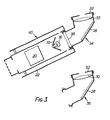
- a single torch as shown in Figure 3 is used, the torch having two interchangeable angled extensions 34 and 36 which are selectively coupled, for example by use of a bayonet fitting, to the cylindrical casing of the torch 40.
- the heat-absorbing filter 38 instead of being housed in the cylindrical casing is held at the inner end of the angled extension 34 which is fitted to the torch when a visual examination of the patient's breasts is to be made.
- the angled extension 36 which is used when a permanent record of the transilluminated image. is to be made contains no infra-red absorber.
- the construction of the torch of Figure 3 is the same as that of the torch of Figure 2.
- the apparatus may be provided with a number of angled-extensions 34, 36 such as those described above, but having different diameters and different shapes, the operator selecting the angled-extension having dimensions most appropriate for the size and shape of the breast under examination.
- the apparatus of the present invention thus enables a thorough examination of the patient to be performed easily with no fear of causing discomfort to the patient by undue heating of the breast tissue.
- the torch used for recording the image of the transilluminated tissue need only be switched on for a few milliseconds or less, there is no likelihood of burning the breast tissue while taking a permanent record of the transilluminated breast tissue.
Landscapes
- Health & Medical Sciences (AREA)
- Life Sciences & Earth Sciences (AREA)
- Surgery (AREA)
- Biophysics (AREA)
- Pathology (AREA)
- Engineering & Computer Science (AREA)
- Biomedical Technology (AREA)
- Heart & Thoracic Surgery (AREA)
- Medical Informatics (AREA)
- Molecular Biology (AREA)
- Physics & Mathematics (AREA)
- Animal Behavior & Ethology (AREA)
- General Health & Medical Sciences (AREA)
- Public Health (AREA)
- Veterinary Medicine (AREA)
- Gynecology & Obstetrics (AREA)
- Reproductive Health (AREA)
- Measuring And Recording Apparatus For Diagnosis (AREA)
- Endoscopes (AREA)
- Investigating, Analyzing Materials By Fluorescence Or Luminescence (AREA)
- Investigating Or Analysing Materials By Optical Means (AREA)
- Medicines Containing Antibodies Or Antigens For Use As Internal Diagnostic Agents (AREA)
Priority Applications (1)
| Application Number | Priority Date | Filing Date | Title |
|---|---|---|---|
| AT83306705T ATE25815T1 (de) | 1982-11-03 | 1983-11-03 | Diaphanographiegeraet mit fernsehkette. |
Applications Claiming Priority (2)
| Application Number | Priority Date | Filing Date | Title |
|---|---|---|---|
| GB8231404 | 1982-11-03 | ||
| GB8231404 | 1982-11-03 |
Publications (2)
| Publication Number | Publication Date |
|---|---|
| EP0108617A1 true EP0108617A1 (de) | 1984-05-16 |
| EP0108617B1 EP0108617B1 (de) | 1987-03-11 |
Family
ID=10534011
Family Applications (1)
| Application Number | Title | Priority Date | Filing Date |
|---|---|---|---|
| EP83306705A Expired EP0108617B1 (de) | 1982-11-03 | 1983-11-03 | Diaphanographiegerät mit Fernsehkette |
Country Status (4)
| Country | Link |
|---|---|
| US (1) | US4600011A (de) |
| EP (1) | EP0108617B1 (de) |
| AT (1) | ATE25815T1 (de) |
| DE (1) | DE3370135D1 (de) |
Cited By (6)
| Publication number | Priority date | Publication date | Assignee | Title |
|---|---|---|---|---|
| EP0447708A3 (en) * | 1990-03-17 | 1991-11-21 | Long Xing Mesd Co. Hai Dien Beijing | Apparatus and method for quantitative examination and high-resolution imaging of human tissue |
| WO1993011704A1 (fr) * | 1991-12-17 | 1993-06-24 | Eduard Emmanuilovich Godik | Procede et dispositif pour le diagnostic d'un organisme vivant |
| US5865743A (en) * | 1994-02-23 | 1999-02-02 | Dynamics Imaging, Inc. | Method of living organism multimodal functional mapping |
| US6002958A (en) * | 1992-12-24 | 1999-12-14 | Dynamics Imaging, Inc. | Method and apparatus for diagnostics of internal organs |
| WO2000042902A1 (en) * | 1999-01-22 | 2000-07-27 | Art Advanced Research Technologies Inc. | Depth discrimination of hetorogeneities in turbid media |
| US6192262B1 (en) | 1994-02-23 | 2001-02-20 | Dobi Medical Systems, Llc | Method of living organism multimodal functional mapping |
Families Citing this family (24)
| Publication number | Priority date | Publication date | Assignee | Title |
|---|---|---|---|---|
| US4768516A (en) * | 1983-10-14 | 1988-09-06 | Somanetics Corporation | Method and apparatus for in vivo evaluation of tissue composition |
| US5140989A (en) | 1983-10-14 | 1992-08-25 | Somanetics Corporation | Examination instrument for optical-response diagnostic apparatus |
| US5139025A (en) | 1983-10-14 | 1992-08-18 | Somanetics Corporation | Method and apparatus for in vivo optical spectroscopic examination |
| US4817623A (en) | 1983-10-14 | 1989-04-04 | Somanetics Corporation | Method and apparatus for interpreting optical response data |
| EP0187283B1 (de) * | 1984-12-26 | 1989-04-26 | Nivarox-FAR S.A. | Vorrichtung zum Auffinden in situ der Querbohrungen eines im Markkanal implantierten hohlen Stiftes für das Zusammenhalten der Fragmente eines gebrochenen Knochens |
| WO1988001485A1 (en) * | 1986-09-02 | 1988-03-10 | Singer Jerome R | Infrared radiation imaging system and method |
| US5146923A (en) * | 1986-12-18 | 1992-09-15 | Dhawan Atam P | Apparatus and method for skin lesion examination |
| US4810875A (en) * | 1987-02-02 | 1989-03-07 | Wyatt Technology Corporation | Method and apparatus for examining the interior of semi-opaque objects |
| US5259380A (en) * | 1987-11-04 | 1993-11-09 | Amcor Electronics, Ltd. | Light therapy system |
| US4945239A (en) * | 1989-03-29 | 1990-07-31 | Center For Innovative Technology | Early detection of breast cancer using transillumination |
| US5079698A (en) * | 1989-05-03 | 1992-01-07 | Advanced Light Imaging Technologies Ltd. | Transillumination method apparatus for the diagnosis of breast tumors and other breast lesions by normalization of an electronic image of the breast |
| JP3433508B2 (ja) * | 1993-12-01 | 2003-08-04 | 浜松ホトニクス株式会社 | 散乱吸収体計測方法及び散乱吸収体計測装置 |
| US5730133A (en) * | 1994-05-20 | 1998-03-24 | Dynamics Imaging, Inc. | Optical functional mamoscope |
| US5657754A (en) * | 1995-07-10 | 1997-08-19 | Rosencwaig; Allan | Apparatus for non-invasive analyses of biological compounds |
| US5833634A (en) * | 1995-11-09 | 1998-11-10 | Uromed Corporation | Tissue examination |
| US5989199A (en) * | 1996-11-27 | 1999-11-23 | Assurance Medical, Inc. | Tissue examination |
| US6091981A (en) * | 1997-09-16 | 2000-07-18 | Assurance Medical Inc. | Clinical tissue examination |
| US5916180A (en) * | 1997-10-03 | 1999-06-29 | Uromed Corporation | Calibrating pressure sensors |
| US6063031A (en) * | 1997-10-14 | 2000-05-16 | Assurance Medical, Inc. | Diagnosis and treatment of tissue with instruments |
| USD425980S (en) * | 1997-10-20 | 2000-05-30 | Assurance Medical, Inc. | Hand-held tissue examination device |
| US6179790B1 (en) | 1997-10-20 | 2001-01-30 | Assurance Medical, Inc. | Layer of material for use with tissue examination device |
| CN102883658B (zh) * | 2009-11-19 | 2016-06-22 | 调节成像公司 | 用于使用结构化照明经由单元件检测来分析浑浊介质的方法和设备 |
| US8586924B2 (en) * | 2010-09-13 | 2013-11-19 | Lawrence Livermore National Security, Llc | Enhancement of the visibility of objects located below the surface of a scattering medium |
| EP3401678B1 (de) | 2012-11-07 | 2020-04-08 | Modulated Imaging Inc. | Effiziente modulierte bildgebung |
Citations (8)
| Publication number | Priority date | Publication date | Assignee | Title |
|---|---|---|---|---|
| US2867209A (en) * | 1955-05-04 | 1959-01-06 | Fr De Construction D App Medic | Endoscopic devices |
| US3806633A (en) * | 1972-01-18 | 1974-04-23 | Westinghouse Electric Corp | Multispectral data sensor and display system |
| US3971954A (en) * | 1973-11-12 | 1976-07-27 | Alphametrics Ltd. | Ultraviolet camera system for dental photography and mouthpieces therefor |
| US4213462A (en) * | 1977-08-25 | 1980-07-22 | Nobuhiro Sato | Optical assembly for detecting an abnormality of an organ or tissue and method |
| GB2068537A (en) * | 1980-02-04 | 1981-08-12 | Energy Conversion Devices Inc | Examining biological materials |
| GB2092856A (en) * | 1981-02-11 | 1982-08-18 | Watmough David John | Transillumination of tissue by visible, UV or IR light |
| EP0075800A1 (de) * | 1981-09-29 | 1983-04-06 | Valter Guidetti | Verfahren und Gerät zur Beobachtung mittels Licht von Neoplasmen nahe der Oberfläche von bestimmten Körperteilen |
| GB2111794A (en) * | 1981-11-17 | 1983-07-06 | David John Watmough | Transillumination of tissue |
Family Cites Families (13)
| Publication number | Priority date | Publication date | Assignee | Title |
|---|---|---|---|---|
| US2098990A (en) * | 1934-03-10 | 1937-11-16 | Phillip S Newton | Therapeutic lamp and method |
| US3335716A (en) * | 1965-01-18 | 1967-08-15 | Gen Electric | Diagnostic thermography method and means |
| US3674008A (en) * | 1970-07-13 | 1972-07-04 | Battelle Development Corp | Quantitative pulsed transilluminator and method of operation |
| US3798366A (en) * | 1972-03-06 | 1974-03-19 | R Winkler | Infrared imaging system |
| DE2609273A1 (de) * | 1976-03-05 | 1977-09-08 | Mutzhas Maximilian F | Bestrahlungseinrichtung mit ultraviolett-strahlenquelle |
| SE7613587L (sv) * | 1976-12-03 | 1978-06-04 | Andersson Torsten | Metod for diagnostiserande av sjukliga forendringar i kvinnobrost |
| JPS54148586A (en) * | 1978-05-15 | 1979-11-20 | Minolta Camera Co Ltd | Jaundice meter |
| US4286602A (en) * | 1979-06-20 | 1981-09-01 | Robert Guy | Transillumination diagnostic system |
| JPS5647014A (en) * | 1979-09-25 | 1981-04-28 | Olympus Optical Co Ltd | Connector for lighting light transmission |
| US4336809A (en) * | 1980-03-17 | 1982-06-29 | Burleigh Instruments, Inc. | Human and animal tissue photoradiation system and method |
| DE3044184A1 (de) * | 1980-11-24 | 1982-06-16 | Mutzhas Maximilian F | Vorrichtung zur phototherapeutischen behandlung der hyperbilirubinaemie |
| US4479499A (en) * | 1982-01-29 | 1984-10-30 | Alfano Robert R | Method and apparatus for detecting the presence of caries in teeth using visible light |
| US4467812A (en) * | 1982-07-19 | 1984-08-28 | Spectrascan, Inc. | Transillumination apparatus |
-
1983
- 1983-11-01 US US06/547,500 patent/US4600011A/en not_active Expired - Fee Related
- 1983-11-03 DE DE8383306705T patent/DE3370135D1/de not_active Expired
- 1983-11-03 EP EP83306705A patent/EP0108617B1/de not_active Expired
- 1983-11-03 AT AT83306705T patent/ATE25815T1/de not_active IP Right Cessation
Patent Citations (8)
| Publication number | Priority date | Publication date | Assignee | Title |
|---|---|---|---|---|
| US2867209A (en) * | 1955-05-04 | 1959-01-06 | Fr De Construction D App Medic | Endoscopic devices |
| US3806633A (en) * | 1972-01-18 | 1974-04-23 | Westinghouse Electric Corp | Multispectral data sensor and display system |
| US3971954A (en) * | 1973-11-12 | 1976-07-27 | Alphametrics Ltd. | Ultraviolet camera system for dental photography and mouthpieces therefor |
| US4213462A (en) * | 1977-08-25 | 1980-07-22 | Nobuhiro Sato | Optical assembly for detecting an abnormality of an organ or tissue and method |
| GB2068537A (en) * | 1980-02-04 | 1981-08-12 | Energy Conversion Devices Inc | Examining biological materials |
| GB2092856A (en) * | 1981-02-11 | 1982-08-18 | Watmough David John | Transillumination of tissue by visible, UV or IR light |
| EP0075800A1 (de) * | 1981-09-29 | 1983-04-06 | Valter Guidetti | Verfahren und Gerät zur Beobachtung mittels Licht von Neoplasmen nahe der Oberfläche von bestimmten Körperteilen |
| GB2111794A (en) * | 1981-11-17 | 1983-07-06 | David John Watmough | Transillumination of tissue |
Cited By (7)
| Publication number | Priority date | Publication date | Assignee | Title |
|---|---|---|---|---|
| EP0447708A3 (en) * | 1990-03-17 | 1991-11-21 | Long Xing Mesd Co. Hai Dien Beijing | Apparatus and method for quantitative examination and high-resolution imaging of human tissue |
| WO1993011704A1 (fr) * | 1991-12-17 | 1993-06-24 | Eduard Emmanuilovich Godik | Procede et dispositif pour le diagnostic d'un organisme vivant |
| US6002958A (en) * | 1992-12-24 | 1999-12-14 | Dynamics Imaging, Inc. | Method and apparatus for diagnostics of internal organs |
| US5865743A (en) * | 1994-02-23 | 1999-02-02 | Dynamics Imaging, Inc. | Method of living organism multimodal functional mapping |
| US6192262B1 (en) | 1994-02-23 | 2001-02-20 | Dobi Medical Systems, Llc | Method of living organism multimodal functional mapping |
| WO2000042902A1 (en) * | 1999-01-22 | 2000-07-27 | Art Advanced Research Technologies Inc. | Depth discrimination of hetorogeneities in turbid media |
| US6415172B1 (en) | 1999-01-22 | 2002-07-02 | Art Advanced Research Technologies, Inc. | Optical imaging of turbid media with depth-related discrimination |
Also Published As
| Publication number | Publication date |
|---|---|
| ATE25815T1 (de) | 1987-03-15 |
| US4600011A (en) | 1986-07-15 |
| DE3370135D1 (de) | 1987-04-16 |
| EP0108617B1 (de) | 1987-03-11 |
Similar Documents
| Publication | Publication Date | Title |
|---|---|---|
| EP0108617B1 (de) | Diaphanographiegerät mit Fernsehkette | |
| USRE32718E (en) | Transillumination diagnostic method and apparatus | |
| Heijblom et al. | The state of the art in breast imaging using the Twente Photoacoustic Mammoscope: results from 31 measurements on malignancies | |
| Bartrum Jr et al. | Transillumination lightscanning to diagnose breast cancer: a feasibility study | |
| US5007428A (en) | Apparatus for examining a body of living tissues | |
| US3335716A (en) | Diagnostic thermography method and means | |
| JPH03274444A (ja) | ヒトの組織を定量検査し高解像度影像を得る方法と装置 | |
| Watmough | Transillumination of breast tissues: factors governing optimal imaging of lesions. | |
| GB2068537A (en) | Examining biological materials | |
| JPH0654792A (ja) | 撮像装置 | |
| JP2016538095A (ja) | 所定の生物学的構造体の非侵襲的検知装置 | |
| EP1620003A2 (de) | System und verfharen zur identifizierung und klassifizierung von dynamischen thermodynamischen verfahren in säugetieren und unterscheidung zwischen und innerhalb dieser verfahren | |
| US7041109B2 (en) | Apparatus and method for interstitial laser therapy of small breast cancers and adjunctive therapy | |
| Jackson et al. | The development of a system for transillumination computed tomography | |
| US7543988B2 (en) | X-ray device for imaging at least one part of an examination object | |
| JP6794438B2 (ja) | 乳房検診を容易にする装置を備える画像診断システム | |
| CA2337871A1 (en) | Optical mammography | |
| JP2009545359A (ja) | 拡散光トモグラフィのための高速蛍光可視化素子 | |
| WO2012075704A1 (zh) | 一体化红外线热扫描食管镜系统 | |
| KR100804809B1 (ko) | 유방암 검사장치 | |
| US20050010114A1 (en) | Optical mammography | |
| Watmough | A light torch for the transillumination of female breast tissues | |
| CN109259763B (zh) | 一种可调辐射范围的成像系统 | |
| KR200294495Y1 (ko) | 일루미노미터의 근적외선을 이용한 유방암 진단기 | |
| CN213606289U (zh) | 一种基于红外线的检测设备 |
Legal Events
| Date | Code | Title | Description |
|---|---|---|---|
| PUAI | Public reference made under article 153(3) epc to a published international application that has entered the european phase |
Free format text: ORIGINAL CODE: 0009012 |
|
| AK | Designated contracting states |
Designated state(s): AT BE CH DE FR GB IT LI LU NL SE |
|
| 17P | Request for examination filed |
Effective date: 19841115 |
|
| 17Q | First examination report despatched |
Effective date: 19860520 |
|
| GRAA | (expected) grant |
Free format text: ORIGINAL CODE: 0009210 |
|
| AK | Designated contracting states |
Kind code of ref document: B1 Designated state(s): AT BE CH DE FR GB IT LI LU NL SE |
|
| REF | Corresponds to: |
Ref document number: 25815 Country of ref document: AT Date of ref document: 19870315 Kind code of ref document: T |
|
| ITF | It: translation for a ep patent filed | ||
| REF | Corresponds to: |
Ref document number: 3370135 Country of ref document: DE Date of ref document: 19870416 |
|
| ET | Fr: translation filed | ||
| PG25 | Lapsed in a contracting state [announced via postgrant information from national office to epo] |
Ref country code: LU Free format text: LAPSE BECAUSE OF NON-PAYMENT OF DUE FEES Effective date: 19871130 |
|
| PLBE | No opposition filed within time limit |
Free format text: ORIGINAL CODE: 0009261 |
|
| STAA | Information on the status of an ep patent application or granted ep patent |
Free format text: STATUS: NO OPPOSITION FILED WITHIN TIME LIMIT |
|
| 26N | No opposition filed | ||
| PGFP | Annual fee paid to national office [announced via postgrant information from national office to epo] |
Ref country code: LU Payment date: 19900905 Year of fee payment: 8 |
|
| PGFP | Annual fee paid to national office [announced via postgrant information from national office to epo] |
Ref country code: BE Payment date: 19900913 Year of fee payment: 8 |
|
| PGFP | Annual fee paid to national office [announced via postgrant information from national office to epo] |
Ref country code: FR Payment date: 19900925 Year of fee payment: 8 |
|
| PGFP | Annual fee paid to national office [announced via postgrant information from national office to epo] |
Ref country code: AT Payment date: 19901017 Year of fee payment: 8 |
|
| PGFP | Annual fee paid to national office [announced via postgrant information from national office to epo] |
Ref country code: SE Payment date: 19901022 Year of fee payment: 8 |
|
| PGFP | Annual fee paid to national office [announced via postgrant information from national office to epo] |
Ref country code: CH Payment date: 19901115 Year of fee payment: 8 |
|
| ITTA | It: last paid annual fee | ||
| PGFP | Annual fee paid to national office [announced via postgrant information from national office to epo] |
Ref country code: NL Payment date: 19901130 Year of fee payment: 8 |
|
| PG25 | Lapsed in a contracting state [announced via postgrant information from national office to epo] |
Ref country code: AT Effective date: 19911103 |
|
| PG25 | Lapsed in a contracting state [announced via postgrant information from national office to epo] |
Ref country code: SE Effective date: 19911104 |
|
| PG25 | Lapsed in a contracting state [announced via postgrant information from national office to epo] |
Ref country code: LI Effective date: 19911130 Ref country code: CH Effective date: 19911130 Ref country code: BE Effective date: 19911130 |
|
| BERE | Be: lapsed |
Owner name: THE UNIVERSITY COURT OF THE UNIVERSITY OF ABERDEE Effective date: 19911130 |
|
| PG25 | Lapsed in a contracting state [announced via postgrant information from national office to epo] |
Ref country code: NL Effective date: 19920601 |
|
| NLV4 | Nl: lapsed or anulled due to non-payment of the annual fee | ||
| PG25 | Lapsed in a contracting state [announced via postgrant information from national office to epo] |
Ref country code: FR Effective date: 19920731 |
|
| REG | Reference to a national code |
Ref country code: CH Ref legal event code: PL |
|
| REG | Reference to a national code |
Ref country code: FR Ref legal event code: ST |
|
| PGFP | Annual fee paid to national office [announced via postgrant information from national office to epo] |
Ref country code: GB Payment date: 19941117 Year of fee payment: 12 |
|
| PGFP | Annual fee paid to national office [announced via postgrant information from national office to epo] |
Ref country code: DE Payment date: 19950118 Year of fee payment: 12 |
|
| EUG | Se: european patent has lapsed |
Ref document number: 83306705.1 Effective date: 19920604 |
|
| PG25 | Lapsed in a contracting state [announced via postgrant information from national office to epo] |
Ref country code: GB Effective date: 19951103 |
|
| GBPC | Gb: european patent ceased through non-payment of renewal fee |
Effective date: 19951103 |
|
| PG25 | Lapsed in a contracting state [announced via postgrant information from national office to epo] |
Ref country code: DE Effective date: 19960801 |