EP0117486A2 - Méthode de croissance et de purification de lignées de cellules humaines à partir de populations de cellules hétérogènes de tissu en culture - Google Patents
Méthode de croissance et de purification de lignées de cellules humaines à partir de populations de cellules hétérogènes de tissu en culture Download PDFInfo
- Publication number
- EP0117486A2 EP0117486A2 EP84101589A EP84101589A EP0117486A2 EP 0117486 A2 EP0117486 A2 EP 0117486A2 EP 84101589 A EP84101589 A EP 84101589A EP 84101589 A EP84101589 A EP 84101589A EP 0117486 A2 EP0117486 A2 EP 0117486A2
- Authority
- EP
- European Patent Office
- Prior art keywords
- melanocytes
- cells
- tissue culture
- cell
- melanocyte
- Prior art date
- Legal status (The legal status is an assumption and is not a legal conclusion. Google has not performed a legal analysis and makes no representation as to the accuracy of the status listed.)
- Withdrawn
Links
Images

Classifications
-
- C—CHEMISTRY; METALLURGY
- C12—BIOCHEMISTRY; BEER; SPIRITS; WINE; VINEGAR; MICROBIOLOGY; ENZYMOLOGY; MUTATION OR GENETIC ENGINEERING
- C12N—MICROORGANISMS OR ENZYMES; COMPOSITIONS THEREOF; PROPAGATING, PRESERVING, OR MAINTAINING MICROORGANISMS; MUTATION OR GENETIC ENGINEERING; CULTURE MEDIA
- C12N5/00—Undifferentiated human, animal or plant cells, e.g. cell lines; Tissues; Cultivation or maintenance thereof; Culture media therefor
- C12N5/06—Animal cells or tissues; Human cells or tissues
- C12N5/0602—Vertebrate cells
- C12N5/0625—Epidermal cells, skin cells; Cells of the oral mucosa
- C12N5/0626—Melanocytes
-
- G—PHYSICS
- G01—MEASURING; TESTING
- G01N—INVESTIGATING OR ANALYSING MATERIALS BY DETERMINING THEIR CHEMICAL OR PHYSICAL PROPERTIES
- G01N33/00—Investigating or analysing materials by specific methods not covered by groups G01N1/00 - G01N31/00
- G01N33/48—Biological material, e.g. blood, urine; Haemocytometers
- G01N33/50—Chemical analysis of biological material, e.g. blood, urine; Testing involving biospecific ligand binding methods; Immunological testing
- G01N33/5005—Chemical analysis of biological material, e.g. blood, urine; Testing involving biospecific ligand binding methods; Immunological testing involving human or animal cells
- G01N33/5008—Chemical analysis of biological material, e.g. blood, urine; Testing involving biospecific ligand binding methods; Immunological testing involving human or animal cells for testing or evaluating the effect of chemical or biological compounds, e.g. drugs, cosmetics
- G01N33/5014—Chemical analysis of biological material, e.g. blood, urine; Testing involving biospecific ligand binding methods; Immunological testing involving human or animal cells for testing or evaluating the effect of chemical or biological compounds, e.g. drugs, cosmetics for testing toxicity
- G01N33/5017—Chemical analysis of biological material, e.g. blood, urine; Testing involving biospecific ligand binding methods; Immunological testing involving human or animal cells for testing or evaluating the effect of chemical or biological compounds, e.g. drugs, cosmetics for testing toxicity for testing neoplastic activity
-
- G—PHYSICS
- G01—MEASURING; TESTING
- G01N—INVESTIGATING OR ANALYSING MATERIALS BY DETERMINING THEIR CHEMICAL OR PHYSICAL PROPERTIES
- G01N33/00—Investigating or analysing materials by specific methods not covered by groups G01N1/00 - G01N31/00
- G01N33/48—Biological material, e.g. blood, urine; Haemocytometers
- G01N33/50—Chemical analysis of biological material, e.g. blood, urine; Testing involving biospecific ligand binding methods; Immunological testing
- G01N33/53—Immunoassay; Biospecific binding assay; Materials therefor
- G01N33/569—Immunoassay; Biospecific binding assay; Materials therefor for microorganisms, e.g. protozoa, bacteria, viruses
- G01N33/56966—Animal cells
-
- G—PHYSICS
- G01—MEASURING; TESTING
- G01N—INVESTIGATING OR ANALYSING MATERIALS BY DETERMINING THEIR CHEMICAL OR PHYSICAL PROPERTIES
- G01N33/00—Investigating or analysing materials by specific methods not covered by groups G01N1/00 - G01N31/00
- G01N33/48—Biological material, e.g. blood, urine; Haemocytometers
- G01N33/50—Chemical analysis of biological material, e.g. blood, urine; Testing involving biospecific ligand binding methods; Immunological testing
- G01N33/53—Immunoassay; Biospecific binding assay; Materials therefor
- G01N33/575—Immunoassay; Biospecific binding assay; Materials therefor for cancer
- G01N33/5751—Immunoassay; Biospecific binding assay; Materials therefor for cancer of the skin, e.g. melanoma
-
- G—PHYSICS
- G01—MEASURING; TESTING
- G01N—INVESTIGATING OR ANALYSING MATERIALS BY DETERMINING THEIR CHEMICAL OR PHYSICAL PROPERTIES
- G01N33/00—Investigating or analysing materials by specific methods not covered by groups G01N1/00 - G01N31/00
- G01N33/48—Biological material, e.g. blood, urine; Haemocytometers
- G01N33/50—Chemical analysis of biological material, e.g. blood, urine; Testing involving biospecific ligand binding methods; Immunological testing
- G01N33/53—Immunoassay; Biospecific binding assay; Materials therefor
- G01N33/577—Immunoassay; Biospecific binding assay; Materials therefor involving monoclonal antibodies binding reaction mechanisms characterised by the use of monoclonal antibodies
-
- C—CHEMISTRY; METALLURGY
- C12—BIOCHEMISTRY; BEER; SPIRITS; WINE; VINEGAR; MICROBIOLOGY; ENZYMOLOGY; MUTATION OR GENETIC ENGINEERING
- C12N—MICROORGANISMS OR ENZYMES; COMPOSITIONS THEREOF; PROPAGATING, PRESERVING, OR MAINTAINING MICROORGANISMS; MUTATION OR GENETIC ENGINEERING; CULTURE MEDIA
- C12N2501/00—Active agents used in cell culture processes, e.g. differentation
- C12N2501/90—Polysaccharides
Definitions
- This invention relates to tissue culture of normal human cells and more particularly to a method for the culture and purification of normal human melanocytes.
- Melanocytes, cultured by this method are of use in, 1) diagnostic assays for malignancies involving melanocytes, 2) assays for toxic and/or drug agents, 3) therapy of skin diseases, 4) assay for melanocyte-specific monoclonal antibodies, 5) comparison of the differentiation antigens of melanomas and melanocytes.
- melanocytes In normal epidermis, melanocytes replicate and are shed slowly whereas keratinocytes replicate more rapidly to replace cells which have been shed.
- the method of the invention can be used for separating out and obtaining cells of interest in pure form from tissue cultures containing a heterogeneous cell population.
- the method of the invention is used to obtain pure human melanocytes in tissue culture.
- Human post-natal foreskin and facial skin are the best sources of epidermal tissue.
- the dermal tissue is removed entirely by forceps and trypsin; steps known in the art.
- the present invention succeeds in a stepwise fashion in selecting out and purifying melanocytes i.e. target cells, from a mixed population of epidermal cells which include fibroblasts, keratinocytes and Langerhans cells.
- target cells i.e. target cells
- the method successfully effects use of specific growth promoters for the target cell, growth suppressor for unwanted cell types and immunoreagent specific for the target cell.
- Epidermal cells are seeded and initially grown in growth medium containing PMA [phorbol 12-myristate, 13-acetatel 10 mg/ml in dimethyl sulfoxide to enhance melanocyte growth.
- PMA is a tumor growth promoter.
- Sequential trypsinization with trypsin/ethylene- diaminetetraacetic acid (EDTA) separates melanocytes from keratinocytes.
- This essential trypsinization step involves a brief contact of the cells with trypsin/EDTA once or twice.
- the enriched melanocytes, harvested after the trypsin step, are replated and grown ideally in PMA (10 ng/ml) and cholera toxin (10- 8 m). Tumor growth promoter enhances melanocyte doubling time while cholera toxin suppresses fibroblast growth.
- Different growth promoters lead to expression of early, intermediate and late antigens on the melanocyte cell surface such as M 3 , M 4 , M 5 & M 6 (Table I).
- a pH of 7.2 supports melanocyte growth while inhibiting that of keratinocytes.
- the growth medium was Eagle's minimal essential medium with 5% fetal calf serum, 2mM 1-glutamine, 100 u/ml penicillin, 0.1 mg/ml streptomycin and 0.25 ug/ml Fungizone.
- a further purification step is immune-rosetting of melanocytes with a specific mouse monoclonal antibody (mAb) which recognizes human melanocytes as opposed to keratinocytes or fibroblasts.
- mAb mouse monoclonal antibody
- Visualization of the antigen-antibody complex is done with sheep red blood cells coupled with Protein A. This is followed by separation of the melanocytes on a Percoll (Trademark of Pharmacia Fine Chemicals) density gradient.
- the invention enables assay of cell samples for melanocytes. It also serves to produce melanocyte differentiation antigens.
- the method can be used generally to isolate and purify cell lines from a heterogeneous tissue culture containing a mixed cell population. What is effective for melanocytes can be used for tissue culture of heterogeneous cells from other tissue sources such as blood, kidney, thymus, etc. Indeed, the method is especially useful for minor or trace cells present in tissue culture, i.e. those cells normally present as only a small fraction of the overall cell population as is the case with melanocytes in epidermal tissue. The method is also useful for other slow-growing cells.
- Fibroblast overgrowth of tissue culture has long been a research problem and it is especially acute with respect to slow-growing cells. Therefore, the present purification can be used to solve the fibroblast problem in many tissue culture situations where monoclonal antibody, specific for the target cell or the unwanted cell, is available.
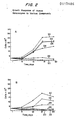
- Fig. 2A plots the growth response of human melanocytes in tissue culture medium: lacking tumor promoters (curve 20); containing 4- ⁇ - phorbol didecanoate (100 ng/ml) (curve 22); containing phorbol 12-myristate 13-acetate (PMA) (10 ng/ml) (curve 24); containing mezerein (10 ng/ml) (curve 26); containing teleocidin (10 ng/ml) (curve 28); and containing PMA (10 ng/ml) plus cholera toxin (10 -8 M) (curve 30).
- Fig. 2B plots the growth response of human melanocytes in tissue culture medium: lacking tumor promoters (curve 20); containing phorbol (10 ng/ml) (curve 22); containing debronoaplysiatoxin (10 ng/ml) (curve 24); containing lyngbyatoxin (10 ng/ml) (curve 26); containing phorbol dibutyrate (10 ng/ml) (curve 28); and containing aplysiatoxin (10 ng/ml) (curve 30).
- the human melanocyte cell strain MC752, passage 30, was derived from a normal human foreskin specimen as previously described.
- the stock cultures were grown in Minimal Essential Medium (Eagle) with Earle's Salts (EMEM) containing 0.01 mM MEM non-essential amino acids (Gibco Lab, Grand Island, NY), 2 mM L-glutamine (Gibco Lab), 100 units/ml penicillin, 0.1 mg/ml streptomycin, 0.25 ug/ml Fungizone (Gibco Lab), 5% fetal calf serum, 10 mg/ml PMA (Consolidated Midland Corp., Brewster, N Y) and 10 -8 M cholera toxin (Schwarz/Mann, Orangeburg, NY), at pH 7.2.
- EMEM Eagle's Salts
- Cells were grown-in this medium for 29 passages and split regularly (1:2) at weekly intervals. For specific experiments the cells were suspended with trypsin and resuspended in the above medium but in the absence of P M A and cholera toxin. The test compounds were then added at the indicated concentrations and 2.5 x 10 4 cells were plated in 2 ml of medium in multiwell dishes (Costar, Cambridge, MA) Trypsinized cells were counted at 5 days. Each experiment was performed in triplicate.
- Epidermal single-cell Suspensions were prepared by the method of Eisinger, M., et al. (1979) Proc. Nat'1. Acad. Sc. USA 76, 5340-5344, and Madin, S.H. and Darby, N.B. (1958) Proc. Soc. Exp. Biol. Med. 98, 574-576. Also see Eisinger, et al. patent No. 4254226.
- tissue sources are facial skin reduced to 15/1000 in. by means of a hand dermatome with a disposable blade, or foreskin samples freed of fatty tissue and washed with antibiotic-containing medium.
- samples were cut into 2 x 5 mm pieces and washed with 0.02% EDTA.
- 0.5 - 1.5g tissue were placed in 2.5 ml of 0.25% trypsin (diluted 1:250) for 12 - 19 hr at 4°C.
- Other tissue sources are adult abdominal skin, adult or fetal trunk, breast epidermis, or thigh skin.
- a fine forceps was used to detach the epidermis from the dermis of each piece of tissue. These isolated epidermal samples were pooled in trypsin/EDTA solution. By pippeting, single-cell epidermal suspensions were prepared. These separated epidermal cells were collected in newborn calf serum. Trypsinization of these epidermal cells was repeated until only stratum corneum cells remained essen - tially. These trypsinized cells were pooled, concentrated by centifugation and resuspended in tissue culture. Thus dermal tissue is entirely absent.
- Epidermal cell suspensions were grown in tissue culture flasks at cell seeding densities of 0.8 x 10 5 /cm 2 (foreskin) and 2.5 x 10 5 /cm 2 (facial skin) in growth medium containing phorbol 12-myristate, 13-acetate (PMA) 10ng/ml in 0.001% dimethyl sulfoxide.
- PMA is also known as 12-0-tetradeconoyl phorbol 13-acetate (TPA). This compound is a murine tumor growth promoter. It also suppresses growth of keratinocytes.
- cholera toxin (10 -8 M) is also present at this first step.
- the medium was changed 24 - 48 hours after cell seeding and unattached cells were removed. Then the medium was discarded 2 - 3 days later and the attached epidermal cells, now free of Langerhans cells, were washed, first with phosphate-buffered saline, and then with trypsin/EDTA.
- This selective differential trypsinization treatment step is necessary to detach melanocytes preferentially. It may be necessary to repeat this step once or twice.
- the detached melanocytes are replated in growth medium alone or in growth medium containing either 10nM cholera toxin or 10ng/ml PMA (TPA) or in growth medium containing both cholera toxin plus PMA in the aforesaid concentrations.
- TPA 10nM cholera toxin or 10ng/ml PMA
- the aforesaid concentrations are optimal.
- the cells are replated at 0.8 - 2 x 10 4 cells/cm 2 densities.
- other phorbol compounds as mezerein and phorbol dibutyrate (PDBu) were tested.
- Indole alkaloids such as teleocidin or lyngbyatoxin and polyactetates such as aplysiatoxin were tried as well. All of these aforesaid compounds have been used as tumor growth promoters. This invention uses such compounds for the first time to promote growth of a normal cell line. (See figures 1 & 2).
- monoclonal antibody which recognizes melanocytes but not fibroblasts or keratinocytes is used to purify melanocyte cultures.
- Mouse monoclonal antibody, R 24 recognizes antigens on melanomas and melanocytes but not antigens on fibroblasts or keratinocytes (Houghton, A.N., et al. J. Exp. Med. 156, 1755 (1982) and Dippold, W.G. et al. (1980) Proc. Nat'l.
- melanocytes are separated out from minor contaminants by means of an immune rosetting technique combined with a Percoll gradient. In general, this technique can be applied to purify subpopulations of cells in mixed culture. Illustration of the method for melanocytes is not meant to limit the invention. The method is especially useful where the target cell is a minor component of the cell population, or is a slow-growing cell, or where both of the aforesaid situations apply.
- Hybridomas are produced by the method of Kohler and Millstein, Nature (London) 256,495-497 (1975).
- the antibody R 24 is produced from hybridoma cells which cells are injected intraperitoneally into Swiss Background nu/nu mice. Ascities produced by this procedure are used as the source of antibody. Dippold, W.G., et al. Supra, Houghton, A.N. et al., Supra. Also see Old, et al. Monoclonal antibodies to Cell Surface Antigens of Human Malignant Melanoma, U.S. Patent Application Serial No. 307,060 filed Sept. 30, 1981 and Albino, et al. Monoclonal Antibodies against Melanocytes and Melanomas, U.S. Patent Application Serial No. 445,561, Filed November 30, 1982.
- Protein A (PA) indicator cells are prepared by conjugating Protein A ( P harmacia) to human erythrocytes with 0.01% chromium chloride. PA cells washed 2x with antibiotic solution (1000 u/ml penicillin, 1 mg/ml streptomycin and 2.5 ug/ml fungizone) are suspended in a final concentration of 10% (v/v).
- antibiotic solution 1000 u/ml penicillin, 1 mg/ml streptomycin and 2.5 ug/ml fungizone
- Melanoma cell line SK-MEL-28 was used as a reference for R 24 antibody titration. The lowest dilution of R 24 which rosetted 90-100% of the reference cells was used for melanocyte purification. This dilution was somewhere between 1/100 - 1/200.
- melanocytes appear bipolar or dendritic whereas keratinocytes have a round or polygonal shape.
- Melanin granules can be visualized under the light microscope in living tissue and when the tissue is fixed, the granules appear as 3,
- the electron microscope visualizes the melanosome and premelanosome structures in melanocytes.
- Leucine amino peptidase is an enzyme characteristic for fibroblasts, and a stain for said enzyme (Wolf, K. (1964) Arch. Kun. Exp. Dermatol. 218 446-460) revealed insignificant fibroblast contamination of these melanocyte cultures. Reliability of this marker for fibroblasts was shown by Regnier, M., et al., Acta. Dermatovener (Stockholm) 53, (1973) 241.
- Mouse monoclonal antibodies which recognize as many as 14 cell surface antigens present on human melanocyte or human melanoma cells are prepared by methods known in the art (Houghton, Supra, Dippold Supra, Albino, Supra). Some of these antigens probably distinguish early, intermediate or late stages of melanocyte differentiation.
- Melanoma antigens M-1, M-2 and HLA-DR were not found to be induced on melanocytes by the tumor growth promoters. That the method is best illustrated by mouse tumor growth promoters is not meant to limit same only to those specific tumor growth promoters.
- Neonatal foreskin melanocytes increase 6 fold in the presence of PMA and cholera toxin but in the same 11 day period, adult sources showed only a single doubling. Higher seeding densities are necessary for adult skin.
- the seeding density range was 0.8 - 2.5 x 10⁵ cells/cm 2 .
- an important part of this invention was the selective differential trypsinization step following cell seeding and attachment. Washing in trypsin/EDTA preferentially detaches melanocytes which are collected and further incubated after replating at cell densities of 0.8 - 2 x 10 4 cells/cm 2 .
- PMA was selective for melanocytes at 1 - 10 ng/ml and as with all the tumor growth promoting substances, showed a preferential toxic effect by preventing attachment and growth of keratinocytes while stimulating those characteristics for melanocytes.
- mouse monoclonal antibodies that recognize melanocyte and melanoma cell surface antigens distinguished several markers present in purified melanocyte cultures namely M-3, M-4, M-5, M-6. These presently denote early and intermediate differentiation of the cell, Houghton, et al., Supra. Not all tumor growth promoters induced all of the above-named antigens, only some of these. Aplysiatoxin, teleocidin, mezerein and PDBu showed positive effects.
- melanocytes will grow in 5-20% fetal calf serum.
- cholera toxin suppressed the growth of fibroblasts, and therefore extended the life span of melanocytes up to 32 weeks, allowed 112 split ratio at each transfer, and permitted a 6-fold increase in the number of melanocytes. Cholera toxin alone gives a 2-fold increase in cell number.
- the R 24 -Percoll step can also be used with melanoma - fibroblast mixed cell populations even in the presence of as high as 85% fibroblasts.
- the present invention is successful in obtaining pure melanocytes in cell culture via the use of:
- melanocytes have been obtained in culture from greater than 50 sources with melanin still present in all cells after three weeks in culture. Long-term culturing of up to eight months has been achieved. Cell viability was not affected by the presence of erythrocytes or Percoll as tested by the trypan blue exclusion test. Growth stopped when cultures reached a density of 8 - 11 x 10 4 cells/cm 2 .
- the R 24 -Percoll step is unsuccessful with some melanoma cells and foreskin cells_which lack R 24 antigen.
- Human red cells were conjugated to purified anti-mouse immunoglobulin (Dako) using 0.01% chromium chloride. Assays were performed in Falcon 3040 Microtest II plates. Sera were incubated with melanocytes for 1 hr. Target cells were then washed, indicator cells added for 1 hr and target cells evaluated for rosetting by light microscopy. Antigen expression was related to the titer endpoint giving 50% rosetted melanocytes. Titers are presented as follows: +++ (>1/500,000), ++ (1/50,000 to 1/500,000), + (1/100 to 1/25,000), - ( ⁇ 1/100).
- the invention involves the use of a micro-organism (monoclonal antibody), which is deposited at the American Type Culture Collection (ATCC)
Landscapes
- Health & Medical Sciences (AREA)
- Life Sciences & Earth Sciences (AREA)
- Engineering & Computer Science (AREA)
- Immunology (AREA)
- Biomedical Technology (AREA)
- Chemical & Material Sciences (AREA)
- Hematology (AREA)
- Molecular Biology (AREA)
- Urology & Nephrology (AREA)
- Biotechnology (AREA)
- Cell Biology (AREA)
- Biochemistry (AREA)
- Microbiology (AREA)
- General Health & Medical Sciences (AREA)
- Food Science & Technology (AREA)
- Medicinal Chemistry (AREA)
- Physics & Mathematics (AREA)
- Analytical Chemistry (AREA)
- General Physics & Mathematics (AREA)
- Pathology (AREA)
- Bioinformatics & Cheminformatics (AREA)
- Zoology (AREA)
- Tropical Medicine & Parasitology (AREA)
- Toxicology (AREA)
- Genetics & Genomics (AREA)
- Wood Science & Technology (AREA)
- Organic Chemistry (AREA)
- Virology (AREA)
- Chemical Kinetics & Catalysis (AREA)
- Dermatology (AREA)
- General Engineering & Computer Science (AREA)
- Micro-Organisms Or Cultivation Processes Thereof (AREA)
- Preparation Of Compounds By Using Micro-Organisms (AREA)
- Medicines That Contain Protein Lipid Enzymes And Other Medicines (AREA)
- Medicines Containing Material From Animals Or Micro-Organisms (AREA)
Applications Claiming Priority (2)
| Application Number | Priority Date | Filing Date | Title |
|---|---|---|---|
| US46985483A | 1983-02-25 | 1983-02-25 | |
| US469854 | 1990-01-23 |
Publications (2)
| Publication Number | Publication Date |
|---|---|
| EP0117486A2 true EP0117486A2 (fr) | 1984-09-05 |
| EP0117486A3 EP0117486A3 (fr) | 1987-05-13 |
Family
ID=23865294
Family Applications (1)
| Application Number | Title | Priority Date | Filing Date |
|---|---|---|---|
| EP84101589A Withdrawn EP0117486A3 (fr) | 1983-02-25 | 1984-02-16 | Méthode de croissance et de purification de lignées de cellules humaines à partir de populations de cellules hétérogènes de tissu en culture |
Country Status (3)
| Country | Link |
|---|---|
| EP (1) | EP0117486A3 (fr) |
| JP (1) | JPS6156130A (fr) |
| CA (1) | CA1233135A (fr) |
Cited By (4)
| Publication number | Priority date | Publication date | Assignee | Title |
|---|---|---|---|---|
| EP0212465A3 (fr) * | 1985-08-14 | 1989-03-01 | Sloan-Kettering Institute For Cancer Research | Méthode pour augmenter la croissance de cellules humaines en culture |
| FR2734825A1 (fr) * | 1995-05-31 | 1996-12-06 | Oreal | Procede de separation de la melanine presente dans des cellules |
| EP0929661A4 (fr) * | 1995-11-15 | 2000-01-12 | Univ Utah | Nouveaux procedes de mise en culture de cellules epitheliales humaines pour l'identification d'une therapeutique et d'un diagnostic du cancer |
| CN104877953A (zh) * | 2014-02-28 | 2015-09-02 | 上海尚瑞生物医药科技有限公司 | 促进黑色素细胞色素产生的皮肤制剂 |
Families Citing this family (1)
| Publication number | Priority date | Publication date | Assignee | Title |
|---|---|---|---|---|
| CN104039953B (zh) * | 2011-09-30 | 2016-08-17 | 株式会社爱茉莉太平洋 | 适应于角质形成细胞的黑素细胞或其祖细胞、以及其制备方法 |
-
1984
- 1984-02-16 EP EP84101589A patent/EP0117486A3/fr not_active Withdrawn
- 1984-02-24 CA CA000448278A patent/CA1233135A/fr not_active Expired
- 1984-02-25 JP JP59035875A patent/JPS6156130A/ja active Pending
Non-Patent Citations (4)
| Title |
|---|
| CHEMICAL ABSTRACTS, vol. 95, no. 21, 23th November 1981, page 180, abstract no. 182025d, Columbus, Ohio, US; H. FUJIKI et al.: "Indole alkaloids: Dihydroteleocidin B, teleocidin, and lyngbyatoxin A as members of a new class of tumor promoters" & PROC. NATL. ACAD. SCI. USA 1981, 78(6), 3872-6 * |
| CHEMICAL ABSTRACTS, vol. 97, no. 11, 13th September 1982, page 267, abstract no. 86807f, Columbus, Ohio, US; M. SUGANUMA et al.: "The third class of new tumor promoters, polyacetates (debromoaplysiatoxin and aplysiatoxin), can differentiate biological actions relevant to tumor promoters." & GANN 1982, 73(3), 495-7 * |
| EXPERIMENTAL CELL RESEARCH, vol. 142, 1982, pages 309-315, Academic Press Inc.; O. MARKO et al.: "Purification of human melanocytes by monoclonal antibody combined with percoll TM gradients" * |
| PROC. NATL. ACAD. SCI. USA, vol. 79, March 1982, pages 2018-2022, US; E. EISINGER et al.: "Selective proliferation of normal human melanocytes in vitro in the presence of phorbol ester and cholera toxin" * |
Cited By (5)
| Publication number | Priority date | Publication date | Assignee | Title |
|---|---|---|---|---|
| EP0212465A3 (fr) * | 1985-08-14 | 1989-03-01 | Sloan-Kettering Institute For Cancer Research | Méthode pour augmenter la croissance de cellules humaines en culture |
| FR2734825A1 (fr) * | 1995-05-31 | 1996-12-06 | Oreal | Procede de separation de la melanine presente dans des cellules |
| EP0929661A4 (fr) * | 1995-11-15 | 2000-01-12 | Univ Utah | Nouveaux procedes de mise en culture de cellules epitheliales humaines pour l'identification d'une therapeutique et d'un diagnostic du cancer |
| CN104877953A (zh) * | 2014-02-28 | 2015-09-02 | 上海尚瑞生物医药科技有限公司 | 促进黑色素细胞色素产生的皮肤制剂 |
| CN104877953B (zh) * | 2014-02-28 | 2018-06-01 | 上海尚瑞生物医药科技有限公司 | 促进黑色素细胞色素产生的皮肤制剂的制备方法 |
Also Published As
| Publication number | Publication date |
|---|---|
| EP0117486A3 (fr) | 1987-05-13 |
| JPS6156130A (ja) | 1986-03-20 |
| CA1233135A (fr) | 1988-02-23 |
Similar Documents
| Publication | Publication Date | Title |
|---|---|---|
| RAPOPORT | Dog thyroid cells in monolayer tissue culture: adenosine 3', 5'-cyclic monophosphate response to thyrotropic hormone | |
| Knutsen et al. | Thymosin-α1 stimulates maturation of CD34+ stem cells into CD3+ 4+ cells in an in vitro thymic epithelia organ coculture model | |
| Haynes et al. | Early events in human T cell ontogeny. Phenotypic characterization and immunohistologic localization of T cell precursors in early human fetal tissues. | |
| Chu et al. | Thymopoietin-like substance in human skin | |
| Wagner et al. | The isolation and culture of capillary endothelium from epididymal fat | |
| Coulomb et al. | A new method for studying epidermalization in vitro | |
| Raghavachar et al. | T lymphocyte control of human eosinophilic granulopoiesis. Clonal analysis in an idiopathic hypereosinophilic syndrome. | |
| Champion et al. | The embryonic thymus produces chemotactic peptides involved in the homing of hemopoietic precursors | |
| US5804446A (en) | Blood-borne mesenchymal cells | |
| Norris et al. | Antibody-dependent cellular cytotoxicity and skin disease | |
| Paige et al. | Differentiation of murine B cell precursors in agar culture. Frequency, surface marker analysis and requirements for growth of clonable pre‐B cells | |
| JPH07504398A (ja) | 真皮に対する表皮ケラチン細胞の接着を改善する生成物および方法 | |
| CA1341401C (fr) | Facteur immunosuppresseur derive de cellules de glioblastome humain | |
| Patterson et al. | Monoclonal antibody 4F2 reactive with basal layer keratinocytes: studies in the normal and a hyperproliferative state | |
| WO1997007200A9 (fr) | Compositions cellulaires precurseurs d'oligodendrocytes adultes et procedes | |
| WO1997007200A1 (fr) | Compositions cellulaires precurseurs d'oligodendrocytes adultes et procedes | |
| Konstantinov et al. | Expression of ƐBP, a Β‐galactoside‐binding Soluble Lectin, in Normal and Neoplastic Epidermis | |
| US4757019A (en) | Method for obtaining pure, stable tissue cultures of human melanocytes | |
| EP0117486A2 (fr) | Méthode de croissance et de purification de lignées de cellules humaines à partir de populations de cellules hétérogènes de tissu en culture | |
| US5024946A (en) | Human monoclonal antibody to antigen of gastric cancer and B-cell line for producing this antibody, method for preparing this B-cell line and antibody, antigen and method of preparation of this antigen | |
| KR940003650B1 (ko) | 흉선 간질-유래 t세포 성장인자 및 그의 제조방법 | |
| Subrahmanyan et al. | Effects of culture substrates and normal hepatic sinusoidal cells on in vitro hepatocyte synthesis of Apo‐SAA | |
| Wittek et al. | Propagation and properties of Kaposi's sarcoma-derived cell lines obtained from patients with AIDS: similarity of cultured cells to smooth muscle cells | |
| Rucheton et al. | Human autologous rosettes: II. Further characterization: Markers and functions | |
| Gottlieb et al. | Purification and in vitro growth of human epidermal basal keratinocytes using a monoclonal antibody |
Legal Events
| Date | Code | Title | Description |
|---|---|---|---|
| PUAI | Public reference made under article 153(3) epc to a published international application that has entered the european phase |
Free format text: ORIGINAL CODE: 0009012 |
|
| AK | Designated contracting states |
Designated state(s): DE FR GB |
|
| PUAL | Search report despatched |
Free format text: ORIGINAL CODE: 0009013 |
|
| AK | Designated contracting states |
Kind code of ref document: A3 Designated state(s): DE FR GB |
|
| 17P | Request for examination filed |
Effective date: 19871022 |
|
| 17Q | First examination report despatched |
Effective date: 19890418 |
|
| STAA | Information on the status of an ep patent application or granted ep patent |
Free format text: STATUS: THE APPLICATION IS DEEMED TO BE WITHDRAWN |
|
| 18D | Application deemed to be withdrawn |
Effective date: 19891010 |
|
| RIN1 | Information on inventor provided before grant (corrected) |
Inventor name: EISINGER, MAGDALENA Inventor name: MARKO, OLGA |