EP0126549A2 - Dispositif de balayage rétinien centré sur la fovée - Google Patents
Dispositif de balayage rétinien centré sur la fovée Download PDFInfo
- Publication number
- EP0126549A2 EP0126549A2 EP84302658A EP84302658A EP0126549A2 EP 0126549 A2 EP0126549 A2 EP 0126549A2 EP 84302658 A EP84302658 A EP 84302658A EP 84302658 A EP84302658 A EP 84302658A EP 0126549 A2 EP0126549 A2 EP 0126549A2
- Authority
- EP
- European Patent Office
- Prior art keywords
- eye
- fixation
- pattern
- fovea
- light
- Prior art date
- Legal status (The legal status is an assumption and is not a legal conclusion. Google has not performed a legal analysis and makes no representation as to the accuracy of the status listed.)
- Granted
Links
Images
Classifications
-
- A—HUMAN NECESSITIES
- A61—MEDICAL OR VETERINARY SCIENCE; HYGIENE
- A61B—DIAGNOSIS; SURGERY; IDENTIFICATION
- A61B1/00—Instruments for performing medical examinations of the interior of cavities or tubes of the body by visual or photographical inspection, e.g. endoscopes; Illuminating arrangements therefor
-
- A—HUMAN NECESSITIES
- A61—MEDICAL OR VETERINARY SCIENCE; HYGIENE
- A61B—DIAGNOSIS; SURGERY; IDENTIFICATION
- A61B3/00—Apparatus for testing the eyes; Instruments for examining the eyes
- A61B3/10—Objective types, i.e. instruments for examining the eyes independent of the patients' perceptions or reactions
- A61B3/14—Arrangements specially adapted for eye photography
-
- A—HUMAN NECESSITIES
- A61—MEDICAL OR VETERINARY SCIENCE; HYGIENE
- A61B—DIAGNOSIS; SURGERY; IDENTIFICATION
- A61B5/00—Measuring for diagnostic purposes; Identification of persons
- A61B5/117—Identification of persons
- A61B5/1171—Identification of persons based on the shapes or appearances of their bodies or parts thereof
-
- G—PHYSICS
- G06—COMPUTING OR CALCULATING; COUNTING
- G06V—IMAGE OR VIDEO RECOGNITION OR UNDERSTANDING
- G06V40/00—Recognition of biometric, human-related or animal-related patterns in image or video data
- G06V40/10—Human or animal bodies, e.g. vehicle occupants or pedestrians; Body parts, e.g. hands
- G06V40/18—Eye characteristics, e.g. of the iris
- G06V40/19—Sensors therefor
Definitions
- Prior U.S. Patent No. 4,109,237 discloses a basic method and apparatus for identifying individuals through their retinal vasculature patterns.
- EP-A-0061832 discloses using a rotating beam scanner for obtaining the identification pattern from the fundus of an eye.
- the identification pattern carrying sufficient information, could be obtained best by centering on the optic nerve.
- the blood vessels emerging from the optic nerve and forming the retinal vasculature could be used for obtaining the individual identification pattern.
- the vasculature of the choroid becomes readily and accurately detectable.
- the choroidal vasculature forms a matting behind the retina even in the area of the macula and fovea, where retinal blood vessels are very small or non-existent.
- the blood vessels of the choroid are stable, as are those of the retina, and thus may be used for obtaining information as to an individual's identity.
- the present invention overcomes this difficulty.
- the present invention is an apparatus and method for identifying individuals through the ocular light reflection pattern from the fundus of the eye.
- the device provides a fixation beam along the visual line of the eye, and a scanner for obtaining an identification pattern in a substantially circular pattern centered around the fovea.
- the apparatus makes use of infrared light in order to observe the vasculature of the choroid as well as the vasculature of the retina.
- a circular scan centered on the fovea, and therefore about the visula axis is used such that eye rotation about the visual axis will not result in the generation of substantially different patterns from a given individual when the individual tilts his head. This is because the area scanned on the fundus varies only in rotation; the sequence of the data remains the same.
- the identification process involves two basic steps. The first is an enrollment wherein the individual learns how to use the device and a reference pattern is acquired and stored.
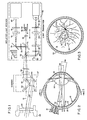
- Figs. 2 and 3 illustrate an eye into which the scanner beam is projected.
- the eye is centered on visual line or axis 10. which intersects fundus 12 at the fovea 14.
- Optic nerve 16 is approximately 15.5 degrees off the visual line.
- the scanner produces a substantially collimated source beam 26 which enters the pupil 28 of the eye and is refracted by lens 30 to substantially focus on fundus 12.
- the scan forms a circular locus of points 32 substantially centered on fovea 14.
- Infrared source means is provided to create a beam of infrared light or radiation to be directed into the subject's eye.
- this includes an incandescent tungsten bulb 40 which produces light, part of which is in the infrared spectrum, having a wave length of preferably, from about 0.7 to about 0.95 microns.
- the light passes through a spatial filter 42 and is refracted by a lens 44.
- An infrared filter 46 passes only the infrared portion of the beam.
- the beam passes through another pinhole or spatial filter 48.
- the beam is then reflected by a mirror 50 onto a beam splitter 52 which is coincident with the fixation optics and the visual line.
- a scanner means is provided for directing the beam into the fixated eye from a plurality of sequential, angularly divergent positions.
- the scanner means includes a rotatable housing 57 and optics which rotate with the housing, as indicated by the circular arrow of Fig 1.
- a hot mirror 58 which is a mirror which reflects infrared light while passing visible light, is located in the path of the source beam and the fixation beam.
- the fixation beam of visible light is substantially passed through the hot mirror, while the source beam of infrared light is substantially reflect- ej off axis.
- a scanner mirror 60 is mounted in the housing at a point spaced from the axis and is oriented to direct the beam into the eye as the housing rotates. The beam is directed to an apex at the pupil of the eye.
- an infrared filter 62 which eliminates any visible light which may have been reflected from the fixation beam by hot mirror 58.
- Hot mirror 58 causes a slight off axis displacement of the fixation beam and an offset plate 64 is provided to null the displacement of the fixation beam.
- An objective lens 66 is mounted in an eyepiece 68 and substantially collimates the beam and directs it into the eye.
- the objective lens at this location serves to provide easier focusing of the device for those individuals who-do not have exactly 20-20 vision, as will be subsequently discussed.
- the beam directed into the eye forms a circular scan, or locus of points, substantially centered on the fovea.
- the scan circle may be of any size selected, and is represented by the circle shown in Fig. 3. at 32.
- the light reflected out of the eye varies in intensity depending on the structures encountered in the scan. Reflected light is re-collimated by the lens of the eye 30 and directed. out of the pupil in a reverse direction.
- the reflected beam passes back through objective lens 66 and filter 62 and reflects off of scanner mirror 60 and hot mirror 58. It is then focused by objective lens 56.
- Beam splitter 52 passes a portion of the reflected scanning beam.
- a hot mirror 70 reflects this portion of the reflected beam to the receiver.
- the reflected beam passes through a spatial filter 72 and is reflected by a mirror 74.
- the beam then is refracted by a lens 76 and passed through another spatial filter 78 to a detector 80.
- the fixation mounting 36 with its pinhole 37 preferably is secured to the carriage in such a manner that it can be spatially adjusted a minute amount. This adjustment allows fine tuning of the fixation axis to coincide with the scanner axis as set forth in EP-A-0061832.
- the light from the fixation light-emitting diode 34 is preferably variable in intensity to vary the pupil size of the individual.
- the size of the subject's pupil will vary, depending on the intensity of the visible light from the fixation target, but will not be affected by the infrared radiation.
- the individual to be identified must first enroll himself in the data base of qualified users of the system. In the enrollment process an initial scan pattern is recorded.
- the individual presents his eye to the apparatus by moving his head adjacent eyepiece 68 and peering into lens 66.
- the fixation reticle 38 is visible and the user fixates his eye on this target. If the reticle appears out of focus, the focus • may be adjusted by moving carriage 82.
- Scanner 57 rotates, and when it is up to speed the infrared source bulb 40 is turned on.
- the source beam is directed into the eye from a plurality of sequential angu- larly divergent positions as the scanner rotates.
- the beam impinges on the fundus.and sequentially forms a circular locus of points substantially centered on the fovea.
- a portion of the light is reflected from the fundus forming a reflected beam.
- the intensity of the reflected beam is determined by the vasculature pattern and measured by detector 80 at each sequential point. The amounts of light thus sensed form the identification pattern.
- the pattern then obtained will be rotated with respect to the reference pattern.
- the sequence of the data remains the same.
- the two patterns are compared while rotating one pattern with respect to the other pattern until it is noted that the two patterns match. Such a comparison can be done very quickly by a computer.
Landscapes
- Health & Medical Sciences (AREA)
- Life Sciences & Earth Sciences (AREA)
- Engineering & Computer Science (AREA)
- General Health & Medical Sciences (AREA)
- Physics & Mathematics (AREA)
- Surgery (AREA)
- Veterinary Medicine (AREA)
- Animal Behavior & Ethology (AREA)
- Biophysics (AREA)
- Biomedical Technology (AREA)
- Heart & Thoracic Surgery (AREA)
- Medical Informatics (AREA)
- Molecular Biology (AREA)
- Public Health (AREA)
- Ophthalmology & Optometry (AREA)
- Pathology (AREA)
- Human Computer Interaction (AREA)
- Multimedia (AREA)
- General Physics & Mathematics (AREA)
- Theoretical Computer Science (AREA)
- Nuclear Medicine, Radiotherapy & Molecular Imaging (AREA)
- Optics & Photonics (AREA)
- Radiology & Medical Imaging (AREA)
- Eye Examination Apparatus (AREA)
- Measurement Of The Respiration, Hearing Ability, Form, And Blood Characteristics Of Living Organisms (AREA)
Applications Claiming Priority (2)
| Application Number | Priority Date | Filing Date | Title |
|---|---|---|---|
| US486014 | 1983-04-18 | ||
| US06/486,014 US4620318A (en) | 1983-04-18 | 1983-04-18 | Fovea-centered eye fundus scanner |
Publications (3)
| Publication Number | Publication Date |
|---|---|
| EP0126549A2 true EP0126549A2 (fr) | 1984-11-28 |
| EP0126549A3 EP0126549A3 (en) | 1986-12-30 |
| EP0126549B1 EP0126549B1 (fr) | 1990-02-14 |
Family
ID=23930269
Family Applications (1)
| Application Number | Title | Priority Date | Filing Date |
|---|---|---|---|
| EP19840302658 Expired - Lifetime EP0126549B1 (fr) | 1983-04-18 | 1984-04-18 | Dispositif de balayage rétinien centré sur la fovée |
Country Status (7)
| Country | Link |
|---|---|
| US (1) | US4620318A (fr) |
| EP (1) | EP0126549B1 (fr) |
| JP (1) | JPS59200628A (fr) |
| KR (1) | KR840008585A (fr) |
| AU (1) | AU570445B2 (fr) |
| CA (1) | CA1211218A (fr) |
| DE (1) | DE3481368D1 (fr) |
Cited By (4)
| Publication number | Priority date | Publication date | Assignee | Title |
|---|---|---|---|---|
| WO1997043677A1 (fr) * | 1996-05-15 | 1997-11-20 | Sensar, Inc. | Dispositif compact d'orientation et de mise au point d'image |
| WO1997046980A1 (fr) * | 1996-06-06 | 1997-12-11 | British Telecommunications Public Limited Company | Identification personnelle |
| EP0872814A1 (fr) * | 1997-04-15 | 1998-10-21 | BRITISH TELECOMMUNICATIONS public limited company | Appareil optique |
| EP0910986A1 (fr) * | 1997-10-24 | 1999-04-28 | BRITISH TELECOMMUNICATIONS public limited company | Appareil d'imagerie |
Families Citing this family (51)
| Publication number | Priority date | Publication date | Assignee | Title |
|---|---|---|---|---|
| FR2555039B1 (fr) * | 1983-11-21 | 1986-04-04 | Centre Nat Rech Scient | Ophtalmoscope catadioptrique a balayage |
| US4717952A (en) * | 1985-06-14 | 1988-01-05 | Canon Kabushiki Kaisha | Medical television system |
| JP2561828B2 (ja) * | 1987-01-26 | 1996-12-11 | キヤノン株式会社 | 眼底面検査装置 |
| US5055658A (en) * | 1988-07-25 | 1991-10-08 | Cockburn John B | Security system employing digitized personal physical characteristics |
| US5063603A (en) * | 1989-11-06 | 1991-11-05 | David Sarnoff Research Center, Inc. | Dynamic method for recognizing objects and image processing system therefor |
| US5206672A (en) * | 1990-09-05 | 1993-04-27 | Nestle S.A. | Surgical optometer |
| US5291560A (en) * | 1991-07-15 | 1994-03-01 | Iri Scan Incorporated | Biometric personal identification system based on iris analysis |
| US5359669A (en) * | 1992-04-13 | 1994-10-25 | Motorola, Inc. | Remote retinal scan identifier |
| DE4326716B4 (de) * | 1992-08-04 | 2005-03-03 | Kabushiki Kaisha Topcon | Anordnung zur Verarbeitung eines ophthalmologischen Bildes |
| US5471542A (en) * | 1993-09-27 | 1995-11-28 | Ragland; Richard R. | Point-of-gaze tracker |
| US5532771A (en) * | 1993-12-17 | 1996-07-02 | Edi Of Louisiana, Inc. | Eye fundus optical scanner system and method |
| US6714665B1 (en) * | 1994-09-02 | 2004-03-30 | Sarnoff Corporation | Fully automated iris recognition system utilizing wide and narrow fields of view |
| US5572596A (en) * | 1994-09-02 | 1996-11-05 | David Sarnoff Research Center, Inc. | Automated, non-invasive iris recognition system and method |
| US6320610B1 (en) | 1998-12-31 | 2001-11-20 | Sensar, Inc. | Compact imaging device incorporating rotatably mounted cameras |
| US6160907A (en) * | 1997-04-07 | 2000-12-12 | Synapix, Inc. | Iterative three-dimensional process for creating finished media content |
| US6084590A (en) * | 1997-04-07 | 2000-07-04 | Synapix, Inc. | Media production with correlation of image stream and abstract objects in a three-dimensional virtual stage |
| US6124864A (en) * | 1997-04-07 | 2000-09-26 | Synapix, Inc. | Adaptive modeling and segmentation of visual image streams |
| US6064752A (en) * | 1997-11-04 | 2000-05-16 | Sensar, Inc. | Method and apparatus for positioning subjects before a single camera |
| US6069967A (en) * | 1997-11-04 | 2000-05-30 | Sensar, Inc. | Method and apparatus for illuminating and imaging eyes through eyeglasses |
| US6266053B1 (en) | 1998-04-03 | 2001-07-24 | Synapix, Inc. | Time inheritance scene graph for representation of media content |
| US6249285B1 (en) | 1998-04-06 | 2001-06-19 | Synapix, Inc. | Computer assisted mark-up and parameterization for scene analysis |
| US6297825B1 (en) | 1998-04-06 | 2001-10-02 | Synapix, Inc. | Temporal smoothing of scene analysis data for image sequence generation |
| CA2335932C (fr) * | 1998-07-09 | 2007-09-18 | Bruce L. Golden | Appareil et procede d'acquisition d'images du systeme vasculaire retinien |
| US20040208343A1 (en) * | 1998-07-09 | 2004-10-21 | Colorado State University Research Foundation | Apparatus and method for creating a record using biometric information |
| US6532298B1 (en) | 1998-11-25 | 2003-03-11 | Iridian Technologies, Inc. | Portable authentication device and method using iris patterns |
| US6377699B1 (en) | 1998-11-25 | 2002-04-23 | Iridian Technologies, Inc. | Iris imaging telephone security module and method |
| JP2001034754A (ja) * | 1999-07-19 | 2001-02-09 | Sony Corp | 虹彩認証装置 |
| US6299306B1 (en) | 2000-03-31 | 2001-10-09 | Sensar, Inc. | Method and apparatus for positioning subjects using a holographic optical element |
| US7043048B1 (en) * | 2000-06-01 | 2006-05-09 | Digimarc Corporation | Capturing and encoding unique user attributes in media signals |
| DE10050358A1 (de) * | 2000-10-11 | 2002-04-18 | Wolfdietrich Steinhuber | Abbildendes optisches Gerät |
| US7224822B2 (en) * | 2000-11-02 | 2007-05-29 | Retinal Technologies, L.L.C. | System for capturing an image of the retina for identification |
| US6453057B1 (en) | 2000-11-02 | 2002-09-17 | Retinal Technologies, L.L.C. | Method for generating a unique consistent signal pattern for identification of an individual |
| WO2002065443A1 (fr) * | 2001-02-15 | 2002-08-22 | Tveye Inc. | Procede et appareil de transmission a faible largeur de bande de donnees se servant de l'anatomie de l'oeil humain |
| US7331669B2 (en) * | 2001-10-16 | 2008-02-19 | Indiana University Research And Technology Corporation | Device for digital retinal imaging |
| JP3916482B2 (ja) * | 2002-02-27 | 2007-05-16 | 株式会社ニデック | 眼科装置 |
| US6996251B2 (en) | 2002-09-30 | 2006-02-07 | Myport Technologies, Inc. | Forensic communication apparatus and method |
| US7778438B2 (en) | 2002-09-30 | 2010-08-17 | Myport Technologies, Inc. | Method for multi-media recognition, data conversion, creation of metatags, storage and search retrieval |
| US10721066B2 (en) | 2002-09-30 | 2020-07-21 | Myport Ip, Inc. | Method for voice assistant, location tagging, multi-media capture, transmission, speech to text conversion, photo/video image/object recognition, creation of searchable metatags/contextual tags, storage and search retrieval |
| EP1696382A1 (fr) * | 2003-12-19 | 2006-08-30 | Matsushita Electric Industries Co., Ltd. | Camera de prise d'images d'iris et systeme d'authentification par l'iris |
| US7252661B2 (en) * | 2003-12-23 | 2007-08-07 | Alcon Refractivehorizons, Inc. | Method and system for patient optical fixation |
| BRPI0509707A (pt) * | 2004-04-08 | 2007-09-18 | Optibrand Ltd Llc | método de processamento de um registro de idade auditável para um animal |
| WO2005122872A2 (fr) * | 2004-06-10 | 2005-12-29 | Optimedica Corporation | Procede et appareil de fixation ophtalmique par balayage |
| US20060147095A1 (en) * | 2005-01-03 | 2006-07-06 | Usher David B | Method and system for automatically capturing an image of a retina |
| US20070092115A1 (en) * | 2005-10-26 | 2007-04-26 | Usher David B | Method and system for detecting biometric liveness |
| US8488895B2 (en) * | 2006-05-31 | 2013-07-16 | Indiana University Research And Technology Corp. | Laser scanning digital camera with pupil periphery illumination and potential for multiply scattered light imaging |
| ES2326205B1 (es) * | 2007-11-27 | 2010-06-29 | Universidad Complutense De Madrid | Metodo y dispositivo para el reconocimiento de individuos basado en la imagen de la retina que incorpora como constante biometrica el area imagen del punto de fijacion. |
| JP2014079517A (ja) * | 2012-10-18 | 2014-05-08 | Canon Inc | 眼科装置 |
| CN105496352A (zh) * | 2014-10-09 | 2016-04-20 | 安尼迪斯公司 | 对脉络膜成像的方法和装置 |
| JP6651747B2 (ja) * | 2015-09-02 | 2020-02-19 | 株式会社ニデック | 走査型レーザ検眼鏡 |
| EP3692889B1 (fr) | 2017-10-05 | 2024-01-03 | QD Laser, Inc. | Dispositif d'examen de la vue |
| US11523095B2 (en) * | 2020-12-21 | 2022-12-06 | Infineon Technologies Ag | Mems mirror-based extended reality projection with eye-tracking |
Family Cites Families (19)
| Publication number | Priority date | Publication date | Assignee | Title |
|---|---|---|---|---|
| US2940371A (en) * | 1957-04-19 | 1960-06-14 | Raymond C Thurow | Focusing apparatus for camera |
| CH436769A (de) * | 1965-12-15 | 1967-05-31 | Haag Ag Streit | Ophthalmologisches Gerät |
| US3611290A (en) * | 1968-06-03 | 1971-10-05 | North American Rockwell | Fingerprint minutiae reading device |
| JPS49136227U (fr) * | 1973-03-22 | 1974-11-22 | ||
| US3869694A (en) * | 1973-10-26 | 1975-03-04 | Honeywell Inc | Ultrasonic control apparatus for an oculometer |
| US4026638A (en) * | 1974-12-02 | 1977-05-31 | Varian Associates | Reduced glare scanner |
| GB1540992A (en) * | 1975-04-22 | 1979-02-21 | Smiths Industries Ltd | Display or other systems and equipment for use in such systems |
| US4068932A (en) * | 1975-05-23 | 1978-01-17 | Canon Kabushiki Kaisha | Optical instrument for examining the eye fundus |
| GB1564315A (en) * | 1976-04-01 | 1980-04-10 | Secr Defence | Method and apparatus for measuring retinal blood flow |
| US4109237A (en) * | 1977-01-17 | 1978-08-22 | Hill Robert B | Apparatus and method for identifying individuals through their retinal vasculature patterns |
| US4253743A (en) * | 1977-05-17 | 1981-03-03 | Canon Kabushiki Kaisha | Eye testing instrument |
| US4213678A (en) * | 1977-09-29 | 1980-07-22 | Retina Foundation | Scanning ophthalmoscope for examining the fundus of the eye |
| DE2843287A1 (de) * | 1977-10-05 | 1979-04-19 | Canon Kk | Augenuntersuchungsinstrument |
| JPS6054053B2 (ja) * | 1977-11-15 | 1985-11-28 | ミノルタ株式会社 | 瞳位置合わせの容易な眼底カメラ |
| US4169663A (en) * | 1978-02-27 | 1979-10-02 | Synemed, Inc. | Eye attention monitor |
| US4287410A (en) * | 1979-02-28 | 1981-09-01 | Sri International | Double Purkinje eye tracker |
| US4304483A (en) * | 1979-12-13 | 1981-12-08 | Whitten Mark E | Hand-held retina photographing apparatus |
| JPS5717810A (en) * | 1980-07-07 | 1982-01-29 | Nippon Denso Co Ltd | Alarm method and device for vehicle |
| US4393366A (en) * | 1981-02-17 | 1983-07-12 | Eye-D Development Ii Ltd. | Rotating beam ocular identification apparatus and method |
-
1983
- 1983-04-18 US US06/486,014 patent/US4620318A/en not_active Expired - Lifetime
-
1984
- 1984-04-04 AU AU26428/84A patent/AU570445B2/en not_active Ceased
- 1984-04-04 CA CA000451263A patent/CA1211218A/fr not_active Expired
- 1984-04-12 KR KR1019840001944A patent/KR840008585A/ko not_active Ceased
- 1984-04-18 EP EP19840302658 patent/EP0126549B1/fr not_active Expired - Lifetime
- 1984-04-18 DE DE8484302658T patent/DE3481368D1/de not_active Expired - Fee Related
- 1984-04-18 JP JP59078304A patent/JPS59200628A/ja active Granted
Cited By (8)
| Publication number | Priority date | Publication date | Assignee | Title |
|---|---|---|---|---|
| WO1997043677A1 (fr) * | 1996-05-15 | 1997-11-20 | Sensar, Inc. | Dispositif compact d'orientation et de mise au point d'image |
| WO1997046980A1 (fr) * | 1996-06-06 | 1997-12-11 | British Telecommunications Public Limited Company | Identification personnelle |
| WO1997046979A1 (fr) * | 1996-06-06 | 1997-12-11 | British Telecommunications Public Limited Company | Appareil d'identification personnelle |
| WO1997046978A1 (fr) * | 1996-06-06 | 1997-12-11 | British Telecommunications Public Limited Company | Appareil d'identification personnelle |
| US6333988B1 (en) | 1996-06-06 | 2001-12-25 | British Telecommunications Plc | Personal identification |
| EP0872814A1 (fr) * | 1997-04-15 | 1998-10-21 | BRITISH TELECOMMUNICATIONS public limited company | Appareil optique |
| EP0910986A1 (fr) * | 1997-10-24 | 1999-04-28 | BRITISH TELECOMMUNICATIONS public limited company | Appareil d'imagerie |
| WO1999021479A1 (fr) * | 1997-10-24 | 1999-05-06 | British Telecommunications Public Limited Company | Appareil de formation d'images |
Also Published As
| Publication number | Publication date |
|---|---|
| JPS642371B2 (fr) | 1989-01-17 |
| EP0126549A3 (en) | 1986-12-30 |
| AU2642884A (en) | 1984-10-25 |
| KR840008585A (ko) | 1984-12-17 |
| AU570445B2 (en) | 1988-03-17 |
| JPS59200628A (ja) | 1984-11-14 |
| CA1211218A (fr) | 1986-09-09 |
| EP0126549B1 (fr) | 1990-02-14 |
| DE3481368D1 (de) | 1990-03-22 |
| US4620318A (en) | 1986-10-28 |
Similar Documents
| Publication | Publication Date | Title |
|---|---|---|
| EP0126549B1 (fr) | Dispositif de balayage rétinien centré sur la fovée | |
| EP0061832B1 (fr) | Dispositif et méthode de vérification d'identité à l'aide du patron du fond oculaire | |
| US8113657B2 (en) | Device and method for determining the orientation of an eye | |
| US6296358B1 (en) | Ocular fundus auto imager | |
| US6027216A (en) | Eye fixation monitor and tracker | |
| US7025459B2 (en) | Ocular fundus auto imager | |
| JP4689141B2 (ja) | 目およびその屈折性成分の全屈折不均質性を同期マッピングする方法および装置 | |
| CA1244552A (fr) | Systeme d'identification de l'iris de l'oeil | |
| US5632282A (en) | Ocular disease detection apparatus | |
| KR100342159B1 (ko) | 홍채영상 포착장치 및 홍채영상 포착방법 | |
| US6116738A (en) | Corneal topographer with central and peripheral measurement capability | |
| US6830336B2 (en) | Automated generation of fundus images based on processing of acquired images | |
| US5355895A (en) | Ocular disease detection apparatus | |
| KR20020059635A (ko) | 맞춤식 각막 프로파일링 | |
| US7360895B2 (en) | Simplified ocular fundus auto imager | |
| US5532771A (en) | Eye fundus optical scanner system and method | |
| WO2000004820A1 (fr) | Acquisition, analyse et imagerie de donnees tridimensionnelles relatives a la retine | |
| CN101219077B (zh) | 用于光学治疗的虹膜识别和跟踪 | |
| US6802837B2 (en) | Device used for the photorefractive keratectomy of the eye using a centering method |
Legal Events
| Date | Code | Title | Description |
|---|---|---|---|
| PUAI | Public reference made under article 153(3) epc to a published international application that has entered the european phase |
Free format text: ORIGINAL CODE: 0009012 |
|
| AK | Designated contracting states |
Designated state(s): CH DE FR GB IT LI NL SE |
|
| PUAL | Search report despatched |
Free format text: ORIGINAL CODE: 0009013 |
|
| AK | Designated contracting states |
Kind code of ref document: A3 Designated state(s): CH DE FR GB IT LI NL SE |
|
| 17P | Request for examination filed |
Effective date: 19870619 |
|
| 17Q | First examination report despatched |
Effective date: 19880624 |
|
| GRAA | (expected) grant |
Free format text: ORIGINAL CODE: 0009210 |
|
| AK | Designated contracting states |
Kind code of ref document: B1 Designated state(s): CH DE FR GB IT LI NL SE |
|
| PG25 | Lapsed in a contracting state [announced via postgrant information from national office to epo] |
Ref country code: NL Effective date: 19900214 Ref country code: IT Free format text: LAPSE BECAUSE OF FAILURE TO SUBMIT A TRANSLATION OF THE DESCRIPTION OR TO PAY THE FEE WITHIN THE PRESCRIBED TIME-LIMIT;WARNING: LAPSES OF ITALIAN PATENTS WITH EFFECTIVE DATE BEFORE 2007 MAY HAVE OCCURRED AT ANY TIME BEFORE 2007. THE CORRECT EFFECTIVE DATE MAY BE DIFFERENT FROM THE ONE RECORDED. Effective date: 19900214 |
|
| ET | Fr: translation filed | ||
| REF | Corresponds to: |
Ref document number: 3481368 Country of ref document: DE Date of ref document: 19900322 |
|
| PG25 | Lapsed in a contracting state [announced via postgrant information from national office to epo] |
Ref country code: GB Effective date: 19900418 |
|
| NLV1 | Nl: lapsed or annulled due to failure to fulfill the requirements of art. 29p and 29m of the patents act | ||
| PLBE | No opposition filed within time limit |
Free format text: ORIGINAL CODE: 0009261 |
|
| STAA | Information on the status of an ep patent application or granted ep patent |
Free format text: STATUS: NO OPPOSITION FILED WITHIN TIME LIMIT |
|
| GBPC | Gb: european patent ceased through non-payment of renewal fee | ||
| PG25 | Lapsed in a contracting state [announced via postgrant information from national office to epo] |
Ref country code: FR Effective date: 19901228 |
|
| 26N | No opposition filed | ||
| REG | Reference to a national code |
Ref country code: FR Ref legal event code: ST |
|
| PGFP | Annual fee paid to national office [announced via postgrant information from national office to epo] |
Ref country code: SE Payment date: 19910415 Year of fee payment: 8 |
|
| PGFP | Annual fee paid to national office [announced via postgrant information from national office to epo] |
Ref country code: CH Payment date: 19910417 Year of fee payment: 8 |
|
| PGFP | Annual fee paid to national office [announced via postgrant information from national office to epo] |
Ref country code: DE Payment date: 19910419 Year of fee payment: 8 |
|
| PG25 | Lapsed in a contracting state [announced via postgrant information from national office to epo] |
Ref country code: SE Effective date: 19920419 |
|
| PG25 | Lapsed in a contracting state [announced via postgrant information from national office to epo] |
Ref country code: LI Effective date: 19920430 Ref country code: CH Effective date: 19920430 |
|
| REG | Reference to a national code |
Ref country code: CH Ref legal event code: PL |
|
| PG25 | Lapsed in a contracting state [announced via postgrant information from national office to epo] |
Ref country code: DE Effective date: 19930101 |
|
| EUG | Se: european patent has lapsed |
Ref document number: 84302658.4 Effective date: 19921108 |