EP0135840A2 - Perinatales Oxymeter - Google Patents
Perinatales Oxymeter Download PDFInfo
- Publication number
- EP0135840A2 EP0135840A2 EP84110306A EP84110306A EP0135840A2 EP 0135840 A2 EP0135840 A2 EP 0135840A2 EP 84110306 A EP84110306 A EP 84110306A EP 84110306 A EP84110306 A EP 84110306A EP 0135840 A2 EP0135840 A2 EP 0135840A2
- Authority
- EP
- European Patent Office
- Prior art keywords
- sensor
- light
- fetal
- probe
- fetal tissue
- Prior art date
- Legal status (The legal status is an assumption and is not a legal conclusion. Google has not performed a legal analysis and makes no representation as to the accuracy of the status listed.)
- Ceased
Links
Images


Classifications
-
- A—HUMAN NECESSITIES
- A61—MEDICAL OR VETERINARY SCIENCE; HYGIENE
- A61B—DIAGNOSIS; SURGERY; IDENTIFICATION
- A61B5/00—Measuring for diagnostic purposes; Identification of persons
- A61B5/145—Measuring characteristics of blood in vivo, e.g. gas concentration or pH-value ; Measuring characteristics of body fluids or tissues, e.g. interstitial fluid or cerebral tissue
- A61B5/1455—Measuring characteristics of blood in vivo, e.g. gas concentration or pH-value ; Measuring characteristics of body fluids or tissues, e.g. interstitial fluid or cerebral tissue using optical sensors, e.g. spectral photometrical oximeters
- A61B5/1464—Measuring characteristics of blood in vivo, e.g. gas concentration or pH-value ; Measuring characteristics of body fluids or tissues, e.g. interstitial fluid or cerebral tissue using optical sensors, e.g. spectral photometrical oximeters specially adapted for foetal tissue
-
- A—HUMAN NECESSITIES
- A61—MEDICAL OR VETERINARY SCIENCE; HYGIENE
- A61B—DIAGNOSIS; SURGERY; IDENTIFICATION
- A61B5/00—Measuring for diagnostic purposes; Identification of persons
- A61B5/145—Measuring characteristics of blood in vivo, e.g. gas concentration or pH-value ; Measuring characteristics of body fluids or tissues, e.g. interstitial fluid or cerebral tissue
- A61B5/14542—Measuring characteristics of blood in vivo, e.g. gas concentration or pH-value ; Measuring characteristics of body fluids or tissues, e.g. interstitial fluid or cerebral tissue for measuring blood gases
-
- A—HUMAN NECESSITIES
- A61—MEDICAL OR VETERINARY SCIENCE; HYGIENE
- A61B—DIAGNOSIS; SURGERY; IDENTIFICATION
- A61B5/00—Measuring for diagnostic purposes; Identification of persons
- A61B5/24—Detecting, measuring or recording bioelectric or biomagnetic signals of the body or parts thereof
- A61B5/25—Bioelectric electrodes therefor
- A61B5/251—Means for maintaining electrode contact with the body
- A61B5/252—Means for maintaining electrode contact with the body by suction
-
- A—HUMAN NECESSITIES
- A61—MEDICAL OR VETERINARY SCIENCE; HYGIENE
- A61B—DIAGNOSIS; SURGERY; IDENTIFICATION
- A61B5/00—Measuring for diagnostic purposes; Identification of persons
- A61B5/24—Detecting, measuring or recording bioelectric or biomagnetic signals of the body or parts thereof
- A61B5/25—Bioelectric electrodes therefor
- A61B5/279—Bioelectric electrodes therefor specially adapted for particular uses
- A61B5/28—Bioelectric electrodes therefor specially adapted for particular uses for electrocardiography [ECG]
- A61B5/283—Invasive
- A61B5/288—Invasive for foetal cardiography, e.g. scalp electrodes
-
- A—HUMAN NECESSITIES
- A61—MEDICAL OR VETERINARY SCIENCE; HYGIENE
- A61B—DIAGNOSIS; SURGERY; IDENTIFICATION
- A61B5/00—Measuring for diagnostic purposes; Identification of persons
- A61B5/43—Detecting, measuring or recording for evaluating the reproductive systems
- A61B5/4306—Detecting, measuring or recording for evaluating the reproductive systems for evaluating the female reproductive systems, e.g. gynaecological evaluations
- A61B5/4343—Pregnancy and labour monitoring, e.g. for labour onset detection
- A61B5/4362—Assessing foetal parameters
-
- A—HUMAN NECESSITIES
- A61—MEDICAL OR VETERINARY SCIENCE; HYGIENE
- A61B—DIAGNOSIS; SURGERY; IDENTIFICATION
- A61B5/00—Measuring for diagnostic purposes; Identification of persons
- A61B5/68—Arrangements of detecting, measuring or recording means, e.g. sensors, in relation to patient
- A61B5/6801—Arrangements of detecting, measuring or recording means, e.g. sensors, in relation to patient specially adapted to be attached to or worn on the body surface
- A61B5/683—Means for maintaining contact with the body
- A61B5/6834—Means for maintaining contact with the body using vacuum
Definitions
- This invention relates to a sensor for use on a fetus during birth. Specifically, this sensor measures arterial oxygen saturation, pulse amplitude, pulse rate, rhythm, electrocardiogram and temperature using non-invasive photoelectric determination.
- EKG electrocardiogram
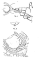
- One representative technique for following fetal heart rate involves a fetal scalp cutaneous EKG electrode device that screws into the fetal scalp (see Fig. 1).
- a vaginal probe is introduced onto the fetal scalp after the membranes have ruptured.
- Fetal EKG can be detected and recorded.
- Fetal well being can be ascertained by changes in fetal heart rate concomitant with uterine contractions. Unusually low fetal heart rates or unusually high fetal heart rates may be indicative of fetal distress.
- Unusually low fetal heart rates or unusually high fetal heart rates may be indicative of fetal distress.
- the ability to assess definite fetal distress from fetal heart rate pattern is restricted and limited.
- hypoxia is a condition of oxygen deprivation which may occur undetected during birth resulting in brain damage (viz. cerebral palsy) and other vital organ deterioration.
- a fetus suffers modest oxygen deprivation during uterine contractions but recovers an adequate oxygen level between contractions.
- the metabolism of the fetus is changed to a non-oxygen- requiring (anaerobic) process that produces lactic acid.
- This anaerobic change effects depression of the pH level of the fetal tissue from alkaline to acid.
- physicians have monitored fetal oxygen levels indirectly by measuring the tissue and/or blood pH level, which indeed becomes substantially acidic after hypoxia persists for several minutes.
- a fetal scalp cutaneous pH electrode device similar to the fetal cutaneous EKG electrode device (see Fig. 1) is used in monitoring fetal pH.
- Clark electrode Another technique for following fetal oxygen level involves the Clark electrode (see Figs. 2 and 3).
- This technique requires an exposed, relatively accessible presenting portion of fetal tissue through the dilated cervix, upon which an airtight dam is adhesively fixed.
- the airtight dam serves to capture oxygen as it diffuses through the fetal cutaneous layer.
- the accumulated diffused oxygen is electrochemically measured by the Clark electrode to provide an indication of oxygen in the fetal tissue.
- This technique suffers from the slowness of oxygen diffusion from the arterial blood stream out to the cutaneous layer. Further this technique suffers from significant incidence of imperfect air seal or an interruption of air seal that renders measurement errant and thus without utility.
- the objective is to provide sufficiently early warning of fetal distress such that appropriate alteration or initiation of treatment (e.g., administration of intravenous fluids and drugs, Caesarian section surgery) can prevent the tragedy of cerebral palsy or other hypoxic organ damage.
- appropriate alteration or initiation of treatment e.g., administration of intravenous fluids and drugs, Caesarian section surgery
- An intrauterine sensor for transillumination of a blood-perfused portion of fetal tissue to measure light extinction is disclosed.
- the sensor may be mounted upon a paddle-shaped probe provided with signal connections contained in an insulated cable leading to a measurement device.
- the sensor can be fastened by surgical glue or adhesive material or a cutaneous screw attachment for disposition on a portion of the fetus accessible through the dilated cervix.
- the senor includes a housing having an upper portion and a lower portion.
- a light source for transilluminating the fetal tissue is contained in the upper portion.
- the lower portion includes a cavity which is open at one end adapted for placement against the fetal tissue.
- a light sensor for receiving the light having transilluminated the fetal tissue is mounted in the cavity.
- attachment means communicate with the cavity for connecting the sensor to a vacuum source to provide a suction force between the sensor and the fetal presenting portion when the open end of the cavity is placed against the fetal presenting portion. Attachment can also be facilitated by the use of medical glue or other adhesive as an adjunct to the vacuum.
- At least one light source is mounted on the first side which lies against the fetal tissue in such a manner as to illuminate the fetal tissue.
- At least one photo-sensor is mounted on the same first side to receive the light after it has passed through or been reflected from the fetal tissue.
- At least one light source is mounted to the first portion of the concavity and at least one photo-sensor is mounted to the second portion of the same concavity.
- At least one light source is mounted to the first portion of a thin cylindrical, blunt-ended probe.
- At least one photo-sensor is mounted on the same first portion of the probe to receive light after it has passed from the light source through the fetal tissue.
- At least one light source is mounted to the first portion of a probe.
- At least one photo-sensor is mounted on the same first portion of the probe to receive light after it has passed from the light source through the fetal tissue.
- the apparatus is introduced into the vagina after the membranes have ruptured.
- the apparatus may be moved up the vagina using semi-flexible columnar tubing, through the dilated cervix, for disposition on a presenting portion of blood-perfused fetal tissue.
- Each device remains adherent to that cutaneous layer during birth until delivery is complete.
- Measurement from the modulated output of the photo-sensor yields information about the pulsating blood supply to the transilluminated fetal tissue, including but not limited to: oxygen saturation of the hemoglobin in the blood; the volume of individual blood pulsations supplying the * flesh; and the rate of blood pulsations occurring with each fetal heartbeat.
- the apparatus may also include, in addition to the aforementioned light sources and sensors, means for measuring heart electrical activity on the fetus and means for measuring the temperature of the fetus.
- the apparatus may include, in addition to the aforementioned fetal sensing means, a similar means for measurint heart electrical actvity, blood pulsation and oxygen saturation in the overlying maternal tissue.
- An object of this invention is to provide an apparatus with light source and a photo-sensor for transillumminating a blood-perfused portion of the fetus.
- the light source and the photo-sensor are mounted on a probe.
- This probe and its attaching signal transmission cable are introduced through the vagina and attached to a presenting portion of the fetus.
- the probe conforms closely and non-invasively to the fetal cutaneous layer.
- An advantage of the apparatus closely bonding to the fetal cutaneous layer is that measurement distortion, commonly caused by motion artifact, is minimized.
- the suppression of motion artifact renders measurement more accurate than a sensor with higher mass and aspect ratio.
- a further advantage of this invention is that the probe is attached superficially to the fetal head and neither invades nor occludes the blood flow to be interrogated.
- a further advantage of the disclosed invention is that the mother's hearbeat may simultaneously be measured and easily be distinguished from the fetal heartbeat whereby confusion of measurement resulting from erroneous placement of the probe is immediately apparent and will not cause error.
- the probe may be provided with two sensor sets: one facing the mother's body, the other facing the fetus.
- a further advantage of the disclosed invention is that the resultant direct and continuous measurements of the fetal arterial oxygen saturation afford the physician the opportunity to diagnose and take immediate responsive action and corrective therapy. For instance, unlike the prior art which had a limited capability to make delayed, indirect measurement of oxygen deprivation already having deleterious effects upon the fetus, the disclosed invention will allow the physician to know immediately when a fetus is initially oxygen deprived. This allows the physician to take steps to prevent the deterioration of brain and other vital organs.
- a further advantage of the disclosed invention is that the temperature of the area of the fetus may be taken simultaneously with the recording of the arterial oxygen saturation.
- a further advantage of the disclosed invention is that a patient's mechanical blood pumping activity (pulse) and oxygen saturation may be instantaneously recorded and compared for diagnosis and early treatment.
- a further advantage of the disclosed invention is that a physician may compare simultaneous instantaneous readings from both the fetus and the mother of their individual heart rate, electrocardiogram, oxygen saturation, and pulse amplitude.
- a further advantage is that the disclosed invention accurately measures arterial saturation even in circumstances when the partial pressure of oxygen is extremely low.
- a fetus receives oxygen from the maternal circulation, the oxygen diffusing from the mother's blood through the placenta into the fetal blood.
- the prior art Clark electrode has a high incidence of inaccurate (i.e. excessively low) oxygen saturation estimation when the partial pressure of the oxygen is low.
- the disclosed invention does not suffer from this handicap.
- a further advantage is the complete absence of erroneous measurement by the disclosed invention due to an imperfect seal between the sensor and the fetus.
- the prior art relied for accurate measurement upon creating and maintaining an absolutely perfect airtight seal between the apparatus and the fetus. Any leak resulted not only in erroneous measurement but the deceptive measurement could lead the physician to conclude in error that the fetus was receiving sufficient oxygen when it was not.
- a further advantage is the elimination of the risk of brain damage, eye damage or serious infection in the fetus.
- the sharp corkscrew pH or EKG electrode physically penetrates the presenting portion of cutaneous fetal tissue.
- presenting portion is the fetal eye or fontanel (the soft bone-free midscalp spot over the brain).
- fontanel or an eyelid is pierced, additional complications can occur.
- the disclosed invention does not expose the fetus to infection risk from skin penetration.
- a further advantage of the disclosed invention is that the parameters indicative of fetal condition are measured without the fetal head hair obstructing true readings.
- the disclosed invention measures accurately without regard t Q the texture of the fetal presenting portion.
- a further advantage of the disclosed invention is the ability to measure fetal arterial oxygen saturation directly rather than indirectly.
- the ability to directly and instantly measure blood oxygen content will accelerate positive diagnosis and appropriate treatment of fetal hypoxia. This is in contrast to the prior art where the fetal arterial oxygen level could only be measured by monitoring indirect effects (e.g., acidosis) of deteriorative changes already in progress.
- the invention disclosed herein gives direct measurement of fetal arterial oxygen saturation.
- a further object of this invention is to provide a combination of sensors mounted on a single probe to provide simultaneous and continuous fetal monitoring of several parameters, including but not limited to arterial hemoglobin oxygen saturation, pulse amplitude, pulse rate and rhythm, fetal electrocardiogram and fetal body temperature.
- the simultaneous and continuous availability of multiple important physiologic paraments provides instant means to cross-validate and corroborate measurements obtained with this probe, thereby improving substantially the clinical utility of the obstetric monitor herein disclosed compared to traditional single parameter (acoustic, electric or pH) monitors.
- a further object of this invention is to provide a method for making a form-fitting sensor.
- the light source and photo-sensor are mounted to thin substrates, being of such small dimension as to tightly conform to the probe surface with low aspect ratio.
- a further advantage of this invention is that it is fully sterilizable and low in cost.
- the resultant apparatus is sanitary, non-invasive, in full conformance to the skin and provides accurate measurement of heartbeat, arterial oxygen saturation and other parameters in both fetus and mother.
- a small, paddle-shaped probe is provided with four apertures, two on either side, for reposit of two sets of paired electrical components.
- a suitable substance for the probe composition that is commecially available is sold under the trademark SILASTIC, by Dow Corning. This is a moldable, translucent, silicon rubber which acts as a spatially diffused light source when in contact with an illuminated light-emitting diode.
- At least one light source 21 or 23, and at least one photo-sensor 20 or 22, the photo-sensors having shielding 30, 32, are located respectively in the two apertures on each side as shown (see Fig. 8).
- FIG. 8 illustrates the resulting assembly of one light source 21 and a photo-sensor 22 mounted to one side of the elongated paddle with another light source 23 and another photo-sensor 20 preferably mounted to the other side of the paddle-shaped probe.
- the paddle side making contact with the fetus is provided with two rows of gripping bumps 34 for traction on the fetal skin and to aid in separating any hair thereon to expose the fetal skin.
- the light sources 21, 23 are preferably light-emitting diodes.
- a light-emitting diode that is suitable and commercially available is manufactured by General Instrument Company with a typical wavelength between 650 and 950 nanometers.
- Each light-emitting diode 21, 23 is mounted on a ceramic substrate by gluing, with electrical connections provided by wire bonding.
- the light sources 21, 23 are each recessed into an aperture 32 (see Fig. 8), fastened to the paddle-shaped probe such that the light-emitting diode conforms to the planar surface of the probe.
- the photo-sensors 20, 22 shown each include a ceramic substrate electrical connections and a light sensitive surface fastened to the ceramic substrate by gluing and by wire bonding.
- the photo-sensors 20, 22 are recessed into the apertures 33 and are fastened to the paddle-shaped probe, such that the photo-sensors conform to the planar surface of the probe (Figs. 6A, 6B).
- each of the ceramic substrates shown in Figs. 8 (i.e., 20, 21, 22, 23) is 4mm x 6mm or of such other appropriately small dimensions.
- Electrical connections 25, 26 shown in Fig. 7 are provided leading from the probe, down detachable columnar tubing 27, which tubing aids in placing the probe up the vagina to the fetus, to a measuring device.
- An electrical connection that is suitable and commercially available is silicone rubber coated shielded multiconductor miniature cable.
- the mounting of the photo-sensors 20, 22 and the light sources (light-emitting diodes) 21, 23 onto the substrates herein disclosed can be accomplished through other configurations.
- an integrated chip or a thin film construction may be desirable for mass production of either the light source or photo-sensor.
- Fiber optic connection between the substrates and the measuring device is also suitable.
- the paddle-shaped probe has planar conformity wide enough to incorporate the two sets of paired photoelectric components 20, 21, 22, 23. Suitable dimensions of the paddle-shaped probe are 15mm x 15mm.
- the two sets of paired photo-sensors 20, 22, preferably photo-diodes, and light sources 21, 23 are mounted into the probe facing off opposite sides of the transparent paddle-shaped probe.
- the light source is preferably a 5mm distance away from the photo-sensor.
- the photo-diodes 20, 22 are encased in metal shielding shaped into boxes 30, with edges extending into the paddle at each corner.
- the shielding is composed of silver or some other composite being an electrical conductor. This shielding serves the dual purpose of excluding ambient light and of providing a metal surface suitable for an electrocardiogram sensor, i.e., by serving as electrodes connected to EKG equipment by electrical leads (not shown).
- the resulting probe may be provided with a combination of sensors including, but not limited to, two arterial oxygen sensors, two electrocardiogram sensors, and one thermistor (not shown).
- the electrical signals provided by the arterial oxygen sensors can be used to determine fetal and material pulse rate, rhythm and amplitude, as well as oxygen level in the blood.
- a small suction cup is provided with one set of photoelectric components: a light source 22 and a photo-sensor 21 mounted on the same concave side of the cup.
- a light source 22 is mounted on the same concave side of the cup.
- two ceramic substrate portions are indicated.
- a photo-sensor 22 is mounted to one ceramic substrate.
- a light source 22 is mounted to the other ceramic substrate.
- the light source 22 may be a light-emitting diode mounted to a ceramic substrate by gluing and by wire bonding. This light-emitting diode 22 conforms to a thin planar layer which is fastened to the concave side of the cup.
- the photo-sensor 21 shown includes a ceramic substrate, electrical connections and a light sensitive surface fastened to the ceramic substrate by gluing and by wire bonding.
- This photo-sensor 21 conforms to the planar layer of the concavity.
- each of the two ceramic substrates is 4mm x 6mm or other appropriately small dimensions.
- Electrical connections 35 are provided leading from the probe, down the columnar tubing 37 (Fig. 10).
- the mounting of the photo-sensor 21 and the light-emitting diode 20 onto the substrates herein disclosed can be accomplished through other configurations. For instance, an integrated chip or a thin film construction may be desirable for mass production of either the light source or photo-sensor. Fiber optic connection between the substrates and the measuring device is also suitable.
- the probe with suction cup 39 is shown placed cutaneously on the fetal presenting portion 38.
- the suction cup is seated on the presenting portion of the fetus 38, and as pressure is exerted from the columnar tubing toward the fetal head air is expelled from the top of the concavity of the cup, at which point the tissue moves to fill the small space where air is expelled creating a suction between the cutaneous layer and the cup.
- arterial oxygen sensors i.e., two sets of photo-sensors and light sources
- the light sources and photo-sensors on each side would preferably be adapted to operate at different wavelengths, in accordance with techniques for measuring the oxygen saturation.
- FIG. 13-16 An alternative and most preferred embodiment of the invention which uses a low-level vacuum to secure the sensor to the fetal tissue is illustrated in Figs. 13-16.
- Fig. 13 the probe or sensor 40 is shown held in place against fetal presenting portion 41.
- Suction tube 48 connected to the sensor terminates in fixture 49 for connecting the sensor to a suction source (not shown) such as those which are typically available in hospitals.
- electrical cable 46 attached to the sensor terminates in a connecting member 47 for connecting the sensor to electrical measuring equipments
- Electrical cables 53, 54 provide attachment means to an EKG monitor, as described below.
- the sensor comprises a relatively small, semi-hollow cylinder made from an optically transparent material such as LEXAN polycarbonate or SILASTIC silicone rubber.
- LEXAN and SILASTIC are trademarks of DuPont and Dow Corning, respectively.
- Electrical cable 53 connects the electrode 42 to the EKG monitoring equipment, as mentioned above.
- the optical components of the sensor comprise a light source 50, preferably a light-emitting diode, mounted on substrate 51, and a light sensor 52, preferably a photo-diode (see Fig. 16).
- the photo-diode is mounted in a stainless steel can 55, which is mounted concentrically in the sensor housing 43, as best shown in Figs. 14 and 15.
- Can 55 extends downwardly into a hollow space or cavity 44 formed in the lower sensor housing. This cavity is open at one end, as shown, for placement against the fetal skin (Figs. 15, 16). With the sensor in place, can 55 contacts the fetal tissue and serves as an electrode for the fetal EKG. Electrical cable 54 connects this electrode 55 to be EKG monitoring equipment, as mentioned above.
- the photo-diode 52 is mounted such that it is spaced from the inner surface of the sensor, a distance which is preferably 0.1".
- the substrate-mounted light source, light-emitting diode 50 is positioned directly above the light sensor, photo-diode 52. This reduces the possibility of undesirable interference or cross-talk caused by extraneous light originating from the light source reaching the light sensor without passing through the fetal flesh.
- the sensor is potted in an optically clear material such as medical grade epoxy or silicone rubber.
- Suction tube 48 is channeled through the upper portion of the sensor housing 43 and is ported to the interior cavity 44 of the sensor through opening 56 (see Figs. 14, 15).
- the sensor is also provided with a beveled edge 45 around the inner diameter of the cavity opening, to facilitate sealing when the vacuum is applied.
- the fetal presenting part 41 In use during childbirth, once the membranes have ruptured the sensor can be manually placed on the fetal presenting part 41, usually the head, through the vagina (see Fig. 16) and a low level vacuum applied through fixture 49 and suction tube 48. The skin of the fetal presenting part is sucked up around the sensor, as shown, to form an optical barrier substantially preventing ambient light from entering the interior portion of the sensor. This assures that the photo-diode 50 contained in the can 55 receives light which has passed through fetal tissue only.
- the structure and arrangement of the sensor is such that when the light source is operating, the entire sensor functions in the manner of a light- transmissive pipe, assuring that an adequate level of light is transmitted to the fetal skin despite the presence of hair, blood, mucous or other substances which tend to decrease the light level.
- the sensor may be provided with two light sources, both located in the upper portion of the sensor 40 as in Fig. 16, and two light sensors, both located in can 55.
- the light source-light sensor pairs would preferably operate at different wavelengths.
- an intravaginal and intrauterine sensor for transillumination of a blood-perfused portion of fetal tissue to measure light extinction.
- the sensor may be mounted onto a paddle-shaped probe provided with signal connections contained in an insulated cable leading to a measurement device.
- the sensor may include an opaque suction cup or other attaching apparatus for disposition on a protruding portion of the fetus.
- the sensor includes a housing having an upper portion and a lower portion. The lower portion includes a cavity which is open at one end for placement against the fetal tissue. Attachment means communicate with the cavity for connecting the sensor to a vacuum source to provide a suction force to secure the sensor against the fetal tissue.
- a light source is mounted on the first side of the probe that is designated to be placed against the fetal tissue in such a manner as to illuminate said tissue.
- a photo-sensor is mounted on same first side in such a manner as to receive said illumination after the light has passed through said fetal tissue.
- a light source is mounted to the first side of the concavity of the suction cup and a photo-sensor is mounted to the opposite side of the same concavity.
- a light source is mounted in the upper housing portion and a light sensor is mounted in the cavity.
- the housing is optically transmissive and adapted to provide an optical path between the light source and the fetal tissue.
Landscapes
- Health & Medical Sciences (AREA)
- Life Sciences & Earth Sciences (AREA)
- Physics & Mathematics (AREA)
- Medical Informatics (AREA)
- Surgery (AREA)
- Biophysics (AREA)
- Pathology (AREA)
- Engineering & Computer Science (AREA)
- Biomedical Technology (AREA)
- Heart & Thoracic Surgery (AREA)
- Veterinary Medicine (AREA)
- Molecular Biology (AREA)
- Public Health (AREA)
- Animal Behavior & Ethology (AREA)
- General Health & Medical Sciences (AREA)
- Pediatric Medicine (AREA)
- Cardiology (AREA)
- Optics & Photonics (AREA)
- Pregnancy & Childbirth (AREA)
- Gynecology & Obstetrics (AREA)
- Reproductive Health (AREA)
- Spectroscopy & Molecular Physics (AREA)
- Measurement Of The Respiration, Hearing Ability, Form, And Blood Characteristics Of Living Organisms (AREA)
Applications Claiming Priority (4)
| Application Number | Priority Date | Filing Date | Title |
|---|---|---|---|
| US52772683A | 1983-08-30 | 1983-08-30 | |
| US527726 | 1983-08-30 | ||
| US64405184A | 1984-08-24 | 1984-08-24 | |
| US644051 | 1984-08-24 |
Publications (2)
| Publication Number | Publication Date |
|---|---|
| EP0135840A2 true EP0135840A2 (de) | 1985-04-03 |
| EP0135840A3 EP0135840A3 (de) | 1986-06-11 |
Family
ID=27062497
Family Applications (1)
| Application Number | Title | Priority Date | Filing Date |
|---|---|---|---|
| EP84110306A Ceased EP0135840A3 (de) | 1983-08-30 | 1984-08-29 | Perinatales Oxymeter |
Country Status (1)
| Country | Link |
|---|---|
| EP (1) | EP0135840A3 (de) |
Cited By (35)
| Publication number | Priority date | Publication date | Assignee | Title |
|---|---|---|---|---|
| EP0199213A3 (de) * | 1985-04-25 | 1989-02-22 | Westinghouse Electric Corporation | Abgeschirmte gebrauchsfertige Elektrode zur zweidimensionalen Aufzeichnung von Elektroenzephalogrammen |
| WO1989009016A1 (fr) * | 1988-03-24 | 1989-10-05 | Johannes Buschmann | Procede et dispositif pour mesurer l'absorption de radiations par un tissu |
| WO1990001293A1 (en) * | 1988-08-12 | 1990-02-22 | Jason Otto Gardosi | Fetal probe |
| WO1990004352A1 (en) * | 1988-10-28 | 1990-05-03 | Nellcor Incorporated | Improved perinatal pulse oximetry sensor |
| EP0301165A3 (de) * | 1987-03-10 | 1991-05-02 | Wagner, Wolfgang, Dr.med. | Einrichtung zur Stoffwechselkontrolle |
| US5024226A (en) * | 1989-08-17 | 1991-06-18 | Critikon, Inc. | Epidural oxygen sensor |
| EP0442011A1 (de) | 1990-02-15 | 1991-08-21 | Hewlett-Packard GmbH | Fühler, Gerät und Verfahren zur nichtinvasiven Messung der Sauerstoffsättigung |
| WO1991015151A1 (en) | 1990-04-04 | 1991-10-17 | Nellcor Incorporated | Improved perinatal pulse oximetry probe |
| WO1991015996A1 (en) * | 1990-04-19 | 1991-10-31 | Egnell Ameda Limited | Non-invasive medical probe provided with suction cup |
| US5080098A (en) * | 1989-12-18 | 1992-01-14 | Sentinel Monitoring, Inc. | Non-invasive sensor |
| EP0471898A1 (de) * | 1990-08-22 | 1992-02-26 | Nellcor Incorporated | Fetales Puls-Sauerstoffmessgerät und Anwendung |
| US5109849A (en) * | 1983-08-30 | 1992-05-05 | Nellcor, Inc. | Perinatal pulse oximetry sensor |
| WO1993018705A1 (de) * | 1992-03-20 | 1993-09-30 | Gerhard Rall | Verfahren und vorrichtung zum messen von vitalen fetalen parametern während der geburt |
| EP0611548A1 (de) * | 1993-02-16 | 1994-08-24 | Gerhard Dipl.-Ing. Rall | Sensoreinrichtung zum Messen von vitalen Parametern eines Feten während der Geburt |
| DE4407541A1 (de) * | 1993-04-02 | 1994-10-06 | Mipm Mammendorfer Inst Fuer Ph | Vorrichtung zum Messen der Sauerstoffsättigung von Feten während der Geburt |
| US5551424A (en) * | 1990-05-29 | 1996-09-03 | Phox Medical Optics, Inc. | Fetal probe apparatus |
| EP0756848A1 (de) * | 1995-07-31 | 1997-02-05 | JOHNSON & JOHNSON MEDICAL, INC. | Instrument zur unblutigen Messung im menschlichen oder tierischen Körper |
| US5662103A (en) * | 1993-02-17 | 1997-09-02 | Utah Medical Products, Inc. | Subcutaneous radiation reflection probe |
| US5839439A (en) * | 1995-11-13 | 1998-11-24 | Nellcor Puritan Bennett Incorporated | Oximeter sensor with rigid inner housing and pliable overmold |
| US5911690A (en) * | 1994-12-01 | 1999-06-15 | Reinhold Kintza | Use of a pulse oxymetry sensor device |
| WO2000025664A1 (en) * | 1998-10-30 | 2000-05-11 | Medtronic, Inc. | Multiple sensor assembly for medical electrical lead |
| US6432051B1 (en) | 1998-03-13 | 2002-08-13 | Instrumentarium Corp. | Tonometric measuring head and measuring method |
| WO2002098272A2 (en) | 2001-06-05 | 2002-12-12 | Barnev Ltd. | Probe anchor |
| US6731976B2 (en) | 1997-09-03 | 2004-05-04 | Medtronic, Inc. | Device and method to measure and communicate body parameters |
| WO2006079862A3 (en) * | 2005-01-31 | 2006-12-21 | Hunor Santha | Pulse oximeter and casing for anchoring a sensor |
| US7165552B2 (en) | 2003-03-27 | 2007-01-23 | Cierra, Inc. | Methods and apparatus for treatment of patent foramen ovale |
| US7186251B2 (en) | 2003-03-27 | 2007-03-06 | Cierra, Inc. | Energy based devices and methods for treatment of patent foramen ovale |
| US7293562B2 (en) | 2003-03-27 | 2007-11-13 | Cierra, Inc. | Energy based devices and methods for treatment of anatomic tissue defects |
| US7311701B2 (en) | 2003-06-10 | 2007-12-25 | Cierra, Inc. | Methods and apparatus for non-invasively treating atrial fibrillation using high intensity focused ultrasound |
| US7367975B2 (en) | 2004-06-21 | 2008-05-06 | Cierra, Inc. | Energy based devices and methods for treatment of anatomic tissue defects |
| WO2007130508A3 (en) * | 2006-05-02 | 2008-06-12 | Nellcor Puritan Bennett Llc | Medical sensor with non-adhesive skin gripping contact |
| US7469158B2 (en) | 1997-06-17 | 2008-12-23 | Ric Investments, Llc | Fetal oximetry system and sensor |
| US7637924B2 (en) | 2003-03-27 | 2009-12-29 | Terumo Kabushiki Kaisha | Methods and apparatus for treatment of patent foramen ovale |
| US9468437B2 (en) | 1996-08-22 | 2016-10-18 | The Trustees Of Columbia University In The City Of New York | Endovascular flexible stapling device |
| WO2025056526A1 (en) * | 2023-09-12 | 2025-03-20 | Institut National De La Sante Et De La Recherche Medicale | Intra-uterine probe |
Family Cites Families (6)
| Publication number | Priority date | Publication date | Assignee | Title |
|---|---|---|---|---|
| DE1909882B2 (de) * | 1969-02-27 | 1976-11-11 | Arnold, Karl M., Dr., 8000 München; Beck, Harald, Prof., 6078 Neu-Isenburg | Einrichtung zum messen von koerperfunktionen bei mensch und tier durch messung der optischen reflexionseigenschaften des wechselnd durchbluteten gewebes und verfahren zu ihrer herstellung |
| DE2830412A1 (de) * | 1978-07-07 | 1980-01-17 | Erich Prof Dr Med Saling | Geraet zum ansetzen von abnehmern an koerpern |
| US4537197A (en) * | 1981-03-06 | 1985-08-27 | Hulka Jaroslav F | Disposable fetal oxygen monitor |
| IE53020B1 (en) * | 1981-08-05 | 1988-05-11 | Ici Plc | Reflected light measuring apparatus |
| US4541439A (en) * | 1982-04-23 | 1985-09-17 | American Home Products Corporation (Del.) | Monitoring of capillary blood flow |
| JPS5980230A (ja) * | 1982-09-24 | 1984-05-09 | アボット・ラボラトリーズ | 心電図用電極とpHプロ−ブとの結合体 |
-
1984
- 1984-08-29 EP EP84110306A patent/EP0135840A3/de not_active Ceased
Cited By (54)
| Publication number | Priority date | Publication date | Assignee | Title |
|---|---|---|---|---|
| US4938218A (en) * | 1983-08-30 | 1990-07-03 | Nellcor Incorporated | Perinatal pulse oximetry sensor |
| US5109849A (en) * | 1983-08-30 | 1992-05-05 | Nellcor, Inc. | Perinatal pulse oximetry sensor |
| EP0199213A3 (de) * | 1985-04-25 | 1989-02-22 | Westinghouse Electric Corporation | Abgeschirmte gebrauchsfertige Elektrode zur zweidimensionalen Aufzeichnung von Elektroenzephalogrammen |
| EP0301165A3 (de) * | 1987-03-10 | 1991-05-02 | Wagner, Wolfgang, Dr.med. | Einrichtung zur Stoffwechselkontrolle |
| WO1989009016A1 (fr) * | 1988-03-24 | 1989-10-05 | Johannes Buschmann | Procede et dispositif pour mesurer l'absorption de radiations par un tissu |
| DE3810008C1 (de) * | 1988-03-24 | 1989-10-26 | Johannes Dr. 8000 Muenchen De Buschmann | |
| US6298253B1 (en) * | 1988-03-24 | 2001-10-02 | Johannes Paul Buschmann | Method and device for measuring the absorption of radiation in a portion of tissue |
| WO1990001293A1 (en) * | 1988-08-12 | 1990-02-22 | Jason Otto Gardosi | Fetal probe |
| US5099842A (en) * | 1988-10-28 | 1992-03-31 | Nellcor Incorporated | Perinatal pulse oximetry probe |
| WO1990004352A1 (en) * | 1988-10-28 | 1990-05-03 | Nellcor Incorporated | Improved perinatal pulse oximetry sensor |
| AU645962B2 (en) * | 1988-10-28 | 1994-02-03 | Nellcor Incorporated | Improved perinatal pulse oximetry sensor |
| US5024226A (en) * | 1989-08-17 | 1991-06-18 | Critikon, Inc. | Epidural oxygen sensor |
| US5127407A (en) * | 1989-08-17 | 1992-07-07 | Critikon, Inc. | Epidural oxygen sensor |
| US5080098A (en) * | 1989-12-18 | 1992-01-14 | Sentinel Monitoring, Inc. | Non-invasive sensor |
| US5285783A (en) * | 1990-02-15 | 1994-02-15 | Hewlett-Packard Company | Sensor, apparatus and method for non-invasive measurement of oxygen saturation |
| EP0442011A1 (de) | 1990-02-15 | 1991-08-21 | Hewlett-Packard GmbH | Fühler, Gerät und Verfahren zur nichtinvasiven Messung der Sauerstoffsättigung |
| US5188108A (en) * | 1990-02-15 | 1993-02-23 | Hewlett-Packard Company | Sensor, apparatus and method for non-invasive measurement of oxygen saturation |
| WO1991015151A1 (en) | 1990-04-04 | 1991-10-17 | Nellcor Incorporated | Improved perinatal pulse oximetry probe |
| WO1991015996A1 (en) * | 1990-04-19 | 1991-10-31 | Egnell Ameda Limited | Non-invasive medical probe provided with suction cup |
| US5345935A (en) * | 1990-04-19 | 1994-09-13 | Egnell Ameda Limited | Non-invasive medical probe provided with suction cup |
| US5551424A (en) * | 1990-05-29 | 1996-09-03 | Phox Medical Optics, Inc. | Fetal probe apparatus |
| US5228440A (en) * | 1990-08-22 | 1993-07-20 | Nellcor, Inc. | Fetal pulse oximetry apparatus and method of use |
| EP0471898A1 (de) * | 1990-08-22 | 1992-02-26 | Nellcor Incorporated | Fetales Puls-Sauerstoffmessgerät und Anwendung |
| US6671530B2 (en) | 1990-08-22 | 2003-12-30 | Nellcor Puritan Bennett Incorporated | Positioning method for pulse oximetry fetal sensor |
| US5743260A (en) * | 1990-08-22 | 1998-04-28 | Nellcor Puritan Bennett Incorporated | Fetal pulse oximetry apparatus and method of use |
| WO1993018705A1 (de) * | 1992-03-20 | 1993-09-30 | Gerhard Rall | Verfahren und vorrichtung zum messen von vitalen fetalen parametern während der geburt |
| US5746212A (en) * | 1992-03-20 | 1998-05-05 | Rall; Gerhard | Process and device for measuring vital fetal parameters during labor and delivery |
| EP0611548A1 (de) * | 1993-02-16 | 1994-08-24 | Gerhard Dipl.-Ing. Rall | Sensoreinrichtung zum Messen von vitalen Parametern eines Feten während der Geburt |
| DE4304693C2 (de) * | 1993-02-16 | 2002-02-21 | Gerhard Rall | Sensoreinrichtung zum Messen von vitalen Parametern eines Feten während der Geburt |
| US5529064A (en) * | 1993-02-16 | 1996-06-25 | Rall; Gerhard | Sensor device for measuring vital parameters of a fetus during labor and delivery |
| US5662103A (en) * | 1993-02-17 | 1997-09-02 | Utah Medical Products, Inc. | Subcutaneous radiation reflection probe |
| DE4407541A1 (de) * | 1993-04-02 | 1994-10-06 | Mipm Mammendorfer Inst Fuer Ph | Vorrichtung zum Messen der Sauerstoffsättigung von Feten während der Geburt |
| US5911690A (en) * | 1994-12-01 | 1999-06-15 | Reinhold Kintza | Use of a pulse oxymetry sensor device |
| EP0756848A1 (de) * | 1995-07-31 | 1997-02-05 | JOHNSON & JOHNSON MEDICAL, INC. | Instrument zur unblutigen Messung im menschlichen oder tierischen Körper |
| US5908384A (en) * | 1995-07-31 | 1999-06-01 | Critikon Company, L.L.C. | Apparatus for non-invasive measurement within a human or animal body |
| US5839439A (en) * | 1995-11-13 | 1998-11-24 | Nellcor Puritan Bennett Incorporated | Oximeter sensor with rigid inner housing and pliable overmold |
| US9468437B2 (en) | 1996-08-22 | 2016-10-18 | The Trustees Of Columbia University In The City Of New York | Endovascular flexible stapling device |
| US7469158B2 (en) | 1997-06-17 | 2008-12-23 | Ric Investments, Llc | Fetal oximetry system and sensor |
| US6731976B2 (en) | 1997-09-03 | 2004-05-04 | Medtronic, Inc. | Device and method to measure and communicate body parameters |
| US6432051B1 (en) | 1998-03-13 | 2002-08-13 | Instrumentarium Corp. | Tonometric measuring head and measuring method |
| US6144866A (en) * | 1998-10-30 | 2000-11-07 | Medtronic, Inc. | Multiple sensor assembly for medical electric lead |
| WO2000025664A1 (en) * | 1998-10-30 | 2000-05-11 | Medtronic, Inc. | Multiple sensor assembly for medical electrical lead |
| EP1420693A4 (de) * | 2001-06-05 | 2007-05-30 | Barnev Ltd | Sondenanker |
| WO2002098272A2 (en) | 2001-06-05 | 2002-12-12 | Barnev Ltd. | Probe anchor |
| US7165552B2 (en) | 2003-03-27 | 2007-01-23 | Cierra, Inc. | Methods and apparatus for treatment of patent foramen ovale |
| US7293562B2 (en) | 2003-03-27 | 2007-11-13 | Cierra, Inc. | Energy based devices and methods for treatment of anatomic tissue defects |
| US7186251B2 (en) | 2003-03-27 | 2007-03-06 | Cierra, Inc. | Energy based devices and methods for treatment of patent foramen ovale |
| US8852181B2 (en) | 2003-03-27 | 2014-10-07 | Terumo Kabushiki Kaisha | Energy based devices and methods for treatment of anatomic tissue defects |
| US7637924B2 (en) | 2003-03-27 | 2009-12-29 | Terumo Kabushiki Kaisha | Methods and apparatus for treatment of patent foramen ovale |
| US7311701B2 (en) | 2003-06-10 | 2007-12-25 | Cierra, Inc. | Methods and apparatus for non-invasively treating atrial fibrillation using high intensity focused ultrasound |
| US7367975B2 (en) | 2004-06-21 | 2008-05-06 | Cierra, Inc. | Energy based devices and methods for treatment of anatomic tissue defects |
| WO2006079862A3 (en) * | 2005-01-31 | 2006-12-21 | Hunor Santha | Pulse oximeter and casing for anchoring a sensor |
| WO2007130508A3 (en) * | 2006-05-02 | 2008-06-12 | Nellcor Puritan Bennett Llc | Medical sensor with non-adhesive skin gripping contact |
| WO2025056526A1 (en) * | 2023-09-12 | 2025-03-20 | Institut National De La Sante Et De La Recherche Medicale | Intra-uterine probe |
Also Published As
| Publication number | Publication date |
|---|---|
| EP0135840A3 (de) | 1986-06-11 |
Similar Documents
| Publication | Publication Date | Title |
|---|---|---|
| EP0135840A2 (de) | Perinatales Oxymeter | |
| EP0440741B1 (de) | Verbesserter perinataler pulsierter Oximeterfühler | |
| US5109849A (en) | Perinatal pulse oximetry sensor | |
| US5823952A (en) | Pulse oximeter sensor with differential slip coefficient | |
| US4537197A (en) | Disposable fetal oxygen monitor | |
| US5727547A (en) | Presenting part fetal oximeter sensor with securing mechanism for providing tension to scalp attachment | |
| US6285896B1 (en) | Fetal pulse oximetry sensor | |
| US5911690A (en) | Use of a pulse oxymetry sensor device | |
| US5776058A (en) | Pressure-attached presenting part fetal pulse oximetry sensor | |
| US5833622A (en) | Non-invasive fetal probe having improved mechanical and electrical properties | |
| US20040082842A1 (en) | System for monitoring fetal status | |
| US5193542A (en) | Peripartum oximetric monitoring apparatus | |
| US5851179A (en) | Pulse oximeter sensor with articulating head | |
| US5916155A (en) | Fetal sensor with securing balloons remote from optics | |
| US20130109937A1 (en) | Two-part patch sensor for monitoring vital signs | |
| ATE141768T1 (de) | Nicht-invasive medizinische sonde mit saugnapf | |
| US20040015067A1 (en) | Noninvasive, intrauterine fetal egg strip electrode | |
| US6298253B1 (en) | Method and device for measuring the absorption of radiation in a portion of tissue | |
| US20230320634A1 (en) | System for monitoring fetal status during child birth | |
| JPS6090535A (ja) | 分娩時の酸素計 | |
| US20130102863A1 (en) | Non-Invasive Trans-Reflective Monitoring Apparatus | |
| Aarnoudse et al. | Subcutaneous oxygen tension in the fetal scalp during labour continuous monitoring with a needle electrode | |
| GB2482758A (en) | Monitoring uterine activity and fetal heart rate | |
| Unger et al. | Instrumentation for fetal electrocardiography and intrauterine pressure: A new scalp electrode and radiotelemetry system | |
| Svenningsen et al. | Application of fiberoptics to the clinical measurement of intra-uterine pressure in labor |
Legal Events
| Date | Code | Title | Description |
|---|---|---|---|
| PUAI | Public reference made under article 153(3) epc to a published international application that has entered the european phase |
Free format text: ORIGINAL CODE: 0009012 |
|
| AK | Designated contracting states |
Designated state(s): AT BE CH DE FR GB IT LI LU NL SE |
|
| PUAL | Search report despatched |
Free format text: ORIGINAL CODE: 0009013 |
|
| AK | Designated contracting states |
Kind code of ref document: A3 Designated state(s): AT BE CH DE FR GB IT LI LU NL SE |
|
| 17P | Request for examination filed |
Effective date: 19861210 |
|
| 17Q | First examination report despatched |
Effective date: 19880903 |
|
| STAA | Information on the status of an ep patent application or granted ep patent |
Free format text: STATUS: THE APPLICATION HAS BEEN REFUSED |
|
| 18R | Application refused |
Effective date: 19900129 |
|
| RIN1 | Information on inventor provided before grant (corrected) |
Inventor name: NEW, WILLIAM, JR. Inventor name: YELDERMAN, MARK Inventor name: CORENMAN, JAMES E. Inventor name: GOODMAN, DAVID E. |