EP0141632A2 - Optische Prüfeinrichtung für aufbewahrte menschliche Thrombozyten - Google Patents
Optische Prüfeinrichtung für aufbewahrte menschliche Thrombozyten Download PDFInfo
- Publication number
- EP0141632A2 EP0141632A2 EP84307422A EP84307422A EP0141632A2 EP 0141632 A2 EP0141632 A2 EP 0141632A2 EP 84307422 A EP84307422 A EP 84307422A EP 84307422 A EP84307422 A EP 84307422A EP 0141632 A2 EP0141632 A2 EP 0141632A2
- Authority
- EP
- European Patent Office
- Prior art keywords
- pack
- platelets
- light
- bag
- channel
- Prior art date
- Legal status (The legal status is an assumption and is not a legal conclusion. Google has not performed a legal analysis and makes no representation as to the accuracy of the status listed.)
- Granted
Links
Images

Classifications
-
- G—PHYSICS
- G01—MEASURING; TESTING
- G01N—INVESTIGATING OR ANALYSING MATERIALS BY DETERMINING THEIR CHEMICAL OR PHYSICAL PROPERTIES
- G01N21/00—Investigating or analysing materials by the use of optical means, i.e. using sub-millimetre waves, infrared, visible or ultraviolet light
- G01N21/17—Systems in which incident light is modified in accordance with the properties of the material investigated
- G01N21/47—Scattering, i.e. diffuse reflection
- G01N21/49—Scattering, i.e. diffuse reflection within a body or fluid
- G01N21/51—Scattering, i.e. diffuse reflection within a body or fluid inside a container, e.g. in an ampoule
-
- G—PHYSICS
- G01—MEASURING; TESTING
- G01N—INVESTIGATING OR ANALYSING MATERIALS BY DETERMINING THEIR CHEMICAL OR PHYSICAL PROPERTIES
- G01N15/00—Investigating characteristics of particles; Investigating permeability, pore-volume or surface-area of porous materials
- G01N15/01—Investigating characteristics of particles; Investigating permeability, pore-volume or surface-area of porous materials specially adapted for biological cells, e.g. blood cells
- G01N2015/018—Platelets
-
- G—PHYSICS
- G01—MEASURING; TESTING
- G01N—INVESTIGATING OR ANALYSING MATERIALS BY DETERMINING THEIR CHEMICAL OR PHYSICAL PROPERTIES
- G01N15/00—Investigating characteristics of particles; Investigating permeability, pore-volume or surface-area of porous materials
- G01N15/02—Investigating particle size or size distribution
- G01N15/0205—Investigating particle size or size distribution by optical means
- G01N15/0211—Investigating a scatter or diffraction pattern
- G01N2015/0222—Investigating a scatter or diffraction pattern from dynamic light scattering, e.g. photon correlation spectroscopy
-
- G—PHYSICS
- G01—MEASURING; TESTING
- G01N—INVESTIGATING OR ANALYSING MATERIALS BY DETERMINING THEIR CHEMICAL OR PHYSICAL PROPERTIES
- G01N21/00—Investigating or analysing materials by the use of optical means, i.e. using sub-millimetre waves, infrared, visible or ultraviolet light
- G01N21/01—Arrangements or apparatus for facilitating the optical investigation
- G01N21/03—Cuvette constructions
- G01N2021/0364—Cuvette constructions flexible, compressible
Definitions
- the ratio of the intensity of light scattered in a particular direction when the platelets are moving, divided by the intensity of light scattered in the same direction when the platelets are still, may be less than 0.7 when the platelets are functional but approaches unity as the platelets die.
- This technique is of theoretical interest but its application would be difficult in clinical use as it would be necessary to remove a pack of platelets from the rocking tray, on which such packs are normally stored to keep the platelets in motion, and to transfer it to some device in which the light scattering properties could be measured both when the platelets are in motion and stationary.
- the invention thus also includes a blood platelet monitoring assembly for carrying out the new method, the assembly comprising at least one translucent pack of blood platelets; means for agitating the pack; a light source for irradiating the pack, while the pack is being agitated, with a beam of light; a photoelectric element for collecting light scattered from the pack in a particular direction; and means responsive to an AC component, corresponding to fluctuations in the intensity of the scattered light, in the output signal from the photoelectric element.
- the particular advantage of the new method is that the viability of the new platelets can be monitored while the packs are being agitated, which is the accepted condition in order to prolong the life of the platelets.
- the pack is agitated to cause the platelets to flow continually to and fro across a zone through which the pack is irradiated with the light beam.
- the agitation could be provided by rocking the pack, for example in association with a tray similar to those on which platelet packs are conventionally stored.
- the frequency of the fluctuations in the amplitude of the scattered light will be twice the frequency of the oscillating flow.
- the pack comprises a bag with two major, generally translucent, opposed walls
- the bag may be supported so that these walls are pinched together to divide the interior of the bag into two parts connected by a channel constituting the zone; one of the parts being periodically squeezed to cause the to and fro flow through the channel.
- the channel is then preferably positioned adjacent to the bottom of the bag, as oriented in its support, so that any tendency of the platelets to sediment at the bottom of the bag is avoided by the continual disturbance of the platelets by the oscillating flow.
- the earlier proposed optical technique used, as a light source, a bulky laser which was housed, together with plates between which the pack under test was sandwiched, in a light proof housing.
- the laser beam was directed through windows in the plates, and through the intervening pack, to the photoelectric element.
- This is all unacceptably cumbersome for clinical use.
- This device is extremely compact, requires no screening from exterior light, and ensures that the optical system is correctly aligned as soon as the pack is gripped between its surfaces.
- the datum reading might be read by a technician and recorded when the pack is fresh, preferably within twelve hours of the platelets having been obtained from a human body. Prior to clinical use, a second reading would be taken by the technician, and the platelets may then be considered to be sufficiently non-functional for clinical use, if the amplitude of the fluctuations has decreased to say 25% or less of the datum figure for fresh platelets.
- An appropriate display would then merely need to provide a numerical reading representing the amplitude of the AC signal.
- appropriate circuitry may incorporate a microprocessor or other intelligent circuit which stores the datum signal corresponding to the pack when fresh, and provides a continuous display representing the ratio of the current signal to the datum signal. In the simplest case, this may be a green light which is illuminated when the platelets are still functional and a red light when they are not.
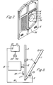
- the system incorporates a device comprising a first, vertical fixed plate 6, and a similar plate 7 which is connected to it by a hinge 8.
- Each plate consists of a peripheral frame formed integrally with a central cruciform portion defining with the peripheral frame, four windows. In each case the upper windows are open. The lower windows of the plate 7 and one of the windows of the plate 6 are partially closed by screens 9.
- the plate 7 is arranged to be swung up generally parallel to the plate 6, about the hinge 8, to grip between the two jaw-like plates a conventional bag 10 of blood platelets in plasma,
- the bag has, in a peripheral seam, holes 11 which are fitted onto pegs 12 projecting from the plate 7.
- the two plates 6 and 7 are held closed, with the bag 10 gripped between them, by means of side catches 13.
- Figure 1 shows the bag in full lines separate from the plates and, in chain dotted lines, in position on the plate 7.
- an L-shaped seal 14 pinches the bag along a pinch line shown at 14' in dotted lines in Figure 1, to divide the interior of the bag into parts 15 and 16, which are interconnected by a channel 17 adjacent to the bottom of the bag.
- a cylindrical housing 18 in which there slides axially a plunger 19 carrying at one end a disc 20 and at the other end a disc 21.
- a helically coiled compression spring 22 acts between the disc 21 and an end wall of the housing 18 to urge the disc 20 to a withdrawn position away from the plate 6.
- the plunger 19 and disc 20 are able to move back out of the plate 6 under the action of the spring 22, thus releasing the squeeze pressure on the part 15 of the bag 10.
- the differential hydrostatic pressure at the channel 17, resulting from the previous displacement of the contents into the part 16 of the bag 10, then causes the contents to flow back through the channel 17 into the part 15.
- This periodic rotation of the cam 23 thus causes to and fro flow in the bag 10 through the channel 17, as indicated by the arrows, and keeps all the contents in continual motion.
- the frequency of the cam 23 may be about 2Hz and the stroke of the disc 20 about 3 ml.
- the plate 6 In alignment with the channel 17, the plate 6 carries a cylindrical mounting 27, incorporating a light emitting diode (Hewlett-Packard HLMP 3750, wavelength 635 nm), which is energised through a line 28.
- the LED directs its beam through an opening 36 in the plate 6, and through the channel 17 in the bag 10, where it is scattered by the platelets. Any light which is ultimately scattered in the straight ahead direction, passes through a complimentary opening in the plate 7, and is picked up by a light sensitive resistor in a cylindrical mounting 29.
- the electrical output of the photoresistor which will correspond directly to the amplitude of the light scattered at an angle of substantially 0° to the beam from the LED, is transmitted along a line 30 to a microprocessor 31. This microprocessor deduces the AC component in the signal by sensing and subtracting the maximum and minimum signal levels.
- the platelets in the bag 10 are caused to flow with an oscillating motion to and fro through the channel 17, the platelets will, at maximum velocity, be caused to stream in mutual alignment through the channel, and, upon reversal of the direction of flow, will adopt a relatively random orientation. This is what leads to the fluctuations of the amplitude in the intensity of light received from the LED by the photoresistor.
- a display of the microprocessor 31 may be preset to respond to an AC signal having an amplitude above or below a certain threshold corresponding to an acceptable or unacceptable number of viable platelets in the bag 10, respectively.
- the microprocessor 31 may be instantly calibrated to a corresponding threshold, by recording the datum reading by, for example, depression of a switch 32.
- a green lamp 33 may be illuminated.
- the amplitude will fall below the threshold, and a red lamp 34 will be illuminated. In this way any particular bag of platelets can be used up to a last possible moment when a satisfactory proportion of its platelets are viable for clinical use.
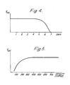
- Figure 4 shows a graph of the variation in a voltage V osc corresponding to the amplitude of the A C component in the signal from the photoresistor, with a period of days. The sharp cut off after, on average, five days, is very noticeable.
- Figure 5 shows a variation in a similar voltage with a variation in the density of viable platelets in the bag 10. This shows that the amplitude of the AC signal is substantially independent of the density when the density is between 300 and 700 times 10 9 /1, and falls off very sharply at lower densities.
Landscapes
- Immunology (AREA)
- Pathology (AREA)
- Life Sciences & Earth Sciences (AREA)
- Chemical & Material Sciences (AREA)
- Analytical Chemistry (AREA)
- Biochemistry (AREA)
- Health & Medical Sciences (AREA)
- General Health & Medical Sciences (AREA)
- Physics & Mathematics (AREA)
- General Physics & Mathematics (AREA)
- Investigating Or Analysing Biological Materials (AREA)
- Optical Head (AREA)
- Glass Compositions (AREA)
- Laser Surgery Devices (AREA)
- Investigating Or Analysing Materials By Optical Means (AREA)
- External Artificial Organs (AREA)
Priority Applications (1)
| Application Number | Priority Date | Filing Date | Title |
|---|---|---|---|
| AT84307422T ATE42147T1 (de) | 1983-10-31 | 1984-10-29 | Optische pruefeinrichtung fuer aufbewahrte menschliche thrombozyten. |
Applications Claiming Priority (2)
| Application Number | Priority Date | Filing Date | Title |
|---|---|---|---|
| GB8328979 | 1983-10-31 | ||
| GB838328979A GB8328979D0 (en) | 1983-10-31 | 1983-10-31 | Optical assay |
Publications (3)
| Publication Number | Publication Date |
|---|---|
| EP0141632A2 true EP0141632A2 (de) | 1985-05-15 |
| EP0141632A3 EP0141632A3 (en) | 1986-06-04 |
| EP0141632B1 EP0141632B1 (de) | 1989-04-12 |
Family
ID=10550966
Family Applications (1)
| Application Number | Title | Priority Date | Filing Date |
|---|---|---|---|
| EP84307422A Expired EP0141632B1 (de) | 1983-10-31 | 1984-10-29 | Optische Prüfeinrichtung für aufbewahrte menschliche Thrombozyten |
Country Status (6)
| Country | Link |
|---|---|
| US (2) | US4682887A (de) |
| EP (1) | EP0141632B1 (de) |
| JP (1) | JPS60113153A (de) |
| AT (1) | ATE42147T1 (de) |
| DE (1) | DE3477692D1 (de) |
| GB (1) | GB8328979D0 (de) |
Cited By (3)
| Publication number | Priority date | Publication date | Assignee | Title |
|---|---|---|---|---|
| WO1986002545A1 (en) * | 1984-10-29 | 1986-05-09 | Bellhouse Medical Products Limited | Blood bag |
| WO1999026066A1 (en) * | 1997-11-18 | 1999-05-27 | Gematron Medical Ab | The handling of blood in bags by means of rotating disc |
| EP2115424A4 (de) * | 2007-02-02 | 2017-07-26 | Canadian Blood Services | Verfahren zum nachweis von bakterienkontamination mittels dynamischer lichtstreuung |
Families Citing this family (20)
| Publication number | Priority date | Publication date | Assignee | Title |
|---|---|---|---|---|
| JPS62220833A (ja) * | 1986-03-22 | 1987-09-29 | Sigma Tec:Kk | 光散乱式微粒子センサ |
| US4810090A (en) * | 1987-08-24 | 1989-03-07 | Cobe Laboratories, Inc. | Method and apparatus for monitoring blood components |
| JPH03500573A (ja) * | 1988-04-20 | 1991-02-07 | フセソユズニ カルディオロジチェスキ ナウチニ ツェントル アカデミイ メディツィンスキフ ナウク エスエスエスエル | 血小板凝集の分析方法及びその装置 |
| US5510621A (en) * | 1994-10-03 | 1996-04-23 | Optical Solutions, Inc. | Apparatus and method for measuring components in a bag |
| US6984526B2 (en) * | 1995-02-08 | 2006-01-10 | University Of South Florida | Spectrophotometric method for determining the viability of a sample containing platelets |
| ES2149087B1 (es) * | 1998-02-26 | 2001-05-16 | Grifols Grupo Sa | Procedimiento para el control repetitivo de la estabilidad de componentes sanguineos y dispositivo para su realizacion. |
| RU2148257C1 (ru) * | 1999-06-01 | 2000-04-27 | Тверская государственная медицинская академия | Способ исследования крови |
| DE10223450A1 (de) * | 2002-05-23 | 2003-12-04 | Laser & Med Tech Gmbh | Optisches Verfahren zur Bestimmung des extrazellulären Hämoglobingehaltes in Blutkonserven |
| AU2003298801B9 (en) * | 2002-12-02 | 2008-07-31 | Gi Dynamics, Inc. | Bariatric sleeve |
| JP2013509618A (ja) | 2009-10-28 | 2013-03-14 | アレンティック マイクロサイエンス インコーポレイテッド | 顕微撮像法 |
| US20140152801A1 (en) | 2009-10-28 | 2014-06-05 | Alentic Microscience Inc. | Detecting and Using Light Representative of a Sample |
| US9075225B2 (en) | 2009-10-28 | 2015-07-07 | Alentic Microscience Inc. | Microscopy imaging |
| US9448151B2 (en) | 2011-10-22 | 2016-09-20 | Lightintegra Technology Inc. | Apparatus and method for platelet monitoring and for assessing the quality of platelets |
| US10502666B2 (en) | 2013-02-06 | 2019-12-10 | Alentic Microscience Inc. | Sample processing improvements for quantitative microscopy |
| WO2014205576A1 (en) * | 2013-06-26 | 2014-12-31 | Alentic Microscience Inc. | Sample processing improvements for microscopy |
| DE102017218846A1 (de) | 2017-10-23 | 2019-04-25 | Fraunhofer-Gesellschaft zur Förderung der angewandten Forschung e.V. | Nicht-invasive Blutanalytik von Blutkonserven |
| EP3700419A4 (de) * | 2017-10-26 | 2021-11-10 | Essenlix Corporation | Schnelltest von blutplättchen |
| CN111498304B (zh) * | 2019-01-31 | 2022-04-19 | 青岛海特生物医疗有限公司 | 血液袋存储盒及血液质量检测方法 |
| EP4078137A4 (de) * | 2019-12-18 | 2024-02-21 | Cyteguard AB | Thrombozytenkonzentrat-kontrolle |
| CN111907824B (zh) * | 2020-08-28 | 2022-01-07 | 山东一达食品有限公司 | 一种快递打包用预防物件损坏辅助设备 |
Family Cites Families (13)
| Publication number | Priority date | Publication date | Assignee | Title |
|---|---|---|---|---|
| US3733136A (en) * | 1971-07-12 | 1973-05-15 | Furedi A Porath | Method and apparatus for measuring the degree of assymetry of non-spherical particles |
| JPS5418596B2 (de) * | 1971-09-07 | 1979-07-09 | ||
| US3893766A (en) * | 1973-06-14 | 1975-07-08 | Coulter Electronics | Apparatus for orienting generally flat particles for slit-scan photometry |
| US3891325A (en) * | 1973-09-20 | 1975-06-24 | Ovutime Inc | Mucus sampling and measuring systems, devices and processes |
| US4135818A (en) * | 1975-01-22 | 1979-01-23 | Bio/Data Corporation | Platelet aggregation monitoring device |
| US4139303A (en) * | 1977-01-21 | 1979-02-13 | The Board Of Regents Of The University Of Washington | Adaptive coherent optical processing method and apparatus for recognizing and counting objects |
| US4201470A (en) * | 1977-02-24 | 1980-05-06 | Albrecht Ehrly | Method and apparatus for measurement and registration of the aggregation rate of particles suspended in a liquid |
| US4227814A (en) * | 1979-02-01 | 1980-10-14 | Baxter Travenol Laboratories, Inc. | Optical density detector |
| US4380169A (en) * | 1981-01-21 | 1983-04-19 | The Curators Of The University Of Missouri | Method and apparatus for measuring moisture tension in leaves of growing plants |
| IT1208062B (it) * | 1981-03-23 | 1989-06-06 | Croce F Dr Salus Ricerca | Dei fenomeni di emocoagulazione, dispositivo di rivelazione ottica concentrazione, ed aggregazione delle piastrine o simili. |
| DE3176091D1 (en) * | 1981-09-15 | 1987-05-14 | Mueller Arno | Method and device for the quantitative determination of dissolved substances in single- or multicomponent systems of laser light scattering |
| NZ200732A (en) * | 1982-05-25 | 1987-05-29 | New Zealand Dev Finance | Deriving sperm motility indication by laser light scattering measurement |
| US4522494A (en) * | 1982-07-07 | 1985-06-11 | The United States Of America As Represented By The Department Of Health And Human Services | Non-invasive optical assessment of platelet viability |
-
1983
- 1983-10-31 GB GB838328979A patent/GB8328979D0/en active Pending
-
1984
- 1984-10-29 EP EP84307422A patent/EP0141632B1/de not_active Expired
- 1984-10-29 AT AT84307422T patent/ATE42147T1/de not_active IP Right Cessation
- 1984-10-29 DE DE8484307422T patent/DE3477692D1/de not_active Expired
- 1984-10-30 JP JP59226958A patent/JPS60113153A/ja active Pending
- 1984-10-30 US US06/666,390 patent/US4682887A/en not_active Expired - Fee Related
- 1984-10-30 US US06/666,357 patent/US4657383A/en not_active Expired - Fee Related
Cited By (5)
| Publication number | Priority date | Publication date | Assignee | Title |
|---|---|---|---|---|
| WO1986002545A1 (en) * | 1984-10-29 | 1986-05-09 | Bellhouse Medical Products Limited | Blood bag |
| US4675019A (en) * | 1984-10-29 | 1987-06-23 | Bellhouse Medical Products Limited | Blood monitoring system |
| US4758083A (en) * | 1984-10-29 | 1988-07-19 | Bellhouse Technology Limited | Blood monitoring system |
| WO1999026066A1 (en) * | 1997-11-18 | 1999-05-27 | Gematron Medical Ab | The handling of blood in bags by means of rotating disc |
| EP2115424A4 (de) * | 2007-02-02 | 2017-07-26 | Canadian Blood Services | Verfahren zum nachweis von bakterienkontamination mittels dynamischer lichtstreuung |
Also Published As
| Publication number | Publication date |
|---|---|
| US4682887A (en) | 1987-07-28 |
| DE3477692D1 (en) | 1989-05-18 |
| EP0141632A3 (en) | 1986-06-04 |
| JPS60113153A (ja) | 1985-06-19 |
| EP0141632B1 (de) | 1989-04-12 |
| ATE42147T1 (de) | 1989-04-15 |
| US4657383A (en) | 1987-04-14 |
| GB8328979D0 (en) | 1983-11-30 |
Similar Documents
| Publication | Publication Date | Title |
|---|---|---|
| US4682887A (en) | Optical assay for stored human platelets | |
| US4830510A (en) | Optical assay method for stored human platelets | |
| US4822568A (en) | Apparatus for measuring aggregation rate of whole blood red blood cells | |
| US5279150A (en) | Automated miniature centrifuge | |
| KR100304144B1 (ko) | 비침습혈액분석장치와그방법 | |
| ES2990057T3 (es) | Método y sistema para mediciones ópticas o eléctricas en fluidos dispersos | |
| US5071247A (en) | Method for analysis of blood platelet aggregations and apparatus therefor | |
| EP2185927B1 (de) | Vorrichtung und verfahren zur bestimmung der erythrozyten-sedimentierungsrate in einer blutprobe | |
| US4710874A (en) | Method and apparatus for displaying particle sedimentation rates in liquids | |
| NL8202218A (nl) | Werkwijze en inrichting voor het meten van de bloedstollingstijd. | |
| JP7382341B2 (ja) | 赤血球沈降速度及び他の関連パラメータを決定するための装置及び方法 | |
| EP2926115B1 (de) | Verfahren und systeme zur bestimmung der physikalischer eigenschaften einer probe in einer tragbaren point-of-care-diagnosevorrichtung | |
| KR20030020946A (ko) | 테스트 스트립상의 유체 존재 여부를 검출하는 장치 및 방법 | |
| CA2492486A1 (en) | Photometric determination of coagulation time in undiluted whole blood | |
| IL119904A0 (en) | An automated method for discriminating platelets from cells and apparatus for carrying out same | |
| US4374644A (en) | Blood cell volume monitoring | |
| US3463614A (en) | Method and apparatus for use in promoting agglomeration | |
| ES2244021T3 (es) | Metodo y aparato para la medicion rapida de capas de celulas. | |
| EP0099309B1 (de) | Laser-Nephelometer zur Detektion der Antigene und Antikörper | |
| US5491095A (en) | System and method for aggulutination detection | |
| EP0021236A1 (de) | Verfahren zur optischen Prüfung flüssiger Proben | |
| JPS61124867A (ja) | 免疫学的試験用試験カード | |
| US4813420A (en) | Hemolysis reaction measuring device | |
| CN1206832A (zh) | 快速测量细胞层的方法 | |
| US4411523A (en) | Cuvette for blood oxygen analysis apparatus |
Legal Events
| Date | Code | Title | Description |
|---|---|---|---|
| PUAI | Public reference made under article 153(3) epc to a published international application that has entered the european phase |
Free format text: ORIGINAL CODE: 0009012 |
|
| AK | Designated contracting states |
Designated state(s): AT BE CH DE FR GB IT LI LU NL SE |
|
| PUAL | Search report despatched |
Free format text: ORIGINAL CODE: 0009013 |
|
| AK | Designated contracting states |
Kind code of ref document: A3 Designated state(s): AT BE CH DE FR GB IT LI LU NL SE |
|
| 17P | Request for examination filed |
Effective date: 19861125 |
|
| 17Q | First examination report despatched |
Effective date: 19880418 |
|
| GRAA | (expected) grant |
Free format text: ORIGINAL CODE: 0009210 |
|
| AK | Designated contracting states |
Kind code of ref document: B1 Designated state(s): AT BE CH DE FR GB IT LI LU NL SE |
|
| PG25 | Lapsed in a contracting state [announced via postgrant information from national office to epo] |
Ref country code: AT Effective date: 19890412 |
|
| REF | Corresponds to: |
Ref document number: 42147 Country of ref document: AT Date of ref document: 19890415 Kind code of ref document: T |
|
| ITF | It: translation for a ep patent filed | ||
| REF | Corresponds to: |
Ref document number: 3477692 Country of ref document: DE Date of ref document: 19890518 |
|
| ET | Fr: translation filed | ||
| PG25 | Lapsed in a contracting state [announced via postgrant information from national office to epo] |
Ref country code: GB Effective date: 19891029 |
|
| PG25 | Lapsed in a contracting state [announced via postgrant information from national office to epo] |
Ref country code: SE Effective date: 19891030 |
|
| PG25 | Lapsed in a contracting state [announced via postgrant information from national office to epo] |
Ref country code: LU Free format text: LAPSE BECAUSE OF NON-PAYMENT OF DUE FEES Effective date: 19891031 Ref country code: LI Effective date: 19891031 Ref country code: CH Effective date: 19891031 Ref country code: BE Effective date: 19891031 |
|
| PLBE | No opposition filed within time limit |
Free format text: ORIGINAL CODE: 0009261 |
|
| STAA | Information on the status of an ep patent application or granted ep patent |
Free format text: STATUS: NO OPPOSITION FILED WITHIN TIME LIMIT |
|
| 26N | No opposition filed | ||
| BERE | Be: lapsed |
Owner name: BELLHOUSE BRIAN JOHN Effective date: 19891031 |
|
| PG25 | Lapsed in a contracting state [announced via postgrant information from national office to epo] |
Ref country code: NL Effective date: 19900501 |
|
| NLV4 | Nl: lapsed or anulled due to non-payment of the annual fee | ||
| GBPC | Gb: european patent ceased through non-payment of renewal fee | ||
| PG25 | Lapsed in a contracting state [announced via postgrant information from national office to epo] |
Ref country code: FR Effective date: 19900629 |
|
| REG | Reference to a national code |
Ref country code: CH Ref legal event code: PL |
|
| PG25 | Lapsed in a contracting state [announced via postgrant information from national office to epo] |
Ref country code: DE Effective date: 19900703 |
|
| REG | Reference to a national code |
Ref country code: FR Ref legal event code: ST |
|
| EUG | Se: european patent has lapsed |
Ref document number: 84307422.0 Effective date: 19900706 |