EP0148551A2 - Méthode pour mesurer la fusion de membranes - Google Patents
Méthode pour mesurer la fusion de membranes Download PDFInfo
- Publication number
- EP0148551A2 EP0148551A2 EP84303614A EP84303614A EP0148551A2 EP 0148551 A2 EP0148551 A2 EP 0148551A2 EP 84303614 A EP84303614 A EP 84303614A EP 84303614 A EP84303614 A EP 84303614A EP 0148551 A2 EP0148551 A2 EP 0148551A2
- Authority
- EP
- European Patent Office
- Prior art keywords
- liposomes
- porins
- ionophore
- dye
- porin
- Prior art date
- Legal status (The legal status is an assumption and is not a legal conclusion. Google has not performed a legal analysis and makes no representation as to the accuracy of the status listed.)
- Withdrawn
Links
Images


Classifications
-
- A—HUMAN NECESSITIES
- A61—MEDICAL OR VETERINARY SCIENCE; HYGIENE
- A61K—PREPARATIONS FOR MEDICAL, DENTAL OR TOILETRY PURPOSES
- A61K9/00—Medicinal preparations characterised by special physical form
- A61K9/10—Dispersions; Emulsions
- A61K9/127—Synthetic bilayered vehicles, e.g. liposomes or liposomes with cholesterol as the only non-phosphatidyl surfactant
- A61K9/1277—Preparation processes; Proliposomes
-
- G—PHYSICS
- G01—MEASURING; TESTING
- G01N—INVESTIGATING OR ANALYSING MATERIALS BY DETERMINING THEIR CHEMICAL OR PHYSICAL PROPERTIES
- G01N21/00—Investigating or analysing materials by the use of optical means, i.e. using sub-millimetre waves, infrared, visible or ultraviolet light
- G01N21/62—Systems in which the material investigated is excited whereby it emits light or causes a change in wavelength of the incident light
- G01N21/63—Systems in which the material investigated is excited whereby it emits light or causes a change in wavelength of the incident light optically excited
- G01N21/64—Fluorescence; Phosphorescence
- G01N21/6428—Measuring fluorescence of fluorescent products of reactions or of fluorochrome labelled reactive substances, e.g. measuring quenching effects, using measuring "optrodes"
-
- Y—GENERAL TAGGING OF NEW TECHNOLOGICAL DEVELOPMENTS; GENERAL TAGGING OF CROSS-SECTIONAL TECHNOLOGIES SPANNING OVER SEVERAL SECTIONS OF THE IPC; TECHNICAL SUBJECTS COVERED BY FORMER USPC CROSS-REFERENCE ART COLLECTIONS [XRACs] AND DIGESTS
- Y10—TECHNICAL SUBJECTS COVERED BY FORMER USPC
- Y10S—TECHNICAL SUBJECTS COVERED BY FORMER USPC CROSS-REFERENCE ART COLLECTIONS [XRACs] AND DIGESTS
- Y10S436/00—Chemistry: analytical and immunological testing
- Y10S436/829—Liposomes, e.g. encapsulation
Definitions
- This invention relates to a method of measuring membrane fusions among liposomes made of phospholipids whether or not the latter are bound to protein.
- Liposomes made of phospholipids e.g. phosphatidylcholine
- Liposomes made of phospholipids have been used by a number of investigators as a good model for studying the structure and the function of biological membranes. Recently, a number of workers have proposed that liposomes be used as vehicles for drug delivery. If the liposomes can be fused with cell membranes in vivo, the scope for administration of drugs in the form of liposome drug combinations will be unlimited. For example, if liposomes containing anti-tumor drug(s) in their intraliposomal space can be delivered specifically to tumor cells and be fused with them, the adverse effects caused by the drug can be minimized. Therefore there has developed a need for establishing working methods that can be used in research into the mechanism of membrane fusion.
- a method of measuring membrane fusion which comprises fusing liposomes containing porins with cation-loaded liposomes lacking porins, mixing the reaction mixture obtained with an ionophore and with a fluorescent dye that is sensitive to the negative potential of liposomes lacking porins induced by the ionophore and measuring the change in fluorescence intensity of the dye which has occurred as a measure of the extent of membrane fusion.
- the present invention provides for measurement of membrane fusion by fusing liposomes containing porins (hereinafter termed porin-liposomes) with cation loaded, preferably K +- loaded, liposomes lacking porin (hereinafter termed porinless-liposomes).
- porin-liposomes liposomes containing porins
- cation loaded preferably K +- loaded, liposomes lacking porin
- porinless-liposomes The existence of unfused porinless-liposomes at any time is quantified by determination of the change in extent of quenching of fluorescence of the fluorescent dye upon dilution of the mixture with cation-containing medium in the presence of an ionophore, the cation being different from that used to load the porinless liposome, e.g. Na to K + of the latter.
- the porin-containing liposomes (porin-liposomes) do not accumulate the fluorescent dye due to the instantaneous equilibrium of
- phospholipids may be used to produce the two types of liposome. They include phosphatidylcholine, phosphatidylserine, phosphatidylinositol, phosphatidylethanolamine, phosphatidic acid, cardiolipin and sphingomyelin. Two or more phospholipids can be mixed to prepare each liposome type. It is not important whether or not the phospholipids species for the two types of liposomes are the same or not.
- the pore forming porin material is composed of three identical subunits.
- the porin is obtainable in pure form from the outer membrane of gram-negative bacteria such as Escherichia coli and Salmonella, and the outer membrane of mitochondria. Porins are transmembraneous proteins that form pores in the membrane.
- Both types of liposomes to be used are usually spherical and their intraliposomal space is filled with aqueous solution.
- the internal solution of the porinless-liposome should contain the cation which is specific for the ionophore to be used.
- Such cations include hydrogen ion, alkali metal ions, i.e. potassium ion, sodium ion, rubidium ion, caesium ion and lithium ion and alkaline earth metal ion, in particular calcium ion.
- a salt of one of these ions is entrapped in the internal space of the liposomes.
- Such salts may be inorganic salts, for example chloride, nitrate, sulphate, phosphate or carbonate or organic salts, for example acetate, citrate, succinate and gluconate.
- the salt concentration should be 20 to 200mM.
- the two types of liposomes can be prepared by following conventional methods. For example, phospholipids dissolved in an organic solvent such as chloroform or ether are placed in a spitz tube and dried to form a film. The dried phospholipid film is suspended in the above salt solution and sonicated. For the preparation of porin-liposomes, phospholipid film dried as above is resuspended in distilled water by sonic oscillation and then porins are added. The mixture is dried again at say 45 0 C under an inert gas stream, for example a nitrogen stream, and kept in an evacuated desiccator. Then the dried film is resuspended in the above salt solution by oscillation.
- an organic solvent such as chloroform or ether
- Membrane fusion between two types of liposome can be initiated by added a fusogen, for example calcium ion, polyethylene glycol or Sendai virus, or by imposing an electric field. Calcium-induced membrane fusions can be terminated by dilution of the mixture or by addition of chelates such as EDTA or EGTA.
- a fusogen for example calcium ion, polyethylene glycol or Sendai virus
- reaction mixtures are diluted with a medium free from the cations of porin-lacking liposome and containing a different cation, a fluorescent dye and an ionophore.
- ionophore can be used when carrying out method of the invention. These include valinomycin (ionophore for potassium ion, rubidium ion and caesium ion), gramicidin A (ionophore for hydrogen ion, lithium ion, sodium ion and potassium ion), nonactin, monactin and dicyclohexyl 18-crown-6 (ionophores for potassium ion and sodium ion), cryptate 211 (ionophore for calcium ion, lithium ion and sodium ion), nigericin (ionophore for calcium ion), monensin (ionophore for sodium ion), A23187 and X537A (ionophores for calcium ion) and alamethecin (ionophore for potassium ion).
- valinomycin ionophore for potassium ion, rubidium ion and caesium ion
- the fluorescent dyes to use include cyanine dyes, for example 3,3'-dipropylthiodicarbocyanine iodide (diS-C 3 -(5)), 3,3'-dipropyloxadicarbocyanine iodide EdiO-C 3 -(5)) and 3,3'-dipropylindotricarbocyanine iodide (diI-C 5 -(7)), merocyanine dyes, for example 5-13-sodium sulphopropyl-2(3H)-benzoxazolylidene)-2-hutenylidenel-1,3- dibutyl-2-thiobarbituric acid (Merocyanine 540), 5-[(1- ⁇ -triethylammonium sulphopropyl-4-(lH)-quinolylidenel-3-ethylrhodamine (WW375), 5-[(3- ⁇ -sodium sulphopropyl-2
- the ionophores and the fluorescent dyes will generally be dissolved in an organic solvent, for example ethanol or acetone.
- the change in fluorescence intensity can be monitored using a fluorophotometer.
- the optimum excitation and emission wave lengths to be used depend on the fluorescent dyes to be employed.
- the extent of the fluorescence quenching is a function of the concentration of porinless-liposomes
- the decrease in the concentration of porinless-liposomes due to the fusion thereof with porin-liposomes results in a decrease of the quenching of the dye.
- the extent of membrane fusions can be determined from the extent of fluorescence quenching of the dye.
- the method of this invention enables membrane fusions induced by many kinds of fusogens to be measured simply and accurately. Therefore the method of this invention finds practical application in the elucidation of the mechanism of the membrane fusion, in the application of the liposomes to drug administration and in investigations into fusogens.
- phosphatidylserine and 0.45 mg of phosphatidylcholine both dissolved in chloroform were mixed in a spitz tube and the solvent was removed under a nitrogen gas stream at ambient temperature.
- the tube was placed in an evacuated desiccator containing silica gel for one hour to dry the phospholipid mixture completely.
- the dried film produced was resuspended in 150 ⁇ l of 10mM Tris-HC1 buffer (pH 8.0) containing 100 mM potassium gluconate by application of sonic vibration for 10 min using the above procedure.
- the porinless-liposomes initially produced and the porin-liposomes subsequently produced by the 0 aforesaid procedure sized about 250 to 500 A in diameter.
- reaction was terminated by diluting the reaction mixture with 280 ⁇ l of 10mM potassium gluconate/10 mM Tris-HC1 buffer (pH 8.0).
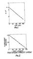
- Figure 1 of the accompanying drawings shows the change with time of the fluorescence intensity after adding valinomycin. More particularly, the logarithms of the differences of fluorescence intensities between a particular time and at equilibrium (Pn ⁇ F) were plotted against time. The maximum fluorescence quenching indicated by the point A in Figure 1 is obtained by extrapolating the line to time 0.
- FIG. 2 of the accompanying drawings shows the relationship between the quantity of porin-liposomes and the extent of fluorescence quenching.
- the porin-liposomes and the porinless-liposomes were mixed in various ratios and the fluorescence intensity was recorded as described above.
- the extent of fluorescence quenching by the porinless-liposomes only was normalized to 1.0.
- the extent of fluorescence quenching was shown to be linearly related to the population of porinless-liposomes.
- the membrane fusions induced by 10 mM Ca ++ were carried out at ambient temperature.
- Figure 3 of the accompanying drawings shows the time course of membrane fusions with the two aforesaid types of liposomes. The extent of membrane fusion can be expressed by the fusion index computed according to the following equation.
- F o the extent of fluorescence quenching of the dye without a fusogen.
- F the extent of fluorescence quenching of the dye with a fusogen.
- Figure 4 shows the effect of the presence of calcium ions on the membrane fusions.
- the reaction mixture was held at ambient temperature for 3 minutes before determining the fusion index.
- Figure 5 of the accompanying drawings shows the effect of incubation temperature on membrane fusions.
- the reaction mixture contained 12.5 mM calcium and was incubated for 3 min.
- polyethylene glycol 4000 (PEG-4000) was used as a fusogen instead of calcium ions.
- a mixture containing 10 ⁇ l each of porin- and porinless-phosphatidylcholine liposomes produced in analogous manner to that described in Example 1 was mixed with 40 ul of 37.5% w/v of PEG-4000 in 100 mM potassium gluconate/10 mM Tris-HC1 buffer (pH 8.0). The mixture was incubated at 35 0 C and diluted with 540 ul of the same buffer. Fluorescence intensity was measured as described in Example 1 using 20 ⁇ l of the mixture.
- Figure 6 of the accompanying drawings shows how the extent of PEG-induce membrane fusions varied with time.
- Figure 7 of the accompanying drawings shows the relationship between PEG concentration and the extent of membrane fusions. Reaction was carried out at 35°C for 10 min before fluorescence quenching (and hence fusion index) was determined.
Landscapes
- Health & Medical Sciences (AREA)
- Chemical & Material Sciences (AREA)
- Life Sciences & Earth Sciences (AREA)
- General Health & Medical Sciences (AREA)
- Immunology (AREA)
- Physics & Mathematics (AREA)
- Optics & Photonics (AREA)
- Dispersion Chemistry (AREA)
- Analytical Chemistry (AREA)
- Biochemistry (AREA)
- Chemical Kinetics & Catalysis (AREA)
- General Physics & Mathematics (AREA)
- Pathology (AREA)
- Nuclear Medicine, Radiotherapy & Molecular Imaging (AREA)
- Medicinal Chemistry (AREA)
- Pharmacology & Pharmacy (AREA)
- Epidemiology (AREA)
- Animal Behavior & Ethology (AREA)
- Public Health (AREA)
- Veterinary Medicine (AREA)
- Investigating Or Analysing Materials By The Use Of Chemical Reactions (AREA)
- Investigating Or Analysing Biological Materials (AREA)
Applications Claiming Priority (2)
| Application Number | Priority Date | Filing Date | Title |
|---|---|---|---|
| JP58092489A JPS59218956A (ja) | 1983-05-27 | 1983-05-27 | 膜融合測定方法 |
| JP92489/83 | 1983-05-27 |
Publications (2)
| Publication Number | Publication Date |
|---|---|
| EP0148551A2 true EP0148551A2 (fr) | 1985-07-17 |
| EP0148551A3 EP0148551A3 (fr) | 1987-04-15 |
Family
ID=14055707
Family Applications (1)
| Application Number | Title | Priority Date | Filing Date |
|---|---|---|---|
| EP84303614A Withdrawn EP0148551A3 (fr) | 1983-05-27 | 1984-05-29 | Méthode pour mesurer la fusion de membranes |
Country Status (4)
| Country | Link |
|---|---|
| US (1) | US4560665A (fr) |
| EP (1) | EP0148551A3 (fr) |
| JP (1) | JPS59218956A (fr) |
| ES (1) | ES8605101A1 (fr) |
Cited By (3)
| Publication number | Priority date | Publication date | Assignee | Title |
|---|---|---|---|---|
| WO1996040060A1 (fr) * | 1995-06-07 | 1996-12-19 | President And Fellows Of Harvard College | Administration intracellulaire de macromolecules |
| US11439708B2 (en) * | 2014-10-06 | 2022-09-13 | Lawrence Livermore National Security, Llc | Nanotube trans-membrane channels mimicking biological porins |
| US12311053B2 (en) | 2019-12-13 | 2025-05-27 | Lawrence Livermore National Security, Llc | Nanotube-vesicle compositions and uses thereof |
Families Citing this family (17)
| Publication number | Priority date | Publication date | Assignee | Title |
|---|---|---|---|---|
| US4873089A (en) * | 1985-07-12 | 1989-10-10 | Cornell Research Foundation Inc., Cornell University | Proteoliposomes as drug carriers |
| US5023086A (en) * | 1987-03-13 | 1991-06-11 | Micro-Pak, Inc. | Encapsulated ionophore growth factors |
| IL91414A0 (en) * | 1988-08-31 | 1990-04-29 | Zynaxis Technologies Inc | Reagents and methods for the determination of analytes |
| US5820848A (en) * | 1990-01-12 | 1998-10-13 | The Liposome Company, Inc. | Methods of preparing interdigitation-fusion liposomes and gels which encapsulate a bioactive agent |
| IE910086A1 (en) * | 1990-01-12 | 1991-07-17 | Liposome Company | Interdigitation-fusion liposomes and gels |
| US6596522B2 (en) | 1997-05-08 | 2003-07-22 | The Regents Of The University Of California | Detection of transmembrane potentials by optical methods |
| US6342379B1 (en) * | 1995-06-07 | 2002-01-29 | The Regents Of The University Of California | Detection of transmembrane potentials by optical methods |
| US5661035A (en) * | 1995-06-07 | 1997-08-26 | The Regents Of The University Of California | Voltage sensing by fluorescence resonance energy transfer |
| US5840859A (en) * | 1995-07-06 | 1998-11-24 | Research Corporation Technologies, Inc. | (Aminostyryl)pyridinium compounds for radiolabelling cell membranes |
| US5837282A (en) * | 1996-10-30 | 1998-11-17 | University Of British Columbia | Ionophore-mediated liposome loading |
| US7306930B2 (en) * | 2001-11-30 | 2007-12-11 | California Institute Of Technology | Method bacterial endospore quantification using lanthanide dipicolinate luminescence |
| WO2003065009A2 (fr) | 2002-02-01 | 2003-08-07 | California Institute Of Technology | Procede et dispositif pour dosages biologiques de spores bacteriennes |
| US7563615B2 (en) * | 2005-04-15 | 2009-07-21 | California Institute Of Technology | Apparatus and method for automated monitoring of airborne bacterial spores |
| US7611862B2 (en) * | 2004-11-12 | 2009-11-03 | California Institute Of Technology | Method and apparatus for detecting and quantifying bacterial spores on a surface |
| US7608419B2 (en) * | 2003-11-13 | 2009-10-27 | California Institute Of Technology | Method and apparatus for detecting and quantifying bacterial spores on a surface |
| US8329390B2 (en) * | 2004-10-21 | 2012-12-11 | Anaspec Incorporated | Detection of transmembrane potentials using N,N,N′-trialkyl thiobarbituric acid-derived polymethine oxonols |
| WO2007084180A2 (fr) * | 2005-06-17 | 2007-07-26 | California Institute Of Technology | Spores bactériens atmosphériques utilisés comme indicateur de biomasse dans un environnement intérieur |
Family Cites Families (1)
| Publication number | Priority date | Publication date | Assignee | Title |
|---|---|---|---|---|
| US4343782A (en) * | 1978-04-20 | 1982-08-10 | Shapiro Howard M | Cytological assay procedure |
-
1983
- 1983-05-27 JP JP58092489A patent/JPS59218956A/ja active Pending
-
1984
- 1984-05-24 US US06/613,893 patent/US4560665A/en not_active Expired - Fee Related
- 1984-05-25 ES ES532814A patent/ES8605101A1/es not_active Expired
- 1984-05-29 EP EP84303614A patent/EP0148551A3/fr not_active Withdrawn
Cited By (4)
| Publication number | Priority date | Publication date | Assignee | Title |
|---|---|---|---|---|
| WO1996040060A1 (fr) * | 1995-06-07 | 1996-12-19 | President And Fellows Of Harvard College | Administration intracellulaire de macromolecules |
| US5643599A (en) * | 1995-06-07 | 1997-07-01 | President And Fellows Of Harvard College | Intracellular delivery of macromolecules |
| US11439708B2 (en) * | 2014-10-06 | 2022-09-13 | Lawrence Livermore National Security, Llc | Nanotube trans-membrane channels mimicking biological porins |
| US12311053B2 (en) | 2019-12-13 | 2025-05-27 | Lawrence Livermore National Security, Llc | Nanotube-vesicle compositions and uses thereof |
Also Published As
| Publication number | Publication date |
|---|---|
| JPS59218956A (ja) | 1984-12-10 |
| EP0148551A3 (fr) | 1987-04-15 |
| ES8605101A1 (es) | 1986-02-16 |
| ES532814A0 (es) | 1986-02-16 |
| US4560665A (en) | 1985-12-24 |
Similar Documents
| Publication | Publication Date | Title |
|---|---|---|
| EP0148551A2 (fr) | Méthode pour mesurer la fusion de membranes | |
| Kano et al. | Pyranine as a sensitive pH probe for liposome interiors and surfaces. pH gradients across phospholipid vesicles | |
| Marchi‐Artzner et al. | Selective adhesion, lipid exchange and membrane‐fusion processes between vesicles of various sizes bearing complementary molecular recognition groups | |
| Clement et al. | Pyranine (8-hydroxy-1, 3, 6-pyrenetrisulfonate) as a probe of internal aqueous hydrogen ion concentration in phospholipid vesicles | |
| Eisenberg et al. | Adsorption of monovalent cations to bilayer membranes containing negative phospholipids | |
| Szoka Jr et al. | The use of aqueous space markers to determine the mechanism of interaction between phospholipid vesicles and cells | |
| Vanderkooi et al. | Fluorescent probe analysis of the lipid architecture of natural and experimental cholesterol-rich membranes | |
| Huang et al. | [45] Preparation of homogeneous, single-walled phosphatidylcholine vesicles | |
| Cranney et al. | Fluorescence lifetime and quenching studies on some interesting diphenylhexatriene membrane probes | |
| Loew et al. | Diffusion potential cascade. Convenient detection of transferable membrane pores | |
| US4978625A (en) | Fluorescence immunoassay using water insoluble dyes | |
| Hildenbrand et al. | Nanosecond fluorescence anisotropy decays of 1, 6-diphenyl-1, 3, 5-hexatriene in membranes | |
| DE69316757T2 (de) | Photoaktivierbare chemiluminizierende matrices | |
| Ploier et al. | A fluorescence-based assay of phospholipid scramblase activity | |
| Karczmar et al. | The interaction of adriamycin with small unilamellar vesicle liposomes. A fluorescence study | |
| Cafiso et al. | [16] Measuring electrostatic potentials adjacent to membranes | |
| Bally et al. | Dopamine accumulation in large unilamellar vesicle systems induced by transmembrane ion gradients | |
| Grethen et al. | Role of coulombic repulsion in collisional lipid transfer among SMA (2: 1)-bounded nanodiscs | |
| Georgallas et al. | The diffusion of small ions through phospholipid bilayers | |
| Cortese et al. | Ionic strength of the intermembrane space of intact mitochondria as estimated with fluorescein-BSA delivered by low pH fusion. | |
| MacDonald et al. | Lipid mixing during freeze-thawing of liposomal membranes as monitored by fluorescence energy transfer | |
| Javed et al. | Resilient Membranized Coacervates Formed through Spontaneous Wrapping of Heat‐Destabilized Lipid Bilayers around Coacervate Droplets | |
| Liu et al. | Antibiotic assisted molecular ion transport across a membrane in real time | |
| Katsu et al. | Precise size determination of amphotericin B and nystatin channels formed in erythrocyte and liposomal membranes based on osmotic protection experiments | |
| Lee et al. | Asymmetric labeling of amino lipids in liposomes |
Legal Events
| Date | Code | Title | Description |
|---|---|---|---|
| PUAI | Public reference made under article 153(3) epc to a published international application that has entered the european phase |
Free format text: ORIGINAL CODE: 0009012 |
|
| AK | Designated contracting states |
Designated state(s): CH DE FR GB IT LI NL |
|
| PUAL | Search report despatched |
Free format text: ORIGINAL CODE: 0009013 |
|
| AK | Designated contracting states |
Kind code of ref document: A3 Designated state(s): CH DE FR GB IT LI NL |
|
| 17P | Request for examination filed |
Effective date: 19870810 |
|
| 17Q | First examination report despatched |
Effective date: 19890420 |
|
| STAA | Information on the status of an ep patent application or granted ep patent |
Free format text: STATUS: THE APPLICATION IS DEEMED TO BE WITHDRAWN |
|
| 18D | Application deemed to be withdrawn |
Effective date: 19890831 |
|
| RIN1 | Information on inventor provided before grant (corrected) |
Inventor name: NAKAE, TAIJI Inventor name: RYO, EISAKU |