EP0169266A2 - Caméra pour tomographie à positrons - Google Patents
Caméra pour tomographie à positrons Download PDFInfo
- Publication number
- EP0169266A2 EP0169266A2 EP84110511A EP84110511A EP0169266A2 EP 0169266 A2 EP0169266 A2 EP 0169266A2 EP 84110511 A EP84110511 A EP 84110511A EP 84110511 A EP84110511 A EP 84110511A EP 0169266 A2 EP0169266 A2 EP 0169266A2
- Authority
- EP
- European Patent Office
- Prior art keywords
- ring
- crystals
- detectors
- septa
- rings
- Prior art date
- Legal status (The legal status is an assumption and is not a legal conclusion. Google has not performed a legal analysis and makes no representation as to the accuracy of the status listed.)
- Granted
Links
Images

Classifications
-
- A—HUMAN NECESSITIES
- A61—MEDICAL OR VETERINARY SCIENCE; HYGIENE
- A61B—DIAGNOSIS; SURGERY; IDENTIFICATION
- A61B6/00—Apparatus or devices for radiation diagnosis; Apparatus or devices for radiation diagnosis combined with radiation therapy equipment
-
- G—PHYSICS
- G01—MEASURING; TESTING
- G01T—MEASUREMENT OF NUCLEAR OR X-RADIATION
- G01T1/00—Measuring X-radiation, gamma radiation, corpuscular radiation, or cosmic radiation
- G01T1/29—Measurement performed on radiation beams, e.g. position or section of the beam; Measurement of spatial distribution of radiation
- G01T1/2914—Measurement of spatial distribution of radiation
- G01T1/2985—In depth localisation, e.g. using positron emitters; Tomographic imaging (longitudinal and transverse section imaging; apparatus for radiation diagnosis sequentially in different planes, steroscopic radiation diagnosis)
-
- A—HUMAN NECESSITIES
- A61—MEDICAL OR VETERINARY SCIENCE; HYGIENE
- A61B—DIAGNOSIS; SURGERY; IDENTIFICATION
- A61B6/00—Apparatus or devices for radiation diagnosis; Apparatus or devices for radiation diagnosis combined with radiation therapy equipment
- A61B6/02—Arrangements for diagnosis sequentially in different planes; Stereoscopic radiation diagnosis
- A61B6/03—Computed tomography [CT]
- A61B6/037—Emission tomography
-
- G—PHYSICS
- G01—MEASURING; TESTING
- G01T—MEASUREMENT OF NUCLEAR OR X-RADIATION
- G01T1/00—Measuring X-radiation, gamma radiation, corpuscular radiation, or cosmic radiation
- G01T1/16—Measuring radiation intensity
- G01T1/161—Applications in the field of nuclear medicine, e.g. in vivo counting
- G01T1/164—Scintigraphy
- G01T1/1641—Static instruments for imaging the distribution of radioactivity in one or two dimensions using one or several scintillating elements; Radio-isotope cameras
- G01T1/1644—Static instruments for imaging the distribution of radioactivity in one or two dimensions using one or several scintillating elements; Radio-isotope cameras using an array of optically separate scintillation elements permitting direct location of scintillations
Definitions
- positron emission tomography camera for assessing in-vivo biochemical behavior and its usefulness for clinic diagnosis of the human body is well known as set forth in my co-pending patent application Serial No. 396,098, filed July 7, 1982, entitled Three-Dimensional Time-of-Flight Positron Emission Camera System.
- Conventional positron cameras are limited to planar cross-sectional imaging without using a multi-step scanning process. As the cameras become more utilized in clinical medicine, three dimensional imaging is required which has high resolution in all three dimensions, multiple image planes, and higher sensitivity. However in the past, the positron camera has been limited to a few research centers due to its high cost.
- the present invention is directed to various improvements in a positron emission tomography camera which results in improved performance, high resolution, high efficiency, and lower costs.
- PET positron emission tomography
- detectors which are formed in rings around a circumference surrounding a patient area to detect the radiation emitted by the patient.
- Conventional PET cameras require septas which are directional shields placed along the outer edge of each detector ring extending inwardly towards the patient to reduce the unwanted radiation and which increase the diameter of the detector rings because of their finite length.
- One feature of the present invention is the use of an additional septum ring placed between the conventional septas. The additional septum allows the inward extent of all of the septas to be reduced thereby reducing the diameter or inner circumference of the ring of detectors thereby reducing the number of detectors which provides a considerable reduction in the cost of the camera.
- Another feature of the present invention is the offsetting of the detectors in a ring to provide better resolution, an increased number of image planes, better data sampling, and a reduction in photomultiplier tubes, and a reduction in scintillation crystal size, with a consequent reduction in cost. That is, typically detectors consist of a photomultiplier tube connected to a single scintillation crystal for detecting and converting the patient radiation into electrical pulses.
- a new feature of the present invention is positioning the crystals relative to the rings of photomultiplier tubes which allows more than one crystal to share a single photomultiplier tube and in which the crystals on one side of the photomultiplier tube are offset from the crystals on the second side of the photomultiplier tube to provide the above described advantages.
- Still a further object of the present invention is the improvement in a positron emission tomography camera having a plurality of detector rings positioned side by side around a patient area to detect radiation therefrom and in which each ring has an inner circumference and each ring contains a plurality of scintillation detectors which are positioned around the circumference and are pointed inwardly for detecting radiation.
- a septum ring extends inwardly from the inner circumference at each outer edge of each ring for directing the area of detection of the detectors.
- An extra septum ring is positioned in the middle of each ring of detectors and parallel to the other septa rings and extends inwardly whereby the inward extent of all of the septa rings are reduced one-half whereby the inner circumference of the detector rings is reduced by twice the reduction in the extent of the septas thereby reducing the number of detectors required in each ring.
- Yet a further object of the present invention is the improvement in a positron emission tomography camera having a plurality of detector rings positioned side by side around a patient area to detect radiation from the patient.
- Each ring contains a plurality of scintillation detectors pointed to the patient area and each detector ring defines a plane slice through the patient area by the detectors in each ring, and each two adjacent detector rings defines an interplane slice to the patient area.
- Each detector ring includes a plurality of photomultiplier tubes around the circumference and a plurality of scintillation crystals positioned around each ring.
- One-half of each of the multiplier tubes in each detector ring is responsive to a single crystal and the second half of each of the multiplier tubes in each detector ring is responsive to the adjacent staggered crystal which is also seen by the tube in the next ring.
- the crystals on the first half of the multiplier tube are offset from the crystals on the second half of the multiplier tube whereby the detectors will detect more interplane slices.
- each ring of detectors includes a septum ring extending inwardly from the inner circumference of each outer edge of each ring and a third septum ring extends inwardly from the inner circumference and is positioned to divide the first half of each photomultiplier tube in the ring from the second half of each photomultiplier tube.
- Yet a further object of the present invention is the provision of offset scintillation crystals relative to photomultiplier tubes in which a first ring of crystals is positioned on one-half of each photomultiplier tubes in each detector ring. Each crystal in the first ring is positioned on a single photomultiplier tube. A second ring of crystals is positioned on the second half of each photomultiplier tube in each ring and positioned between the crystals on the first ring. Each of the crystals on the second ring is positioned on two photomultiplier tubes in different detector rings whereby the crystals on the second ring are offset relative to the crystals on the first ring.
- first ring of crystals and the second ring of crystals are aligned with the first ring of crystals and the second ring of crystals on adjacent detector rings. Because of the offset detector geometry, more interplane coincidence information can be used in the image reconstruction and therefore increasing the total sensitivity of the camera.
- Another object is the provision of a first ring of crystals positioned on one-half of each photomultiplier tube in each detector ring. Each crystal in the first ring is positioned on a single photomultiplier tube. A second ring of crystals is positioned on the second half of each photomultiplier tube in each ring. Each of the crystals on the second ring is positioned on two photomultiplier tubes in the same ring whereby the crystals on the second ring are offset relative to the crystals on the first ring.
- Still a further object is the provision of a plurality of rings of photomultiplier tubes in which each photomultiplier tube is responsive to third scintillation crystals.
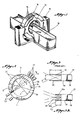
- a cross section of a typical positron camera has an opening 16 for insertion of the patient to be scanned, a plurality of detectors 22 for each detector ring which are mounted around the patient area 16 from an inner circumference 24 for detecting radiation from the patient area 16.
- Extending inwardly from the detector circumference 24 to the outside patient circumference 26 of the patient area 16 is a first 30 and second 32 septum.
- the septa 30 and 32 extend inwardly from the inner circumference 24 of the detector rings and from the outer edges of the detector rings 14 and are used to reduce unwanted signals such as random and scattered signals which would adversely affect the collected data. That is, septa are installed in the PET cameras to act as "blinders" for the detectors 22. Septa are normally made out of lead or tungsten and extend from the detectors 22 towards the patient.
- the effectiveness of a septum is characterized by its length L, thickness and the separation S between septa.
- the ratio t defines the solid angle, or the opening of the detector 22 and determines the amount of scattered and random radiation reaching the detectors 22.
- the diameter D of the detector inner circumference 24 is determined by the diameter d of the patient opening 16, the septa length L and the wobble diameter w.
- diameter D d + 2L + 2w.
- the patient opening d may be reduced such as when the camera 10 is to be used for imaging of only the brain instead of any part of a whole body.
- One feature of the present invention is the provision of an extra septum 36 which, as best seen in Fig. 3A, is placed in between the position of the normal septa 30a and 32b which allows the total length of all of the septa to be reduced to approxi- mately .
- the scattered and random signals are still maintained at approximately similar levels to the prior art structure of Fig. 3, but the sensitivity of the detectors 22 is slightly increased since the outer circumference 24 will be reduced by the distance L allowing the detectors 22 to be closer to the object being imaged.
- the diameter D of the inner circumference ring 24 can be reduced from 100 centimeters to 80 centimeters for a 20% decrease in the number of detectors 22. This can result in a cost savings of approximately twenty percent in the cost of the detectors.
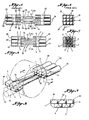
- a detector 22 typically consists of a photomultiplier tube 38 and a scintillation crystal 40.
- the crystal 40 converts the detected radiation into light which is transmitted to the photomultiplier tube 38 for converting the detected radiation into electrical pulses.
- crystal 4 is positioned adjacent to and is seen by photomultiplier tubes 60 and 62 while crystal 5 is seen by multiplier tube 62 and 64.
- the identification of the crystal can readily be made. That is, if crystal 1 is actuated only photomultiplier 60 is responsive. Similarly, if crystal 2 is actuated only photomultiplier 62 is responsive, and if crystal 3 is actuated only photomultiplier 64 is responsive. However, if both photomultiplier 60 and 62 respond, this is an identification of crystal 4. Similarly, crystal 5 will create a response in both photomultiplier tubes 62 and 64.
- Figs. 4 and 5 indicated that a three row detector camera will provide three straight-on slices and two effective in-between slices.
- the present improvement as best seen in Figs. 6 and 7, will because crystals 4 and 5 are offset, and behind in dotted outlines, the crystals 1, 2 and 3, will provide nine effective slices consisting of five straight on slices and four effective "in between" slices.
- the straight-on slices are numbered 70 and the effective in-between slices are numbered 72.
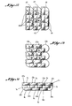
- Fig. 10 is the prior art structure of Figs. 4 and 5.
- detector rings 22a are provided in which the different rings include photomultiplier tubes 60, 62, 64, respectively.
- the arrangement of crystals la, 2a, 3a, 4a, and 5a are rotated ninety degrees compared to the embodiment of Fig. 9.
- the arrangement of Fig. 12 provides similar identification of the crystals and advantages of the arrangement of Fig. 9, but in Fig. 12 the in-plane slices are thinner and high interplane resolution is obtained.
- a still different detector arrangement is provided in which rings 22a are shown.
- crystals lb, 2b, 4b and 5b are provided. This arrangement with less crystal is not as efficient although the offset rows of crystals are advantageous over the conventional detectors in some respects.
- the rings 22c contain photomultiplier tubes 60c, 62c, 64c, and 65c, respectively, which are responsive to the three crystals.
- the crystals are lc, 2c, 3c, 4c, 5c, 6c, 7c, 8c, 9c, and lOc which are positioned in horizontal rows across the rings 22c. If desired, the crystals could be positioned around each of the rings 22c instead of across the plurality of rings 22c.
- crystal lc is identified by a signal from only tube 60c.
- Crystal 5c is identified by a signal of tube 62c being larger than a signal from tube 60c.
- crystal 8c is identified by a signal from tube 60c being larger than a signal from tube 62c. Identification of the other crystals is similar.
- Fig. 14 has the advantages of higher resolution, more effective sampling in the image planes, and lower costs because of sharing of the photomultiplier tubes.
- the positron emission tomography camera of the present invention provides a higher resolution, increased number of image planes, requires less photomultiplier tubes, provides better data sampling in the slice direction, has fewer gaps, the whole object is sampled in three dimensions for accurate representation, and this is accomplished at less cost.
Landscapes
- Health & Medical Sciences (AREA)
- Life Sciences & Earth Sciences (AREA)
- Physics & Mathematics (AREA)
- Engineering & Computer Science (AREA)
- Medical Informatics (AREA)
- High Energy & Nuclear Physics (AREA)
- Molecular Biology (AREA)
- Nuclear Medicine, Radiotherapy & Molecular Imaging (AREA)
- Optics & Photonics (AREA)
- Biomedical Technology (AREA)
- General Health & Medical Sciences (AREA)
- General Physics & Mathematics (AREA)
- Spectroscopy & Molecular Physics (AREA)
- Heart & Thoracic Surgery (AREA)
- Pathology (AREA)
- Radiology & Medical Imaging (AREA)
- Biophysics (AREA)
- Surgery (AREA)
- Animal Behavior & Ethology (AREA)
- Public Health (AREA)
- Veterinary Medicine (AREA)
- Nuclear Medicine (AREA)
- Analysing Materials By The Use Of Radiation (AREA)
- Medicines Containing Antibodies Or Antigens For Use As Internal Diagnostic Agents (AREA)
- Luminescent Compositions (AREA)
- Studio Devices (AREA)
- Investigating Or Analysing Materials By The Use Of Chemical Reactions (AREA)
- Measuring And Recording Apparatus For Diagnosis (AREA)
- Magnetic Resonance Imaging Apparatus (AREA)
- Apparatus For Radiation Diagnosis (AREA)
- Radiography Using Non-Light Waves (AREA)
- Organic Low-Molecular-Weight Compounds And Preparation Thereof (AREA)
Priority Applications (1)
| Application Number | Priority Date | Filing Date | Title |
|---|---|---|---|
| AT84110511T ATE50367T1 (de) | 1984-05-24 | 1984-09-04 | Positronenemissionstomographie-kamera. |
Applications Claiming Priority (2)
| Application Number | Priority Date | Filing Date | Title |
|---|---|---|---|
| US613699 | 1984-05-24 | ||
| US06/613,699 US4563582A (en) | 1984-05-24 | 1984-05-24 | Positron emission tomography camera |
Related Child Applications (2)
| Application Number | Title | Priority Date | Filing Date |
|---|---|---|---|
| EP88115476A Division EP0307004A3 (fr) | 1984-05-24 | 1984-09-04 | Caméra pour tomographie à positrons |
| EP88115476.9 Division-Into | 1988-09-21 |
Publications (3)
| Publication Number | Publication Date |
|---|---|
| EP0169266A2 true EP0169266A2 (fr) | 1986-01-29 |
| EP0169266A3 EP0169266A3 (en) | 1986-12-17 |
| EP0169266B1 EP0169266B1 (fr) | 1990-02-07 |
Family
ID=24458356
Family Applications (2)
| Application Number | Title | Priority Date | Filing Date |
|---|---|---|---|
| EP84110511A Expired - Lifetime EP0169266B1 (fr) | 1984-05-24 | 1984-09-04 | Caméra pour tomographie à positrons |
| EP88115476A Withdrawn EP0307004A3 (fr) | 1984-05-24 | 1984-09-04 | Caméra pour tomographie à positrons |
Family Applications After (1)
| Application Number | Title | Priority Date | Filing Date |
|---|---|---|---|
| EP88115476A Withdrawn EP0307004A3 (fr) | 1984-05-24 | 1984-09-04 | Caméra pour tomographie à positrons |
Country Status (14)
| Country | Link |
|---|---|
| US (1) | US4563582A (fr) |
| EP (2) | EP0169266B1 (fr) |
| JP (1) | JPS60250280A (fr) |
| KR (1) | KR910005917B1 (fr) |
| AT (1) | ATE50367T1 (fr) |
| AU (1) | AU567580B2 (fr) |
| BR (1) | BR8404913A (fr) |
| CA (1) | CA1221178A (fr) |
| DE (1) | DE3481346D1 (fr) |
| GB (2) | GB2159379B (fr) |
| HK (1) | HK84388A (fr) |
| MX (1) | MX157832A (fr) |
| SU (1) | SU1487800A3 (fr) |
| ZA (2) | ZA846324B (fr) |
Cited By (3)
| Publication number | Priority date | Publication date | Assignee | Title |
|---|---|---|---|---|
| EP0205791A1 (fr) * | 1985-05-13 | 1986-12-30 | Clayton Foundation for Research | Caméra pour tomographie à positrons avec plusieurs ensembles coplanaires de détecteurs |
| EP0212631A1 (fr) * | 1985-08-23 | 1987-03-04 | Clayton Foundation for Research | Caméra pour tomographie à positrons |
| EP0249695A1 (fr) * | 1986-06-18 | 1987-12-23 | Clayton Foundation for Research | Guide de lumière pour une caméra à tomographie par émission de positrons |
Families Citing this family (40)
| Publication number | Priority date | Publication date | Assignee | Title |
|---|---|---|---|---|
| US4749863A (en) * | 1984-12-04 | 1988-06-07 | Computer Technology And Imaging, Inc. | Two-dimensional photon counting position encoder system and process |
| US4743764A (en) * | 1984-12-04 | 1988-05-10 | Computer Technology And Imaging, Inc. | Two dimensional photon counting position encoder system and process |
| US4677299A (en) * | 1985-05-13 | 1987-06-30 | Clayton Foundation For Research | Multiple layer positron emission tomography camera |
| US4755679A (en) * | 1986-06-19 | 1988-07-05 | Wong Wai Hoi | Method and apparatus for maximizing counts of a PET camera |
| US4883966A (en) * | 1988-05-05 | 1989-11-28 | Wong Wai Hoi | Pet camera with crystal masking |
| US4864138A (en) * | 1988-07-14 | 1989-09-05 | Clayton Foundation For Research | Positron emission tomography camera |
| US5027817A (en) * | 1989-06-22 | 1991-07-02 | New York University | Statistical based display for positron emission tomography scans |
| US5224037A (en) * | 1991-03-15 | 1993-06-29 | Cti, Inc. | Design of super-fast three-dimensional projection system for Positron Emission Tomography |
| US5138165A (en) * | 1991-10-31 | 1992-08-11 | Rockwell International Corporation | Three dimensional positron emission tomography system |
| US5210420A (en) * | 1991-12-19 | 1993-05-11 | Positron Corporation | Positron emission tomography scanner |
| US5471061A (en) * | 1994-03-21 | 1995-11-28 | Cti Pet Systems, Inc. | Method and apparatus for transmission measurements to form a 2-d or a 3-d image in tomography applications |
| DE69622759T2 (de) * | 1995-11-22 | 2003-03-20 | Picker International Inc., Cleveland | Streuungs-filter für emissionstomographie |
| US5798527A (en) * | 1996-10-21 | 1998-08-25 | Ugm Laboratory, Inc. | Tomographic emission scanner having curved element detectors |
| NL1014401C2 (nl) * | 2000-02-17 | 2001-09-04 | Stichting Tech Wetenschapp | Ceriumhoudend anorganisch scintillatormateriaal. |
| WO2001087140A2 (fr) * | 2000-05-16 | 2001-11-22 | Crosetto Dario B | Procede et appareil pour l'imagerie medicale anatomique et fonctionnelle |
| US7132664B1 (en) | 2002-11-09 | 2006-11-07 | Crosetto Dario B | Method and apparatus for improving PET detectors |
| US20040164249A1 (en) * | 2003-02-26 | 2004-08-26 | Crosetto Dario B. | Method and apparatus for determining depth of interactions in a detector for three-dimensional complete body screening |
| US7329874B2 (en) | 2003-09-24 | 2008-02-12 | Radiation Monitoring Devices, Inc. | Lu1-xI3:Cex-a scintillator for gamma-ray spectroscopy and time-of-flight pet |
| US7173247B2 (en) * | 2003-09-24 | 2007-02-06 | Radiation Monitoring Devices, Inc. | Lu1-xI3:Cex—a scintillator for gamma ray spectroscopy and time-of-flight PET |
| US8017915B2 (en) | 2008-03-14 | 2011-09-13 | Reflexion Medical, Inc. | Method and apparatus for emission guided radiation therapy |
| US8809791B2 (en) | 2009-05-20 | 2014-08-19 | Koninklijke Philips N.V. | Continuous time-of-flight scatter simulation method |
| JP5751944B2 (ja) * | 2010-06-16 | 2015-07-22 | 株式会社東芝 | Tof―pet装置、検出器リング、及び検出器 |
| KR101232124B1 (ko) * | 2010-09-16 | 2013-02-15 | 박순애 | 양전자 방출 단층 촬영장치 및 그 촬영방법 |
| US8304738B2 (en) | 2010-10-19 | 2012-11-06 | Kabushiki Kaisha Toshiba | Pet detector scintillation light guiding system having fiber-optics plates |
| EP2461183B1 (fr) * | 2010-10-19 | 2018-10-03 | Toshiba Medical Systems Corporation | Module détecteur de tomographie à émission de positrons, détecteur de radiations, système de balayage de tomographie à émission de positrons, procédé de traitement de signaux et procédé pour la fabrication du module détecteur de radiations |
| US8357903B2 (en) * | 2010-10-19 | 2013-01-22 | Kabushiki Kaisha Toshiba | Segmented detector array |
| CN102451017B (zh) * | 2010-10-19 | 2017-10-31 | 东芝医疗系统株式会社 | Pet检测器模块、pet扫描仪系统、核医学图像摄影检测器模块 |
| US9283403B2 (en) | 2011-03-31 | 2016-03-15 | Reflexion Medical, Inc. | Systems and methods for use in emission guided radiation therapy |
| US9599731B2 (en) * | 2013-03-14 | 2017-03-21 | Koninklijke Philips N.V. | Positron emission tomography and/or single photon emission tomography detector |
| CN104367332B (zh) * | 2014-12-09 | 2017-02-22 | 武汉数字派特科技有限公司 | 一种pet探测器的探测器环 |
| CN107924730B (zh) | 2015-06-10 | 2021-09-28 | 反射医疗公司 | 高带宽双态多叶式准直器设计 |
| CN109152928B (zh) | 2016-03-09 | 2021-05-28 | 反射医疗公司 | 用于计算辐射治疗的注量图的方法和系统 |
| CN110248604B (zh) | 2016-11-15 | 2023-07-21 | 反射医疗公司 | 放射治疗患者平台 |
| WO2018093849A1 (fr) | 2016-11-15 | 2018-05-24 | Reflexion Medical, Inc. | Procédés destinés à l'administration d'un rayonnement en radiothérapie guidée par émission |
| WO2018093933A1 (fr) | 2016-11-15 | 2018-05-24 | Reflexion Medical, Inc. | Système d'administration de photons à haute énergie guidé par émission |
| WO2018183748A1 (fr) | 2017-03-30 | 2018-10-04 | Reflexion Medical, Inc. | Systèmes et procédés de radiothérapie avec suivi de tumeur |
| JP7672126B2 (ja) | 2017-07-11 | 2025-05-07 | リフレクション メディカル, インコーポレイテッド | Pet検出器残光管理のための方法 |
| CN111148471B (zh) | 2017-08-09 | 2023-08-22 | 反射医疗公司 | 用于发射引导放射治疗中的故障检测的系统和方法 |
| US11029418B2 (en) | 2017-09-06 | 2021-06-08 | The United States Of America, As Represented By The Secretary, Department Of Health And Human Services | Micro-dose calibrator |
| US11369806B2 (en) | 2017-11-14 | 2022-06-28 | Reflexion Medical, Inc. | Systems and methods for patient monitoring for radiotherapy |
Family Cites Families (11)
| Publication number | Priority date | Publication date | Assignee | Title |
|---|---|---|---|---|
| GB1406685A (en) * | 1972-12-13 | 1975-09-17 | Baird Atomic Inc | Sensing matrices for radioactivity-distribution detectors |
| US4057727A (en) * | 1976-10-22 | 1977-11-08 | G. D. Searle & Co. | Positron imaging system with improved count rate and tomographic capability |
| US4150292A (en) * | 1977-02-18 | 1979-04-17 | Ter Pogossian Michel M | Imaging device for computerized emission tomography |
| GB2034148B (en) * | 1978-08-30 | 1983-06-15 | Gen Electric | Multi element high resolution scintillator structure |
| CA1117228A (fr) * | 1979-08-27 | 1982-01-26 | Montreal Neurological Institute | Dispositif de visualisation d'annihilations positoniques utilisant plusieurs anneaux de detecteurs decales |
| US4259578A (en) * | 1979-08-27 | 1981-03-31 | Thompson Christopher J | Movable collimator for positron annihilation imaging device |
| JPS5648560A (en) * | 1979-09-29 | 1981-05-01 | Kagaku Gijutsucho Hoshasen Igaku Sogo Kenkyusho | Position detector for radiant ray |
| JPS5814071A (ja) * | 1981-07-20 | 1983-01-26 | Agency Of Ind Science & Technol | ポジトロンct装置における同時計数回路 |
| JPS5870188A (ja) * | 1981-10-22 | 1983-04-26 | Agency Of Ind Science & Technol | ポジトロンct用検出装置 |
| JPS5892973A (ja) * | 1981-11-30 | 1983-06-02 | Shimadzu Corp | エミツシヨンct装置用放射線検出装置 |
| US4559597A (en) * | 1982-07-07 | 1985-12-17 | Clayton Foundation For Research | Three-dimensional time-of-flight positron emission camera system |
-
1984
- 1984-05-24 US US06/613,699 patent/US4563582A/en not_active Expired - Lifetime
- 1984-08-15 ZA ZA846324A patent/ZA846324B/xx unknown
- 1984-08-29 GB GB08421844A patent/GB2159379B/en not_active Expired
- 1984-08-31 KR KR1019840005353A patent/KR910005917B1/ko not_active Expired
- 1984-09-04 AT AT84110511T patent/ATE50367T1/de not_active IP Right Cessation
- 1984-09-04 DE DE8484110511T patent/DE3481346D1/de not_active Expired - Lifetime
- 1984-09-04 EP EP84110511A patent/EP0169266B1/fr not_active Expired - Lifetime
- 1984-09-04 EP EP88115476A patent/EP0307004A3/fr not_active Withdrawn
- 1984-09-06 CA CA000462587A patent/CA1221178A/fr not_active Expired
- 1984-09-07 AU AU32826/84A patent/AU567580B2/en not_active Ceased
- 1984-09-17 JP JP59192741A patent/JPS60250280A/ja active Pending
- 1984-09-28 BR BR8404913A patent/BR8404913A/pt unknown
- 1984-10-09 SU SU843798783A patent/SU1487800A3/ru active
- 1984-10-22 MX MX203139A patent/MX157832A/es unknown
-
1986
- 1986-06-26 GB GB08615592A patent/GB2175780B/en not_active Expired
- 1986-08-15 ZA ZA863600A patent/ZA863600B/xx unknown
-
1988
- 1988-10-20 HK HK843/88A patent/HK84388A/xx unknown
Cited By (3)
| Publication number | Priority date | Publication date | Assignee | Title |
|---|---|---|---|---|
| EP0205791A1 (fr) * | 1985-05-13 | 1986-12-30 | Clayton Foundation for Research | Caméra pour tomographie à positrons avec plusieurs ensembles coplanaires de détecteurs |
| EP0212631A1 (fr) * | 1985-08-23 | 1987-03-04 | Clayton Foundation for Research | Caméra pour tomographie à positrons |
| EP0249695A1 (fr) * | 1986-06-18 | 1987-12-23 | Clayton Foundation for Research | Guide de lumière pour une caméra à tomographie par émission de positrons |
Also Published As
| Publication number | Publication date |
|---|---|
| GB2159379B (en) | 1988-05-11 |
| ZA846324B (en) | 1986-08-27 |
| GB2159379A (en) | 1985-11-27 |
| EP0307004A3 (fr) | 1989-11-15 |
| ZA863600B (en) | 1987-12-30 |
| KR850008617A (ko) | 1985-12-21 |
| JPS60250280A (ja) | 1985-12-10 |
| AU567580B2 (en) | 1987-11-26 |
| GB8421844D0 (en) | 1984-10-03 |
| GB8615592D0 (en) | 1986-07-30 |
| CA1221178A (fr) | 1987-04-28 |
| GB2175780B (en) | 1988-05-11 |
| MX157832A (es) | 1988-12-15 |
| US4563582A (en) | 1986-01-07 |
| DE3481346D1 (de) | 1990-03-15 |
| ATE50367T1 (de) | 1990-02-15 |
| KR910005917B1 (ko) | 1991-08-08 |
| EP0307004A2 (fr) | 1989-03-15 |
| HK84388A (en) | 1988-10-28 |
| EP0169266B1 (fr) | 1990-02-07 |
| AU3282684A (en) | 1985-11-28 |
| SU1487800A3 (ru) | 1989-06-15 |
| BR8404913A (pt) | 1986-04-29 |
| GB2175780A (en) | 1986-12-03 |
| EP0169266A3 (en) | 1986-12-17 |
Similar Documents
| Publication | Publication Date | Title |
|---|---|---|
| EP0169266B1 (fr) | Caméra pour tomographie à positrons | |
| EP0212631B1 (fr) | Caméra pour tomographie à positrons | |
| EP0205791B1 (fr) | Caméra pour tomographie à positrons avec plusieurs ensembles coplanaires de détecteurs | |
| EP0219648B1 (fr) | Caméra pour tomographie à positrons avec plusieurs ensembles coplanaires de détecteurs | |
| US5032728A (en) | Single photon emission computed tomography system | |
| US4095107A (en) | Transaxial radionuclide emission camera apparatus and method | |
| US7817827B2 (en) | Enhanced planar single photon emission imaging | |
| US6232605B1 (en) | Flat scintillation gamma camera, with very high spatial resolution, with modular structure | |
| US7485866B2 (en) | Method and apparatus for increasing spatial resolution of a pet scanner | |
| US20180292548A1 (en) | Apparatus and methods for depth-of-interaction positron tomography detector using dichotomous sensing | |
| US4843245A (en) | Scintillation detector for tomographs | |
| US7102135B2 (en) | PET scanner | |
| EP0249695B1 (fr) | Guide de lumière pour une caméra à tomographie par émission de positrons | |
| ATE128557T1 (de) | Hochauflösender gamma-strahlungsdetektor für positronemissionstomographie (pet) und einzelne photonemissionscomputertomographie (spect). | |
| CN210626673U (zh) | 射线探测器及核医学成像设备 | |
| US6429434B1 (en) | Transmission attenuation correction method for PET and SPECT | |
| US8063377B2 (en) | Crystal identification for high resolution nuclear imaging | |
| US4864138A (en) | Positron emission tomography camera | |
| US6535571B2 (en) | Detector for an X-ray computed tomography apparatus | |
| US6718006B2 (en) | Fiber-optic encoding for dual transmission measurements in positron emission tomography | |
| JPH05264736A (ja) | ポジトロンct装置 | |
| JPH02285278A (ja) | リング型ect装置 |
Legal Events
| Date | Code | Title | Description |
|---|---|---|---|
| PUAI | Public reference made under article 153(3) epc to a published international application that has entered the european phase |
Free format text: ORIGINAL CODE: 0009012 |
|
| AK | Designated contracting states |
Designated state(s): AT BE CH DE FR IT LI LU NL SE |
|
| PUAL | Search report despatched |
Free format text: ORIGINAL CODE: 0009013 |
|
| AK | Designated contracting states |
Kind code of ref document: A3 Designated state(s): AT BE CH DE FR IT LI LU NL SE |
|
| 17P | Request for examination filed |
Effective date: 19870126 |
|
| 17Q | First examination report despatched |
Effective date: 19880510 |
|
| GRAA | (expected) grant |
Free format text: ORIGINAL CODE: 0009210 |
|
| AK | Designated contracting states |
Kind code of ref document: B1 Designated state(s): AT BE CH DE FR IT LI LU NL SE |
|
| ITF | It: translation for a ep patent filed | ||
| REF | Corresponds to: |
Ref document number: 50367 Country of ref document: AT Date of ref document: 19900215 Kind code of ref document: T |
|
| REF | Corresponds to: |
Ref document number: 3481346 Country of ref document: DE Date of ref document: 19900315 |
|
| ET | Fr: translation filed | ||
| PGFP | Annual fee paid to national office [announced via postgrant information from national office to epo] |
Ref country code: CH Payment date: 19900814 Year of fee payment: 7 |
|
| PGFP | Annual fee paid to national office [announced via postgrant information from national office to epo] |
Ref country code: SE Payment date: 19900816 Year of fee payment: 7 Ref country code: FR Payment date: 19900816 Year of fee payment: 7 Ref country code: AT Payment date: 19900816 Year of fee payment: 7 |
|
| PGFP | Annual fee paid to national office [announced via postgrant information from national office to epo] |
Ref country code: LU Payment date: 19900821 Year of fee payment: 7 |
|
| PGFP | Annual fee paid to national office [announced via postgrant information from national office to epo] |
Ref country code: BE Payment date: 19900829 Year of fee payment: 7 |
|
| ITTA | It: last paid annual fee | ||
| PG25 | Lapsed in a contracting state [announced via postgrant information from national office to epo] |
Ref country code: LU Free format text: LAPSE BECAUSE OF NON-PAYMENT OF DUE FEES Effective date: 19900930 |
|
| PGFP | Annual fee paid to national office [announced via postgrant information from national office to epo] |
Ref country code: NL Payment date: 19900930 Year of fee payment: 7 |
|
| PLBE | No opposition filed within time limit |
Free format text: ORIGINAL CODE: 0009261 |
|
| STAA | Information on the status of an ep patent application or granted ep patent |
Free format text: STATUS: NO OPPOSITION FILED WITHIN TIME LIMIT |
|
| 26N | No opposition filed | ||
| PG25 | Lapsed in a contracting state [announced via postgrant information from national office to epo] |
Ref country code: AT Effective date: 19910904 |
|
| PG25 | Lapsed in a contracting state [announced via postgrant information from national office to epo] |
Ref country code: SE Effective date: 19910905 |
|
| PG25 | Lapsed in a contracting state [announced via postgrant information from national office to epo] |
Ref country code: LI Effective date: 19910930 Ref country code: CH Effective date: 19910930 Ref country code: BE Effective date: 19910930 |
|
| BERE | Be: lapsed |
Owner name: CLAYTON FOUNDATION FOR RESEARCH Effective date: 19910930 |
|
| PG25 | Lapsed in a contracting state [announced via postgrant information from national office to epo] |
Ref country code: NL Effective date: 19920401 |
|
| NLV4 | Nl: lapsed or anulled due to non-payment of the annual fee | ||
| PG25 | Lapsed in a contracting state [announced via postgrant information from national office to epo] |
Ref country code: FR Effective date: 19920529 |
|
| REG | Reference to a national code |
Ref country code: CH Ref legal event code: PL |
|
| REG | Reference to a national code |
Ref country code: FR Ref legal event code: ST |
|
| EUG | Se: european patent has lapsed |
Ref document number: 84110511.7 Effective date: 19920408 |
|
| PGFP | Annual fee paid to national office [announced via postgrant information from national office to epo] |
Ref country code: DE Payment date: 20030911 Year of fee payment: 20 |