EP0172616A2 - Tomographie - Google Patents
Tomographie Download PDFInfo
- Publication number
- EP0172616A2 EP0172616A2 EP85304157A EP85304157A EP0172616A2 EP 0172616 A2 EP0172616 A2 EP 0172616A2 EP 85304157 A EP85304157 A EP 85304157A EP 85304157 A EP85304157 A EP 85304157A EP 0172616 A2 EP0172616 A2 EP 0172616A2
- Authority
- EP
- European Patent Office
- Prior art keywords
- electrodes
- potentials
- pairs
- isopotentials
- impedance
- Prior art date
- Legal status (The legal status is an assumption and is not a legal conclusion. Google has not performed a legal analysis and makes no representation as to the accuracy of the status listed.)
- Withdrawn
Links
- 238000003325 tomography Methods 0.000 title description 2
- 238000000034 method Methods 0.000 claims abstract description 15
- 230000003247 decreasing effect Effects 0.000 claims abstract description 5
- 238000010276 construction Methods 0.000 claims abstract description 3
- 230000000007 visual effect Effects 0.000 claims description 4
- 238000011835 investigation Methods 0.000 claims description 2
- 210000000056 organ Anatomy 0.000 claims description 2
- 210000002784 stomach Anatomy 0.000 description 4
- 230000030136 gastric emptying Effects 0.000 description 3
- 238000005259 measurement Methods 0.000 description 3
- 210000001015 abdomen Anatomy 0.000 description 2
- 210000004072 lung Anatomy 0.000 description 2
- 230000029058 respiratory gaseous exchange Effects 0.000 description 2
- 230000003068 static effect Effects 0.000 description 2
- 230000000740 bleeding effect Effects 0.000 description 1
- 239000008280 blood Substances 0.000 description 1
- 210000004369 blood Anatomy 0.000 description 1
- 238000004364 calculation method Methods 0.000 description 1
- 210000000038 chest Anatomy 0.000 description 1
- 230000007547 defect Effects 0.000 description 1
- 238000002059 diagnostic imaging Methods 0.000 description 1
- 238000010586 diagram Methods 0.000 description 1
- 230000000694 effects Effects 0.000 description 1
- 235000013305 food Nutrition 0.000 description 1
- 238000003384 imaging method Methods 0.000 description 1
- 238000002347 injection Methods 0.000 description 1
- 239000007924 injection Substances 0.000 description 1
- 235000012054 meals Nutrition 0.000 description 1
- 230000002285 radioactive effect Effects 0.000 description 1
- 230000035945 sensitivity Effects 0.000 description 1
- 210000000813 small intestine Anatomy 0.000 description 1
- 239000008399 tap water Substances 0.000 description 1
- 235000020679 tap water Nutrition 0.000 description 1
- 238000009423 ventilation Methods 0.000 description 1
Images


Classifications
-
- A—HUMAN NECESSITIES
- A61—MEDICAL OR VETERINARY SCIENCE; HYGIENE
- A61B—DIAGNOSIS; SURGERY; IDENTIFICATION
- A61B5/00—Measuring for diagnostic purposes; Identification of persons
- A61B5/05—Detecting, measuring or recording for diagnosis by means of electric currents or magnetic fields; Measuring using microwaves or radio waves
- A61B5/053—Measuring electrical impedance or conductance of a portion of the body
- A61B5/0536—Impedance imaging, e.g. by tomography
Definitions
- This invention relates to tomography and is concerned with the provision of a method of obtaining tomographic images of a body or a portion thereof (hereinafter referred to simply as a body), more particularly - but not exclusively - of any part of a live human body.
- X-ray computed tompography has encouraged the proposal of other medical imaging techniques not fraught with the dangers of X-rays.
- the use of low frequency electric currents has been suggested although no practical results have been published, but some literature exists on methods which involve measurements of impedance between electrodes on the surface of the body and propose methods for reconstruction of spatial impedance variations.
- GL-PS 2 119 250A gives examples to which may be added 'Electrical impedance imaging of the thorax' by Y. Kim, J.G. Webster and W.J. Topkins (Jn.
- GB-PS 2 119 250A also discloses a contrasting method which involves placing a plurality of surface electrodes at spaced intervals on the body, causing currents to flow in the body, and measuring the potential between pairs of electrodes, calculating the potential in each case on the assumption that the body consists of one uniform medium, obtaining the ratio between the measured potential and the calculated potential in each case, calculating the isopotentials expected within the uniform medium between the pairs of electrodes to create an assumed image of the body, modifying the assumed imag by back projecting the respective ratios long the appropriate isopotentials, by increasing the impedance along an isopotential in proportion to a ratio greater than unity or decreasing the impedance in proportion to a ratio less than unity, and plotting the modified impedances to create a tomographic image.
- back projection The modifying of the impedance distribution in this manner is known as "back projection", and the execution of the back projection (or the superimposition of the modified impedance along isopotentials) results in a tomographic image of the distribution of impedance over the cross- sectional area of the body in the plane containing the electrodes.
- the resolution of the tomographic . image can be improved by "iteration” (i.e., by. recalculating the potentials in each case using the modified impedance distribution as an approximate guide to the actual 1 distribution of impedance, obtaining the ratio between the recalculated potential and the measured potential in each case, and modifying the modified impedance distribution accordingly) and/or by "weightin” the "back projection” ratios in accordance with changing distribution of isopotentials through the body.
- iteration i.e., by. recalculating the potentials in each case using the modified impedance distribution as an approximate guide to the actual 1 distribution of impedance, obtaining the ratio between the recalculated potential and the measured potential in each case, and modifying the modified impedance distribution accordingly
- weightin the "back projection” ratios in accordance with changing distribution of isopotentials through the body.
- the object .of the invention is, therefore, the provision of a method of obtaining topographic images of a body by causing currents to flow in the body and measuring potentials between pairs of electrodes in an array of spaced electrodes and affording a picture of the changes in the state of the body.
- a method for the construction of tonographic images of a body comprises placing an array of spaced electrodes in contact with the body, causing currents to flow in the body, by applying an electrical potential between pairs of electrodes in turn, calculating the potentials between other pairs of electrodes on the assumption that the body consists of one uniform medium, plotting the isopotentials corresponding to the calculated results to create an assumed image of the body, measuring initial potentials between those other pairs of electrodes in sequence over the array of electrodes, measuring subsequent potentials between the same pairs of electrodes in the sequence after a change in the internal state of the body, determining the ratios between the initial potentials and the subsequent potentials in each case, and modifying the image by back projecting the respective ratios along the appropriate isopotentials and thereby increasing the impedance along an isopotential in proportion to a ratio greater than unity or decreasing the impedance in proportion to a ratio less than unity.
- the image produced by this method does not ccntain the static structures within the body but gives a picture of the internal state of the body after the change of state, which may be effected by means of an injection into the blood stream and/or by ingesting food and/or drink, or which may be the result of respiration, gastric emptying, internal bleeding, or the cardial cycle, and the plotting of the isopotentials may be effected by a visual display unit (VDU) and/or a print-out unit, forming images by intensity of dots in proportion to impedance.
- VDU visual display unit
- print-out unit forming images by intensity of dots in proportion to impedance.
- the invention is particularly advantageous in being able to afford a "dynamic" picture of the continually changing state within a body by repeating the potential measuring sequence and potential difference determinations at regular and/or frequent intervals, and recording and/or viewing the successive modified images corresponding thereto. Changes in impedance distribution can be seen even in the presence of very much larger static impedance variations.
- a rate of change of state within the body may be obtained by plotting a graph of the intensity of dots corresponding to the organ under investigation.
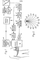
- an array of sixteen Ag/AgCL electrodes equi-spaced round the abdomen of a human body is coupled to equipment by means of which an applied potential of approximately 3 volts at 4 milliamps is produced by a waveform generator A at 50kHz and applied through a voltage to current, converter B and a multiplexer C in turn between adjacent pairs of electrodes in sequence and in each and every case the resultant potential between every adjacent pair of electrodes'is fed through an amplifier D and a phase sensitive detector E and is recorded by a sample and hold unit F, from which the data is fed through a 12-Bit analogue to digital converter G to a computer H.
- the units A, E, F and G of the equipment are all controlled by a master clock J, which also controls the multiplexer through a unit K which stores the electrode combinations.
- the sixteen electrodes shown in Figure 2 give rise to 1456 potential measurements which can be recorded in 1.456 seconds or less.
- Figure 2 also shows the assumed image of isopotentials to be expected when current is applied between electrodes 1 and 2, with the body assumed to consist of one uniform medium and circular in cross-section.
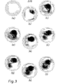
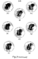
- Initial potentials between adjacent pairs of electrodes are measured in sequence over the array of electrodes, subsequent potentials between the same pairs of electrodes are measured in the same sequence after a change in the internal state of the body (which in this case results from having a drink - as indicated previously and as will be referred to again presently with reference to Figures 3 and 4), the subsequent potentials are compared by the computer H with the respective initial potentials, and the ratios are back projected along the appropriate isopotentials shown in Figure 2, by increasing the impedance along an isopotential in proportion to a ratio greater than unity or decreasing the impedance in proportion to a ratio less than unity.
- Figure 5 the sixteen electrodes are disposed round the torso
- Figure 6 shows a resulting sequence of six print-outs at intervals of 1 second during inspiration after inhalation respectively of (a) 0.45, (b) 1.0, (c) 1.5, (d) 2.0, (e) 2.7 and (f) 4.2 litres of air.
- the lungs are clearly seen, with anterior at the bottom of each image. Any defect in ventilation will show as a distortion of the image of the lungs.
Landscapes
- Health & Medical Sciences (AREA)
- Life Sciences & Earth Sciences (AREA)
- Nuclear Medicine, Radiotherapy & Molecular Imaging (AREA)
- Medical Informatics (AREA)
- Surgery (AREA)
- Biophysics (AREA)
- Pathology (AREA)
- Engineering & Computer Science (AREA)
- Biomedical Technology (AREA)
- Heart & Thoracic Surgery (AREA)
- Radiology & Medical Imaging (AREA)
- Molecular Biology (AREA)
- Physics & Mathematics (AREA)
- Animal Behavior & Ethology (AREA)
- General Health & Medical Sciences (AREA)
- Public Health (AREA)
- Veterinary Medicine (AREA)
- Measurement And Recording Of Electrical Phenomena And Electrical Characteristics Of The Living Body (AREA)
- Measurement Of The Respiration, Hearing Ability, Form, And Blood Characteristics Of Living Organisms (AREA)
- Apparatus For Radiation Diagnosis (AREA)
Applications Claiming Priority (2)
| Application Number | Priority Date | Filing Date | Title |
|---|---|---|---|
| GB8415236 | 1984-06-14 | ||
| GB848415236A GB8415236D0 (en) | 1984-06-14 | 1984-06-14 | Tomography |
Publications (2)
| Publication Number | Publication Date |
|---|---|
| EP0172616A2 true EP0172616A2 (de) | 1986-02-26 |
| EP0172616A3 EP0172616A3 (de) | 1987-03-04 |
Family
ID=10562452
Family Applications (1)
| Application Number | Title | Priority Date | Filing Date |
|---|---|---|---|
| EP85304157A Withdrawn EP0172616A3 (de) | 1984-06-14 | 1985-06-12 | Tomographie |
Country Status (4)
| Country | Link |
|---|---|
| EP (1) | EP0172616A3 (de) |
| JP (1) | JPH0636787B2 (de) |
| CA (1) | CA1220863A (de) |
| GB (2) | GB8415236D0 (de) |
Cited By (2)
| Publication number | Priority date | Publication date | Assignee | Title |
|---|---|---|---|---|
| WO1997015825A1 (en) * | 1995-10-27 | 1997-05-01 | Disperse Technologies Limited | Characterisation of flowing dispersions |
| CN102379697A (zh) * | 2011-10-12 | 2012-03-21 | 中国人民解放军第四军医大学 | 电阻抗扫描成像前级信号调理模块的检测装置及标校方法 |
Families Citing this family (10)
| Publication number | Priority date | Publication date | Assignee | Title |
|---|---|---|---|---|
| GB9113830D0 (en) * | 1991-06-27 | 1991-08-14 | Brown Brian H | Applied potential tomography |
| GB9222888D0 (en) * | 1992-10-30 | 1992-12-16 | British Tech Group | Tomography |
| DE4332257C2 (de) * | 1993-09-22 | 1996-09-19 | P Osypka Gmbh Medizintechnik D | Vorrichtung zum Erzeugen tomografischer Bilder |
| RU2127075C1 (ru) | 1996-12-11 | 1999-03-10 | Корженевский Александр Владимирович | Способ получения томографического изображения тела и электроимпедансный томограф |
| EP1681016B1 (de) | 1999-10-15 | 2009-08-26 | Kao Corporation | Vorrichtung zur Messung der Verteilung von Körperfettgehalt |
| EP1347706B1 (de) | 2000-12-30 | 2008-05-21 | The University Of Leeds | Elektrische impedanztomografie |
| EP1363531B1 (de) | 2001-02-22 | 2010-10-20 | Kao Corporation | Körperfettmessgerät |
| US6980852B2 (en) * | 2002-01-25 | 2005-12-27 | Subqiview Inc. | Film barrier dressing for intravascular tissue monitoring system |
| RU2223511C1 (ru) * | 2002-07-16 | 2004-02-10 | Научно-исследовательский институт интроскопии при Томском политехническом университете | Бесконтактный способ определения потенциалов заряженной поверхности объекта и устройство для его осуществления |
| TWI461180B (zh) * | 2011-12-30 | 2014-11-21 | Univ Nat Chiao Tung | 增進電阻抗斷層攝影影像解析度的方法 |
Family Cites Families (4)
| Publication number | Priority date | Publication date | Assignee | Title |
|---|---|---|---|---|
| US3971365A (en) | 1973-02-12 | 1976-07-27 | Beckman Instruments, Inc. | Bioelectrical impedance measuring system |
| DE2813068A1 (de) * | 1978-03-25 | 1979-10-04 | Philips Patentverwaltung | Verfahren und vorrichtung zur ermittlung von inneren koerperstrukturen |
| GB2119520B (en) * | 1982-04-30 | 1985-05-15 | Brian Hilton Brown | Tomography |
| GB2138148B (en) * | 1983-04-13 | 1986-07-30 | Denis Nigel Smith | Method and apparatus for deriving currents and potentials representative of the impedance of zones of a body |
-
1984
- 1984-06-14 GB GB848415236A patent/GB8415236D0/en active Pending
-
1985
- 1985-06-04 GB GB08514045A patent/GB2160323B/en not_active Expired
- 1985-06-11 CA CA000483687A patent/CA1220863A/en not_active Expired
- 1985-06-12 EP EP85304157A patent/EP0172616A3/de not_active Withdrawn
- 1985-06-14 JP JP60128380A patent/JPH0636787B2/ja not_active Expired - Lifetime
Cited By (3)
| Publication number | Priority date | Publication date | Assignee | Title |
|---|---|---|---|---|
| WO1997015825A1 (en) * | 1995-10-27 | 1997-05-01 | Disperse Technologies Limited | Characterisation of flowing dispersions |
| US6210972B1 (en) | 1995-10-27 | 2001-04-03 | Disperse Technologies Limited | Characterization of flowing dispersions |
| CN102379697A (zh) * | 2011-10-12 | 2012-03-21 | 中国人民解放军第四军医大学 | 电阻抗扫描成像前级信号调理模块的检测装置及标校方法 |
Also Published As
| Publication number | Publication date |
|---|---|
| CA1220863A (en) | 1987-04-21 |
| GB8514045D0 (en) | 1985-07-10 |
| GB2160323B (en) | 1987-12-09 |
| JPS6176131A (ja) | 1986-04-18 |
| EP0172616A3 (de) | 1987-03-04 |
| GB2160323A (en) | 1985-12-18 |
| GB8415236D0 (en) | 1984-07-18 |
| JPH0636787B2 (ja) | 1994-05-18 |
Similar Documents
| Publication | Publication Date | Title |
|---|---|---|
| US4617939A (en) | Tomography | |
| Eyuboglu et al. | In vivo imaging of cardiac related impedance changes | |
| Barber | A review of image reconstruction techniques for electrical impedance tomography | |
| Frerichs et al. | Regional lung perfusion as determined by electrical impedance tomography in comparison with electron beam CT imaging | |
| Battista et al. | Computed tomography for radiotherapy planning | |
| Robb et al. | High-speed three-dimensional x-ray computed tomography: The dynamic spatial reconstructor | |
| Barber | Quantification in impedance imaging | |
| RU2127075C1 (ru) | Способ получения томографического изображения тела и электроимпедансный томограф | |
| US6167300A (en) | Electric mammograph | |
| Pierce et al. | Estimation of lung volumes from chest radiographs using shape information. | |
| Holt Jr et al. | A study of the human heart as a multiple dipole electrical source: I. normal adult male subjects | |
| Ritman et al. | Quantitative imaging of the structure and function of the heart, lungs, and circulation | |
| EP0172616A2 (de) | Tomographie | |
| Cherepenin et al. | Preliminary static EIT images of the thorax in health and disease | |
| Zhang et al. | Computation of a 3-D model for lung imaging with electrical impedance tomography | |
| EP0094113A2 (de) | Tomographie | |
| Mori et al. | Accurate contiguous sections without breath-holding on chest CT: value of respiratory gating and ultrafast CT. | |
| Robb et al. | The DSR: a high-speed three-dimensional X-ray computed tomography system for dynamic spatial reconstruction of the heart and circulation | |
| Tobis et al. | Measurement of left ventricular ejection fraction by videodensitometric analysis of digital subtraction angiograms | |
| US6941165B2 (en) | Cardiac magnetic field diagnosing apparatus by late ventricular potential and method of locating intramyocardial excitement uneven propagation portion | |
| Keller et al. | Optimum energy for performing CT iodinated constrast studies | |
| Zhao et al. | Individual thorax geometry reduces position and size differences in reconstructed images of electrical impedance tomography | |
| Wood | Cardiovascular and pulmonary dynamics by quantitative imaging. | |
| Ro et al. | Computed masks in coronary subtraction imaging | |
| Frerichs et al. | Electrical impedance tomography and its perspectives in intensive care medicine |
Legal Events
| Date | Code | Title | Description |
|---|---|---|---|
| PUAI | Public reference made under article 153(3) epc to a published international application that has entered the european phase |
Free format text: ORIGINAL CODE: 0009012 |
|
| AK | Designated contracting states |
Designated state(s): AT BE CH DE FR GB IT LI LU NL SE |
|
| PUAL | Search report despatched |
Free format text: ORIGINAL CODE: 0009013 |
|
| AK | Designated contracting states |
Kind code of ref document: A3 Designated state(s): AT BE CH DE FR GB IT LI LU NL SE |
|
| STAA | Information on the status of an ep patent application or granted ep patent |
Free format text: STATUS: THE APPLICATION IS DEEMED TO BE WITHDRAWN |
|
| 18D | Application deemed to be withdrawn |
Effective date: 19870907 |
|
| RIN1 | Information on inventor provided before grant (corrected) |
Inventor name: BROWN, BRIAN HILTON Inventor name: BARBER, DAVID CHARLES |