EP0178841A2 - Protéine régulatrice des gonades - Google Patents
Protéine régulatrice des gonades Download PDFInfo
- Publication number
- EP0178841A2 EP0178841A2 EP85307212A EP85307212A EP0178841A2 EP 0178841 A2 EP0178841 A2 EP 0178841A2 EP 85307212 A EP85307212 A EP 85307212A EP 85307212 A EP85307212 A EP 85307212A EP 0178841 A2 EP0178841 A2 EP 0178841A2
- Authority
- EP
- European Patent Office
- Prior art keywords
- grp
- activity
- determined
- inhibition
- protein
- Prior art date
- Legal status (The legal status is an assumption and is not a legal conclusion. Google has not performed a legal analysis and makes no representation as to the accuracy of the status listed.)
- Granted
Links
Images












Classifications
-
- C—CHEMISTRY; METALLURGY
- C07—ORGANIC CHEMISTRY
- C07K—PEPTIDES
- C07K16/00—Immunoglobulins [IG], e.g. monoclonal or polyclonal antibodies
- C07K16/18—Immunoglobulins [IG], e.g. monoclonal or polyclonal antibodies against material from animals or humans
-
- C—CHEMISTRY; METALLURGY
- C07—ORGANIC CHEMISTRY
- C07K—PEPTIDES
- C07K14/00—Peptides having more than 20 amino acids; Gastrins; Somatostatins; Melanotropins; Derivatives thereof
- C07K14/435—Peptides having more than 20 amino acids; Gastrins; Somatostatins; Melanotropins; Derivatives thereof from animals; from humans
- C07K14/46—Peptides having more than 20 amino acids; Gastrins; Somatostatins; Melanotropins; Derivatives thereof from animals; from humans from vertebrates
- C07K14/47—Peptides having more than 20 amino acids; Gastrins; Somatostatins; Melanotropins; Derivatives thereof from animals; from humans from vertebrates from mammals
- C07K14/4701—Peptides having more than 20 amino acids; Gastrins; Somatostatins; Melanotropins; Derivatives thereof from animals; from humans from vertebrates from mammals not used
- C07K14/4702—Regulators; Modulating activity
-
- G—PHYSICS
- G01—MEASURING; TESTING
- G01N—INVESTIGATING OR ANALYSING MATERIALS BY DETERMINING THEIR CHEMICAL OR PHYSICAL PROPERTIES
- G01N33/00—Investigating or analysing materials by specific methods not covered by groups G01N1/00 - G01N31/00
- G01N33/48—Biological material, e.g. blood, urine; Haemocytometers
- G01N33/50—Chemical analysis of biological material, e.g. blood, urine; Testing involving biospecific ligand binding methods; Immunological testing
- G01N33/74—Chemical analysis of biological material, e.g. blood, urine; Testing involving biospecific ligand binding methods; Immunological testing involving hormones or other non-cytokine intercellular protein regulatory factors such as growth factors, including receptors to hormones and growth factors
-
- A—HUMAN NECESSITIES
- A61—MEDICAL OR VETERINARY SCIENCE; HYGIENE
- A61K—PREPARATIONS FOR MEDICAL, DENTAL OR TOILETRY PURPOSES
- A61K38/00—Medicinal preparations containing peptides
-
- G—PHYSICS
- G01—MEASURING; TESTING
- G01N—INVESTIGATING OR ANALYSING MATERIALS BY DETERMINING THEIR CHEMICAL OR PHYSICAL PROPERTIES
- G01N30/00—Investigating or analysing materials by separation into components using adsorption, absorption or similar phenomena or using ion-exchange, e.g. chromatography or field flow fractionation
- G01N30/02—Column chromatography
Definitions
- This invention relates generally to the field of biochemistry, and more particularly to the chemistry and physiology of gonadal function.
- gonad refers to the mammalian reproduction system, and includes both the testis and the ovary. The function of these glands is dependent upon the interrelationship of a number of hormones and proteins, many of which are found, in varying amounts, in both sexes.
- the ovary is the essential female reproductive organ in which eggs are produced.
- vertebrates there are commonly two ovaries, suspended from the dorsal surface of the broad ligaments, one on each side of the uterus.
- Adult human ovaries are composed of a fibrous vascular stroma in which are imbedded the Graafian follicles, which contain the eggs.
- the eggs are discharged by the bursting of the Graafian follicles on the surface of the ovary and are then immediately received into the mouth of the oviduct. Thereafter, the eggs flow through the fallopian tube to the uterus, covered by a mucous membrane known as the endometrium, where the fertilized ovum develops.
- the egg cells or ova are periodically matured in the ovaries at intervals of approximately four weeks. At the end of each four week period, one egg reaches maturity and passes into one of the fallopian tubes. The egg descends gradually and remains viable for a short while, and during this time fertilization may take place.
- the germinal epithelium there is a layer of cells called the germinal epithelium.
- the potential egg begins its existence and continues to develop until a primary follicle, i.e., a group of cells isolated from the main layer, is formed around the potential egg.
- a primary follicle i.e., a group of cells isolated from the main layer.
- each human ovary forms between 200,000 and 400,000 follicles. Of all these potential eggs only a few develop into mature eggs, most of them degenerating.
- the primary follicle that does not degenerate increases in size, and the egg cell itself enlarges up to thirty times its original size.
- the developing follicle produces sex hormones by metabolizing pre-hormones using a series of enzymes: 3p-ol dehydrogenase, 17 ⁇ -hydroxylase, hydroxysteroid dehydrogenase, and aromatase.
- Aromatase is a central enzyme in the production of sex hormones referred to as estrogens (estradiol, estrone and estriol).
- Estrogenic hormones play a particularly important role in both the menstrual cycle and the reproductive cycle. Although 17/-estradiol is the principal estrogenic hormone, a number of other estrogenic substances have been isolated, including estriol and estrone. These hormones induce the growth of the vaginal epithilium, secretion of mucous by the glands of the cervix and initiate the growth of the endometrium.
- the corpus luteum which fills a ruptured Graafian follicle in the mammalian ovary, produces at least three hormones, progesterone, 17!-estradiol and relaxin.
- Progesterone acts to complete the proliferation of the endometrium, which was initiated by the estrogenic hormones, and to prepare it for the implantation of the ovum.
- This reproductive cycle is well regulated as long as the production and secretion of both the sex hormones from the ovaries and the gonadotropic hormones of the pituitary gland are within normal limits.
- the anterior lobe of the pituitary by manufacturing and secreting the gonadotropic hormones, controls the production of the sexual hormones in the ovaries and stimulates the development of the reproductive organs and the maintenance of their structure.
- the ovaries
- the gonadotropic hormones have been determined to be proteins, with variable amounts of carbohydrates, and their structures are known.
- the molecular weight of human LH is about 26,000, and that of human FSH is about 30,000.
- the cellular response to gonadotropic hormones is translated through cellular receptors. These receptors are located within the cell membrane and are specific for each gonadotropin, thus, LH only activates LH receptors and FSH only activates FSH receptors.
- Non-abortifacient means for the avoidance of pregnancy include oral contraceptive medications which contain estrogen and/or progesterone-like steroidal sex hormones. These medications, by raising the level of sex hormones in the blood stream, generate a cervical mucous which is hostile to spermatazoa. With increased levels of such hormones the endometrium tends to resist implantation of the fertilized ova. Further, the excess hormones provided by oral contraceptives directly inhibit the release of LH and FSH by the pituitary. As such, the ovarian cycle is disrupted and ovulation does not occur.
- steroidal contraceptive medications e.g., nausea, vomiting, weight gain, hypertension and tumor stimulation, adverse effects on calcium and phosphate metabolism
- these side effects result not only from the abnormally high levels of steroidal hormones in the blood stream, but from disruption of the hormonic homeostatis of the organism, i.e., the cycle of hormone adjustment between the ovary and the pituitary gland.
- the gonadotropic hormones under control of the gonadotropic hormones, produce the female sexual hormones.
- the rate of production of gonadotropins by the pituitary is influenced by the production of sex hormones. The effects are-mutual and the two glands maintain an exact balance in hormone production.
- the follicle-stimulating hormone (FSH) from the pituitary stimulates the Graafian follicles, which thus produce estrogens.
- Estrogens not only inhibit FSH production through negative feedback on the pituitary, but also stimulate the pituitary to increase its production of luteinizing hormone (LH) through positive feedback.
- LH luteinizing hormone
- This hormone (LH) in turn brings about ovulation of the Graafian follicle.
- the LH stimulates the empty follicle, now the corpus luteum, to produce progesterone.
- This hormone brings about the changes in the reproductive organs required for the development of the embryo.
- the progesterone then partly inhibits the pituitary from producing more LH. Thus, there is no further ovulation.
- the subsequent fall in progesterone then releases the pituitary inhibition allowing for the production of FSH to begin the process anew.
- hCG human chorionic gonadotropin
- sex hormones are commonly referred to as male (androgenic) and female (estrogenic) hormones
- these substances including the proteinaceous substances associated therewith, are each important in the regulation of both the male and the female reproductive systems.
- the luteinizing hormone (LH) and follicle stimulating hormone (FSH) play important roles in the regulation of the testis.
- LH luteinizing hormone
- FSH follicle stimulating hormone
- gonadotropins are synthesized and released in the male pituitary under the regulation of a hypothalamic peptide (luteinizing hormone releasing hormone) which is synthesized in the hypothalamus.
- LH binds to the surface receptors on the Leydig cells and promotes increased testosterone synthesis.
- testosterone synthesis is also regulated by the prevailing estradiol concentration, with high estradiol levels decreasing testosterone synthesis.
- Spermatogenesis is a complex event where primitive germ cells (the spermatogonia) proceed through multiple cell divisions, in an orderly progression from spermatogonia to spermatocyte to spermatid, in order to form mature spermatozoa.
- the inter- genderal communality of the regulating hormones and proteins in the gonads is demonstrated by the fact that estrogen is a necessary component in the conversion of spermatogonia to spermatocytes.
- the enzyme aromatase is a central enzyme in the conversion of androgens to estrogens. It should be noted that aromatase is a paracrine (intragonadal) enzyme that has been shown to be critical to the regulation of fertility in both the male and female gender of mammalian species.
- the regulation of fertility refers to a method and/or a composition of matter associated therewith, which enables either an increase or a decrease in mammalian fertility, that is, fertility control as opposed to mere contraception.
- the present invention accomplishes the foregoing objectives by providing a protein moiety which enables the intragonadal modulation of the level of aromatase in a mammalian host and thus can 1) regulate the maturation of ovarian follicles and the production of viable ova and 2) regulate spermatogenesis and the production of mature spermatozoa, without disturbing the normal level of sex hormones in the body.
- a proteinaceous material or moiety having a molecular weight of from about 12,000 to about 18,000 and isoelectric points of about pH 4.0 to 4.5 and about pH 6.0 to 6.5 is provided, which inhibits intragonadal aromatase activity, modulates the intragonadal activity of 3p-ol dehydrogenase, inhibits the development of granulosa cell LH receptors, and prevents the maturation of mature ova and the production of mature spermatozoa.
- the term protein moiety refers to a protein, proteins or functional operatives groups thereon which produce the described results. The production and activity of the protein moiety is interspecies, and it is both produced and effective in monotocous and polytocous mammals.
- antibodies to the novel protein are provided which inhibit the natural production of the protein moiety with a corresponding alteration in 3 ⁇ -ol dehydrogenase and increase in aromatase activity resulting in the promotion of follicular development, ovum maturation and spermatogenesis.
- mammalian fertility may be controlled substantially independently of exogenous sex hormones and without modulating the extragonadal levels of estrogens.
- methods are provided for the isolation, purification and production of the protein moiety, and for the use thereof in the regulation of fertility control.
- the intragonadal regulating protein moiety is referred to herein as a gonadal regulating protein, and abbreviated as GRP.
- the embodiment hereinafter set forth relates to the isolation and purification of the intragonadal protein moiety and its use in the inhibition of the activity of the aromatase enzyme, modulation of 3 ⁇ -ol dehyrogenase activity,-regulation of mature ova and sperm formation and the production of the protein moiety by cell cultures.
- the protein moiety is isolated from the ovary or the testis by methods such as salt fractionation and dialysis, and purified by lyophilization and chromatography. Granulosa or Sertoli cells may be cultured to produce substantial amounts of the protein moiety, which is then similarly purified.
- gonadal fluid may be obtained by compression of appropriate mammalian gonads, and purified by chromatographic separation to yield GRP, although initial salt fractionation and subsequent electrophoretic separation vastly improve the separation procedure.
- GRP may be extracted from biological fluids including ovaries, testis, rete testis fluid, lymph, blood, and ovarian follicular fluid from mammalian species.
- biological fluids including ovaries, testis, rete testis fluid, lymph, blood, and ovarian follicular fluid from mammalian species.
- the biological effect of the moiety was assessed in laboratory animals by determining variations in ovarian weight and analyzing body fluids for steriod hormones and enzyme activities.
- Aromatase inhibition was determined by incubating granulosa cells in the presence of a substrate and GRP and measuring the amount of estrogen formed.
- the 3 ⁇ -ol dehydrogenase was found to be modulated by incubating granulosa cells in the presence of a substrate and GRP and measuring the amount of progesterone formed.
- the addition of labelled LH and FSH to in vitro granu--losa cell cultures showed a decrease in LH receptors in the cells, and no change in FSH receptors.
- compositions may be administered to individuals orally, by injection, membrane absorption or other means which will be apparent to those skilled in the art.
- a particularly advantageous method of ⁇ administration comprises the introduction of an effective amount of the moiety to the absorbent mucous membranes.
- GRP inhibits aromatase and normal follicle maturation, it plays a central role in the ovulatory process. Accordingly blockage of the GRP activity allows for the maturation of multiple follicles to ovulatory status.
- the protein may be coincubated with hybrid cells in vitro to produce antibodies to proteins from different species.
- the antibody produced by the hybrid cultures and collected by chromatography may be injected into females thus blocking the action of the follicular protein and allowing for development and eventual ovulation of one or more follicles.
- Agricultural uses of a similarly prepared antibody allow for the maturation of multiple follicles in livestock which could be collected, fertilized in vitro and reinserted into a properly prepared surrogate.
- Ovarian venous blood (5 ml) was collected from six women (aged 26, 28, 31, 36, 37 and 41 years) undergoing laparotomy for indications not related to ovarian dysfunction on days 12-14 after the onset of the last menstrual period.
- Patients 1-3 maintained regular menstrual cycles, while patients 4-6 were anovulatory, as evidenced by oligomenonrrhea and a lack of large antral follicles in the ovarian cortex.
- a 25-gauge needle was inserted into the venous drainage within the infundibulo-pelvic ligament, and free-flowing blood was aspirated.
- the locus of the preovulatory follicle was determined by direct visual inspection.
- Peripheral serum (10 ml) was collected concurrently from an antecubital vein. Serum was separated by centrifugation (800 x g for fifteen minutes) of the clotted specimen and stored frozen (-35 * C) until fractionation. Concentrations of 17 ⁇ - estradiol in ovarian (1340, 886, and 470 pg/ml) and peripheral (248, 261, and 201 pg/ml) venous samples collected from patients 1, 2 and 3, respectively, were consistent with the preovulatory 17 ⁇ -estradiol levels reported for normal women.
- comparable samples from anovulatory patients 4-6 contained low levels of 17 ⁇ -estradiol in both ovarian ( 200 pg/ml) .and peripheral ( 50 pg/ml) sera.
- Slowly thawed serum was fractionated by the dropwise addition of an equal volume of saturated ammonium sulfate under persistent agitation at 4 * C. After twelve hours, the precipitate was resuspended (2:1, vol/vol) with 10% ammonium sulfate.
- the supernatant was dialyzed with 10,000 molecular weight exclusion membranes against Dulbecco's phosphate-buffered saline (PBS; 0.025 M; pH 6.8) for 16 hours.
- PBS Dulbecco's phosphate-buffered saline
- the retentate was passed through a Concanavalin A-linked Sepharose 4B (Con A ) column (5 ml; Pharmacia, Piscataway, New Jersey) which was washed with 5 vol 0.5 M NaCl, pH 7.4, then further eluted with 0.5 M ⁇ -methyl-D-mannoside in PBS at a flow rate of 20 ml/h at 4'C.
- Rat trunk serum 17 ⁇ -estradiol determinations were performed by methods described in Goeblesmann et al., in Leprow et al (Eds.) Vasectomy: Immunological and Pathologic Effects in Animals and Man, Academic Press, New York, p. 165. Control determination of chromatography fractions containing inhibitory activity was performed by heating (56°C; for thirty minutes) or trypsin digestion (20 mg/100 ml) of representative samples for 4 hours.
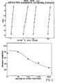
- Figure 1 compares absorbency, at 254 nm, of the Sephadex G-50 fractions from a preovulatory ovarian venous sample of patient 1 to the bioassay results of the sample, as determined by rat ovarian weight and serum 17f-estradiol concentrations.
- V e /V o 1.42-1.55
- fractions with a Ve/Vo of 1.42-1.55 corresponded to an inhibition of hMG-induced ovarian stimulation in the bioassay, as evidenced by a decrease in ovarian weight (59 + 0.5 mg) and a significant (P ⁇ 0.01, by paired t test) decrease in serum 17p-estradiol to levels less than 25 pg/ml.
- these fractions were pooled and processed for dose response activity.
- Peripheral and ovarian venous blood collected from the ovary contralateral to the site of ovulation in patient 1 demonstrated similar G-50 elution profiles (data not shown).
- the isoelectric point of active fractions eluted from the Sephadex G-50 column was estimated by ampholyte displacement chromatography. Pooled aliquots (1 ml) of active fractions were layered on a Polybuffer Exchanger 94 (25 ml; equilibrated to pH 7.4 with 0.025 M Imidazole HC1) column (0.6 x 30 cm). Fractions were eluted with Pharmalyte(TM) PolybufferTM 74-HC1 adjusted to pH 4.0 with HC1 1 N at 10 ml/h at 4'C.
- the bioassay results from ovarian weight and serum 17p-estradiol concentrations from representative fractions suggested that the isoelectric point of the active Sephadex G-50 eleuents from patient 1 were between pH 6.2-6.5.
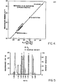
- Figure 4 depicts the K av values for the molecular weight standards and active Sephadex G-50 fractions from patients 1-3. Estimations of molecular weight ranged from 14,000-16,500 for patient 1, from 14,000-17,000 for patient 2, and from 16,000-18,000 for patient 3.
- At least one protein which suppresses the follicle response to gonadotropins is secreted by the preovulatory ovary.
- a heat- and trypsin-labile substance is secreted directly into the venous drainage from the ovary containing the dominant follicle which suppresses the follicular response to gonadotropins. That this protein is not secreted in large amounts by anovulatory ovaries was evidenced by the failure of the bioassay to detect inhibitory activity in the venous drainage of the contralateral ovary of patients 1-3 as well as the ovarian venous effluents from three anovulatory women.
- This potential intra- and/or interovarian regulator of folliculogenesis mediates dominance of the preovulatory follicle by an active process, such that after the selection of the dominant follicle, the gonadotropin responsivity of other follicles on the same and contra- .lateral ovaries is suppressed.
- human follicular fluid was aspirated from regularly menstruating women (aged twenty-five to thirty-five years) undergoing ovarian hyperstimulation during participation in an in vitro fertilization protocol by treatment with clomiphene citrate (150 mg/day for five days, beginning three-eight days after onset of spontaneously occurring menses) and hCG (5000 IU forty-eight hours before aspiration).
- Sera were collected daily and estrogen concentrations were determined.
- serum estrogen concentrations exceeded 800 pg/ml, patients underwent laparoscopy for aspiration of follicles in excess of 20 mm in diameter.
- the follicular aspirate was immediately centrifuged (800 x g), the granulosa cells were removed and the aspirate was then frozen (-57°C). Follicular aspirates from twenty such patients were pooled and provided the .hFF used throughout this study.
- hFF pooled hFF was slowly thawed and fractionated by dropwise addition of an equal volume of saturated ammonium sulfate (SAS) during persistent agitation at 4°C. After a twelve hour incubation at 4 * C, the precipitate was pelleted, the supernatant was discarded, and the pellet was resuspended (2:1, vol/vol) with 10% SAS. An additional twelve hours of agitation was followed by centrifugation at 3,000 x g for thirty minutes. The resulting supernatant was dialized using 10,000 molecular weight exclusion dialysis membranes against three changes of 0.025 M Tris/HC1, pH 7.5, (buffer A) for sixteen hours. Insoluble material was removed from the retentate by centrifugation (3,000 x g for 30 minutes).
- SAS saturated ammonium sulfate
- the column was returned to its upright position and lowered into anode buffer containing 1% sulfuric acid. The remaining upper portion of the column was filled with 1% ethanolamine. The column was cooled by recirculating water at 1-4 * C throughout the procedure. Isoelectric focusing was initated at 800 constant V (16 mA) and allowed to proceed to equilibrium as monitored by an eventual decline in the milliamperage to 2.5 mA (8-12 h). Thereafter, pooled column fractions (3 ml) were dialyzed against buffer A to remove ampholytes before bioassay.
- the rats used in the bioassay were hypophysectomized and implanted as described in Example One, and serum estradiol-17 ⁇ concentration was determined as described previously.
- Control determinations (no injected test fractions) for unstimulated ovarian weight were 34.7 ⁇ 3.2 (SEM) mg/rat and for LH-FSH-stimulated were 192.0 + 30.5 mg/rat.
- Control levels of trunk serum estradiol were 12.5 ⁇ 0.7 pg/ml for unstimulated rats, and 118.5 + 21 pg/ml for LH-FSH-stimulated. Where indicated, 100% inhibition equals ovarian weight and/or serum estradiol concentration of mean unstimulated control values.
- Zero percent inhibition equals ovarian weight or serum estradiol concentration of LH-FSH-stimulated control rats.
- Protein separation was performed using a Waters HPLC/ GPC Model 244 Liquid Chromatograph equipped with a 0.75 x 50 cm TSK 3000 SW gel exclusion column. A 100 -ul aliquot of the dialyzed, Orange A-dye-matrix eluent was separated on each high performance liquid chromatographic (HPLC) run. The proteins were eluted from the TSK column using an isocratic gradient of 0.02 M PBS (pH 7.0) at a flow of 0.5 ml/min.
- the protein peaks were detected by absorbance at 280 nm with a Waters variable wavelength detector (Model M-450) and molecular weight estimates of the specific follicular fluid proteins were performed using highly purified molecular weight chromatography standards of ribonuclease-A, chymotrypsin, ovalbumin, and bovine serum albumin (BSA).
- BSA bovine serum albumin
- Table 1 summarizes the results of Dye-Ligand matrix chromatography of the 10-55% SAS-dialyzed hFF fraction. Although Orange A bound only 17% of the charged protein, 1.5 M KC1 eluted bioactive material that contained the greatest inhibition of the hMG-induced rat ovarian weight gain (89 + 6.8% SEM; P 0.05) when compared to the other Dye-Ligand eluents.
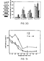
- Figure 5 depicts the chromatographic elution profile of hFF developed through Sephadex G-50 at 254 nm after SAS 10-55% cut, dialysis (?10,000 molecular weight), and elution of Orange A-bound material with 1.5 M KCl.
- Fractions (2 ml) were tested in the LH-FSH-stimulated hypophysectomized, immature, DES-treated rat for inhibition of ovarian weight, and serum estradiol concentration (mean + SEM).
- An initial peak in absorbance can be seen at V e -V O ratio of 1.0-1.1, which after descending, reaches a gradual ascending second plateau (V e -V O of 1.58-1.68).
- FIG 8 depicts the HPCL elution profile of the Orange A-bound extracted hFF material eluted through a TSK 3,000 analytical molecular weight exclusion column.
- Figure 9 indicates the elution profile of the same material eluted through a TSK 3,000 preparative molecular weight exclusion column.
- Each of these elution profiles displays unique chromatographic patterns of GRP.
- the HPLC eluent was divided into ten fractions based on peak absorbances [0: 18.5-21.0 minutes (void volume); 1: 21.0-23.0 minutes; 2: 23.0-25.0 minutes; 3: 25.0-28.5 minutes; 4: 28.5-32.2 minutes; 5: 32.2-34.0 minutes; 6: 34.0-40.0 minutes; 7: 40.0-45.0 minutes; 8: 45.0-49.0 minutes; 9: 49.0-53.0 minutes].
- Aromatase inhibiting activity elutes at 40-45 minutes. These fractions corresponded to the following molecular weight ranges: 1, 100,000; 2, 100,000; 3, 70,000-100,000; 7: 5,500-18,000; 8: 2,500-5,500; 9, 2,500.
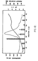
- FIG. 11 shows a polyacrylamide gel, with sodium dodecyl sulfate, of the fractions shown in Figure 8 after HPLC.
- Columns 1-5 in Figure 11 correspond, respectively, to the columns 3-7 in Figure 8.
- the GRP is shown at the bottom of column 5, with a molecular weight of 15,000 to 16,000 when compared to the molecular weight standards to the right of column 5.
- the standards include ribonuclease A at 14,000, and chymo- tripsinogen A at 22,000.
- Example One there is described a protein, in the venous drainage of the human ovary which contains the preovulatory follicle, which suppresses the follicular response to gonadotropins. That this protein was not secreted in large amounts by anovulatory ovaries was evidenced by failure of the bioassay to detect inhibitory activity in the venous drainage of the contralateral ovary in ovulatory patients as well as both ovarian vein effluents from anovulatory women.
- hFF human follicular fluid
- the rat bioassay employed to identify material in hFF which inhibits LH-FSH-mediated follicular stimulation was the same as that reported for human ovarian vein blood extracts.
- Follicular inhibitory activity in hFF had a molecular weight, determined by HPLC size exclusion chromatography (10,000-18,000) that is similar to that of follicular inhibitory activity recovered from human ovarian vein serum (14,000-17,000).
- the follicular inhibitory substance reported here was derived from follicular aspirates of women during spontaneous ovarian cycles whose follicles were hyerstimulated by clomiphene and hCG therapy. Consequently, no conclusion can be drawn regarding this material in normally developing fo-llicles. However, since a similar activity has been identified in the ovarian vein serum draining the spontaneous preovulatory ovary (Example One) these data indicate this inhibitory protein to be a product of the dominant follicle itself.
- Example Two details the isolation of a .protein fraction (GRP) from human follicular fluid, which suppresses follicular response to gonadotropins.
- GFP .protein fraction
- the present example employs an identical isolation, bioassay and HPLC procedures with'respect to porcine follicular fluid (pFF). Isoelectric focusing demonstrated inhibitory activity at about pH 4.2-4.5 and pH 6.27 and peak activity was found in the molecular weight range 12,000-18,000 daltons.
- Granulosa cells were isolated from rat ovaries and FSH binding was determined by the procedure of Erickson, as described in Example Four. Aromatase activity of the rat granulosa cells was determined in a procedure identical to that described therein.
- Table 2 sumarizes the results of dye-ligand matrix chromatography of the 10-55% saturated ammonium sulfate porcine follicular fluid fraction. Although Orange A bound only 13% of the charged protein, 1.5 M KC1 elution recovered bioactive material which contained the greatest inhibition of hMG-induced rat ovarian weight response (p ⁇ .05) when compared to the other dye-ligand eluents. When these same fractions were treated with heat (56°C) or trypsin (10 mg%), no suppression in ovarian weight was found.
- Figure 14 depicts the chromatographic elution profile of pFF at 280 nm developed through Sephadex G-100 following the' 10-55% SAS cut, dialysis, and elution of Orange A-bound material with 1.5 M KC1.
- An initial peak can be seen at a V e /V o of 1.1 to 1.17, thereafter trailing, followed by a gradual ascending second plateau at V e/ V o 1.7 to 1.9.
- biological activity was assessed in the HIFR-hMG bioassay, 95% inhibition of both ovarian weight and trunk serum estradiol-17-B levels were found at V e/ V o of 1.5.
- Figure 16 depicts the HPLC elution profile of the Orange A-bound extracted PFF material after separation through the Sephadex G-100 column (V e /V o 1.3-1.7).
- the HPLC eluent was divided into five fractions based on peak absorbances (1: retention time - 25.0-27.8 minutes; 2: 27.8-31.0 minutes; 3: 31.0-36.0 minutes; 4: 36.0-39.0 minutes; 5: 39.0-43.0 minutes). These fractions corresponded to the followina molecular weight ranges: 1: 100,000-74,000; 2: 74,000-36,000; 3: 36,000-18,000; 4: 18,000-12,000; 5: 12,000-5,800.
- the retention times of peak absorbances after HPLC elution, when correlated to molecular weight standards, were highly reproducible (five runs).
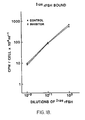
- Figure 18 shows the results of FSH binding studies performed on the granulosa cells removed from the HIFR-hMG rats used in the bioassay of the Sephadex-G100 fraction which contained inhibitory activity. No difference in FSH binding to the rat granulosa cells was evident between the control and inhibitor treated HIFR-hMG rats.
- Follicular fluid was aspirated from regularly menstruating-women (twenty-five to thirty-five years old), undergoing clomiphene citrate (150 mg/day for five days, beginning three-eight days after the onset of spontaneously occurring menses) and hCG (4000 IU, 36 hours before aspiration) therapy during participation in an in vitro fertilization protocol. Serum was collected daily, and estrogen concentrations were determined. When serum estrogen concentrations exceeded 800-1000 pg/ml, patients underwent laparoscopy for aspiration of follicles in excess of 20 mm in diameter. Follicular aspirates were immediately centrifuged (800 x g), granulosa cells were removed for culture, and the supernatant was frozen (-57 . C). Follicular aspirates from seven patients were evaluated.
- the aspirated follicular fluid volume, number of viable granulosa cells, and follicular fluid steriod concentrations from the largest follicle recovered from the seven patients are depicted in Table 2. All of the antral fluids contained high concentrations of progesterone (7-12 ug/ml), indicating premature luteinizations of the. follicles (30-32) as a result of the clomiphene/hCG therapy. Approximately 100,000 viable granulosa cells were obtained from each follicle.
- the retentate (500 mg in 0.5 ml aliquots) was passed through a column (9 x 32 mm; bed volume, 2 ml) containing agarose-immobilized Orange A (Dye matrix, Amicon), which had been equilibrated with 20 mM Tris-HCl, pH 7.5. Unbound material was eluted with 10 ml 20 mM Tris-HCl, pH 7.5 and bound material was eluted with 10 ml 1.5 M KC1 in 20 mM Tris-HCl pH 7.5. Bound eluent fractions were dialyzed overnight against PBS or distilled water. Protein concentrations were determined by the method of Lowry et al., J. Biol. Chem. 143:265.
- results of control determinations were 34.8 + 3.2 mg/rat for unstimulated ovarian weight and 122.0 + 13.5 mg/rat for FSH-stimulated ovarian weight.
- Control levels of trunk serum estradiol were 12.5 + 0.7 pg/ml for unstimulated and 118.5 + 21 pg/ml for LH/FSH stimulated. Where indicated, 100% inhibition equals the ovarian weight and/or serum estradiol concentration of mean unstimulated control values.
- Zero percent inhibition equals the ovarian weight or serum estradiol concentration of LH/FSH-stimulated control rats.
- Figure 19 depicts the effect of extracted follicular fluid (ehFF)and respective granulosa cell culture media (24, 48, and 72 hours) on the inhibition of LH/FSH (2 IU)-stimulated rat ovarian weight augmentation and serum estradiol secretion (2 ml/rat). Each value represents the mean + SEM of three rats.
- All four follicular fluid extracts contained inhibitory activity, as evidenced by inhibition of both rat ovarian weight (40-65%) and trunk . serum estradiol concentrations (85-100%).
- Figure 20 depicts the compiled (mean ⁇ SE) bioassay results of culture media (containing GRP) derived from granulosa cell cultures of patients 5, 6 and 7. All media tested without gonadotropins added to the culture contained inhibitory activity of both ovarian weight and trunk serum estradiol concentrations exceeding 75% throughout the first two days of culture. Thereafter (days 3 and 4), the inhibitory activity of media from unstimulated cultures declined (43% and 37%, and 18% and 12% for ovarian.weight and serum estradiol levels, respectively).
- HPLC high performance liquid chromatographic
- Protein separation was performed using the same Waters HPLC/GPC Model 244 liquid chromatograph equipped with two Waters 1-125 gel exclusion columns connected in series. A 10- ⁇ l aliquot of the dialyzed, Orange A dye matrix-bound eluent was separated on each HPLC run. The proteins were eluted from the 1-125 columns using an isocratic gradient of 0.02 M PBS, pH 7.0, at a flow of 0.5 ml/minute (800 psi). The protein peaks were detected at 280 nm, and molecular weight estimates of the specific follicular fluid proteins were performed using highly purified molecular weight chromatography standards of ribonuclease-A, chymotrypsin, ovalbumin, and BSA.
- Granulosa cells were cultured for up to four days in twenty-four hour intervals using 35 x 10-mm tissue culture dishes and 2 ml medium 199 containing 25 mM Hepes supplemented with 100 U/ml penicillin, 100 ⁇ g streptomycin sulfate, and 15% fetal calf serum (medium A). Cultures were maintained in a humidified, 95% air-5% C0 2 incubator at 37 . C. After each twenty-four hour incubation period, spent medium was collected and stored frozen at -20 * C until bioassay was performed. Where indicated, human menopausal gonadotropin (FSH-LH,1:1) was added to specific cultures at the time of complete medium change.
- FSH-LH,1:1 human menopausal gonadotropin
- Granulosa cells were isolated from rat ovaries and were collected by centrifugation at 800 x g at 4 * C for ten minutes. FSH binding was determined using a modification of known techniques. Rat FSH, provided by the National Pituitary Agency, was labeled by the chloramine-T procedure. Cells were resuspended in appropriate volumes of PBS-01% gelatin (PBS-gel), pH 7.0. All assays were run with three concentration of labeled hormone (100 ⁇ l), buffer (PBS-0.1% gel; 100 d) and 100 ⁇ l cells. Reactions were initiated by the addition of granulosa cells and were carried out for four hours at 25"C.
- Figure 21 depicts the FSH binding studies performed on the granulosa cells removed from the HIFR-hMG-treated rat ovaries used in the bioassay experiments shown in Figure 19. Specific binding of rat FSH (rFSH) to granulosa cells was determined by incubating three concentrations of labeled rFSH in the presence and absence of excess unlabeled rFSH. Rat granulosa cell specific FSH binding was similar whether the rat received injections of spent medium from human granulosa cell cultures or saline. However, a marked difference in the ovarian weight and trunk serum estradiol concentrations of these rats was present.
- rFSH rat FSH
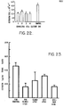
- Rat granulosa cell aromatase activity is shown in Figure 22 and was markedly inhibited (P ⁇ 0.01) by treatment with spent media from all four days of culture (2.4 + 0.4, 2.9 + 0.7, 2.4 + 0.4, and 2.1 + 0.2 pg estrogen/10,000 viable cells/ml, respectively, compared to saline control 4.6 + 0.2 pg estrogen/10,000 viable cells/ml.
- Control determinations were performed on rat granulosa cells collected from HIFR-hMG-treated rats which did not receive culture medium injections. Inhibition of relative estrogen production by rat granulosa cells in the presence of 10- 7 M androstenedione was seen throughout all four days of human granulosa cell culture medium treatment.
- a particularly advantageous method of preparing reproducing granulosa cell cultures which will produce GRP in quantity is to employ mammals which have no antibodies to foreign tissues, such as the laboratory rodent known as the nude mouse, for initial cell growth.
- mammals which have no antibodies to foreign tissues, such as the laboratory rodent known as the nude mouse, for initial cell growth.
- Numerous experiments have been performed wherein an injection of about 10 7 granulosa cells, isolated from human, rat and other species, was administered subcutaneously to nude mice, followed by the administration of 100 mg of diethylstilbestrol (an estrogen) daily. After six to eight weeks, cells were recovered by aspiration with a syringe from the bleb formed by the initial injection, and the recovered cells reproduced in tissue culture.
- nonsteroidal regulators of ovarian function have been identified in a variety of species, including oocyte maturation inhibitor, luteinizing inhibitor, folliculostatin or inhibin, and FSH binding inhibitor.
- the biophysical characteristics of the intraqonadal protein of this invention are different from such substances.
- This material elutes -through gel exclusion chromatography with a molecular weight of 12,000-18,000, binds to Orange A dye matrix, and has an apparent isoelectric point of pH 4.0-4.5 and 6.0-6.5.
- Bovine testis were homogenized and the homogenate precipitated with 55% saturated ammonium sulfate. The precipitate was resuspended and dialyzed against distilled water. The retentate was eluted though Sephadex G-100 with TRIS buffer (20 mM, pH 7.5). The eluant corresonding to a molecular weight range of 12,000-18,000 was collected and charged into an Orange A dye matrix column. The column was eluted with TRIS buffer containing 0.5 M KC1 (20 mM, pH 7.5).
- This eluant contained GRP as evidenced by the inhibition of porcine granulosa cell aromatase activity (Figure 23; bTEc and bTE-OAB columns) and rat serum estradiol levels (Figure 24; BTE + FSH and BTE-OAB + FSH columns). Further, this activity was contained in testicular extract fractions with isoelectric points of pH 4.1-4.5 and 6.0-6.5. This demonstrates that the testis and its secretory product, rete testis fluid, also contains GRP.
- Granulosa cells secrete GRP and Sertoli cells are the embryonic homologue in testis of the granulosa cells in the ovary. Further, the biological activities of granulosa cells are present in the Sertoli cells insofar as they have been identified and are relevant to gonadal function. Thus, the Sertoli cell provides an additional, readily available source of GRP. Accordingly, Sertoli cells may be grown in cell culture and the recovered culture media is extracted by the procedures described in the foregoing examples, principally salt participation, chromatographic procedures and electrophoresis.
- Follicular aspirates were obtained from women (aged twenty-four-thirty-two years) who were participating in an in vitro fertilization protocol. All patients had regular ovulatory menstrual cycles based on monthly vaginal bleeding and at least a single luteal phase serum progesterone in excess of 3 ng/ml.
- serial ultrasonographic examinations ADR Model 2140 real-time sector scanner with a 3.5 mHz rotating head transducer
- Follicular aspirates were immediately transferred to an adjacent laboratory for removal of granulosa cells by centrifugation (600 x G, 15 minutes) and storage of follicular fluid (-37'C) until assay.
- Follicular fluid concentrations of estradiol, progesterone, 17-hydroxy progesterone, androstenedione and testosterone were determined by established radioimmunoassay techniques.
- GRP was isolated from individual follicular fluid samples substantially as described in Example Two.
- Porcine granulosa cells were collected from fresh ovaries obtained at the local slaughterhouse. After washing in serum free HAMS-HEPES tissue culture media, 5 x 10 5 cells in 0.2 ml of medium were pipetted into 12 x 75 mm polystyrene tubes. Triplicate 200 ul portions of each follicular fluid preparation at three different protein concentrations (700-10 ug/ml) were tested. Each tube then received 100 ⁇ l of FSH (10n g) in culture medium and was incubated at 37°C in a shaking water bath for three hours. An atmosphere of - 90% N 2 , 5% 0 2 and 5% C0 2 was maintained throughout 'the incubation.
- the incubation was stopped by the addition of 0.5 ml Hams-Hepes media and centrifugation at 1000 x g for five minutes.
- the granulosa cells were then resuspended in 0.5 ml Hams-Hepes media, whereupon 100 ⁇ l of cells were assayed for armoatase activity.
- Androstenedione, the referent aromatase substrate was added in 0.1 ml medium (final concentration, 1.0 x 10- 7 M). All incubations were performed in duplicate for three hours at 37°C in a shaking water bath (120 cycles/ minute). The reaction was stopped by transferring the tubes to an iced water bath before centrifugation (five minutes at 1000 x g).
- anterior pituitary glands were obtained from 20 cycling female Sprague-Dawly rats (250-300 gm body weight). During the thirty minute interval required for removal of all the pituitary glands, each gland was placed in medium (pH 7.4, 20 . C). Pituitary glands were finely minced with scissors and incubated in a mixture containing 1% viokase, 33% collagenase, and 3% bovine serum albumin in medium buffer at 37°C for thirty to forty-five minutes. The dispersed cells were counted using a hemocytometer and pituicyte viability was determined by 1% trypan blue exclusion. Typically, more than 90% of the dispersed cells were viable.
- the cells were diluted to a concentration of 2.5 x 10 5 viable cells per ml growth medium.
- Growth medium consisted of HAMS F10 containing 10% fetal calf serum with penicillin, fugizone and streptomycin (50 u/ml and 50 mg/ml and 50 mg/ml respectively).
- Cells were added to tissue culture dishes in a volume of 2.0 ml growth media, and attachment of the cells to the well surface was completed by two days. After cell attachment, the original growth media was discarded and the cells were washed twice with additional HAMS balanced salt soluticn. Thereafter, three concentrations of charcoal- treated follicular fluid (.02%, 0.2%, 2%) were added to the plate.
- porcine follicular fluid standard (PFF1 KT-1, provided by CP Channing) was resuspended in water and tested in each assay at .003%, .016%,.08%, .04%, and 2% concentrations. Twenty-four hours later, the spent culture media was assayed in duplicate using the NIH-RIA kit for rFSH.
- the mean estrogen concentration in the serum control tube for each GRPassay was set at 100%, and the response in each test was expressed as a percentage of the control estrogen concentration.
- the coefficient of variation for each group of three replicate tubes was calculated. If the coefficient was 15%, the estrogen assay was repeated and/or one value of the three was discarded.
- a curve was constructed in which the percent inhibition of estrogen in each well was plotted vs the protein concentration of follicular fluid added. Unknown values were determined by plotting the experimentally determined values at three different protein concentrations (700-10 ug/ml) on a log-linear graph and extrapolating the value at 50 ug/ml. The percent inhibition of estradiol at 50 ug of unknown follicular fluid was read off the standard curve and expressed as percent aromatase inhibition for that follicle.
- the response in each test plate was expressed as a percentage of the control FSH concentration, which was set at 100%. The coefficient of variation of each group of three replicate plates was calculated. If the coeffient was 15% the assay was repeated.
- a standard curve was constructed in which the percent inhibition of FSH in standard wells was plotted vs the log of the standard added. Least squares linear regression was used to construct a standard curve in the linear portion of the dose response curve. Unknown follicular fluid inhibin .values were determined by plotting the experimentally .determined values (0.02%, 0.2%, 2%) on a log-linear graph and extrapolating the value at 1%.
- Tests for statistical significance were performed by one-way analysis of variance and Duncan's new multiple range test. Correlation between follicular fluid steroid concentration and GRP activity was performed using regression analysis with tests of statistical significance performed by t test corrected for N.
- porcine granulosa cells were used in each subsequent assay.
- porcine granulosa cell concentrations were used: .5 x 10 6 , 1 x 10 6 , 2 x 10 6 cells/ ml.
- the total amount of estrogen produced following a three-hour incubation was 55 + 9, 140 + 27, 375 + 48 pg/culture dish, respectively. Consequently, 2 x 10 6 porcine granulosa cells were used in each subsequent assay.
- porcine FSH NIH P-2 reagent
- follicular protein 100 ug
- porcine granulosa cell cultures were prepared with or without porcine FSH added to the media (.5 ml final volume), incubated at 37 * C for three hours in a shaker bath with 95% 0 2 and 5% C0 2 , after which androstenedione (10- 6 M) was added in .5 ml of growth media. Cultures were incubated for three hours at 37°C in a shaker bath then centrifuged (1000 x G for fifteen minutes) and media collected for estrogen determination.
- the GRP in elution profiles from Orange A were analyzed using three different concentrations of KC1: 0.17 M KC1 yielded 3.1 mg protein/ml which had a 37% inhibition of aromatase, 0.5 M KC1 eluted 6 mg protein/ml which had an 84% inhibition of aromatase and 1.5M KC1 eluted .05 mg protein/ml which had a 6.7% inhibition of aromatase. Accordingly, the .5 M KC1 fraction was used to elute the active material from the Orange A bound column.
- KC1 (.5M) was found to have no effect in the granulosa cell aromatase assay: control (without KC1, 3 determinations, 710 + 41 ng/ml. with KC1, 712 + 38 ng/ml). Duration of incubation time was assessed with and without FSH. Two hour assay incubations yielded 2 ng estrogen/ ml; twenty-four hours: 5.3 ng estrogen/ml. With 2 ng FSH added, 39 ng estrogen/ml at two hours and 6 ng FSH/ml produced 8 ng estrogen/ml at twenty-four hours.
- estradiol in the follicular fluid of untreated patients was 2.59 + 1.2 ug/ml, for patients receiving hMG therapy, .34 + 0.5 ug/ml, for patients receiving clomiphene, 1.31 + .34 ug/ml. These values were all significantly different (p (.05, untreated vs clomiphene, clomiphene vs hMG; p ⁇ .01 untreated vs hMG).
- Progesterone values for untreated patients were 9.84 ⁇ 3.35 ug/ml, for hMG-treated patients, 5.18 + 1.1 ug/ml, and for clomiphene-treated patients, 11.3 ⁇ 2.3 ug/ml These differences were significant for the unstimulated and hMG-treated patients (p ⁇ .05) and hMG vs clomiphene treated patients (p ⁇ .01).
- the 17-hydroxyprogesterone concentrations for patients receiving no additional therapy were 1.66 + .25 ug/ml, for those receiving clomiphene therapy 2.6 + .3 ug/ml, and for those receiving hMG therapy: .76 ⁇ .11 ug/ml.
- Aromatase incubations were carried out in a total volume of 0.6 ml in 12 x 75 ml glass tubes as described in the above-identified references. Incubations contained 0.3 ml of placental cell-free preparation in buffer A, 0.10 ml of 14 C-androstenedione (A,10- 6 molar), NADPH (10- 6 molar) and nicotinamide (0.4 molar) in buffer A. Gonadal regulatory protein test fractions (0.2 ml), as described above, were preincubated with placental extracts (20 minutes) and then the 14 C-androstenedione-NADPH mixture was added. Reaction at termination/quenching was performed by addition of 100-fold excess unlabeled A. Estrogen concentrations were determined by radioimmunoassay as described in J. Clin. Endocrinol. Metab., 39, 754-760.
- GRP-dose response determinations were performed using a three-minute reaction time.
- the velocities determined over a range of substrate concentrations (0.5-2 mM) demonstrated Michaelis-Menton type kinetics with a Km of 0.8 mM as shown in Figure 27.
- the aromatase velocities were determined over a range of GRP concentrations (0, 62.5, 125, 250, 500 and 1,000 ⁇ g/ml).
- Enzyme inhibition may be of three types.
- First, competitive inhibition describes the competition of the inhibitor for the substrate-specific site on the enzyme.
- Second, non-competitive inhibition describes a direct inhibition of the enzyme without competition for-the substrate-specific sites.
- Third, uncompetitive inhibition describes an indirect inhibition of the enzyme by additional mechanisms.' In Figure 28, the intersection of the lines at the abscissa, as opposed to ordinal intersection (competitive) or non- intersection (uncompetitive) show that GRP is a non-competitive inhibitor of aromatase activity. GRP directly interacts with the aromatase molecule to inhibit its activity.
- GRP was isolated from porcine follicular fluid substantially as described in Example Three.
- the granulosa cells were counted using a hemocytometer and viability was determined by 1% trypan blue exclusion.
- Cells were cultured (2 x 10 5 ) in 2 ml of Medium 100 containing 10% fetal calf serum with penicillin and streptomycin (100 g/ml and 100 U/ml respectively) in 12 x 75 mm Falcon plastic test tubes.
- FSH (10 ng), and/or GRP (1 mg) were added at the initiation of culture. Media was changed after seventy-two hours.
- Porcine granulosa cells were suspended in appropriate volumes of PBS-0.1% gelatin (PBS-gel). All assays were run with five concentrations of labeled hCG (10 ul), buffer (PBS-0.1% gel, 100 ul), and cells. Reactions were initiated by the addition of granulosa cells and were carried out for four hours at 25"C. All reaction tubes were precoated with 5% BSA to reduce non-specific absorption. Reactions were terminated by adding one ml of cold PBS followed by centrifugation at 30,000 x g for ten minutes. The supernatant was carefully aspirated and the pellet rewashed with one ml of PBS. The final pellet was counted in a gamma counter. Specific binding was calculated as the difference between binding in the presence (non- specific) and absence (total) of an excess of unlabeled hormone. Data were analyzed by Scatchard plots. Duplicate determinations were performed in three separate assays at each time interval.
- Follicular protein significantly reduced porcine granulosa cell hCG binding by seventy-four hours of culture. This effect was prevented with the co-administration of FSH.
- Adenylate cyclase activity in the membranes of GRP + FSH and GRP alone treated cells was significantly less than in cells incubated with FSH (p ⁇ .05).
- Adenylate cyclase activity of GRP treated cells was unchanged in the presence of methyl-isobutal-xanthine. Further, when GRP was heated (56C, 45 min.) or precipitated with 10% TCA, it lost- the capability to inhibit adenylate cyclase.
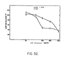
- the 50% inhibitory dose (ID50) for GRP inhibition of Gpp(NH)p stimulated adenylate cyclase activity with preincubation of granulosa cells with FSH was 350 ug/ml and 80 ug/ml without FSH.
- the ID50 for the G RP inhibition of forskolin stimulated adenylate cyclase astivity was 350 ug/ml ( Figure 32).
- Adenylate cyclase activity was determined after a 10 min. incubation with forskolin or Gpp(NH)p. When these responses were compared during a 5-20 min. interval, the Gpp(NH)p stimulated adenylate cyclase activity was more sensitive to inhibition by GRP than forskolin stimulated adenylate cyclase activity (p ⁇ .05).
- Adenylate cyclase activity stimulated by Gpp(NH)p was also, on a molar basis, more sensitive to GRP inhibition than forskolin stimulated activity.
- GRP had no apparent effect on LH or hCG responsive adenylate cyclase activity in these granulosa cell preparations. No other naturally occurring substance has previously been shown to selectively inhibit FSH responsive adenylate cyclase activity in granulosa cells without also inhibiting LH responsive adenylate cyclase activity. In conclusion, these data demonstrate that GRP inhibited FSH responsive adenylate cyclase activity in porcine granulosa cells.
- Follicular fluid extracts containing this activity are shown in said Examples to have,a molecular weight of 12,000-18,000 and isoelectric points of pH 4. 0 to 4.5 and 6.0 to 6.5, showing that the biophysical nature of this protein is not inhibin.
- the effects of this follicular protein fraction on the integrated hypothalamic-pituitary-ovarian axis of the normally cycling monkey are assessed.
- Serum estradiol levels were reduced throughout the interval of GRP treatment in the monkeys without LH surges in the late follicular phase ( Figure 34) and in those with luteal phase defects (Figure 33). Serum FSH and LH levels were within the 95% confidence intervals for GRP treated monkeys ( Figure 33). However, in all five GRP treated monkeys, serum FSH levels rose during the course of therapy. In the monkeys with inadequate luteal phases (Figure 33), serum estradiol levels were below the 95% confidence intervals in the late follicular and mid luteal phases.
- GRP administered to normally cycling monkeys throughout the follicular phase of the menstrual cycle reduced peripheral estradiol levels without markedly affecting FSH, resulting in either apparent anovulation or inadequate luteal phases.
- the mixed response in serum sex steroid levels in the GRP treated monkeys may reflect a variable ovarian sensitivity to GRP. That serum FSH levels were not significantly inhibited indicates that this material has a biological activity different from that of charcoal-extracted whole porcine follicular fluid. Further, when GRP was tested in an inhibin assay, its activity was either at the limits of sensitivity or undetectable. Charcoal-extracted whole porcine follicular fluid.
- inhibin disrupts the normal process of follicular maturation through selective inhibition of FSH secretion.
- normal or rising serum FSH levels during GRP treatment were found. This indicates that the purification of follicular fluid described herein removed the major inhibin activity.
- the molecular weight of the described GRP is less than 20,000 while inhibin activity has been associated with molecular weights greater than 45,000.
- Example Three Male mongrel dogs were injected with 2 mg of G RP derived from porcine follicular fluid, as described in Example Three, at 08:00 and 16:00 hours for 20 days. On Day 20 of therapy the testis were obtained, fixed in formalin, sectioned by microtome (four micron sections), mounted on slides and stained with hematoxylin and eosin. Upon evaluation of the slides, a marked reduction in mature spermatozoa was present in the seminiferous tubules in the GRP-treated dogs as compared to the controls. Moreover, there was an 87% reduction in pacytene spermatocytes and a 44% reduction in mature spermatogonia in the GRP-treated dogs.
- GRP-treated group showed a significant inhibition of transformation from type A to type B spermatogonia during the final stages of spermatogenesis.
- any remaining type B spermatogonia (mature spermatozoa) in the above- described study are solely attributable to transformation prior to treatment, and repeated administration of G R P to male mammals will completely inhibit the production of mature spermatozoa.
- Antibodies (monoclonal and polyclonal) to GRP and its conjuners and analogs may be prepared for diagnostic and therapeutic uses including but not limited to fertility control.
- Antibody in this contex refers to a synthetic protein which binds GRP and alters GRP biological activity.
- Antibodies to GRP are prepared by either polyclonal or monoclonal techniques:
- 1 ⁇ g of GRP was solubilized in 0.5 ml physiological saline and emulsified with an equal volume of Freund's adjuvant to prepare an inoculum.
- New Zealand White, female rabbits weighing 2-1/2-3 kg were bled via the median ear artery for pre-immune serum.
- a 10 x 20 cm area on the back was shaved, then each rabbit was intradermally injected at multiple points.
- Approximately 50-75 ⁇ l of inoculum was injected into 10-25 sites in the shaved area; rabbits 1 and 3 received 0.6 ml and 1.6 ml, respectively.
- the rabbits were boosted in similar fashion six weeks later receiving 0.5 ml and 1.5 ml, respectively.
- Six and ten day post-boost the rabbits were test bled, again through the median artery.
- Sera containing the polyclonal antibodies thus obtained were titred via RIA against 125 I -labelled GRP as follows.
- Rabbit sera were two-fold serially diluted in RIA buffer from 1:1000 to 1:64,000. 100 ⁇ l each of the RIA buffer, diluted serum, and ⁇ 25 I -G RP and 200 ⁇ l RIA buffer were also prepared. The tubes were incubated at room temperature (RT) overnight. One-half ml (0.5 ml) precipitating solution containing 2% goat-anti- rabbit gamma globulin and 4% polyethylene glycol in RIA buffer was then added to each tube except the total tubes. The tubes were vortexed, incubated at room temperature for ten minutes, then centrifuged at 3000 rpm for an additional ten minutes. The supernatant was aspirated and the precipitin-pellet counted for one minute on a gamma counter. The rabbit sera was found to contain antibody which bound 125 I-GRP in.all dilutions tested, including 1:64,000.
- BALB/c mice are immunized with GRP by intraperitoneal injection (250 ug) x 2. Thereafter, the spleens are collected and cell suspensions prepared by perfusion with DMEM. The BALB/c spleen cells are fused with SP 2/0-Ag 14 mouse myeloma cells by PEG and the resultant hybridomas grown in HAT selective tissue culture media + 20% fetal calf serum. The surviving cells are allowed to grow to confluence. The spent culture media is checked for antibody titer, specificity, and affinity.
- mice were immunized with GRP adjuvant emulsion described above. Each mouse first received 0.2 ml of this emulsion intraperitoneally, then were reinjected in similar fashion with 0.1 ml six weeks later. Mouse serum was obtained ten days after the second injection and then tested for anti-GRP activity via ELISA. The mouse exhibiting the highest absolute anti-GRP activity was chosen for cell-fusion.
- the chosen mouse was intravenously injected with 0.1 ml GRP solubilized in physiological saline, and SP2/0-Ag14 BALB/c myeloma cells maintained in log phase culture.
- the mouse was sacrificed and its spleen aseptically removed.
- Spleen cell suspension containing B-lymphocytes and macrophages were prepared by prefusion of the spleen.
- the cell suspension was washed and collected by centrifugation.
- Myeloma cells were also washed in this manner. Live cells were counted and the cells were placed in a 37°C water bath and 1 ml of 50% poly-ethylene glycol in DMEM slowly added.
- the cells were incubated in the PEG for one to one-and-a-half minutes at 37°C, after which the PEG was diluted by the slow addition of media.
- the cells were pelletted and 35 to 40 ml of DMEM containing 10% fetal bovine serum was added.
- the cells were then dispensed into tissue culture plates and incubated overnight in a 37°C, 5% C0 2 , humidified incubator.
- HAT medium hypoxanthine, thymidine, and aminopterin
- the plates were incubated with 1 x HAT medium every three-four days for two weeks. Fused cells were thereafter grown in DMEM-FCS containing hypoxanthine and thymidine. As cell growth became 1/2 to 3/4 confluent on the bottom of the wells, supernatant tissue culture fluid was taken and tested for GRP-specific antibody by ELISA. Positive wells were cloned by limiting dilution over macrophage or thymocyte feeder plates, and cultured in DMEM-FCS. Cloned wells were tested and recloned three times before a statistically significant monoclonal antibody was obtained. Spent culture media from the chosen clone contained antibody which bound 125 I -G RP in all dilutions tested, including 1:64,000.
- GRP antibodies that a wide variety of diagnostic tests is possible using the antibodies of the invention.
- the over-production of GRP by the gonads an indication of the presence of a condition such as ovarian cancer, could easily be detected in body fluids such as serum through the use of an immunoassay which employs the novel antibodies according to methods known in the art.
- an immunoassay to detect decreased levels of GR P in the body would be a useful adjunct to known hormone assays.
- Further uses for the antibodies include the induction of superovulation in livestock.
- the direct administration of GRP antibodies to sheep has been shown to induce multiple ovulations in ewes. It should be noted that other than the novelty of the protein and the preparation of GRP antibodies thereto, techniques required for the preparation and use of diagnostic test kits and super- ovulatory antibodies are known in the art and will not be described in further detail.
- the proteinaceous substance of the invention may be produced by other methods.
- a c-DNA probe can be prepared against the biologically active portion of GRP, and used to identify the GRP genome in granulosa cells or Sertoli cells from any mammalian species.
- the identified genome can then be synthesized into a plasmid which can then be employed to produce recombinant DNA in proliferating bacteria according to methods known in the art.
- granulosa or Sertoli cells may be transformed, e.g. by SV40 virus, to produce GRP in quantity.
- GRP intergonadal protein
- the identifying characteristics, methods of production and uses set forth herein identify a novel and unique proteinaceous substance.
- GRP is described herein to be the only proteinaceous substance known which inhibits aromatase activity by determination of the extent of the conversion of androgens to estrogens. Moreover, such inhibition is unique in that it is non-competitive in placental microsomes. Further, GRP produces a biphasic effect on 3f-ol dehydrogenase activity in cultured granulosa cells as shown in Figures 29 and 30.
- GRP inhibits FSH induction of LH receptor formation as determined by addition to an in vitro culture of granulosa cells, and inhibits FSH responsive adenylate cyclase activity as similarly determined.
- GRP is identified by the elution profile through a molecular weight exclusion column under the conditions set forth in Example Two, as indicated by the designation "GRP" of the elution curve in Figure 9.
- the elution profile through a hydrogen ion exclusion column, particularly the area designated by the indicated peaks numbers 4 and 7 in Figure 15, further identifies identifying characteristics of GRP when conducted under the conditions set forth in the Example Three.
- a polyacrylamide gel electrophoretic pattern from a high pressure liquid chromatographic column as shown by the designation "GRP" in column 6 of Figure 11, uniquely identifies GRP when the pattern is obtained as set forth in Example Two.
Landscapes
- Health & Medical Sciences (AREA)
- Life Sciences & Earth Sciences (AREA)
- Chemical & Material Sciences (AREA)
- Molecular Biology (AREA)
- Organic Chemistry (AREA)
- Medicinal Chemistry (AREA)
- Immunology (AREA)
- Engineering & Computer Science (AREA)
- Biochemistry (AREA)
- General Health & Medical Sciences (AREA)
- Biomedical Technology (AREA)
- Hematology (AREA)
- Proteomics, Peptides & Aminoacids (AREA)
- Biophysics (AREA)
- Urology & Nephrology (AREA)
- Genetics & Genomics (AREA)
- Microbiology (AREA)
- Analytical Chemistry (AREA)
- Cell Biology (AREA)
- Zoology (AREA)
- Toxicology (AREA)
- Gastroenterology & Hepatology (AREA)
- Endocrinology (AREA)
- Food Science & Technology (AREA)
- Physics & Mathematics (AREA)
- Biotechnology (AREA)
- General Physics & Mathematics (AREA)
- Pathology (AREA)
- Medicines That Contain Protein Lipid Enzymes And Other Medicines (AREA)
- Medicines Containing Material From Animals Or Micro-Organisms (AREA)
- Acyclic And Carbocyclic Compounds In Medicinal Compositions (AREA)
- Peptides Or Proteins (AREA)
- Preparation Of Compounds By Using Micro-Organisms (AREA)
Priority Applications (1)
| Application Number | Priority Date | Filing Date | Title |
|---|---|---|---|
| AT85307212T ATE60337T1 (de) | 1984-10-11 | 1985-10-09 | Geschlechtsdruesen regulierendes protein. |
Applications Claiming Priority (2)
| Application Number | Priority Date | Filing Date | Title |
|---|---|---|---|
| US66100284A | 1984-10-11 | 1984-10-11 | |
| US661002 | 1984-10-11 |
Publications (3)
| Publication Number | Publication Date |
|---|---|
| EP0178841A2 true EP0178841A2 (fr) | 1986-04-23 |
| EP0178841A3 EP0178841A3 (en) | 1987-05-27 |
| EP0178841B1 EP0178841B1 (fr) | 1991-01-23 |
Family
ID=24651786
Family Applications (1)
| Application Number | Title | Priority Date | Filing Date |
|---|---|---|---|
| EP85307212A Expired - Lifetime EP0178841B1 (fr) | 1984-10-11 | 1985-10-09 | Protéine régulatrice des gonades |
Country Status (11)
| Country | Link |
|---|---|
| EP (1) | EP0178841B1 (fr) |
| JP (1) | JPS61178923A (fr) |
| AT (1) | ATE60337T1 (fr) |
| AU (1) | AU599306B2 (fr) |
| CA (1) | CA1268417A (fr) |
| DE (1) | DE3581469D1 (fr) |
| DK (1) | DK464585A (fr) |
| GR (1) | GR852463B (fr) |
| IE (1) | IE58721B1 (fr) |
| IL (1) | IL76562A0 (fr) |
| NZ (1) | NZ213660A (fr) |
Cited By (2)
| Publication number | Priority date | Publication date | Assignee | Title |
|---|---|---|---|---|
| US5102868A (en) * | 1990-01-08 | 1992-04-07 | Genentech, Inc. | Method for inhibiting follicular maturation |
| US5166190A (en) * | 1990-01-08 | 1992-11-24 | Genentech, Inc. | Method for increasing fertility in males |
Families Citing this family (3)
| Publication number | Priority date | Publication date | Assignee | Title |
|---|---|---|---|---|
| JP2002031940A (ja) | 2000-07-19 | 2002-01-31 | Fujitsu Ltd | 2成分現像装置、画像形成装置及び攪拌スクリュー |
| JP5513802B2 (ja) * | 2009-08-04 | 2014-06-04 | ホーユー株式会社 | 等電点電気泳動用ゲル及び等電点電気泳動方法 |
| GB201909275D0 (en) * | 2019-06-27 | 2019-08-14 | Ge Healthcare Bio Sciences Ab | Chromatograpy apparatus and method |
Family Cites Families (3)
| Publication number | Priority date | Publication date | Assignee | Title |
|---|---|---|---|---|
| IN140168B (fr) * | 1974-10-14 | 1976-09-25 | All India Inst Med | |
| GB2115281B (en) * | 1982-02-17 | 1984-12-12 | Univ Surrey And Agricultural R | The improvement of animal fecundity by treatment with antisera to gonadal steroids |
| AU4437485A (en) * | 1984-06-08 | 1986-01-10 | Biotechnology Australia Proprietary Limited | Inhibin isolated from ovarian follicular fluid |
-
1985
- 1985-09-25 CA CA000491514A patent/CA1268417A/fr not_active Expired - Lifetime
- 1985-09-26 IE IE238385A patent/IE58721B1/en not_active IP Right Cessation
- 1985-09-30 AU AU48127/85A patent/AU599306B2/en not_active Ceased
- 1985-10-01 NZ NZ213660A patent/NZ213660A/xx unknown
- 1985-10-04 IL IL76562A patent/IL76562A0/xx not_active IP Right Cessation
- 1985-10-09 EP EP85307212A patent/EP0178841B1/fr not_active Expired - Lifetime
- 1985-10-09 DE DE8585307212T patent/DE3581469D1/de not_active Expired - Lifetime
- 1985-10-09 AT AT85307212T patent/ATE60337T1/de not_active IP Right Cessation
- 1985-10-10 DK DK464585A patent/DK464585A/da not_active Application Discontinuation
- 1985-10-10 GR GR852463A patent/GR852463B/el unknown
- 1985-10-11 JP JP60224993A patent/JPS61178923A/ja active Pending
Cited By (2)
| Publication number | Priority date | Publication date | Assignee | Title |
|---|---|---|---|---|
| US5102868A (en) * | 1990-01-08 | 1992-04-07 | Genentech, Inc. | Method for inhibiting follicular maturation |
| US5166190A (en) * | 1990-01-08 | 1992-11-24 | Genentech, Inc. | Method for increasing fertility in males |
Also Published As
| Publication number | Publication date |
|---|---|
| NZ213660A (en) | 1991-08-27 |
| DE3581469D1 (de) | 1991-02-28 |
| JPS61178923A (ja) | 1986-08-11 |
| IE852383L (en) | 1986-04-11 |
| IE58721B1 (en) | 1993-11-03 |
| EP0178841B1 (fr) | 1991-01-23 |
| ATE60337T1 (de) | 1991-02-15 |
| AU4812785A (en) | 1986-05-29 |
| CA1268417A (fr) | 1990-05-01 |
| GR852463B (fr) | 1986-02-11 |
| AU599306B2 (en) | 1990-07-19 |
| EP0178841A3 (en) | 1987-05-27 |
| IL76562A0 (en) | 1986-02-28 |
| DK464585D0 (da) | 1985-10-10 |
| DK464585A (da) | 1986-04-12 |
Similar Documents
| Publication | Publication Date | Title |
|---|---|---|
| Robertson et al. | Comparison of inhibin immunological and in vitro biological activities in human serum | |
| SULLIVAN et al. | Hormonal regulation of immunoglobulins in the rat uterus: uterine response to multiple estradiol treatments | |
| US4764502A (en) | Intragonadal regulatory protein | |
| DIZEREGA et al. | Human granulosa cell secretion of protein (s) which suppress follicular response to gonadotropins | |
| Duignan et al. | Sex hormone levels and gonadotrophin release in the polycystic ovary syndrome | |
| US4734398A (en) | Follicular regulating protein | |
| EP0260306A4 (en) | Method of assay of inhibin | |
| Lamperti et al. | Effects of mercuric chloride on the reproductive cycle of the female hamster | |
| US4676981A (en) | Monoclonal antibodies against GnRH | |
| EP0178841B1 (fr) | Protéine régulatrice des gonades | |
| Ray Kling et al. | Identification of a porcine follicular fluid fraction which suppresses follicular response to gonadotropins | |
| Gilfillan et al. | Development and validation of a radioimmunoassay for follistatin in human serum | |
| US5173404A (en) | Immunoassay for follicular regulatory protein | |
| US4912201A (en) | Monoclonal antibodies and antisera to intragonadal regulatory protein | |
| Yoshida et al. | Monoclonal antibodies to the thyrotropin receptor bind to a 56-kDa subunit of the thyrotropin receptor and show heterogeneous bioactivities. | |
| Genazzani et al. | Acetyl‐1‐carnitine as possible drug in the treatment of hypothalamic amenorrhea | |
| Blackwell et al. | Concomitant release of FSH and LH induced by native and synthetic LRF | |
| Wadia et al. | Effect of the human follicle-stimulating hormone-binding inhibitor and its N-terminal fragment on follicle-stimulating hormone-induced progesterone secretion by granulosa cells in vitro | |
| Buntin et al. | Properties of hepatic binding sites for prolactin in ring doves (Streptopelia risoria) | |
| Dias et al. | Evidence for a high molecular weight follicle-stimulating hormone binding inhibitor in bovine testis | |
| Klootwijk et al. | Urinary excretion of the TRH-like peptide pyroglutamyl-glutamyl-prolineamide in rats | |
| Schneyer et al. | Development of a radioligand receptor assay for measuring follitropin in serum: application to premature ovarian failure | |
| Siler-Khodr et al. | Production and activity of placental releasing hormones | |
| NAKAZAWA et al. | Immunohistochemical study on oxytocin-like substance in the human placenta | |
| LEW et al. | Alteration of follicle regulatory protein levels in human reproductive disorders: Anovulation |
Legal Events
| Date | Code | Title | Description |
|---|---|---|---|
| PUAI | Public reference made under article 153(3) epc to a published international application that has entered the european phase |
Free format text: ORIGINAL CODE: 0009012 |
|
| AK | Designated contracting states |
Kind code of ref document: A2 Designated state(s): AT BE CH DE FR GB IT LI LU NL SE |
|
| PUAL | Search report despatched |
Free format text: ORIGINAL CODE: 0009013 |
|
| AK | Designated contracting states |
Kind code of ref document: A3 Designated state(s): AT BE CH DE FR GB IT LI LU NL SE |
|
| 17P | Request for examination filed |
Effective date: 19871022 |
|
| 17Q | First examination report despatched |
Effective date: 19880506 |
|
| GRAA | (expected) grant |
Free format text: ORIGINAL CODE: 0009210 |
|
| AK | Designated contracting states |
Kind code of ref document: B1 Designated state(s): AT BE CH DE FR GB IT LI LU NL SE |
|
| PG25 | Lapsed in a contracting state [announced via postgrant information from national office to epo] |
Ref country code: SE Effective date: 19910123 Ref country code: AT Effective date: 19910123 |
|
| REF | Corresponds to: |
Ref document number: 60337 Country of ref document: AT Date of ref document: 19910215 Kind code of ref document: T |
|
| REF | Corresponds to: |
Ref document number: 3581469 Country of ref document: DE Date of ref document: 19910228 |
|
| ITF | It: translation for a ep patent filed | ||
| ET | Fr: translation filed | ||
| PGFP | Annual fee paid to national office [announced via postgrant information from national office to epo] |
Ref country code: SE Payment date: 19910919 Year of fee payment: 7 |
|
| PLBE | No opposition filed within time limit |
Free format text: ORIGINAL CODE: 0009261 |
|
| STAA | Information on the status of an ep patent application or granted ep patent |
Free format text: STATUS: NO OPPOSITION FILED WITHIN TIME LIMIT |
|
| 26N | No opposition filed | ||
| PGFP | Annual fee paid to national office [announced via postgrant information from national office to epo] |
Ref country code: BE Payment date: 19921012 Year of fee payment: 8 |
|
| PGFP | Annual fee paid to national office [announced via postgrant information from national office to epo] |
Ref country code: LU Payment date: 19921022 Year of fee payment: 8 |
|
| PGFP | Annual fee paid to national office [announced via postgrant information from national office to epo] |
Ref country code: NL Payment date: 19921031 Year of fee payment: 8 |
|
| PGFP | Annual fee paid to national office [announced via postgrant information from national office to epo] |
Ref country code: CH Payment date: 19921117 Year of fee payment: 8 |
|
| EPTA | Lu: last paid annual fee | ||
| PG25 | Lapsed in a contracting state [announced via postgrant information from national office to epo] |
Ref country code: LU Free format text: LAPSE BECAUSE OF NON-PAYMENT OF DUE FEES Effective date: 19931009 |
|
| PG25 | Lapsed in a contracting state [announced via postgrant information from national office to epo] |
Ref country code: LI Effective date: 19931031 Ref country code: CH Effective date: 19931031 Ref country code: BE Effective date: 19931031 |
|
| BERE | Be: lapsed |
Owner name: UNIVERSITY OF SOUTHERN CALIFORNIA Effective date: 19931031 |
|
| PG25 | Lapsed in a contracting state [announced via postgrant information from national office to epo] |
Ref country code: NL Effective date: 19940501 |
|
| NLV4 | Nl: lapsed or anulled due to non-payment of the annual fee | ||
| REG | Reference to a national code |
Ref country code: CH Ref legal event code: PL |
|
| PGFP | Annual fee paid to national office [announced via postgrant information from national office to epo] |
Ref country code: FR Payment date: 19941012 Year of fee payment: 10 |
|
| PGFP | Annual fee paid to national office [announced via postgrant information from national office to epo] |
Ref country code: GB Payment date: 19941017 Year of fee payment: 10 |
|
| PGFP | Annual fee paid to national office [announced via postgrant information from national office to epo] |
Ref country code: DE Payment date: 19941027 Year of fee payment: 10 |
|
| PG25 | Lapsed in a contracting state [announced via postgrant information from national office to epo] |
Ref country code: GB Effective date: 19951009 |
|
| GBPC | Gb: european patent ceased through non-payment of renewal fee |
Effective date: 19951009 |
|
| PG25 | Lapsed in a contracting state [announced via postgrant information from national office to epo] |
Ref country code: FR Effective date: 19960628 |
|
| PG25 | Lapsed in a contracting state [announced via postgrant information from national office to epo] |
Ref country code: DE Effective date: 19960702 |
|
| REG | Reference to a national code |
Ref country code: FR Ref legal event code: ST |