EP0180878A2 - Heterodimerischer Rezeptor von T-Lymphozyten - Google Patents
Heterodimerischer Rezeptor von T-Lymphozyten Download PDFInfo
- Publication number
- EP0180878A2 EP0180878A2 EP85113658A EP85113658A EP0180878A2 EP 0180878 A2 EP0180878 A2 EP 0180878A2 EP 85113658 A EP85113658 A EP 85113658A EP 85113658 A EP85113658 A EP 85113658A EP 0180878 A2 EP0180878 A2 EP 0180878A2
- Authority
- EP
- European Patent Office
- Prior art keywords
- amino acid
- region
- acid residues
- cysteine
- approximately
- Prior art date
- Legal status (The legal status is an assumption and is not a legal conclusion. Google has not performed a legal analysis and makes no representation as to the accuracy of the status listed.)
- Withdrawn
Links
- 210000001744 T-lymphocyte Anatomy 0.000 title claims abstract description 58
- 239000002299 complementary DNA Substances 0.000 claims abstract description 71
- 108090000623 proteins and genes Proteins 0.000 claims abstract description 60
- 210000001151 cytotoxic T lymphocyte Anatomy 0.000 claims abstract description 37
- 102000016266 T-Cell Antigen Receptors Human genes 0.000 claims abstract description 34
- 235000001014 amino acid Nutrition 0.000 claims abstract description 32
- 108091008874 T cell receptors Proteins 0.000 claims abstract description 30
- 239000002773 nucleotide Chemical group 0.000 claims abstract description 29
- 125000003729 nucleotide group Chemical group 0.000 claims abstract description 29
- 150000001413 amino acids Chemical group 0.000 claims abstract description 27
- 102000004169 proteins and genes Human genes 0.000 claims abstract description 27
- 235000018102 proteins Nutrition 0.000 claims abstract description 26
- 125000000151 cysteine group Chemical group N[C@@H](CS)C(=O)* 0.000 claims abstract description 24
- 230000001086 cytosolic effect Effects 0.000 claims abstract description 19
- 238000005304 joining Methods 0.000 claims abstract description 18
- 108010076504 Protein Sorting Signals Proteins 0.000 claims abstract description 17
- 108020003175 receptors Proteins 0.000 claims abstract description 17
- 125000000539 amino acid group Chemical group 0.000 claims description 47
- 108090000765 processed proteins & peptides Proteins 0.000 claims description 40
- 238000009396 hybridization Methods 0.000 claims description 27
- 239000000523 sample Substances 0.000 claims description 26
- 108091032973 (ribonucleotides)n+m Proteins 0.000 claims description 25
- 125000003275 alpha amino acid group Chemical group 0.000 claims description 25
- 238000000034 method Methods 0.000 claims description 24
- 102000003886 Glycoproteins Human genes 0.000 claims description 20
- 108090000288 Glycoproteins Proteins 0.000 claims description 20
- XUJNEKJLAYXESH-UHFFFAOYSA-N cysteine Natural products SCC(N)C(O)=O XUJNEKJLAYXESH-UHFFFAOYSA-N 0.000 claims description 20
- 230000004988 N-glycosylation Effects 0.000 claims description 19
- 239000000427 antigen Substances 0.000 claims description 19
- 108091007433 antigens Proteins 0.000 claims description 19
- 102000036639 antigens Human genes 0.000 claims description 19
- 235000018417 cysteine Nutrition 0.000 claims description 19
- 102000004196 processed proteins & peptides Human genes 0.000 claims description 19
- 229920001184 polypeptide Polymers 0.000 claims description 17
- 108091034057 RNA (poly(A)) Proteins 0.000 claims description 15
- 108020004705 Codon Proteins 0.000 claims description 14
- 230000002209 hydrophobic effect Effects 0.000 claims description 12
- 210000002443 helper t lymphocyte Anatomy 0.000 claims description 11
- 241000894007 species Species 0.000 claims description 9
- 101150076359 Mhc gene Proteins 0.000 claims description 8
- 208000003950 B-cell lymphoma Diseases 0.000 claims description 7
- 102000016844 Immunoglobulin-like domains Human genes 0.000 claims description 6
- 108050006430 Immunoglobulin-like domains Proteins 0.000 claims description 6
- 239000013612 plasmid Substances 0.000 claims description 5
- 238000010367 cloning Methods 0.000 claims description 4
- 210000002257 embryonic structure Anatomy 0.000 claims description 4
- 230000015572 biosynthetic process Effects 0.000 claims description 3
- 239000002458 cell surface marker Substances 0.000 claims description 3
- 108020004999 messenger RNA Proteins 0.000 claims description 3
- 230000029087 digestion Effects 0.000 claims description 2
- 238000012216 screening Methods 0.000 claims description 2
- 229940024606 amino acid Drugs 0.000 claims 8
- -1 cationic amino acids Chemical class 0.000 claims 3
- ZDXPYRJPNDTMRX-UHFFFAOYSA-N glutamine Natural products OC(=O)C(N)CCC(N)=O ZDXPYRJPNDTMRX-UHFFFAOYSA-N 0.000 claims 3
- 230000000295 complement effect Effects 0.000 claims 2
- DCXYFEDJOCDNAF-UHFFFAOYSA-N Asparagine Natural products OC(=O)C(N)CC(N)=O DCXYFEDJOCDNAF-UHFFFAOYSA-N 0.000 claims 1
- DCXYFEDJOCDNAF-REOHCLBHSA-N L-asparagine Chemical compound OC(=O)[C@@H](N)CC(N)=O DCXYFEDJOCDNAF-REOHCLBHSA-N 0.000 claims 1
- 102000012750 Membrane Glycoproteins Human genes 0.000 claims 1
- 108010090054 Membrane Glycoproteins Proteins 0.000 claims 1
- 229960001230 asparagine Drugs 0.000 claims 1
- 235000009582 asparagine Nutrition 0.000 claims 1
- 230000002194 synthesizing effect Effects 0.000 claims 1
- 102000005962 receptors Human genes 0.000 abstract description 12
- 241001529936 Murinae Species 0.000 abstract description 6
- 102000017727 Immunoglobulin Variable Region Human genes 0.000 abstract description 5
- 108010067060 Immunoglobulin Variable Region Proteins 0.000 abstract description 5
- 238000004519 manufacturing process Methods 0.000 abstract description 5
- 210000004698 lymphocyte Anatomy 0.000 abstract description 3
- 108020004414 DNA Proteins 0.000 description 20
- NHBKXEKEPDILRR-UHFFFAOYSA-N 2,3-bis(butanoylsulfanyl)propyl butanoate Chemical compound CCCC(=O)OCC(SC(=O)CCC)CSC(=O)CCC NHBKXEKEPDILRR-UHFFFAOYSA-N 0.000 description 15
- 210000004027 cell Anatomy 0.000 description 12
- 241000699666 Mus <mouse, genus> Species 0.000 description 10
- 238000004458 analytical method Methods 0.000 description 10
- 210000003719 b-lymphocyte Anatomy 0.000 description 10
- 108060003951 Immunoglobulin Proteins 0.000 description 8
- 108010079585 Immunoglobulin Subunits Proteins 0.000 description 8
- 102000012745 Immunoglobulin Subunits Human genes 0.000 description 8
- 206010035226 Plasma cell myeloma Diseases 0.000 description 8
- 102000018358 immunoglobulin Human genes 0.000 description 8
- 201000000050 myeloid neoplasm Diseases 0.000 description 8
- 210000001161 mammalian embryo Anatomy 0.000 description 7
- 230000008707 rearrangement Effects 0.000 description 6
- 108091028043 Nucleic acid sequence Proteins 0.000 description 5
- 239000012634 fragment Substances 0.000 description 5
- 238000002955 isolation Methods 0.000 description 5
- 239000012528 membrane Substances 0.000 description 5
- 239000000047 product Substances 0.000 description 5
- 108700042075 T-Cell Receptor Genes Proteins 0.000 description 4
- 210000004408 hybridoma Anatomy 0.000 description 4
- 108700005091 Immunoglobulin Genes Proteins 0.000 description 3
- 102000006496 Immunoglobulin Heavy Chains Human genes 0.000 description 3
- 108010019476 Immunoglobulin Heavy Chains Proteins 0.000 description 3
- 206010025323 Lymphomas Diseases 0.000 description 3
- 241001465754 Metazoa Species 0.000 description 3
- 125000000729 N-terminal amino-acid group Chemical group 0.000 description 3
- 206010028980 Neoplasm Diseases 0.000 description 3
- 108700026244 Open Reading Frames Proteins 0.000 description 3
- 108010092262 T-Cell Antigen Receptors Proteins 0.000 description 3
- 210000004899 c-terminal region Anatomy 0.000 description 3
- 125000002091 cationic group Chemical group 0.000 description 3
- 210000000805 cytoplasm Anatomy 0.000 description 3
- 229940079593 drug Drugs 0.000 description 3
- 239000003814 drug Substances 0.000 description 3
- 239000000499 gel Substances 0.000 description 3
- 125000001165 hydrophobic group Chemical group 0.000 description 3
- 230000001900 immune effect Effects 0.000 description 3
- 229940072221 immunoglobulins Drugs 0.000 description 3
- 108091008146 restriction endonucleases Proteins 0.000 description 3
- 239000004475 Arginine Substances 0.000 description 2
- 108020004635 Complementary DNA Proteins 0.000 description 2
- 108091035707 Consensus sequence Proteins 0.000 description 2
- ZHNUHDYFZUAESO-UHFFFAOYSA-N Formamide Chemical compound NC=O ZHNUHDYFZUAESO-UHFFFAOYSA-N 0.000 description 2
- CKLJMWTZIZZHCS-REOHCLBHSA-N L-aspartic acid Chemical compound OC(=O)[C@@H](N)CC(O)=O CKLJMWTZIZZHCS-REOHCLBHSA-N 0.000 description 2
- 108700018351 Major Histocompatibility Complex Proteins 0.000 description 2
- 239000000020 Nitrocellulose Substances 0.000 description 2
- 238000000636 Northern blotting Methods 0.000 description 2
- 241000283973 Oryctolagus cuniculus Species 0.000 description 2
- 101150052863 THY1 gene Proteins 0.000 description 2
- 108091036066 Three prime untranslated region Proteins 0.000 description 2
- 239000011543 agarose gel Substances 0.000 description 2
- ODKSFYDXXFIFQN-UHFFFAOYSA-N arginine Natural products OC(=O)C(N)CCCNC(N)=N ODKSFYDXXFIFQN-UHFFFAOYSA-N 0.000 description 2
- 235000003704 aspartic acid Nutrition 0.000 description 2
- OQFSQFPPLPISGP-UHFFFAOYSA-N beta-carboxyaspartic acid Natural products OC(=O)C(N)C(C(O)=O)C(O)=O OQFSQFPPLPISGP-UHFFFAOYSA-N 0.000 description 2
- 238000010804 cDNA synthesis Methods 0.000 description 2
- 238000002474 experimental method Methods 0.000 description 2
- 102000054766 genetic haplotypes Human genes 0.000 description 2
- 125000000291 glutamic acid group Chemical group N[C@@H](CCC(O)=O)C(=O)* 0.000 description 2
- 125000000404 glutamine group Chemical group N[C@@H](CCC(N)=O)C(=O)* 0.000 description 2
- LEQAOMBKQFMDFZ-UHFFFAOYSA-N glyoxal Chemical compound O=CC=O LEQAOMBKQFMDFZ-UHFFFAOYSA-N 0.000 description 2
- 125000003588 lysine group Chemical group [H]N([H])C([H])([H])C([H])([H])C([H])([H])C([H])([H])C([H])(N([H])[H])C(*)=O 0.000 description 2
- MYWUZJCMWCOHBA-VIFPVBQESA-N methamphetamine Chemical compound CN[C@@H](C)CC1=CC=CC=C1 MYWUZJCMWCOHBA-VIFPVBQESA-N 0.000 description 2
- 230000004048 modification Effects 0.000 description 2
- 238000012986 modification Methods 0.000 description 2
- 238000010369 molecular cloning Methods 0.000 description 2
- 229920001220 nitrocellulos Polymers 0.000 description 2
- 230000004481 post-translational protein modification Effects 0.000 description 2
- 125000002924 primary amino group Chemical group [H]N([H])* 0.000 description 2
- 238000002415 sodium dodecyl sulfate polyacrylamide gel electrophoresis Methods 0.000 description 2
- 230000020382 suppression by virus of host antigen processing and presentation of peptide antigen via MHC class I Effects 0.000 description 2
- 210000001541 thymus gland Anatomy 0.000 description 2
- 102000035160 transmembrane proteins Human genes 0.000 description 2
- 108091005703 transmembrane proteins Proteins 0.000 description 2
- 210000004881 tumor cell Anatomy 0.000 description 2
- 230000003612 virological effect Effects 0.000 description 2
- 241000283690 Bos taurus Species 0.000 description 1
- 108700031361 Brachyury Proteins 0.000 description 1
- 101800001415 Bri23 peptide Proteins 0.000 description 1
- 101150111062 C gene Proteins 0.000 description 1
- 101800000655 C-terminal peptide Proteins 0.000 description 1
- 102400000107 C-terminal peptide Human genes 0.000 description 1
- 101800005309 Carboxy-terminal peptide Proteins 0.000 description 1
- 108010078791 Carrier Proteins Proteins 0.000 description 1
- 102000014914 Carrier Proteins Human genes 0.000 description 1
- 241000272201 Columbiformes Species 0.000 description 1
- 102000018832 Cytochromes Human genes 0.000 description 1
- 108010052832 Cytochromes Proteins 0.000 description 1
- BWGNESOTFCXPMA-UHFFFAOYSA-N Dihydrogen disulfide Chemical compound SS BWGNESOTFCXPMA-UHFFFAOYSA-N 0.000 description 1
- 102000004190 Enzymes Human genes 0.000 description 1
- 108090000790 Enzymes Proteins 0.000 description 1
- 108700024394 Exon Proteins 0.000 description 1
- WHUUTDBJXJRKMK-UHFFFAOYSA-N Glutamic acid Natural products OC(=O)C(N)CCC(O)=O WHUUTDBJXJRKMK-UHFFFAOYSA-N 0.000 description 1
- 101150017040 I gene Proteins 0.000 description 1
- 102100029567 Immunoglobulin kappa light chain Human genes 0.000 description 1
- 101710189008 Immunoglobulin kappa light chain Proteins 0.000 description 1
- 102100029566 Immunoglobulin lambda-1 light chain Human genes 0.000 description 1
- 101710118731 Immunoglobulin lambda-1 light chain Proteins 0.000 description 1
- 102100034343 Integrase Human genes 0.000 description 1
- 102000014150 Interferons Human genes 0.000 description 1
- 108010050904 Interferons Proteins 0.000 description 1
- 101150008942 J gene Proteins 0.000 description 1
- 108091026898 Leader sequence (mRNA) Proteins 0.000 description 1
- 102100026964 M1-specific T cell receptor beta chain Human genes 0.000 description 1
- 108010090665 Mannosyl-Glycoprotein Endo-beta-N-Acetylglucosaminidase Proteins 0.000 description 1
- 108700005081 Overlapping Genes Proteins 0.000 description 1
- 108010092799 RNA-directed DNA polymerase Proteins 0.000 description 1
- 238000002105 Southern blotting Methods 0.000 description 1
- 206010042971 T-cell lymphoma Diseases 0.000 description 1
- 208000027585 T-cell non-Hodgkin lymphoma Diseases 0.000 description 1
- 101150117115 V gene Proteins 0.000 description 1
- 241000251539 Vertebrata <Metazoa> Species 0.000 description 1
- 238000001042 affinity chromatography Methods 0.000 description 1
- 108010087408 alpha-beta T-Cell Antigen Receptors Proteins 0.000 description 1
- 230000000890 antigenic effect Effects 0.000 description 1
- 206010003246 arthritis Diseases 0.000 description 1
- 238000009835 boiling Methods 0.000 description 1
- 210000001185 bone marrow Anatomy 0.000 description 1
- 238000004113 cell culture Methods 0.000 description 1
- 239000003795 chemical substances by application Substances 0.000 description 1
- 230000000973 chemotherapeutic effect Effects 0.000 description 1
- 238000004587 chromatography analysis Methods 0.000 description 1
- 238000004891 communication Methods 0.000 description 1
- 150000001875 compounds Chemical class 0.000 description 1
- 238000010924 continuous production Methods 0.000 description 1
- 238000007796 conventional method Methods 0.000 description 1
- 230000001461 cytolytic effect Effects 0.000 description 1
- 231100000433 cytotoxic Toxicity 0.000 description 1
- 230000001472 cytotoxic effect Effects 0.000 description 1
- 238000001514 detection method Methods 0.000 description 1
- 238000011161 development Methods 0.000 description 1
- 230000018109 developmental process Effects 0.000 description 1
- 238000001976 enzyme digestion Methods 0.000 description 1
- 108020001507 fusion proteins Proteins 0.000 description 1
- 102000037865 fusion proteins Human genes 0.000 description 1
- 108010074605 gamma-Globulins Proteins 0.000 description 1
- 230000013595 glycosylation Effects 0.000 description 1
- 238000006206 glycosylation reaction Methods 0.000 description 1
- 229940015043 glyoxal Drugs 0.000 description 1
- 230000012447 hatching Effects 0.000 description 1
- 230000007062 hydrolysis Effects 0.000 description 1
- 238000006460 hydrolysis reaction Methods 0.000 description 1
- 229910052588 hydroxylapatite Inorganic materials 0.000 description 1
- 238000002347 injection Methods 0.000 description 1
- 239000007924 injection Substances 0.000 description 1
- 229940079322 interferon Drugs 0.000 description 1
- 210000003292 kidney cell Anatomy 0.000 description 1
- 230000003211 malignant effect Effects 0.000 description 1
- 239000000463 material Substances 0.000 description 1
- 239000011159 matrix material Substances 0.000 description 1
- XYJRXVWERLGGKC-UHFFFAOYSA-D pentacalcium;hydroxide;triphosphate Chemical compound [OH-].[Ca+2].[Ca+2].[Ca+2].[Ca+2].[Ca+2].[O-]P([O-])([O-])=O.[O-]P([O-])([O-])=O.[O-]P([O-])([O-])=O XYJRXVWERLGGKC-UHFFFAOYSA-D 0.000 description 1
- 210000002729 polyribosome Anatomy 0.000 description 1
- 239000002244 precipitate Substances 0.000 description 1
- 238000001556 precipitation Methods 0.000 description 1
- 238000002360 preparation method Methods 0.000 description 1
- 238000011451 sequencing strategy Methods 0.000 description 1
- 238000012163 sequencing technique Methods 0.000 description 1
- 210000002966 serum Anatomy 0.000 description 1
- 239000012064 sodium phosphate buffer Substances 0.000 description 1
- 239000007787 solid Substances 0.000 description 1
- 239000000243 solution Substances 0.000 description 1
- 230000000392 somatic effect Effects 0.000 description 1
- 238000010561 standard procedure Methods 0.000 description 1
- 239000007858 starting material Substances 0.000 description 1
- 238000003786 synthesis reaction Methods 0.000 description 1
Images


Classifications
-
- C—CHEMISTRY; METALLURGY
- C07—ORGANIC CHEMISTRY
- C07K—PEPTIDES
- C07K14/00—Peptides having more than 20 amino acids; Gastrins; Somatostatins; Melanotropins; Derivatives thereof
- C07K14/435—Peptides having more than 20 amino acids; Gastrins; Somatostatins; Melanotropins; Derivatives thereof from animals; from humans
- C07K14/705—Receptors; Cell surface antigens; Cell surface determinants
- C07K14/70503—Immunoglobulin superfamily
- C07K14/7051—T-cell receptor (TcR)-CD3 complex
-
- A—HUMAN NECESSITIES
- A61—MEDICAL OR VETERINARY SCIENCE; HYGIENE
- A61K—PREPARATIONS FOR MEDICAL, DENTAL OR TOILETRY PURPOSES
- A61K39/00—Medicinal preparations containing antigens or antibodies
-
- Y—GENERAL TAGGING OF NEW TECHNOLOGICAL DEVELOPMENTS; GENERAL TAGGING OF CROSS-SECTIONAL TECHNOLOGIES SPANNING OVER SEVERAL SECTIONS OF THE IPC; TECHNICAL SUBJECTS COVERED BY FORMER USPC CROSS-REFERENCE ART COLLECTIONS [XRACs] AND DIGESTS
- Y10—TECHNICAL SUBJECTS COVERED BY FORMER USPC
- Y10S—TECHNICAL SUBJECTS COVERED BY FORMER USPC CROSS-REFERENCE ART COLLECTIONS [XRACs] AND DIGESTS
- Y10S930/00—Peptide or protein sequence
- Y10S930/01—Peptide or protein sequence
- Y10S930/26—Containing cys-cys disulfide bridge between nonadjacent cysteine residues
-
- Y—GENERAL TAGGING OF NEW TECHNOLOGICAL DEVELOPMENTS; GENERAL TAGGING OF CROSS-SECTIONAL TECHNOLOGIES SPANNING OVER SEVERAL SECTIONS OF THE IPC; TECHNICAL SUBJECTS COVERED BY FORMER USPC CROSS-REFERENCE ART COLLECTIONS [XRACs] AND DIGESTS
- Y10—TECHNICAL SUBJECTS COVERED BY FORMER USPC
- Y10S—TECHNICAL SUBJECTS COVERED BY FORMER USPC CROSS-REFERENCE ART COLLECTIONS [XRACs] AND DIGESTS
- Y10S930/00—Peptide or protein sequence
- Y10S930/01—Peptide or protein sequence
- Y10S930/30—Signal or leader sequence
Definitions
- the vertebrate immune .system is characterized by its ability to respond to an enormously diverse set of antigenic determinants. This capability is due to the synthesis by the body of a set of glycoproteins whose specificity for a single antigen is determined by a variable sequence of amino acids which binds to the antigen.
- the glycoproteins, which recognize and bind free antigens, are produced by B cells and are called immunoglobulins (Ig).
- the characteristic primary and secondary structure is made up of heavy and light chains which begin with a leader peptide of 17-29 residues, followed by a variable (V) region of 94-97 residues, then a joining region of 13-17 residues (J), then a constant region (C).
- V variable
- J joining region of 13-17 residues
- C constant region
- the domains of the constant regions of the immunoglobulins are encoded by separate exons from those for the variable region and do not appear to rearrange during development.
- T cells or thymus derived lymphocytes are capable of recognizing a wide range of different antigens.
- the ability to recognise a given antigen is also fixed in any particular clonal line of T cell.
- T cells recognize only antigens located on the surface of cells in the specific molecular context of self major histo- compatibility complex (MHC) gene products, not freely circulating antigens.
- MHC self major histo- compatibility complex
- T cell receptors recognize foreign antigens (such as viral antigens) in the molecular context of the T cell host's self-MHC gene products or recognize foreign MHC gene products.
- Cell surface antigens include tumor cell and viral antigens. The ability to recognize cell-bound antigens is acquired when the T cells differentiate in the host thymus.
- T cell receptor a heterodimeric glycoprotein of about 90,000 daltons and consisting of an alpha subunit and a beta subunit held together by an inter-chain disulphide bond(s).
- Peptide fingerprint analysis of alpha and beta chains from several T cell lines suggest that both subunits are composed of variable (V) and constant (C) regions.
- a second cDNA clone pHDS4/203 was identified as the gene coding for the alpha chain of the T cell antigen receptor. However, subsequent studies have shown that this clone lacks sequences that correspond to sites for N-linked glycosylation and it has recently been shown that both the alpha and beta chains of the T cell receptor are N-glycosylated.
- each gene is composed of separate V, J and C gene segments. In the cases of YT35 and TM86, the corresponding genes have also been'shown to have D segments. Their V, (D), and J segments have characteristic signal sequences for gene rearrangement (heptamer and nonamer separated by either 12 or 23 base pairs).
- TM 86, YT35 and pHDSll Due to the substantial homology between the sequences of TM 86, YT35 and pHDSll, it is likely that they represent the beta subunit of the T cell receptor.
- Another object of the present invention is to provide the protein or amino acide sequence of a murine alloreactive cytotoxic T lymphocyte receptor, including both of its subunits.
- the complete nucleotide and amino acid sequences for the T cell receptor derived from the alloreactive cytotoxic T lymphocyte (CTL) clone 2C, of BALB.B origin and specific for the product of a Class I gene at the D end of the BALB/c H-2 complex (d haplotype) is disclosed.
- CTL alloreactive cytotoxic T lymphocyte
- Two related but distinct cDNA clones corresponding to the alpha and beta subunits of the T cell receptor were cloned and sequenced.
- the genes corresponding to these cDNA are rearranged and expressed in CTL clones but not in myelomas and the rearrangement pattern varies with the CTL clone.
- the corresponding poly(A) RNA is composed of 5'- variable and 3'- constant regions.
- the encoded subunits are composed of a signal peptide, two immunoglobulin-like domains, each with one disulphide loop, a transmembrane peptide, and a cytoplasmic peptide.
- the two domains are homologous to variable and constant domains of immunoglobulin chains, particularly those of lambda light chains.
- the encoded subunits Besides having two cysteine residues in each domain, the encoded subunits have a fifth cysteine residue in the region between the disulphide bonded peptide loop of the constant domain and the transmembrane peptide, at a position where the two subunits may be disulphide bonded to each other.
- the constant region sequence of the beta subunit as encoded by the gene pHDSll from a cytotoxic T cell is compared to the constant region sequence of the beta subunit of a T helper cell and found to correspond. It is therefore concluded that the disclosed T cell specific cDNA sequences for the constant region of at least the beta subunit and probably the alpha subunit may be used to produce protein, and antibody to that protein, which are useful in isolation, identification, and other methods for both cytotoxic T lymphocytes and T helper cells. Expected homology among mammalian species, including man, further extends possible applications of the present invention.
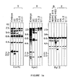
- Fig. 1 is a Southern blot analyses, according to the method taught in Molec. Biol. 98, 503-517 (1975), of DNA from BALB/c embryo, myelomas MOPC315 and MOPC460, both BALB/c derived, and CTL lines 2C, G4, 1.5.2 and 2.1.1, all BALB.B derived, and described by Kranz et al. in Proc. Nat. Acad. Sci. USA, 81, 573-577 (1984). DNA was digested with the indicated restriction enzymes, electrophoresed through 0.8% agarose gels, blotted onto nitrocellulose and hybridized with the 32p-labelled, nick-translated insert from clone pHDS58 (Fig.
- Fig. 2 is a RNA blot analyses, according to the method taught in Meth. Enzym. 100, 255-266 (1983), of poly(A) + RNA from various B- and T-cell lines.
- Probes a, total insert of pHDS58; b, HpaII fragment containing 270 bp at the end of the pHDS58 insert (V-region probe); and C, total insert of pHDSll.
- RNA was extracted from B-cell lymphomas A20-2J and CH1 and alloreactive ( H - 2 b anti-H-2 d ) cytotoxic T-lymphocyte clones 2.1.1, 1.5.2. G4 and 2C.
- Fig. 3 is a restriction map of the insert of cDNA clones pHDS58 (Fig. 3a) and pHDSll (Fig. 3b).
- the map was constructed by a combination of single and double restriction enzyme digestions of the plasmid DNA.
- the V, J, C and 5'- or 3'- untranslated regions are shown by shading, black, hatching and white, respectively.
- the sequencing strategy used to determine the nucleotide sequence shown in Fig. 4 is shown.
- the arrows indicate the directions and the extent of sequence determination.
- the open and closed circles indicate that the ends of the DNA fragments labelled are 5' and 3', respectively.
- Fig. 4 is the nucleotide and predicted amino acid sequences of the cDNA clones pHDS58 (Fig. 4a) and pHDSll (Fig. 4b).
- the nucleotide sequence was determined by the method of Maxam and Gilbert, described in Math. Enzym. 65, 499-560 (1980), according to the strategy shown in Fig. 3. Numbers given above the amino acid sequence designate amino acid residue positions.
- the V, J, C, TM (transmembrane) and CY (cytoplasmic) regions are indicated by horizontal arrows although exact boundaries are ambiguous. Cysteine residues thought to be involved in intra-domain or inter-chain disulphide linkages are encircled.
- the potential N-glycosylation sites (N-X-S or N-X-T) are also indicated.
- Fig. 5 is a comparison of the V and C regions of the predicted amino acid sequence of pHDS58 and the V and C regions, respectively, of the predicted or determined amino acid sequences of five other polypeptide chains: a chain encoded by pHDS4/203, the beta-chain encoded by pHDSll, 93G7 gamma 1 immunoglobulin heavy chain, MOPC603 ka pp a light chain, and MOPC104E lambda l light chain, as taught by Kabat et al. in Sequences of Proteins of Immunological Interest, (NIH, Bethesda (1983). Those residues common between the pHDS58 protein and any one of the other five chains are boxed. * , the residues common among all six chains. Approximate boundaries of framework and hypervariable regions as they appear in immunoglobulin V regions are indicated by horizontal arrows. The amino acid positions refer to those of pHDS58 protein.
- Fig. 6a is a comparison of the J region amino acid sequences of pHDS58, that of pHDS4/203, as taught in Nature 309, 757-762 (1984), and consensus sequences of the T-cell antigen receptor beta-chain, as taught by Gascoigne et al. in Nature 310, 387-391 (1984), and immunoglobulin J H , J kappa and J lambda , as taught by
- Fig. 6b is a comparison of the J region sequence of the amino acid sequence of pHDS4/203, pHDSll. 86T1 and Ig J H , J kappa , and J lambda consensus sequences. Those residues common between pHDS4/203 J and any one of the other 5 J regions are boxed in and shaded.
- Fig. 7 shows the proposed overall structure of the 2C T-cell receptor.
- Each receptor molecule is made up of two chains, alpha and beta, each with two extracellular Ig-like domains, an amino-terminal variable and a carboxy-terminal constant domain. Each of these domains is stabilized by an S-S bond between cysteine residues.
- the two chains are held by a single inter- chain S-S bond located close to the cell's outer membrane.
- the protein is anchored on the membrane by two (one for each chain) hydrophobic transmembrane peptides.
- a short carboxy-terminal peptide rich in cationic residues extends into the cytoplasm in each chain.
- Both alpha and beta chains are N-glycosylated, with the sites indicated on the alpha subunit by CHO.
- the preferred embodiment of the present invention is the structure and amino acid and nucleotide sequences of a heterodimeric glycoprotein which functions as a receptor on the surface of a T lymphocyte and comprises both an alpha and a beta subunit.
- the invention also includes cDNA hybridization probes and antibodies for the identification and isolation of T cell receptors from other species as well as from cytotoxic and helper T cells.
- the T cell receptor and the nucleotide and amino acid sequences required to produce the protein or its components are defined by their substantial identity to the proposed tertiary structure and nucleotide and amino acid sequences of the receptor of a murine alloreactive cytotoxic T lymphocyte.
- the T-cell receptor is made up of two chains, each with two extracellular Ig-like domains, an amino-terminal variable domain, and a carboxy-terminal constant domain. Each of these domains is stabilized by a disulfide bond between cysteine residues that are separated in the linear sequence by 50-70 residues.
- the alpha chain consisting of approximately 248 amino acid residues, has cystein residues at amino acid residues 22, 90 (variable region), 134, 184 (constant region), and 202 (constant region adjacent transmembrane region).
- the beta chain consisting of 282 amino acid residues, has cysteine residues at amino acid residues 23, 91 (variable region), 140, 201 (constant region) and 236 (constant region adjacent transmembrane region).
- the variable region of the alpha subunit consisting of amino acids 1-98, is joined to the constant region, consisting of amino acids 112 to 222, by amino acids 99-111.
- the variable region of the beta subunit consisting of amino acids 1-96, is joined to the constant region, consisting of amino acids 110 to 254, by amino acids 97-109.
- each subunit has at its carboxyl-end a peptide carrying an extra cysteine, a hydrophobic stretch of about 20-22 amino acids followed by a short stretch of approximately 5 amino acid residues in which cationic residues abound.
- These segments correspond, respectively, to the transmembrane and cytoplasmic domains characteristically found ; in transmembrane proteins.
- the alpha chain as described, consists of 248 amino acids and has a calculated relative molecular mass of approximately 28,000.
- the chain contains 23 negatively charged amino acid residues (15 aspartic acid and 8 glutamic acid residues), and 19 positively charged amino acid residues (6 arginine and 13 lysine residues), corresponding to an isoelectric point near neutrality in the absence of post-translation modifications.
- the potential sites for N-glycosylation are located at amino acid residues 70-72, 178-180, 142-144, and 221-223.
- the alpha chain is preceeded by a sequence of 20 amino acid residues.
- variable and constant regions of both chains are coded for by distinct gene segments.
- the same gene appears to code for at least the beta chain constant region of cytotoxic T-lymphocytes and the beta chain constant region of helper T cells.
- the variable regions of cytotoxic T-lymphocytes (CTL) and helper T cells are probably encoded by non-overlapping gene segments.
- CTL cytotoxic T-lymphocytes
- the apparent homology between the amino acid sequences of constant regions of both helper T cells and cytotoxic T cells enables one to use hybridization probes and antibodies directed against the constant region of the beta chain of the T receptor of either cell type to identify and isolate the T receptor of the other cell type. It is probable that the constant region of the alpha chain is also substantially identical for helper T cells and CTLs.
- T cell-specific cDNA clones were isolated from the alloreactive CTL clone 2C, of BALB.B (mouse) origin and specific for the D end of the BALB/c H-2 complex (d haplotype). This clone was described by D. M. Kranz, D. H. Sherman, M. V. Sitkovsky, M. S. Pasternach and H. N. Eisen in Proc. Natl. Acad. Sci. U.S.A., 81, 573-577 (1984).
- the cDNA (complementary DNA) synthesized on the- poly(A) + RNA from 2C was subtracted twice with poly(A) + RNA from a mouse B cell lymphoma, A20-2J, according to the method of Hedrick et al. described in Nature 308, 149-153 (1984).
- the B cell lymphoma described by D. J. McKean, A. J. Infante, A. Nilson, M. Kimoto, C. G. Fathman, E. Walker and N. Warner in J. Exp. Med., 154, 1419-1431 (1981) was used to remove cDNA that cross reacted with B lymphocytes rather than being specific for T lymphocytes.
- the method of Hedrick et al. is as follows:
- a library of cDNA from the CTL clone 2C was constructed from the subtracted cDNA using the vector pBR 322 and a standard dC.dG tailing method, such as the one described by T. Maniatis, E. F. Fritsch, J. Sambrook in Molecular Cloning, A Laboratory Manual, p. 218, 269-307 (Cold Spring Harbor Laboratory, 1982).
- the library was screened using two hybridization probes. The first was the 2C cDNA prepared from the poly(A) RNA of membrane-bound polysomes followed by subtraction with poly(A) + RNA from a second B-cell lymphoma, CH1, described by M. A. Lynes, L. L. Lanier, G. F. Babcock, P. J. Wettstein and G. Haughton in J. Immunol., 121:2352-2357 (1978). The second was the cDNA prepared from the total poly(A) + RNA from the B-cell lymphoma A20-2J.
- a total of 140 putative T-cell-specific cDNA clones were identified and grouped on the basis of the corresponding genes, using the following procedures.
- mouse DNA inserts were dissected from a representative cDNA clone of each group and used as probes for colony hybridization of all of the cDNA clones. This procedure allowed assignment of 105 clones among the 12 groups, as shown in Table 1. The above procedures were repeated for some of the remaining unassigned clones.
- the 2C-specific cDNA clones were identified and their plasmid DNA used as hybridization probes in a series of RNA blotting experiments in order to confirm the T cell-specific expression of the corresponding genes.
- the T cell-specific cDNA were grouped into sets according to the sizes of the corresponding mRNA present in 2C.
- One probe was found to be for the T cell specific cell surface marker Thy-1 on the basis of its hybridization to a previously identified Thy-1 cDNA clone provided by Mark Davis.
- Fig. 1a shows the results of Southern gel blotting analysis using clone pHDS58 as the hybridization probe with DNA from BALB/c embryos, four CTL clones of different specificities and two myelomas digested with KpnI, EcoRI, and BGIII restriction enzymes. With each of the three enzymes, the patterns obtained with 2C DNA are different from the patterns obtained with BALB embryo DNA. Clone 2C showed an extra fragment not present in embryo DNA and lacked fragments which were present in embryo DNA. The myeloma DNA patterns are the same as the embryo DNA patterns.
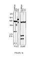
- Fig. 1b shows the results of Southern gel blotting analysis using clone pHDSll as the hybridization probe with DNA from BALB/c embryos, myeloma P3 (BALB/c derived), and CTL 2C (BALB.B) digested with PvuII and EcoRI.
- Fig. 3a shows the region of the cDNA sequence covered by the probe.
- RNA When pHDSll was used as the hybridization probe, the size of the RNA was somewhat variable from one CTL to another. CTL 2C, 1.5.2, and 2.1.1 all gave a major RNA component of about 1,300 bases while CTL G4 contained two major components of 1,400 and 1,200 bases.
- restriction maps of the pHDS58 cDNA clone (Fig. 3a) and the pHDSll cDNA clone (Fig. 3b) were constructed using standard procedures such as the ones described by T. Maniatis, E. F. Fritsch, J. Sambrook in Molecular Cloning, A Laboratory Manual, p. 3-54, 374-401 (Cold Spring Harbor Laboratory 1982).
- the DNA sequence was determined using the method of A. M. Maxam and W. Gilbert in Methods in Enzymology, L. Grossman and V. Moldave, Editors, 65:499-560 (Academic Press, N.Y. 1980).
- Fig. 4b The entire nucleotide sequence of the 1,054 base pair insert of clone pHDS11 is shown in Fig. 4b.
- the longest open reading frame is composed of an 879 nucleotide stretch whose corresponding amino acid sequence of 293 residues is also shown in Fig. 4b.
- the codons are numbered starting with the triplet GAC at nucleotide position 36-38.
- Homology between the pHDSll protein and immunoglobulin variable regions (V), particularly V k regions, suggests that Asp at position 1 is the N-terminal residue.
- the variable region is between codons 1 and 96 and the joining region from codons 97 to 109.
- the major body of the constant region is defined by codons 110 (Glu) and 236 (Cys). Codon 236 (Cys) is prior to the N-terminus of the transmembrane segment (TM) which extends from codon 256 to 277.
- the constant region pHDSll sequence is identical to the sequence of the corresponding region of the thymocyte cDNA clone, 86Tl, described by Hedrick et al., except for one base pair in codon 159.
- T H T helper cell
- the two sequences are identical throughout the constant regions and the entire 3' untranslated region except for two base pair differences at codon 159 and at nucleotide positions 992.
- the joining region from codons 97 to 109 in the pHDSll sequence is distinct from 86T1 and 2B4#71, but homologous to the joining segments of Ig genes. As shown in Fig. 6b, the pHDSll joining sequence corresponds exactly to the sequence of the J T 7 genomic segment recently identified and characterized by Chien et al.
- the pHDSll sequence between codons 1 and 96 is quite different from the corresponding region of either 86T1 or 2B4#71. There are areas of conserved residues between the three sequences, however, and between these sequences and the immunoglobulin variable regions of both heavy and light chains, in particular, the two cysteine residues involved in intradomain disulfide linkages and the Trp residue at residue 34.
- TM transmembrane
- pHDSll can encode a processed protein of 282 residues with a relative molecular mass of 33,000 daltons. There are four potential sites for N-glycosylation.
- Fig. 4a The entire nucleotide sequence of the 1,372 base pair insert of clone pHDS58 is shown in Fig. 4a.
- the longest open reading frame begins with the Met codon at nucleotide positions 56-58, extends over a stretch of 804 bp, and ends at nucleotide position 859.
- the corresponding amino acid sequence of 248 residues is also shown in Fig. 4a.
- the codons and amino acids are numbered starting with the triplet CAG (Gln) at nucleotide positions 116-118.
- the amino acid sequence immediately following the Met and extending to the Gln is highly hydrophobic and probably comprises part of a signal peptide.
- the sequence of clone pHDS58 is significantly homologous to that of pHDSll and the polypeptide chain encoded by pHDS4/203, as well as to the sequence of an immunoglobulin heavy-chain (93G7), an immunoglobulin kappa light chain (MOPC603) and an immunoglobulin lambda 1 light chain (MOPC104E).
- Figs. 5a and 6a The homology is shown in Figs. 5a and 6a, where the pHDS58 amino acid sequence is compared with the V (or V+D), J and C region sequences of the other five polypeptide chains. Sequence homology is evident in all three regions, but is most striking in the J region, where it amounts to 38-62%. In the V regions, the pHDS58 sequence is related to the other five sequences by 22-29% homology. The six amino acid residues that are conserved in this region among pHDSll, pHDS4/203 and the three types of immunoglobulin chains (Gln, Cys, Trp, Gln, Tyr and Cys at positions 5, 22, 34, 37, 88 and 90, respectively) are also shared by pHDS58.
- the sequence homology is least in the C regions, where it ranges over 12-20%. Nevertheless, the relatedness is evident around the two Cys residues that form intra-domain disulphide bonds in immunoglobulin chains.
- the pHDS58 sequence like the pHDSll and pHDS4/203 sequences, is more homologous to the light chains (25% and 27% for kappa and lambda respectively) than to the heavy chain (23%).
- the pHDS58 polypeptide chain exhibits no obvious sequence homology to the corresponding regions of pHDSll or the chain encoded by pHDS4/203. Nevertheless, the three polypeptide chains are organized in a very similar fashion in these regions.
- the C region of the pHDS58 chain is followed by a peptide carrying an extra cysteine,then by a stretch of about 20 hydrophobic residues that corresponds to a transmembrane peptide and finally by a short hydrophilic C-terminal peptide that presumably extrudes into the intracytoplasmic space.
- the proposed processed polypeptide chain is 248 amino acids long.
- the calculated relative molecular mass is approximately 28,000.
- the chain contains 23 negatively charged (15 aspartic acid and 8 glutamic acid residues) and 19 positively charged residues (6 arginine and 13 lysine residues), corresponding to an isoelectric point near neutrality in the absence of post-translation modifications.
- Fig. 4a it has four potential sites for N-glycosylation.
- Endoglycosidase F digestion of the alpha/beta heterodimers of mouse helper T cells in a human T-cell tumour by J. P. Allison suggested that both subunits of these T-cell receptors are N-glycosylated.
- N-glycosylation sites are shown by the tripeptide Asn-X-Ser/Thr. Recent studies have also shown that the alpha subunit of CTL clone 2C is N-glycosylated. As shown in Fig. 4a, the proposed N-glycosylation sites are at amino acid residues 70-72, 178-180, 192-194 and 221-223.
- variable region of the alpha subunit is between amino acids 1 and 98.
- the major body of the constant region is defined by amino acids 112 (Tyr) and 221 (Asn).
- a joining region between the variable region and the constant region extends from amino acids 99 to 111.
- Amino acid 202 (Cys) is prior to the N-terminus of the transmembrane segment (TM) which extends from amino acids 222 to 243.
- the joining region from amino acid residues 99 to 111 is homologous to the joining segments of Ig genes.
- the pHDS58 joining sequence corresponds to the sequences of the T-cell antigen receptor beta-chain described by Gascoigne et al. in Nature 310, 387-391 (1984), and immunoglobulin J H , J kappa , and J lambda described by Kabat et al. in Sequences of Proteins of Immunological Interest, (NIH, Bethesda (1983)).
- the pHDS58 sequence between amino acids 1 and 100 (variable region) and amino acids 110 and 200 (constant region) have areas of conserved residues with a chain encoded by pHDS4/203, the beta-chain encoded by pHDSll, 93G7 gamma 1 immunoglobulin heavy chain, MOPC603kappa light chain, and MOPC 104E lambda l light chain.
- TM transmembrane
- pHDS58 can encode a processed polypeptide chain of 248 amino acid residues with a relative molecular mass of approximately 28,000 daltons. There are four potential sites for N-glycosylation.
- the gene defined by cDNA clones pHDS58 is expressed and rearranged specifically in T cells. This gene and its product share several characteristics with pHDSll and pHDS4/203, and their products. These similarities may be summarized as follows.
- the proposed structure for the T cell receptor is shown in Fig. 7.
- the receptor molecule is made up of two chains, each with extracellular immunoglobulin-like domains, an amino-terminal variable domain and a carboxy-terminal constant domain. Each domain is stabilized by a disulfide bond between cysteine residues that are separated by a linear sequence of 50-70 residues.
- the molecular weights of the alpha and beta subunits are each less by about 10,000 to 15,000 daltons than the apparent molecular weight observed in SDS-polyacrylamide gel electrophoresis. It is possible this difference is due to N-glycosylation of the subunits.
- a stable association between the two subunits may be required to form an effective binding region, as is characteristic of immunoglobulins.
- the obligate participation of two different subunits in the formation of a .,ingle combining site means that combinational variability is likely to contribute to structural and functional diversity of these receptors.
- the T cell receptor is defined in the example by its amino acid sequence, nucleotide sequence of variable, joining, constant, transmembrane,and cytoplasmic segments, arrangement of disulfide bonds and glycosylation sites.
- mouse Ig C k and human Ig C k are 60% homologous
- mouse I g Clambda and human Ig C lambda are 65% homologous
- rabbit C H2 and human C H2 are 63% homologous
- rabbit CH 3 and human CH 3 are 66% homologous (Davis et al, Microbiology,,2nd Ed., p. 441 (Harper & Row Publishers, 1973) and comparing homology between the gene sequence of Hedrick et al. and Yanagi et al.
- the cDNA may be used in various systems known to those skilled in the art to make large quantities of the actual T cell receptor protein. This is of tremendous use since a major limitation on previous work was the small amount of T cell receptor available for study. Both procaryotic and eucaryotic systems are useful for production of T cell receptor protein or the alpha or beta subunits of the receptor.
- the eucaryotic system has the advantage over the procaryotic system that the expressed molecule is glycosylated.
- the T cell receptor protein or polypeptide sequences may be used for further studies, in systems for the detection of anti-T cell antibodies, and in other procedures known to those skilled in the art. They may also be used for the production of specific T cell receptor antibodies which are directed against the entire molecule, the alpha or beta subunit, or specific regions within the molecule, such as the constant region. The antibodies directed against the constant region of the cytotoxic T lymphocyte should be equally effective against the constant region of the T helper cells.
- Antibodies may be produced from the protein or its subunits using conventional techniques known to those skilled in -he art. Nucleotide sequences may also be utilized to produce short peptides or fusion proteins which are then bound to a carrier protein such as bovine gamma globulin (BTG) for injection into an animal for the production of antibodies.
- a carrier protein such as bovine gamma globulin (BTG)
- BCG bovine gamma globulin
- an animal may be immunized against the protein or peptide bound protein and immunoglobulin isolated from the serum.
- the B cells from the immunized animal with the desired specificity may also be fused with a cell line which is maintained in cell culture, such as myelomas or other tumour cell lines, to form hybridomas for the continuous production of antibody.
- Antibodies are useful in isolation procedures such as by affinity chromatography wherein antibody is bound to a solid matrix and by precipitation of soluble antibodies in solution. Antibodies are useful in analysis and identification using any of a number of well known techniques.
- a recently developed use of antibodies involves binding an agent, such as a drug, to the antibody, then injecting the bound antibody into a patient so that the drug is delivered only to the desired site.
- an agent such as a drug
- an example of such a use would be delivery of a chemotherapeutic compound to malignant T cells in a patient with a T cell lymphoma.
Landscapes
- Health & Medical Sciences (AREA)
- Immunology (AREA)
- Life Sciences & Earth Sciences (AREA)
- Chemical & Material Sciences (AREA)
- Organic Chemistry (AREA)
- Zoology (AREA)
- Medicinal Chemistry (AREA)
- Toxicology (AREA)
- Biochemistry (AREA)
- Biophysics (AREA)
- General Health & Medical Sciences (AREA)
- Genetics & Genomics (AREA)
- Gastroenterology & Hepatology (AREA)
- Molecular Biology (AREA)
- Proteomics, Peptides & Aminoacids (AREA)
- Cell Biology (AREA)
- Peptides Or Proteins (AREA)
- Preparation Of Compounds By Using Micro-Organisms (AREA)
- Micro-Organisms Or Cultivation Processes Thereof (AREA)
- Medicines That Contain Protein Lipid Enzymes And Other Medicines (AREA)
- Medicines Containing Antibodies Or Antigens For Use As Internal Diagnostic Agents (AREA)
Applications Claiming Priority (2)
| Application Number | Priority Date | Filing Date | Title |
|---|---|---|---|
| US666988 | 1984-10-31 | ||
| US06/666,988 US4873190A (en) | 1984-06-13 | 1984-10-31 | Heterodimeric T lymphocyte receptor |
Publications (2)
| Publication Number | Publication Date |
|---|---|
| EP0180878A2 true EP0180878A2 (de) | 1986-05-14 |
| EP0180878A3 EP0180878A3 (de) | 1987-05-06 |
Family
ID=24676356
Family Applications (1)
| Application Number | Title | Priority Date | Filing Date |
|---|---|---|---|
| EP85113658A Withdrawn EP0180878A3 (de) | 1984-10-31 | 1985-10-26 | Heterodimerischer Rezeptor von T-Lymphozyten |
Country Status (3)
| Country | Link |
|---|---|
| US (1) | US4873190A (de) |
| EP (1) | EP0180878A3 (de) |
| JP (1) | JPS61257998A (de) |
Cited By (19)
| Publication number | Priority date | Publication date | Assignee | Title |
|---|---|---|---|---|
| EP0174366A4 (de) * | 1984-03-01 | 1987-03-26 | Univ Leland Stanford Junior | T-zellenrezeptor, spezifisch für antigenpolypeptide und verwandte polynukleotide. |
| EP0149548A3 (en) * | 1984-01-13 | 1987-05-20 | Ontario Cancer Institute | Nucleic acid encoding a t-cell antigen receptor polypeptide |
| EP0296786A1 (de) * | 1987-06-23 | 1988-12-28 | The Board Of Trustees Of The Leland Stanford Junior University | Innerhalb des alpha-Locus gelegenes T-Zell-Rezeptor-Gen und DNA-Konstruktionen |
| EP0340793A3 (de) * | 1988-05-04 | 1991-02-27 | Yeda Research And Development Company Limited | Zellen mit ausgestattener Antikörperspezifität |
| US5223426A (en) * | 1988-12-15 | 1993-06-29 | T Cell Sciences, Inc. | Monoclonal antibodies reactive with defined regions of the t-cell antigen receptor |
| US5445940A (en) * | 1991-08-28 | 1995-08-29 | Brigham & Women's Hospital | Methods and compositions for detecting and treating a subset of human patients having an autoimmune disease |
| EP0653493A4 (de) * | 1992-04-30 | 1996-03-27 | Taisho Pharmaceutical Co Ltd | Methode zur detektion der genexpression vom t-zell antigenrezeptor. |
| WO1996015238A1 (en) * | 1994-11-16 | 1996-05-23 | Bradley Michael John Stringer | Targeted t lymphocytes |
| US5747036A (en) * | 1991-08-28 | 1998-05-05 | Brigham & Women's Hospital | Methods and compositions for detecting and treating a subset of human patients having an autoimmune disease |
| US5766947A (en) * | 1988-12-14 | 1998-06-16 | Astra Ab | Monoclonal antibodies reactive with an epitope of a Vβ3.1 variable region of a T cell receptor |
| US5843728A (en) * | 1991-03-07 | 1998-12-01 | The General Hospital Corporation | Redirection of cellular immunity by receptor chimeras |
| US5851828A (en) * | 1991-03-07 | 1998-12-22 | The General Hospital Corporation | Targeted cytolysis of HIV-infected cells by chimeric CD4 receptor-bearing cells |
| US5906936A (en) * | 1988-05-04 | 1999-05-25 | Yeda Research And Development Co. Ltd. | Endowing lymphocytes with antibody specificity |
| US5912170A (en) * | 1991-03-07 | 1999-06-15 | The General Hospital Corporation | Redirection of cellular immunity by protein-tyrosine kinase chimeras |
| US6004811A (en) * | 1991-03-07 | 1999-12-21 | The Massachussetts General Hospital | Redirection of cellular immunity by protein tyrosine kinase chimeras |
| US6753162B1 (en) | 1991-03-07 | 2004-06-22 | The General Hospital Corporation | Targeted cytolysis of HIV-infected cells by chimeric CD4 receptor-bearing cells |
| US7049136B2 (en) | 1991-03-07 | 2006-05-23 | The General Hospital Corporation | Redirection of cellular immunity by receptor chimeras |
| US7741465B1 (en) | 1992-03-18 | 2010-06-22 | Zelig Eshhar | Chimeric receptor genes and cells transformed therewith |
| US8211422B2 (en) | 1992-03-18 | 2012-07-03 | The United States Of America, As Represented By The Secretary, Department Of Health And Human Services | Chimeric receptor genes and cells transformed therewith |
Families Citing this family (11)
| Publication number | Priority date | Publication date | Assignee | Title |
|---|---|---|---|---|
| AU617980B2 (en) * | 1985-12-03 | 1991-12-12 | Astra Aktiebolag | Cell-free t cell antigen receptor and its clinical utilities |
| US5614192A (en) * | 1989-07-19 | 1997-03-25 | Connective Therapeutics, Inc. | T cell receptor peptides as therapeutics for immune-related disease |
| US5776459A (en) * | 1989-07-19 | 1998-07-07 | Connetics Corporation | TCR V beta 5 peptides |
| US5552300A (en) * | 1994-01-13 | 1996-09-03 | T Cell Sciences, Inc. | T cell antigen receptor V region proteins and methods of preparation thereof |
| US6087096A (en) * | 1995-11-13 | 2000-07-11 | Dau; Peter C. | Method of intrafamily fragment analysis of the T cell receptor α and β chain CDR3 regions |
| AU9186598A (en) | 1997-09-26 | 1999-04-23 | Kyowa Hakko Kogyo Co. Ltd. | Killer t cell receptor recognizing human immunodeficiency virus |
| US6759243B2 (en) | 1998-01-20 | 2004-07-06 | Board Of Trustees Of The University Of Illinois | High affinity TCR proteins and methods |
| US20020165149A1 (en) * | 2000-12-08 | 2002-11-07 | Kranz David M. | Mutated class II major histocompatibility proteins |
| US20080064631A1 (en) * | 2006-01-13 | 2008-03-13 | Jeffrey Molldrem | T-cell receptors for use in diagnosis and therapy of cancers and autoimmune disease |
| WO2016014725A1 (en) | 2014-07-22 | 2016-01-28 | The University Of Notre Dame Du Lac | Molecular constructs and uses thereof |
| US10556545B2 (en) | 2015-06-29 | 2020-02-11 | Royal Truck & Equipment, Inc. | Over-cab rack for traffic delineators |
Family Cites Families (3)
| Publication number | Priority date | Publication date | Assignee | Title |
|---|---|---|---|---|
| US4713332A (en) * | 1984-01-13 | 1987-12-15 | The Ontario Cancer Institute | T cell specific CDNA clone |
| ATE188251T1 (de) * | 1984-03-01 | 2000-01-15 | Univ Leland Stanford Junior | T-zellenrezeptor, spezifisch für antigenpolypeptide und verwandte polynukleotide |
| EP0200350A3 (de) * | 1985-04-15 | 1987-05-13 | The Ontario Cancer Institute | Nukleinsäure mit Code für ein T-Zellen-Antigenrezeptor-Polypeptid |
-
1984
- 1984-10-31 US US06/666,988 patent/US4873190A/en not_active Expired - Lifetime
-
1985
- 1985-10-26 EP EP85113658A patent/EP0180878A3/de not_active Withdrawn
- 1985-10-31 JP JP60243027A patent/JPS61257998A/ja active Pending
Cited By (33)
| Publication number | Priority date | Publication date | Assignee | Title |
|---|---|---|---|---|
| EP0149548A3 (en) * | 1984-01-13 | 1987-05-20 | Ontario Cancer Institute | Nucleic acid encoding a t-cell antigen receptor polypeptide |
| EP0570027A1 (de) * | 1984-03-01 | 1993-11-18 | The Board Of Trustees Of The Leland Stanford Junior University | T-Zellenrezeptor, spezifisch für Antigenpolypeptide und verwandte Polynukleotide |
| EP0609901A1 (de) * | 1984-03-01 | 1994-08-10 | The Board Of Trustees Of The Leland Stanford Junior University | T-Zellenrezeptor, spezifisch für Antigenpolypeptide und verwandte Polynukleotiden |
| EP0174366A4 (de) * | 1984-03-01 | 1987-03-26 | Univ Leland Stanford Junior | T-zellenrezeptor, spezifisch für antigenpolypeptide und verwandte polynukleotide. |
| US6180104B1 (en) | 1984-03-01 | 2001-01-30 | The Board Of Trustees Of The Leland Stanford Junior University | T cell receptor beta subunit |
| EP0712928A3 (de) * | 1987-06-23 | 1996-06-05 | Univ Leland Stanford Junior | |
| EP0296786A1 (de) * | 1987-06-23 | 1988-12-28 | The Board Of Trustees Of The Leland Stanford Junior University | Innerhalb des alpha-Locus gelegenes T-Zell-Rezeptor-Gen und DNA-Konstruktionen |
| EP0340793A3 (de) * | 1988-05-04 | 1991-02-27 | Yeda Research And Development Company Limited | Zellen mit ausgestattener Antikörperspezifität |
| US5906936A (en) * | 1988-05-04 | 1999-05-25 | Yeda Research And Development Co. Ltd. | Endowing lymphocytes with antibody specificity |
| US5912172A (en) * | 1988-05-04 | 1999-06-15 | Yeda Research And Development Co. Ltd. | Endowing lymphocytes with antibody specificity |
| US5766947A (en) * | 1988-12-14 | 1998-06-16 | Astra Ab | Monoclonal antibodies reactive with an epitope of a Vβ3.1 variable region of a T cell receptor |
| US6048526A (en) * | 1988-12-15 | 2000-04-11 | Astra Ab | Monoclonal antibodies reactive with defined regions of the T cell antigen receptor |
| US6171799B1 (en) | 1988-12-15 | 2001-01-09 | Astra Ab | Monoclonal antibodies reactive with defined regions of the T cell antigen receptor |
| US5980892A (en) * | 1988-12-15 | 1999-11-09 | Astra Ab | Monoclonal antibodies reactive with defined regions of the T cell antigen receptor |
| US5976533A (en) * | 1988-12-15 | 1999-11-02 | Astra Ab | Monoclonal antibodies reactive with defined regions of the T cell antigen receptor |
| US5223426A (en) * | 1988-12-15 | 1993-06-29 | T Cell Sciences, Inc. | Monoclonal antibodies reactive with defined regions of the t-cell antigen receptor |
| US6284240B1 (en) | 1991-03-07 | 2001-09-04 | The General Hospital Corporation | Targeted cytolysis of HIV-infected cells by chimeric CD4 receptor-bearing cells |
| US7094599B2 (en) | 1991-03-07 | 2006-08-22 | The General Hospital Corporation | Targeted cytolysis of HIV-infected cells by chimeric CD4 receptor-bearing cells |
| US5851828A (en) * | 1991-03-07 | 1998-12-22 | The General Hospital Corporation | Targeted cytolysis of HIV-infected cells by chimeric CD4 receptor-bearing cells |
| US5843728A (en) * | 1991-03-07 | 1998-12-01 | The General Hospital Corporation | Redirection of cellular immunity by receptor chimeras |
| US6004811A (en) * | 1991-03-07 | 1999-12-21 | The Massachussetts General Hospital | Redirection of cellular immunity by protein tyrosine kinase chimeras |
| US7320787B2 (en) | 1991-03-07 | 2008-01-22 | The General Hospital Corporation | Redirection of cellular immunity by protein tyrosine kinase chimeras |
| US5912170A (en) * | 1991-03-07 | 1999-06-15 | The General Hospital Corporation | Redirection of cellular immunity by protein-tyrosine kinase chimeras |
| US7049136B2 (en) | 1991-03-07 | 2006-05-23 | The General Hospital Corporation | Redirection of cellular immunity by receptor chimeras |
| US6753162B1 (en) | 1991-03-07 | 2004-06-22 | The General Hospital Corporation | Targeted cytolysis of HIV-infected cells by chimeric CD4 receptor-bearing cells |
| US6392013B1 (en) | 1991-03-07 | 2002-05-21 | The General Hospital Corporation | Redirection of cellular immunity by protein tyrosine kinase chimeras |
| US6410014B1 (en) | 1991-03-07 | 2002-06-25 | The General Hospital Corporation | Redirection of cellular immunity by protein-tyrosine kinase chimeras |
| US5445940A (en) * | 1991-08-28 | 1995-08-29 | Brigham & Women's Hospital | Methods and compositions for detecting and treating a subset of human patients having an autoimmune disease |
| US5747036A (en) * | 1991-08-28 | 1998-05-05 | Brigham & Women's Hospital | Methods and compositions for detecting and treating a subset of human patients having an autoimmune disease |
| US7741465B1 (en) | 1992-03-18 | 2010-06-22 | Zelig Eshhar | Chimeric receptor genes and cells transformed therewith |
| US8211422B2 (en) | 1992-03-18 | 2012-07-03 | The United States Of America, As Represented By The Secretary, Department Of Health And Human Services | Chimeric receptor genes and cells transformed therewith |
| EP0653493A4 (de) * | 1992-04-30 | 1996-03-27 | Taisho Pharmaceutical Co Ltd | Methode zur detektion der genexpression vom t-zell antigenrezeptor. |
| WO1996015238A1 (en) * | 1994-11-16 | 1996-05-23 | Bradley Michael John Stringer | Targeted t lymphocytes |
Also Published As
| Publication number | Publication date |
|---|---|
| EP0180878A3 (de) | 1987-05-06 |
| JPS61257998A (ja) | 1986-11-15 |
| US4873190A (en) | 1989-10-10 |
Similar Documents
| Publication | Publication Date | Title |
|---|---|---|
| US5189147A (en) | Meterodimeric T lymphocyte receptor antibody | |
| US4873190A (en) | Heterodimeric T lymphocyte receptor | |
| Saito et al. | A third rearranged and expressed gene in a clone of cytotoxic T lymphocytes | |
| US4874845A (en) | T lymphocyte receptor subunit | |
| Samelson et al. | Identification of the components of the murine T cell antigen receptor complex | |
| Born et al. | Peptide sequences of T-cell receptor δ and γ chains are identical to predicted X and γ proteins | |
| Saito et al. | Complete primary structure of a heterodimeric T-cell receptor deduced from cDNA sequences | |
| Loh et al. | Identification and sequence of a fourth human T cell antigen receptor chain | |
| Siu et al. | The human T cell antigen receptor is encoded by variable, diversity, and joining gene segments that rearrange to generate a complete V gene | |
| Kikutani et al. | Molecular structure of human lymphocyte receptor for immunoglobulin E | |
| Nikaido et al. | Molecular cloning of cDNA encoding human interleukin-2 receptor | |
| EP0570027B1 (de) | T-Zellenrezeptor, spezifisch für Antigenpolypeptide und verwandte Polynukleotide | |
| Williams et al. | Structural diversity in domains of the immunoglobulin superfamily | |
| Littman et al. | The isolation and sequence of the gene encoding T8: a molecule defining functional classes of T lymphocytes | |
| Hata et al. | Identification of putative human T cell receptor δ complementary DNA clones | |
| Hayday et al. | Structure, organization, and somatic rearrangement of T cell gamma genes | |
| US5216132A (en) | Soluble t-cell antigen receptor chimeric antigens | |
| CA2021594C (en) | Antibody to human interleukin-6 receptor | |
| Bonyhadi et al. | Delta is the C x-gene product in the γ/δ antigen receptor of dendritic epidermal cells | |
| Sewell et al. | The murine homologue of the T lymphocyte CD2 antigen: molecular cloning, chromosome assignment and cell surface expression | |
| Samelson et al. | Characterization of an anti-peptide antibody that recognizes the murine analogue of the human T cell antigen receptor-T3 delta-chain. | |
| Leiden et al. | The complete primary structure of the T-cell receptor genes from an alloreactive cytotoxic human T-lymphocyte clone | |
| Gebhard et al. | Identification of canine T-lymphocyte subsets with monoclonal antibodies | |
| Costa et al. | Functional reconstitution of an immunoglobulin antigen receptor in T cells. | |
| US4970296A (en) | Heterodimeric T lymphocyte receptor |
Legal Events
| Date | Code | Title | Description |
|---|---|---|---|
| PUAI | Public reference made under article 153(3) epc to a published international application that has entered the european phase |
Free format text: ORIGINAL CODE: 0009012 |
|
| AK | Designated contracting states |
Kind code of ref document: A2 Designated state(s): AT BE CH DE FR GB IT LI LU NL SE |
|
| PUAL | Search report despatched |
Free format text: ORIGINAL CODE: 0009013 |
|
| AK | Designated contracting states |
Kind code of ref document: A3 Designated state(s): AT BE CH DE FR GB IT LI LU NL SE |
|
| STAA | Information on the status of an ep patent application or granted ep patent |
Free format text: STATUS: THE APPLICATION IS DEEMED TO BE WITHDRAWN |
|
| 18D | Application deemed to be withdrawn |
Effective date: 19871107 |
|
| RIN1 | Information on inventor provided before grant (corrected) |
Inventor name: SAITO, HARUO Inventor name: EISEN, HERMAN N. Inventor name: TONEGAWA, SUSUMU Inventor name: KRANZ, DAVID M. |