EP0183780B1 - Procede de differenciation de sous-ensembles de cellules humaines - Google Patents
Procede de differenciation de sous-ensembles de cellules humaines Download PDFInfo
- Publication number
- EP0183780B1 EP0183780B1 EP85902799A EP85902799A EP0183780B1 EP 0183780 B1 EP0183780 B1 EP 0183780B1 EP 85902799 A EP85902799 A EP 85902799A EP 85902799 A EP85902799 A EP 85902799A EP 0183780 B1 EP0183780 B1 EP 0183780B1
- Authority
- EP
- European Patent Office
- Prior art keywords
- cells
- human
- monoclonal antibody
- subset
- reactivity
- Prior art date
- Legal status (The legal status is an assumption and is not a legal conclusion. Google has not performed a legal analysis and makes no representation as to the accuracy of the status listed.)
- Expired - Lifetime
Links
Images

Classifications
-
- C—CHEMISTRY; METALLURGY
- C07—ORGANIC CHEMISTRY
- C07K—PEPTIDES
- C07K16/00—Immunoglobulins [IG], e.g. monoclonal or polyclonal antibodies
- C07K16/18—Immunoglobulins [IG], e.g. monoclonal or polyclonal antibodies against material from animals or humans
- C07K16/28—Immunoglobulins [IG], e.g. monoclonal or polyclonal antibodies against material from animals or humans against receptors, cell surface antigens or cell surface determinants
- C07K16/289—Immunoglobulins [IG], e.g. monoclonal or polyclonal antibodies against material from animals or humans against receptors, cell surface antigens or cell surface determinants against CD45
-
- Y—GENERAL TAGGING OF NEW TECHNOLOGICAL DEVELOPMENTS; GENERAL TAGGING OF CROSS-SECTIONAL TECHNOLOGIES SPANNING OVER SEVERAL SECTIONS OF THE IPC; TECHNICAL SUBJECTS COVERED BY FORMER USPC CROSS-REFERENCE ART COLLECTIONS [XRACs] AND DIGESTS
- Y10—TECHNICAL SUBJECTS COVERED BY FORMER USPC
- Y10S—TECHNICAL SUBJECTS COVERED BY FORMER USPC CROSS-REFERENCE ART COLLECTIONS [XRACs] AND DIGESTS
- Y10S530/00—Chemistry: natural resins or derivatives; peptides or proteins; lignins or reaction products thereof
- Y10S530/868—Chemistry: natural resins or derivatives; peptides or proteins; lignins or reaction products thereof involving autoimmunity, allergy, immediate hypersensitivity, delayed hypersensitivity, immunosuppression, or immunotolerance
Definitions
- This invention relates to monoclonal antibodies.
- T4 and T8 there exists both functional and phenotypic heterogeneity; Thomas et al., J. lmmunol., 125: 2402 (1980); Morimoto et al., J. Immunol., 128; 1645 (1982); Gatenby et al., J. Exp. Med., 156: 55 (1982); and Reinherz et al., J. Immunol., 126: 67 (1981). Interaction between subpopulations of T4 and T8 cells, for example, is required to induce suppression of IgG production in antigen, pokeweed mitogen, or autologous leukocyte reaction-driven systems. Similarly, differentiation of T8 cytotoxic effectors from precytotoxic T8 lymphocytes in mixed leukocyte reactions has been shown to require the presence of T4 cells.
- the invention features a method of distinguishing subsets within a plurality of human cells, preferably T cells such as T4 cells, which method includes producing a monoclonal antibody to a non-human primate cell such as a marmoset or chimpanzee T cell, contacting the monoclonal antibody with the human cells, and distinguishing the subsets on the basis of different degrees of reactivity with the monoclonal antibody.
- T cells such as T4 cells
- the method of the invention permits the division of an otherwise apparently homogeneous population (or "set") of human cells into unique subpopulations (or “subsets).
- this dissection into subsets can be correlated with the existence of polymorphic determinants which can be used as markers of disease susceptibility, in particular autoimmune diseases such as Juvenile Rheumatoid Arthritis (JRA), Sjogren's disease, and Systemic Lupus for Erythermatosis (SLE), in which T cells are implicated.
- JRA Juvenile Rheumatoid Arthritis
- SLE Systemic Lupus for Erythermatosis
- T4 cells can be dissected into subsets, which may be functionally distinct, helps explain the great heterogeneity, in terms of clinical patterns, which exists in diseases such as JRA and SLE, but which do not correlate with simple measurements of T4 cells.
- Immunization with a non-human primate cell thus can produce a monoclonal antibody which reacts to a greater degree with a first subset of a set of human cells than with a second subset, even though the two subsets exhibit substantially the same degree of reactivity with a different monoclonal antibody which defines the set; e.g., an antibody highly reactive with T4 cells but exhibiting little reactivity with T8 cells.
- the human cells are lymphocytes, e.g., B cells or T cells such as T4 or T8 cells.
- antigenic determinants common to a human and a non-human primate cell sometimes can exhibit greater immunogenicity in rodents, e.g., mice, when presented on the non-human cell, compared to the human cell. This may be because of a comparatively greater immunodominance of some determinants on non-human primate cells, perhaps owing to the expression of the determinant on the non-human cell in a more highly antigenic configuration.
- This discovery makes possible increased production of monoclonal antibodies against important but weakly antigenic determinants on human cells, by immunizing with a non-human primate cell also bearing the determinant.
- the method of using non-human primate cells to produce a monoclonal antibody capable of dividing a set of human cells into subsets can be carried out an additional step, to produce a monoclonal antibody which is specific for one of the subsets, as follows.
- a non-human primate-derived monoclonal antibody has been used to identify two distinct subsets of, say, human T4 cells (one subset being more reactive and the other less reactive with the antibody)
- one of the identified subsets say the more reactive subset
- the monoclonal antibodies produced by these hybridomas can then be screened against the immunizing subset and the other subset.
- An antibody more reactive with the immunizing subset than with the other subset defines the polymorphic surface structure or structures which differentiate the two subsets.
- Such an antibody could be useful in the diagnosis and/or treatment of a disease, e.g., JRA, caused or exacerbated by the immunizing subset. Diagnosis could be accomplished using flow cytometry to measure reactivity of cells, with the antibody conjugated with a fluorescent dye. To treat the disease, the antibody could be chemically coupled to a cytotoxic agent and administered to a patient suffering from the disease. The antibody would specifically bind to and destroy the disease-causing cells, but not the normal cells.
- the first step in the method is to select the non-human primate whose cells are to be used for immunization.
- the choice of primate depends in part on how phylogenetically distant the primate is from humans. This phylogenetic distance is generally reflected in the reactivity of a primate's cells with monoclonal antibodies of human origin.
- Table 1 shows the reactivities of T-cells of various species with monoclonal antibodies produced by immunizing Balb/C or CAF1 mice with a variety of human T-cell subsets.
- Common marmoset T-cells are reactive with T4A and T8A, but with none of the other antibodies.
- Non-human primate T cells can be used for immunization as follows. First, the cells are isolated from heparinized blood utilizing Ficoll-Hypague and density gradient centrifugation. The marmoset cells are then treated with 0.15M NH 4 CI to lyse erythrocytes, washed, resuspended in phosphate buffered saline, and used for immunization and frozen for subsequent screening.
- Balb/C or CAF1 mice are then immunized with these cells using standard procedures.
- the splenocytes obtained are fused in PEG with P3/NS1/1-AG4-1 myeloma cells.
- Hybridoma culture supernatants reactive with immunizing cells, but unreactive with human B cells or B cell lines, are then selected, and these lines are cloned and recloned by limiting dilution in the presence of feeder cells using standard techniques.
- the initial screen is meant to identify antibodies reactive with primate T-cells and unreactive with human B lymphocytes. Subsequent screening then involves the characterization of such antibodies on large panels of human T lymphocytes, including freshly isolated T4 cells, T4JRA-TQ1+, T4JRA ⁇ TQ1-, T8 cells, T4 cytotoxic lines, T8 cytotoxic lines, T4 antigen specific inducer T cell lines, T4 antigen specific inducer of suppressor T cell lines, T8 suppressor lines, and freshly isolated activated T cells. Antibodies which are reactive with a fraction of the inducer or suppressor population but unreactive with human B lymphocytes, B cell lines, myeloid cells, and myeloid lines are isolated.
- Such antibodies can then be used to divide a T4 or T8 population into subsets, based on the degree to which cells from each subset react with the antibodies.
- Such different reactivities will indicate either the existence of polymorphic epitopes in the structure of a single surface antigen which defines, say, T4 or T8; or the existence of a family of such surface antigens which define the T4 and T8 population, which family of antigens exhibits heterogeneity.
- the polymorphism or heterogeneity can be detected using primate-derived monoclonal antibodies.
- Antibodies that react with subfractions of T4 and T8 populations of cells are characterized by indirect immunofluorescence, as follows. Approximately 10 6 cells are incubated with either hybridoma supernatants or ascites, washed at 4°C extensively, and then stained with FITC anti-mouse IgG. The fluorescent antibody-coated cells are then analyzed on a FACs I, an EPICS V, or a similar instrument, which allow for a precise quantitative assessment of the number of reactive cells.
- anti-2H4 A particular anti-primate cell monoclonal antibody, designated anti-2H4, was produced using standard techniques, as follows.
- mice (Jackson Laboratories, Bar Harbor, ME) were immunized with T lymphocyte lines from the cotton top tamarin Saquinus oedipus, an herbivorous New World primate species. Peripheral blood lymphocytes from this species were stimulated in vitro with PHA and then maintained in continuous culture with T cell growth factors. Hybridoma cultures containing antibodies reactive with human (E+) cells were selected, cloned, and recloned by limiting dilution methods in the presence of feeder cells; E+ cells are known to be capable of separating T cell derivative antibodies from non-T cell derived antibodies. Malignant ascites were then developed and utilized for analysis.
- E+ human
- the monoclonal antibody anti-2H4 was shown to be of the IgG1 isotype by specificity of straining with fluorescein-labeled goat anti-mouse IgG (Meloy Laboratories, Springfield, VA), and by its failure to be stained by fluorescein-labeled antibodies directed against other subclasses of mouse immunoglobulin.
- Human E+ lymphocytes were treated with anti-T4 or anti-T8 monoclonal antibodies and rabbit complement (C) (Pel-Freeze Biologicals). 2 x 10' cell aliquots were incubated with 1 ml of antibody at a 1:250 dilution for 1 hour at room temperature and then 0.3 ml rabbit C was added to the mixture. The mixture was incubated for another hour in a 37°C shaking water bath, washed, and residual cells cultured overnight at 37°C. After lysis of cells with anti-T4 and C, >90% of the residual cells were T8+ cells and ⁇ 5% were T4+ cells. After lysis with anti-T8 and C, >90% of the remaining cells were T4+ cells and ⁇ 5% were T8+ cells. These two populations will be referred to herein as the T8+ and T4+ sets, respectively.
- T4+ T cells To separate T4+ T cells into 2H4+ and 2H4- subpopulations, 80 x 10 6 T4+ cells which had been cultured overnight were labeled with 4 ml of 1/250 dilution of anti-2H4 and developed with fluorescein-conjugated F(ab') 2 goat anti-mouse F(ab') 2 .
- Cytofluorographic analysis of cell populations was performed by means of indirect immunofluorescence with fluorescein-conjugated F(ab') 2 goat anti-mouse (Fab') 2 on an Epics V cell sorter (Coulter Electronics). Background fluorescence reactivity was determined with control ascites obtained from mice immunized with nonsecreting hybridoma clones. For analysis, all monoclonal antibodies were utilized in antibody excess at dilutions of 1/250 to 1/1000.
- This procedure produced two subsets of T4+ cells, a subset exhibiting high reactivity with anti-2H4 (designated "2H4+”), and a subset exhibiting low reactivity with anti-2H4 ("2H4-").
- Post-sort viability was greater than 95% by trypan blue exclusion in all instances.
- Purity of separated T cell subsets was in excess of 95%.
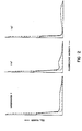
- Fig. 2 is a cytofluorographic analysis of unfractionated T, T4+, and T8+ cells with anti-2H4 monoclonal antibody, displayed in logarithmic scale.
- 2H4+ T cells were found in both T4+ and T8+ subpopulations.
- anti-2H4 antibody The reactivity of anti-2H4 antibody with other human lymphoid cells and cell lines is shown in Table 2, below.
- Anti-2H4 was found to be reactive with over 30% of both peripheral blood B cells and null cells, only slightly reactive with macrophages obtained by adherence techniques, and unreactive with thymic lymphocytes.
- Anti-2H4 was also unreactive with 3 of 4 human T cell lines tested, and weakly reactive with JM cell lines, the most mature of four T cell lines which were tested.
- the data in Table 2 also indicate that reactivity with anti-2H4 was not restricted to cells of the T lineage: four lymphoblastoid B cell lines and two Burkitt's lymphoma lines showed reactivity with anti-2H4.
- T cells were cultured in RPMI 1640 media with 10% human AB serum, 200 mM L-glutamine, 25 mM HEPES buffer (Microbiological Associates), 0.5% sodium bicarbonate and 1% penicillin-streptomycin. 10 5 cells per microculture well were tested for proliferative response to an optimal dose of phytohemagglutinin (PHA) (Burroughs-Wellcome Co., Research Triangle Park, NC) and concanavalin A (Con A) (Calbiochem, San Diego, CA). The alloantigen-driven proliferative response was measured concurrently by stimulating with mitomycin C-treated Laz 156, an Epstein-Barr virus-transformed human B lymphoid line.
- PHA phytohemagglutinin
- Con A Con A
- T4+2H4+ and T4+2H4- cell populations As shown in Table 3, below, differences in response to Con A and soluble antigens were seen in T4+2H4+ and T4+2H4- cell populations. In response to Con A, T4+2H4+ T cells incorporated significantly more H-Tdr than did the T4+2H4- population. In response to soluble antigens such as TT and mumps, T4+2H4- T cells incorporated significantly more 3 H ⁇ TdR than did the T4+2H4+ T cell population. These differences between the proliferative response of T4+2H4+ and T4+2H4- populations were significant (P ⁇ 0.05).
- T cell help for B cell immunoglobulin production was restricted to the T4+2H4+ or T4+2H4- T cell subset.
- unfractionated T4+ T cells or T4+2H4+ and T4+2H4- cells were mixed with autologous B lymphocytes, stimulated with PWM in vitro, and total IgG production was measured after 7 days in culture.
- lymphocytes were cultured in round-bottomed microtiter culture plates (Falcon) at 37°C in a humidified atmosphere with 5% C0 2 for 7 days in RPMI 1640 supplemented with 20% heat-inactivated fetal calf serum (Microbiological Associates), 0.5% sodium bicarbonate, 200 mM L-glutamine, 25 mM HEPES and 1% pencillin-streptomycin.
- T4 cells To determine the effect of various subsets of the T4 cells on secretion of IgG by autologous plasma cells, various numbers of unfractionated T4+ T cells or purified T4+2H4+ and T4+2H4- T cell subsets were added to 5 x 10 4 B cells in a volume of a 1 ml. To this was added 0.1 ml of pokeweed mitogen (PWM) (Gibco Laboratories, Grand Island Biological Co., Grand Island, NY) at a 1:50 dilution. Macrophages were added to all populations at a 5% final concentration at the initiation of in vitro cultures.
- PWM pokeweed mitogen
- T4+2H4+ and T4+2H4- cells were added to separate cultures of autologous B cells, the IgG secretion induced by the T4+2H4- T cell subset was approximately 10 times greater than that obtained with the combination of T4+2H4+ and B cell (27500 ⁇ 1800 ng vs. 2400 ⁇ 120 ng). Furthermore, a quantitative comparison of the helper function provided by T4+2H4+ and T4+2H4- T cells for B cell IgG production (Table 4, below) showed that the helper effect of T4+2H4- T cells was strikingly greater that that of T4+2H4+ T cells at any number of T cells and B cells tested.
- helper activity for antibody production in response to PWM by B cells was found within the T4+2H4- subset of cells, and the T4+2H4+ and minimal helper effect in this interaction. Effect of T4+2H4- cells on the generation of suppressor effector cells
- T4+2H4+ or T4+2H4- cells were added to a constant number of B cells (5 x 10 4 ), T4+2H4- or T4+2H4+ T cells (2 x 10 4 ), and T8 cells (1 x 10°) in the presence of PWM.
- Tables 5 and 6, below when increasing numbers of T4+2H4+ cells were added to a constant number of B cells, T4+2H4- cells, and T8 cells, increasing suppression of IgG production was observed (4800 ng vs 300 ng, 32000 ng vs 5000 ng, 24000 ng vs 4800 ng). In contrast, the addition of increasing numbers of T4+2H4+ T cells resulted in enhanced IgG production.
- T4+2H4+ T cells are themselves suppressor effector cells
- varying numbers of T4+2H4+ or T4+2H4- cells were added to a constant number of B cells (5 x 10 4 ) and T4+2H4+ or T4+2H4- cells (2 x 10 4 ) in the presence or absence of T8+ cells (1 x 10 4 ) with PWM.
- Table 7 shows that when increasing numbers of T4+2H4+ T cells were added to the mixture of B cells and T4+2H4- cells including T8 cells, marked suppression was seen, as previously shown in Table 6.
- T4+2H4- cells were not themselves effectors of suppression, rather they induced or activated T8+ cells to suppress the immune response.
- Hybridoma cells producing anti-2H4 antibody have been deposited in the American Type Culture Collection, Rockville, Maryland, and given ATCC Accession No. HB 8570.
Landscapes
- Health & Medical Sciences (AREA)
- Chemical & Material Sciences (AREA)
- Immunology (AREA)
- Organic Chemistry (AREA)
- Medicinal Chemistry (AREA)
- Biophysics (AREA)
- General Health & Medical Sciences (AREA)
- Genetics & Genomics (AREA)
- Biochemistry (AREA)
- Molecular Biology (AREA)
- Proteomics, Peptides & Aminoacids (AREA)
- Life Sciences & Earth Sciences (AREA)
- Preparation Of Compounds By Using Micro-Organisms (AREA)
- Micro-Organisms Or Cultivation Processes Thereof (AREA)
- Medicines Containing Antibodies Or Antigens For Use As Internal Diagnostic Agents (AREA)
- Peptides Or Proteins (AREA)
- Investigating Or Analysing Biological Materials (AREA)
Abstract
Claims (15)
Applications Claiming Priority (2)
| Application Number | Priority Date | Filing Date | Title |
|---|---|---|---|
| US06/616,284 US4649106A (en) | 1984-06-01 | 1984-06-01 | Distinguishing subsets of human cells |
| US616284 | 1984-06-01 |
Publications (3)
| Publication Number | Publication Date |
|---|---|
| EP0183780A1 EP0183780A1 (fr) | 1986-06-11 |
| EP0183780A4 EP0183780A4 (fr) | 1988-11-02 |
| EP0183780B1 true EP0183780B1 (fr) | 1991-01-23 |
Family
ID=24468783
Family Applications (1)
| Application Number | Title | Priority Date | Filing Date |
|---|---|---|---|
| EP85902799A Expired - Lifetime EP0183780B1 (fr) | 1984-06-01 | 1985-05-10 | Procede de differenciation de sous-ensembles de cellules humaines |
Country Status (6)
| Country | Link |
|---|---|
| US (1) | US4649106A (fr) |
| EP (1) | EP0183780B1 (fr) |
| JP (1) | JPS61502233A (fr) |
| CA (1) | CA1245582A (fr) |
| DE (1) | DE3581505D1 (fr) |
| WO (1) | WO1985005639A1 (fr) |
Families Citing this family (4)
| Publication number | Priority date | Publication date | Assignee | Title |
|---|---|---|---|---|
| US4661446A (en) * | 1985-02-19 | 1987-04-28 | Dana-Farber Cancer Institute, Inc. | Monoclonal antibodies and method of immunizing therewith |
| US5256532A (en) * | 1988-05-02 | 1993-10-26 | Zynaxis Technologies, Inc. | Methods, reagents and test kits for determination of subpopulations of biological entities |
| US6287793B1 (en) | 1988-08-19 | 2001-09-11 | Elan Pharmaceuticals, Inc. | Diagnostic methods for alzheimer's disease |
| US5059518A (en) * | 1988-10-20 | 1991-10-22 | Coulter Corporation | Stabilized lyophilized mammalian cells and method of making same |
Family Cites Families (6)
| Publication number | Priority date | Publication date | Assignee | Title |
|---|---|---|---|---|
| US4363799A (en) * | 1979-03-20 | 1982-12-14 | Ortho Pharmaceutical Corporation | Monoclonal antibody to human T cells, and methods for preparing same |
| US4364932A (en) * | 1979-09-18 | 1982-12-21 | Ortho Pharmaceutical Corporation | Monoclonal antibody to human cytotoxic and suppressor T cells and methods of preparing same |
| US4364937A (en) * | 1980-01-08 | 1982-12-21 | Ortho Pharmaceutical Corporation | Monoclonal antibody to a human T cell antigen and methods of preparing same |
| US4381292A (en) * | 1980-11-14 | 1983-04-26 | The Board Of Trustees Of The Leland Stanford Jr. University | Anti-human T-lymphocyte monoclonal antibody |
| US4474893A (en) * | 1981-07-01 | 1984-10-02 | The University of Texas System Cancer Center | Recombinant monoclonal antibodies |
| US4511662A (en) * | 1982-06-18 | 1985-04-16 | Bio-Rad Laboratories, Inc. | Simultaneous assay for T and B lymphocyte populations and subpopulations |
-
1984
- 1984-06-01 US US06/616,284 patent/US4649106A/en not_active Expired - Fee Related
-
1985
- 1985-05-10 JP JP60502405A patent/JPS61502233A/ja active Pending
- 1985-05-10 EP EP85902799A patent/EP0183780B1/fr not_active Expired - Lifetime
- 1985-05-10 DE DE8585902799T patent/DE3581505D1/de not_active Expired - Lifetime
- 1985-05-10 WO PCT/US1985/000885 patent/WO1985005639A1/fr not_active Ceased
- 1985-05-31 CA CA000482884A patent/CA1245582A/fr not_active Expired
Non-Patent Citations (16)
| Title |
|---|
| N, Fed, Proc., Vol. 80, issued 1981, L.N. Martin et al., Abstract No. 4334, "Hybridomas in Studies of Nonhuman Primates" * |
| N, Immunogenetics, Vol. 11, No. 1, issued 1980, P. Parham et al., "Molecular Characterization of HLA-A, B. Homologues in Owl Monkeys...", pages 131-143 * |
| N, Immunology, Vol. 44, issued 1981, A.R. Sanderson et al., "Epitope Sites on Human Beta 2-Microglobulin...", pages 169-175. * |
| N, J, Immunogenetics, Vol. 8, issued 1981, J.J. Marchalonis et al., "A Marmoset T-Lymhocyte Protein...", pages 165-175 * |
| N, J. Clin., Invest., Vol. 67, issued March 1981, C. Morimoto et al., "Autoantibody to an Immunoregulatory Inducer Population in Patients with Juvenil Rheumatoid Arthritis, pages 753-761 * |
| N, J. Exp. Med., Vol. 156, issued 1982, P.A. Gatenby et al., "Immunoglobulin Secretion in the Human Autologous Mixed Leukocyte Reaction, pages 55-67 * |
| N, J. Immunol., Vol. 120, issued 1978, A.M. Dvorak et al., "Immunologic Rejection of Mammary Adenocarcinoma...", pages 1240-1248. * |
| N, J. Immunol., Vol. 125, issued 1980, Y. Thomas et al., "Functional Analysis of Human T. cell, Subsets Defined by Monoclonal Antibodies, pages 2402-2408 * |
| N, J. Immunol., Vol. 126, issued 1981, E.L. Reinherz et al., "Sub- populations of the T4 Inducer T Cell Subset in Man...", pages 67-70 * |
| N, J. Immunol., Vol. 128, No. 1, issued January 1982, E. L. Reinherz et al., "Heterogeneity of Human T4 Inducer T Cells...", pages 463-468 * |
| N, Proc. Natl. Acad. Sci. USA, Vol. 77, No. 6, J.J. Marchalonis et al., "Surface Component of Primate Thymus-derived Lymphocytes..." pages 3625-3629, issued June 1980 * |
| N, Science, Vol. 215, issued 15 January 1982, B.F. Haynes et al., "Human T Cell Antigen Expression by Primate T Cells", Pages 298-300 * |
| N, Vet. Immunol. and Immunopath., Vol. 2, issued 1981, L.R. Ellingsworth et al.,"Distribution of Helper and Suppressor/Cytotoxic T-Lymphocytes..", pages 541-553 * |
| N,Cell, Vol.19, issued April 1980, E.L. Reinherz et al., "The Differentiation and Function of Human T Lymphocytes" pages 821-827 * |
| N,Febs Letters, Vol. 146, No. 1, issued September 1982, C.E. Rogers et al., "Differentiation of Immunochemically Related Enzymes in Different Primate Species by Monoclonal Antibodies", pages 93-96 * |
| N,J. Immunol., Vol. 129, No.1, issued July 1982, A. Yachie et al., "Ia-Positive Cells Generated by PWM-Stimulation within OKT4 Subset..." pages 103-107 * |
Also Published As
| Publication number | Publication date |
|---|---|
| DE3581505D1 (de) | 1991-02-28 |
| CA1245582A (fr) | 1988-11-29 |
| JPS61502233A (ja) | 1986-10-09 |
| WO1985005639A1 (fr) | 1985-12-19 |
| EP0183780A1 (fr) | 1986-06-11 |
| EP0183780A4 (fr) | 1988-11-02 |
| US4649106A (en) | 1987-03-10 |
Similar Documents
| Publication | Publication Date | Title |
|---|---|---|
| Morimoto et al. | The isolation and characterization of the human suppressor inducer T cell subset. | |
| Morimoto et al. | The isolation and characterization of the human helper inducer T cell subset. | |
| Hansen et al. | Monoclonal antibodies identifying a novel T-cell antigen and Ia antigens of human lymphocytes | |
| EP0030815B1 (fr) | Lignée de cellules hybrides pour la préparation d'anticorps monoclonaux contre un antigène prothymocyte humain; anticorps; procédé pour préparer cet anticorps, applications diagnostiques et thérapeutiques et compositions pharmaceutiques contenant cet anticorps | |
| US4381295A (en) | Monoclonal antibody to human helper T cells and methods of preparing same | |
| EP0030450B1 (fr) | Lignée de cellules hybrides pour la préparation d'anticorps monoclonaux fixant le complément contre les cellules suppresseur T humaines; anticorps; procédé pour préparer cet anticorps; applications diagnostiques et thérapeutiques et compositions pharmaceutiques contenant cet anticorps | |
| EP0033578B1 (fr) | Lignée de cellules hybrides pour la préparation d'anticorps monoclonaux contre un antigène T humain, anticorps et procédés | |
| van Heyningen et al. | Human MHC class II molecules as differentiation markers | |
| EP0030814B1 (fr) | Lignée de cellules hybrides pour la préparation d'anticorps monoclonaux contre un antigène thymocyte précoce humain; anticorps; procédé pour préparer cet anticorps; applications diagnostiques et thérapeutiques et compositions pharmaceutiques contenant cet anticorps | |
| EP0025722B1 (fr) | Anticorps monoclonal contre les cellules T humaines cytotoxiques et suppressives et son procédé de préparation | |
| AU642364B2 (en) | Monoclonal antibody which distinguishes helper inducer and supressor inducer CE4+ lymphocytes | |
| EP0030449B1 (fr) | Lignée de cellules hybrides pour la préparation d'anticorps monoclonaux contre un antigène thymocyte humain, anticorps, procédé pour préparer cet anticorps, applications diagnostiques et thérapeutiques et compositions pharmaceutiques contenant cet anticorps | |
| EP0218587B1 (fr) | Anticorps monoclonaux et procede d'immunisation les utilisant | |
| EP0183780B1 (fr) | Procede de differenciation de sous-ensembles de cellules humaines | |
| US4816404A (en) | Late differentiation antigens associated with helper T lymphocyte function | |
| US4709015A (en) | Monoclonal antibody to human suppressor T cells | |
| Silvian et al. | Lymphocyte subpopulations in benign monoclonal gammopathy | |
| Letvin et al. | Definition of the T-lymphocyte inducer of suppression in primates using a monoclonal antibody | |
| US4691010A (en) | Hybrid cell line for producing monoclonal antibody to a human early thymocyte antigen, antibody, and methods | |
| US4725543A (en) | Hybrid cell line for producing complement-fixing monoclonal antibody to human suppressor T cells, antibody and methods | |
| US4806349A (en) | Hybrid cell line for producing monoclonal antibody to a human prothymocyte antigen, antibody, and methods | |
| US4806629A (en) | Monoclonal antibody to a human thymocyte antigen | |
| US4828995A (en) | Hybrid cell line for producing monoclonal antibody to a human thymocyte antigen, antibody, and methods | |
| JPS6363556B2 (fr) | ||
| JPH01466A (ja) | 急性伝染性単核症を検出する方法 |
Legal Events
| Date | Code | Title | Description |
|---|---|---|---|
| PUAI | Public reference made under article 153(3) epc to a published international application that has entered the european phase |
Free format text: ORIGINAL CODE: 0009012 |
|
| 17P | Request for examination filed |
Effective date: 19860204 |
|
| AK | Designated contracting states |
Kind code of ref document: A1 Designated state(s): CH DE FR GB IT LI |
|
| A4 | Supplementary search report drawn up and despatched |
Effective date: 19881102 |
|
| 17Q | First examination report despatched |
Effective date: 19890830 |
|
| GRAA | (expected) grant |
Free format text: ORIGINAL CODE: 0009210 |
|
| ITF | It: translation for a ep patent filed | ||
| AK | Designated contracting states |
Kind code of ref document: B1 Designated state(s): CH DE FR GB IT LI |
|
| REF | Corresponds to: |
Ref document number: 3581505 Country of ref document: DE Date of ref document: 19910228 |
|
| ET | Fr: translation filed | ||
| PLBE | No opposition filed within time limit |
Free format text: ORIGINAL CODE: 0009261 |
|
| STAA | Information on the status of an ep patent application or granted ep patent |
Free format text: STATUS: NO OPPOSITION FILED WITHIN TIME LIMIT |
|
| 26N | No opposition filed | ||
| PGFP | Annual fee paid to national office [announced via postgrant information from national office to epo] |
Ref country code: FR Payment date: 19930409 Year of fee payment: 9 |
|
| PGFP | Annual fee paid to national office [announced via postgrant information from national office to epo] |
Ref country code: DE Payment date: 19930414 Year of fee payment: 9 Ref country code: CH Payment date: 19930414 Year of fee payment: 9 |
|
| PGFP | Annual fee paid to national office [announced via postgrant information from national office to epo] |
Ref country code: GB Payment date: 19930430 Year of fee payment: 9 |
|
| PG25 | Lapsed in a contracting state [announced via postgrant information from national office to epo] |
Ref country code: GB Effective date: 19940510 |
|
| PG25 | Lapsed in a contracting state [announced via postgrant information from national office to epo] |
Ref country code: LI Effective date: 19940531 Ref country code: CH Effective date: 19940531 |
|
| GBPC | Gb: european patent ceased through non-payment of renewal fee |
Effective date: 19940510 |
|
| PG25 | Lapsed in a contracting state [announced via postgrant information from national office to epo] |
Ref country code: FR Effective date: 19950131 |
|
| REG | Reference to a national code |
Ref country code: CH Ref legal event code: PL |
|
| PG25 | Lapsed in a contracting state [announced via postgrant information from national office to epo] |
Ref country code: DE Effective date: 19950201 |
|
| REG | Reference to a national code |
Ref country code: FR Ref legal event code: ST |