EP0199261A2 - Immunologischer Testkit - Google Patents
Immunologischer Testkit Download PDFInfo
- Publication number
- EP0199261A2 EP0199261A2 EP86105184A EP86105184A EP0199261A2 EP 0199261 A2 EP0199261 A2 EP 0199261A2 EP 86105184 A EP86105184 A EP 86105184A EP 86105184 A EP86105184 A EP 86105184A EP 0199261 A2 EP0199261 A2 EP 0199261A2
- Authority
- EP
- European Patent Office
- Prior art keywords
- test kit
- collagen
- kit according
- enzyme
- antigen
- Prior art date
- Legal status (The legal status is an assumption and is not a legal conclusion. Google has not performed a legal analysis and makes no representation as to the accuracy of the status listed.)
- Withdrawn
Links
- 230000001900 immune effect Effects 0.000 title claims abstract description 10
- 102000008186 Collagen Human genes 0.000 claims abstract description 41
- 108010035532 Collagen Proteins 0.000 claims abstract description 41
- 229920001436 collagen Polymers 0.000 claims abstract description 40
- 239000000427 antigen Substances 0.000 claims abstract description 21
- 102000036639 antigens Human genes 0.000 claims abstract description 21
- 108091007433 antigens Proteins 0.000 claims abstract description 21
- 239000000758 substrate Substances 0.000 claims abstract description 15
- 102000004190 Enzymes Human genes 0.000 claims abstract description 10
- 108090000790 Enzymes Proteins 0.000 claims abstract description 10
- 239000003153 chemical reaction reagent Substances 0.000 claims abstract description 4
- 230000015572 biosynthetic process Effects 0.000 claims abstract description 3
- 239000000872 buffer Substances 0.000 claims description 18
- 239000000975 dye Substances 0.000 claims description 18
- 238000001514 detection method Methods 0.000 claims description 16
- 210000002966 serum Anatomy 0.000 claims description 15
- 239000000243 solution Substances 0.000 claims description 15
- 239000000020 Nitrocellulose Substances 0.000 claims description 9
- 229920001220 nitrocellulos Polymers 0.000 claims description 9
- 238000010790 dilution Methods 0.000 claims description 8
- 239000012895 dilution Substances 0.000 claims description 8
- 239000013642 negative control Substances 0.000 claims description 6
- 238000005406 washing Methods 0.000 claims description 6
- 239000013641 positive control Substances 0.000 claims description 5
- 210000001179 synovial fluid Anatomy 0.000 claims description 5
- 102000002260 Alkaline Phosphatase Human genes 0.000 claims description 4
- 108020004774 Alkaline Phosphatase Proteins 0.000 claims description 4
- 244000309466 calf Species 0.000 claims description 4
- 230000001376 precipitating effect Effects 0.000 claims description 3
- 239000000126 substance Substances 0.000 claims description 3
- 208000025747 Rheumatic disease Diseases 0.000 abstract description 6
- 238000003745 diagnosis Methods 0.000 abstract description 3
- FAPWRFPIFSIZLT-UHFFFAOYSA-M Sodium chloride Chemical compound [Na+].[Cl-] FAPWRFPIFSIZLT-UHFFFAOYSA-M 0.000 description 17
- 102000004169 proteins and genes Human genes 0.000 description 15
- 108090000623 proteins and genes Proteins 0.000 description 15
- 238000000034 method Methods 0.000 description 14
- 238000002965 ELISA Methods 0.000 description 13
- 238000006243 chemical reaction Methods 0.000 description 13
- 238000003556 assay Methods 0.000 description 8
- 239000011780 sodium chloride Substances 0.000 description 7
- 108091003079 Bovine Serum Albumin Proteins 0.000 description 6
- 238000011534 incubation Methods 0.000 description 6
- 239000011534 wash buffer Substances 0.000 description 6
- QKNYBSVHEMOAJP-UHFFFAOYSA-N 2-amino-2-(hydroxymethyl)propane-1,3-diol;hydron;chloride Chemical compound Cl.OCC(N)(CO)CO QKNYBSVHEMOAJP-UHFFFAOYSA-N 0.000 description 5
- 229940098773 bovine serum albumin Drugs 0.000 description 5
- 201000010099 disease Diseases 0.000 description 5
- 208000037265 diseases, disorders, signs and symptoms Diseases 0.000 description 5
- 239000011734 sodium Substances 0.000 description 5
- 108060003951 Immunoglobulin Proteins 0.000 description 4
- 239000000969 carrier Substances 0.000 description 4
- 229940127121 immunoconjugate Drugs 0.000 description 4
- 102000018358 immunoglobulin Human genes 0.000 description 4
- 230000000552 rheumatic effect Effects 0.000 description 4
- 206010036030 Polyarthritis Diseases 0.000 description 3
- 229920001213 Polysorbate 20 Polymers 0.000 description 3
- 238000002474 experimental method Methods 0.000 description 3
- 239000000463 material Substances 0.000 description 3
- 239000000203 mixture Substances 0.000 description 3
- 235000010486 polyoxyethylene sorbitan monolaurate Nutrition 0.000 description 3
- 239000000256 polyoxyethylene sorbitan monolaurate Substances 0.000 description 3
- QRXMUCSWCMTJGU-UHFFFAOYSA-L (5-bromo-4-chloro-1h-indol-3-yl) phosphate Chemical compound C1=C(Br)C(Cl)=C2C(OP([O-])(=O)[O-])=CNC2=C1 QRXMUCSWCMTJGU-UHFFFAOYSA-L 0.000 description 2
- XZKIHKMTEMTJQX-UHFFFAOYSA-N 4-Nitrophenyl Phosphate Chemical compound OP(O)(=O)OC1=CC=C([N+]([O-])=O)C=C1 XZKIHKMTEMTJQX-UHFFFAOYSA-N 0.000 description 2
- 108010001336 Horseradish Peroxidase Proteins 0.000 description 2
- 239000004793 Polystyrene Substances 0.000 description 2
- CDBYLPFSWZWCQE-UHFFFAOYSA-L Sodium Carbonate Chemical compound [Na+].[Na+].[O-]C([O-])=O CDBYLPFSWZWCQE-UHFFFAOYSA-L 0.000 description 2
- UIIMBOGNXHQVGW-UHFFFAOYSA-M Sodium bicarbonate Chemical compound [Na+].OC([O-])=O UIIMBOGNXHQVGW-UHFFFAOYSA-M 0.000 description 2
- 239000012876 carrier material Substances 0.000 description 2
- ZBCBWPMODOFKDW-UHFFFAOYSA-N diethanolamine Chemical compound OCCNCCO ZBCBWPMODOFKDW-UHFFFAOYSA-N 0.000 description 2
- 229940072221 immunoglobulins Drugs 0.000 description 2
- JPXMTWWFLBLUCD-UHFFFAOYSA-N nitro blue tetrazolium(2+) Chemical compound COC1=CC(C=2C=C(OC)C(=CC=2)[N+]=2N(N=C(N=2)C=2C=CC=CC=2)C=2C=CC(=CC=2)[N+]([O-])=O)=CC=C1[N+]1=NC(C=2C=CC=CC=2)=NN1C1=CC=C([N+]([O-])=O)C=C1 JPXMTWWFLBLUCD-UHFFFAOYSA-N 0.000 description 2
- 229920002223 polystyrene Polymers 0.000 description 2
- 238000004445 quantitative analysis Methods 0.000 description 2
- 238000010186 staining Methods 0.000 description 2
- UAIUNKRWKOVEES-UHFFFAOYSA-N 3,3',5,5'-tetramethylbenzidine Chemical compound CC1=C(N)C(C)=CC(C=2C=C(C)C(N)=C(C)C=2)=C1 UAIUNKRWKOVEES-UHFFFAOYSA-N 0.000 description 1
- WJWMRPDAOIYYRX-UHFFFAOYSA-N 4-(4-diazoniophenyl)sulfanylbenzenediazonium Chemical compound C1=CC([N+]#N)=CC=C1SC1=CC=C([N+]#N)C=C1 WJWMRPDAOIYYRX-UHFFFAOYSA-N 0.000 description 1
- 206010002556 Ankylosing Spondylitis Diseases 0.000 description 1
- 108010074051 C-Reactive Protein Proteins 0.000 description 1
- 102100032752 C-reactive protein Human genes 0.000 description 1
- 102000014914 Carrier Proteins Human genes 0.000 description 1
- 108010078791 Carrier Proteins Proteins 0.000 description 1
- -1 Diazobenzyloxymethyl Chemical group 0.000 description 1
- 108010010803 Gelatin Proteins 0.000 description 1
- 102000006947 Histones Human genes 0.000 description 1
- 108010033040 Histones Proteins 0.000 description 1
- 206010039710 Scleroderma Diseases 0.000 description 1
- VMHLLURERBWHNL-UHFFFAOYSA-M Sodium acetate Chemical compound [Na+].CC([O-])=O VMHLLURERBWHNL-UHFFFAOYSA-M 0.000 description 1
- 239000013504 Triton X-100 Substances 0.000 description 1
- 229920004890 Triton X-100 Polymers 0.000 description 1
- 230000003460 anti-nuclear Effects 0.000 description 1
- 230000001580 bacterial effect Effects 0.000 description 1
- VJLOFJZWUDZJBX-UHFFFAOYSA-N bis(2-hydroxyethyl)azanium;chloride Chemical compound [Cl-].OCC[NH2+]CCO VJLOFJZWUDZJBX-UHFFFAOYSA-N 0.000 description 1
- 238000006555 catalytic reaction Methods 0.000 description 1
- 210000003855 cell nucleus Anatomy 0.000 description 1
- 239000007795 chemical reaction product Substances 0.000 description 1
- 238000003748 differential diagnosis Methods 0.000 description 1
- BNIILDVGGAEEIG-UHFFFAOYSA-L disodium hydrogen phosphate Chemical compound [Na+].[Na+].OP([O-])([O-])=O BNIILDVGGAEEIG-UHFFFAOYSA-L 0.000 description 1
- 229910000397 disodium phosphate Inorganic materials 0.000 description 1
- 230000000694 effects Effects 0.000 description 1
- 238000006911 enzymatic reaction Methods 0.000 description 1
- 239000012894 fetal calf serum Substances 0.000 description 1
- 108010074605 gamma-Globulins Proteins 0.000 description 1
- 238000001502 gel electrophoresis Methods 0.000 description 1
- 239000008273 gelatin Substances 0.000 description 1
- 229920000159 gelatin Polymers 0.000 description 1
- 235000019322 gelatine Nutrition 0.000 description 1
- 235000011852 gelatine desserts Nutrition 0.000 description 1
- 210000000987 immune system Anatomy 0.000 description 1
- 238000011835 investigation Methods 0.000 description 1
- 239000004816 latex Substances 0.000 description 1
- 229920000126 latex Polymers 0.000 description 1
- 206010025135 lupus erythematosus Diseases 0.000 description 1
- 239000012528 membrane Substances 0.000 description 1
- 229910000402 monopotassium phosphate Inorganic materials 0.000 description 1
- 238000002264 polyacrylamide gel electrophoresis Methods 0.000 description 1
- 238000002360 preparation method Methods 0.000 description 1
- 206010039073 rheumatoid arthritis Diseases 0.000 description 1
- 229920006395 saturated elastomer Polymers 0.000 description 1
- 238000009738 saturating Methods 0.000 description 1
- 230000035945 sensitivity Effects 0.000 description 1
- 239000001632 sodium acetate Substances 0.000 description 1
- 235000017281 sodium acetate Nutrition 0.000 description 1
- 229910000030 sodium bicarbonate Inorganic materials 0.000 description 1
- 229910000029 sodium carbonate Inorganic materials 0.000 description 1
- 208000011580 syndromic disease Diseases 0.000 description 1
- 238000010998 test method Methods 0.000 description 1
Images
Classifications
-
- G—PHYSICS
- G01—MEASURING; TESTING
- G01N—INVESTIGATING OR ANALYSING MATERIALS BY DETERMINING THEIR CHEMICAL OR PHYSICAL PROPERTIES
- G01N33/00—Investigating or analysing materials by specific methods not covered by groups G01N1/00 - G01N31/00
- G01N33/48—Biological material, e.g. blood, urine; Haemocytometers
- G01N33/50—Chemical analysis of biological material, e.g. blood, urine; Testing involving biospecific ligand binding methods; Immunological testing
- G01N33/53—Immunoassay; Biospecific binding assay; Materials therefor
- G01N33/564—Immunoassay; Biospecific binding assay; Materials therefor for pre-existing immune complex or autoimmune disease, i.e. systemic lupus erythematosus, rheumatoid arthritis, multiple sclerosis, rheumatoid factors or complement components C1-C9
-
- G—PHYSICS
- G01—MEASURING; TESTING
- G01N—INVESTIGATING OR ANALYSING MATERIALS BY DETERMINING THEIR CHEMICAL OR PHYSICAL PROPERTIES
- G01N33/00—Investigating or analysing materials by specific methods not covered by groups G01N1/00 - G01N31/00
- G01N33/48—Biological material, e.g. blood, urine; Haemocytometers
- G01N33/50—Chemical analysis of biological material, e.g. blood, urine; Testing involving biospecific ligand binding methods; Immunological testing
- G01N33/68—Chemical analysis of biological material, e.g. blood, urine; Testing involving biospecific ligand binding methods; Immunological testing involving proteins, peptides or amino acids
- G01N33/6887—Chemical analysis of biological material, e.g. blood, urine; Testing involving biospecific ligand binding methods; Immunological testing involving proteins, peptides or amino acids from muscle, cartilage or connective tissue
Definitions
- the invention relates to an immunological test kit for the detection of autoantibodies against collagen in serum and in synovial fluids.
- This detection of antibodies against the body's own collagens, i.e. autoantibodies allows statements to be made for the diagnosis of diseases, particularly those of the rheumatic type (e.g. chronic polyarthritis, rheumatoid arthritis, lupus erythematosus, Sjörgen syndrome, Bechterew's disease, scleroderma, etc.)
- diseases particularly those of the rheumatic type (e.g. chronic polyarthritis, rheumatoid arthritis, lupus erythematosus, Sjörgen syndrome, Bechterew's disease, scleroderma, etc.)
- a common detection method for autoantibodies to date is the antiglobulin consumption test (AGK test). This method is technical and very complex to implement. Therefore, it often leads to untrustworthy results.
- rheumatoid factors rheumatoid factors
- RF rheumatoid factors
- This is a heterogeneous group of autoantibodies against various endogenous gamma globulins.
- these rheumatoid factors do not occur in all rheumatic affections (seropositive / seronegative). In contrast, they can also be detected in healthy people and in non-rheumatic diseases.
- a large number of qualitative and quantitative methods are known for the diagnostic determination of the rheumatoid factors, which are often very complex and not very specific (Waaler-Rose test, Waller test, latex fixation test, passive hemaglutination test (K. Fehr, A. Böni, " Joints - Chronic Polyarthritis ", in” Immunology ", edited by KO Vorators, 1983, pp. 465-499)).
- the object of the invention is to provide an immunological test kit for the diagnosis of diseases of the rheumatic type, in which an assay can be carried out in a technically simple manner.
- the required amounts of serum or synovial fluids should be small.
- the sensitivity of the detection must be as high as possible and the specificity must be directly verifiable.
- a simple qualitative as well as a quantitative analysis should be possible with the help of this assay.
- the immunological test kit is suitable for different test variants.
- Different collagens can be immobilized on the carriers.
- Human collagens of types I to VI are particularly preferred.
- Calf collagen predominantly type I
- An examination of the collagen type against which the autoantibody is directed allows additional statements to be made for medical diagnostics.
- microtiter plates are particularly suitable as carriers in the immunological test kit according to the invention. These are preferably made of a polystyrene material. Commercially available special tubes can also be used. Carrier materials made of nitrocellulose or other suitable materials which are capable of binding the antigen can also be used, e.g. Diazobenzyloxymethyl (DBM) or diazophenylthioether materials (DPT). All commercially available carriers are suitable for carrying out the ELISA method; the last-mentioned carriers are particularly preferred for the bloting method.
- DBM Diazobenzyloxymethyl
- DPT diazophenylthioether materials
- the detection of the autoantibody bound to the immobilized antigen is carried out by means of an anti-antibody which is additionally provided with an enzyme.
- This enzyme catalyzes the conversion of initially colorless substrate molecules into a colored end product.
- the dye formed here can be determined visually, which gives a purely qualitative yes-no statement. However, it can also be determined quantitatively by suitable measuring methods, which gives a semi-quantitative or quantitative statement.
- the immunological test kit is suitable for different test variants.
- the enzyme-linked immunosorbent assay (ELISA) as well as the protein blot assay and the protein dot blot assay are suitable for the type of detection system.
- the ELISA is based on the following principles (E. Engvall; Methods in Enzymology 70, 419-439 (1980)): An antigen is adsorbed on a suitable surface. The immobilized antigen is incubated with the solutions to be tested for antibodies. Any antibodies that are present bind to the antigen. With the help of an enzyme-labeled anti-antibody, which in turn recognizes the bound antibody, the antibody directed against the antigen can then be detected with an enzyme reaction by a color change. Positive and negative controls carried out in parallel guarantee the accuracy of the evidence.
- the ELISA method does not allow to see directly which antigen an antibody is directed against and therefore only allows certain statements about the specificity of the detection reaction.
- the specificity of the response measured in the ELISA can be examined. Proteins are separated according to their molecular weight by means of polyacrylamide gel electrophoresis and then transferred electrophoretically to nitrocellulose paper to which they bind with high affinity. The remaining free binding sites on the nitrocellulose are then saturated with a solution containing a carrier protein (for example bovine serum albumin). The nitrocellulose is then incubated in an antibody-containing solution. The antibodies bind to the antigen. With the help of the enzyme-labeled anti-antibody already mentioned and a precipitable enzyme substrate, the antigen is detected in a color reaction.
- a carrier protein for example bovine serum albumin
- the substrate for the enzyme-catalyzed dye reaction and thus the resulting dye can be varied.
- substrates which are capable of forming soluble dyes for example 3, 3 ', 5, 5'-tetramethylbenzidine, 0.004% H 2 O 2 in 100 mM sodium acetate pH 6 when using horseradish peroxidase or preferably 0.1% p-nitrophenyl phosphate , 10% Diethanolamine-HC1, pH 9.8, 5 mM MgCl 2 when using alkaline phosphatase.
- substrates are suitable which form precipitating dyes, for example 10% diethanolamine HCl, pH 9.8, 0.01% nitro blue tetrazolium, 0.005% 5-bromo-4-chloro-3-indolyl phosphate, 4 mM MgCl; 0.25 mg / ml 3, 3 1- diaminobenzidine, 0.015% H 2 0 2 in 50 mM Tris-HCl pH 7.
- precipitating dyes for example 10% diethanolamine HCl, pH 9.8, 0.01% nitro blue tetrazolium, 0.005% 5-bromo-4-chloro-3-indolyl phosphate, 4 mM MgCl; 0.25 mg / ml 3, 3 1- diaminobenzidine, 0.015% H 2 0 2 in 50 mM Tris-HCl pH 7.
- the various substrates used are all commercially available.
- Soluble dyes are normally preferred in the ELISA technique, since they can easily be quantified photometrically. Blot techniques, on the other hand, use insoluble dyes so that the individual "bands" remain recognizable and the dye is not distributed over the entire support. In this case, quantitative determinations are more complex.
- Antibodies of the IgM class are basically first formed by the immune system, which are then replaced by IgG antibodies.
- the immunoglobulin class of the autoantibody can be determined with the aid of appropriate anti-antibodies (anti-IgG, anti-IgM). It may be possible to make statements about the course of the disease.
- Antibodies against human IgG (y chain-specific; IgG 1-4 ) or IgM (u chain-specific) are commercially available, for example from Sigma, Dako, Behringwerken and Boehringer, Mannheim.
- test substances for positive and negative controls are the substances customary in the present field.
- a human serum which contains no antibodies against collagens is used as a negative control in the two test systems used here, so that there should be no change in color.
- a mixture of patient sera containing autoantibodies serves as a positive control. This serum mixture is adjusted so that it causes a defined color change. This color change then serves as a reference value for the sera to be measured with an unknown level of autoantibodies.
- the carrier can be provided with any amount of collagen; when using microtiter plates, it is preferred to use at least 400 ng protein per cavity, preferably 400 ng - 1000 ng protein / cavity.
- the cavities of the carrier are washed at least once with a washing buffer, for example 0.12 m NaCl, 0.01 m Na 2 HPO 4 , 0.004 m KH 2 P0 4 , pH 7.8 (PBS solution), or 50 mM Tris -HCl, pH 7.4, 100 mM NaCl.
- a washing buffer for example 0.12 m NaCl, 0.01 m Na 2 HPO 4 , 0.004 m KH 2 P0 4 , pH 7.8 (PBS solution), or 50 mM Tris -HCl, pH 7.4, 100 mM NaCl.
- the wash buffer solutions preferably contain Tween 20 R , for example 0.05% by weight.
- the effect of the washing buffer can be improved even further by adding sodium chloride.
- Tween 20 R can be replaced with 0.1% NP40, 0.1% Triton X-100 or Brij 59.
- the former buffer is preferred.
- Suitable dilution buffers are the above-mentioned washing buffers in combination with 1% bovine serum albumin or 10% fetal calf serum, 100 mM NaCl, 10 mM Tris-HCl, pH 7.4; or 3% gelatin, 100mM NaCl, 10mM Tris-HCl, pH 7.4. After the incubation, wash again with one of the wash buffers mentioned above.
- an anti-antibody conjugate diluted in a buffer e.g. Anti-human-IgG or -IgM conjugated with alkaline phosphatase or a-human-IgG or -IgM conjugated with horseradish peroxidase. Incubation takes place at room temperature, the conjugate dilution is 1: 250-1: 1000, preferably 1: 400-1: 600, most preferably 1: 500.
- the reaction then develops, using the substrates and dyes mentioned above.
- the substrates and dyes are incubated in 10% diethanolamine-HC1 pH 9.8; 5 mM NaCL 2 were dissolved with which in the Previous steps formed antigen-antibody complexes for 20-30 minutes at 37 ° C.
- the detection of autoantibodies against collagen and the titer determination can be carried out easily and reproducibly.
- this test one can differentiate between immunoglobulins of the class IgG and IgM.
- the specificity of this procedure can be checked directly using protein blot technology.
- test kit according to the invention is also suitable for the rapid detection of autoantibodies with a test strip. Here you get a yes-no statement. However, the test kit is also ideally suited for semi-quantitative and quantitative determinations.
- a test system according to the invention was produced. Highly pure human collagens (type I - VI), which were in buffer A (15 mM Na 2 CO 3 ; 34 mM NaHCO 3 ; pH 9.6), were in Greiner microtiter plates (microtiter plate, F-Form 655101) incubated. The wells of the microtiter plate were then washed three times with buffer B (0.12 M NaCL, 0.01 M Na 2 HP0 4 ; 0.004 M KH 2 PO 4 ; 0.05% Tween 20 R ; pH 7.8). The washing process was followed by incubation for two hours with patient sera, diluted in buffer C (buffer B, 1% bovine serum albumin) at room temperature.
- buffer A 15 mM Na 2 CO 3 ; 34 mM NaHCO 3 ; pH 9.6
- Greiner microtiter plates microtiter plate, F-Form 655101
- the wells of the microtiter plate were then washed three times with buffer B (0.12 M Na
- the two-hour incubation was carried out with the anti-antibody conjugate diluted in buffer C (here anti-human-IgG or -IgM conjugated with alkaline phosphatase) at room temperature.
- buffer C here anti-human-IgG or -IgM conjugated with alkaline phosphatase
- the reaction was developed with buffer D (0.1% p-nitrophenyl phosphate; 10% diethanolamine HC1; pH 9.8; 5 mM MgCl 2 ). Both the amount of collagen adsorbed on the cavity and the dilution of the anti-antibody conjugate are decisive parameters for the detection reaction. It could be demonstrated that 400 ng collagen adsorbed on the cavity and a conjugate dilution of 1: 500 lead to good results in the test system described here.
- Example 1 The tests for autoantibodies against collagens of human origin described in Example 1 can also be carried out using the protocol described in 1 in the polystyrene tube (e.g. Nunc Star Tubes). The results are identical to those mentioned in Example 1.
- the level of the serum titer can be important for diagnostic purposes. An assessment of the status of the disease is then possible.
- Calf collagens also allow the display of autoantibodies in the ELISA test system described in Example 1.
- 400 ng of calf collagen in buffer A were incubated for 12 hours at 37 ° C. in the cavities of the microtiter plate referred to in example 1 and the procedure followed. The results thus obtained were in good agreement with those listed in Example 3.
Landscapes
- Health & Medical Sciences (AREA)
- Life Sciences & Earth Sciences (AREA)
- Engineering & Computer Science (AREA)
- Immunology (AREA)
- Biomedical Technology (AREA)
- Hematology (AREA)
- Molecular Biology (AREA)
- Chemical & Material Sciences (AREA)
- Urology & Nephrology (AREA)
- Cell Biology (AREA)
- General Health & Medical Sciences (AREA)
- Biotechnology (AREA)
- Pathology (AREA)
- Food Science & Technology (AREA)
- Medicinal Chemistry (AREA)
- Physics & Mathematics (AREA)
- Analytical Chemistry (AREA)
- Biochemistry (AREA)
- Microbiology (AREA)
- General Physics & Mathematics (AREA)
- Proteomics, Peptides & Aminoacids (AREA)
- Rehabilitation Therapy (AREA)
- Rheumatology (AREA)
- Investigating Or Analysing Materials By The Use Of Chemical Reactions (AREA)
- Investigating Or Analysing Biological Materials (AREA)
- Peptides Or Proteins (AREA)
- Medicines Containing Antibodies Or Antigens For Use As Internal Diagnostic Agents (AREA)
Abstract
- a) einen Träger, auf dem Kollagen als Antigen immobilisiert ist
- b) ein enzymmarkiertes Antiantikörper-Reagenz
- c) ein Substrat, das zur enzymkatalysierten Farbstoffbildung befähigt ist.
Description
- Die Erfindung betrifft einen immunologischen Testkit für den Nachweis von Autoantikörpern gegen Kollagen im Serum und in Synovialflüssigkeiten. Dieser Nachweis von Antikörpern gegen körpereigene Kollagene, also von Autoantikörpern, erlaubt Aussagen für die Diagnostik von Erkrankungen, insbesondere denen des rheumatischen Formenkreises (z.B. chronische Polyarthritis, rheumatoide Arthritis, Lupus erythematodes, Sjörgen Syndrom, Morbus Bechterew, Sklerodermie u.a.)
- Ein bis heute übliches Nachweisverfahren für Autoantikörper ist der Antiglobulin-Konsumptionstest (AGK-Test). Diese Methode ist technisch und in der Durchführung sehr aufwendig. Sie führt deshalb oft zu wenig vertrauenswürdigen Ergebnissen.
- Weitere derzeit durchgeführte Rheumateste beruhen auf dem Nachweis von Rheumafaktoren (RF). Dabei handelt es sich um eine heterogene Gruppe von Autoantikörpern gegen ver-' schiedene körpereigene Gammaglobuline. Allerdings treten diese Rheumafaktoren nicht bei allen rheumatischen Affektionen auf (seropositiv / seronegativ). Dagegen lassen sie sich auch bei Gesunden und bei nichtrheumatischen Erkrankungen nachweisen. Zur diagnostischen Bestimmung der Rheumafaktoren sind eine Vielzahl von qualitativen und quantitativen Methoden bekannt, die oft sehr aufwendig und wenig spezifisch sind (Waaler-Rose-Test, Waller-Test, Latex-Fixationstest, passiver Hämaglutinationstest (K. Fehr, A. Böni, "Gelenke - Chronische Polyarthritis", in "Immunologie", Herausgegeben von K.O. Vorländer, 1983, S. 465-499)).
- Zur Differentialdiagnose werden, insbesondere bei chronischer Polyarthritis (cP), antinukleäre Faktoren, Autoantikörper gegen Zellkernbestandteile (DNS, Histone, Nukleolus-RNS u.a.) bestimmt. Auch Teste auf C-reaktives Protein und zur Bestimmung des Antistreptolysin-Titers werden in der Rheumadiagnostik eingesetzt.
- Es ist aus der Literatur bekannt, daß bei Erkrankungen des rheumatischen Formenkreises sowohl im Serum als auch in Synovialflüssigkeiten Autoantikörper gegen körpereigene Kollagene auftreten (C.Steffen et al., Klin. Wschr. 51, 222-229 (1973); C. Steffen, Z. Rheumatol. 37, 131-147 (1978)). Diese Autoantikörper stellen deshalb einen medizinisch interessanten Diagnoseparameter dar.
- Der Erfindung liegt die Aufgabe zu Grunde, einen immunologischen Testkit für die Diagnostik von Erkrankungen des rheumatischen Formenkreises bereitzustellen, bei dem ein Assay in technisch einfach durchführbarer Form ablaufen kann. Die erforderlichen Mengen an Serum oder Synovialflüssigkeiten sollen gering sein. Die Sensitivität des Nachweises muß möglichst hoch und die Spezifität direkt überprüfbar sein. Außerdem soll sowohl eine einfache qualitative wie auch eine quantitative Analyse mit Hilfe dieses Assays möglich sein.
- Diese Aufgabe wird gelöst mit einem immunologischen Testkit der zuvor genannten Art, umfassend
- a) einen Träger, auf dem das Kollagen immobilisiert ist,
- b) ein enzymmarkiertes Antiantikörper-Reagenz
- c) ein Substrat, das zur enzymkatalysierten Farbstoffbildung befähigt ist.
- Der erfindungsgemäße immunologische Testkit basiert auf der folgenden Testmethode:
- a) Ein Antigen (Kollagen) wird auf einem geeigneten Trägermaterial adsorbiert und immobilisiert.
- b) Im Serum oder der Synovialflüssigkeit der Patienten enthaltene Autoantikörper werden an das immobilisierte Antigen (Kollagen) gebunden.
- c) Der an das Antigen gebundene Autoantikörper wird nachgewiesen.
- Erfindungsgemäß ist der immunologische Testkit für verschiedene Testvarianten geeignet. Es können unterschiedliche Kollagene auf den Trägern immobilisiert sein. Besonders bevorzugt sind menschliche Kollagene der Typen I bis VI. Auch Kalbskollagen (überwiegend Typ I) läßt sich einsetzen. Eine Untersuchung, gegen welchen Kollagentyp der Autoantikörper gerichtet ist, erlaubt zusätzliche Aussagen für die medizinische Diagnostik.
- Als Träger im erfindungsgemäßen immunologischen Testkit besonders geeignet sind käufliche Mikrotiterplatten, (z.B. der Firmen Greiner, Nunc, Dynatech, Miles, Millipore). Diese bestehen vorzugsweise aus einem Polystyrolmaterial. Aber auch kommerziell erhältliche Spezialröhrchen sind einsetzbar. Auch Trägermaterialien aus Nitrocellulose, oder anderen geeigneten Materialien, welche befähigt sind, das Antigen zu binden, können verwandt werden, z.B. Diazobenzyloxymethyl- (DBM) oder Diazophenylthioethermaterialien (DPT). Für die Durchführung des ELISA-Verfahrens eignen sich alle im Handel erhältlichen Träger, für die Blotingverfahren sind insbesondere die zuletzt genannten Träger bevorzugt.
- Der Nachweis des an das immobilisierte Antigen gebundenen Autoantikörpers erfolgt mittels eines Antiantikörpers, der zusätzlich mit einem Enzym versehen ist. Dieses Enzym katalysiert die Umwandlung zunächst farbloser Substratmoleküle in ein farbiges Endprodukt. Der hierbei gebildete Farbstoff kann visuell bestimmt werden, wodurch man eine rein qualitative Ja-Nein-Aussage erhält. Er kann aber auch durch geeignete Meßverfahren quantitativ bestimmt werden, wodurch man eine semi-quantitative oder quantitative Aussage erhält.
- Erfindungsgemäß ist der immunologische Testkit für verschiedene Testvarianten geeignet. So kommen für die Art des Nachweissystems der Enzyme-Linked-Immuno-Sorbent-Assay (ELISA) ebenso wie der Proteinblotassay und der Proteindotblotassay in Betracht.
- Der ELISA beruht auf folgenden Prinzipien (E. Engvall; Methods in Enzymology 70, 419-439 (1980)): Ein Antigen wird an einer geeigneten Oberfläche adsorbiert. Das immobilisierte Antigen wird mit den Lösungen, die auf Antikörper getestet werden sollen, inkubiert. Dabei binden eventuell vorhandene Antikörper an das Antigen. Mit Hilfe eines enzymmarkierten Antiantikörpers, der wiederum den gebundenen Antikörper erkennt, läßt sich dann der gegen das Antigen gerichtete Antikörper mit einer Enzymreaktion durch Farbumschlag nachweisen. Parallel durchgeführte positive und negative Kontrollen garantieren die Richtigkeit des Nachweises.
- Die ELISA-Methode gestattet jedoch nicht direkt zu sehen, gegen welches Antigen ein Antikörper gerichtet ist und läßt deshalb nur bestimmte Aussagen über die Spezifität der Nachweisreaktion zu. Mit Hilfe der Proteinblottechnik kann jedoch die im ELISA gemessene Reaktion hinsichtlich ihrer Spezifität genau untersucht werden. Dabei werden Proteine mittels Polyacrylamidgelelektrophorese nach ihrem Molekulargewicht aufgetrennt und danach elektrophoretisch auf Nitrozellulosepapier transferiert, an das sie mit hoher Affinität binden. Die übrigen freien Bindungsstellen auf der Nitrozellulose werden anschließend mit einer ein Trägerprotein (beispielsweise Rinderserum-Albumin) enthaltenden Lösung abgesättigt. Danach wird die Nitrozellulose in einer antikörperhaltigen Lösung inkubiert. Dabei binden die Antikörper an das Antigen. Mit Hilfe des bereits genannten enzymmarkierten Antiantikörpers und eines präzipitierbaren Enzymsubstrates wird das Antigen in einer Farbreaktion nachgewiesen.
- Im Proteindotblotassay wird das Protein ohne vorherige geleketrophoretische Auftrennung direkt auf Nitrocellulose, an das alle Proteine stark binden, adsorbiert. Anschließend wird wie beim Proteinblotassay weiterverfahren. Das Substrat kann so gewählt werden, daß es bei der enzymkatalysierten Umsetzung einen wasserlöslichen oder einen wasserunlöslichen, präzipitierenden Farbstoff ergibt. Die positive Reaktion ist dann entweder am Farbumschlag der Substratlösung oder an dem sich anfärbenden Proteinfleck auf dem Papier zu erkennen (Teststreifen zur einfachen Ja-Nein-Aussage):
- Erfindungsgemäß sind auch Blot-Varianten brauchbar. Hierbei wird ein Teststreifen mit immobilisiertem Antigen, daran gebundenem Autoantikörper und angelagertem enzymmarkiertem Antiantikörper in eine geeignete Substratlösung eingebracht. Hierbei entsteht ein löslicher Farbstoff, welcher sich nicht an den Teststreifen anlagert. Die entstandene Farbstofflösung kann anschließend problemlos quantitativ ausgewertet werden.
- Das Substrat für die enzymkatalysierte Farbstoffreaktion und damit der resultierende Farbstoff läßt sich variieren. Bevorzugt sind Substrate, welche zur Bildung löslicher Farbstoffe befähigt sind, z.B. 3, 3', 5, 5'-Tetramethylbenzidin, 0,004 % H202 in 100 mM Natriumacetat pH 6 bei Verwendung von Meerettichperoxidase oder vorzugsweise 0,1 % p-Nitrophenylphosphat, 10 % Diethanolamin-HC1, pH 9,8, 5 mM MgCl2 bei Verwendung von alkalischer Phosphatase. In gleicher Weise sind Substrate geeignet, welche präzipitierende Farbstoffe bilden, z.B. 10 % Diethanolamin-HCl, pH 9,8, 0,01 % Nitroblautetrazolium, 0,005 % 5-Brom-4-chlor-3-indolylphosphat, 4 mM MgCl; 0,25 mg/ml 3, 31-Diaminobenzidin, 0,015 % H202 in 50 mM Tris-HCl pH 7. Die verschiedenen eingesetzten Substrate sind alle im Handel erhältlich.
- Lösliche Farbstoffe bevorzugt man normalerweise bei der ELISA-Technik, da diese photometrisch leicht quantitativ bestimmt werden können. Bei den Blottechniken verwendet man dagegen nichtlösliche Farbstoffe, so daß die einzelnen "Banden" erkennbar bleiben, und sich der Farbstoff nicht auf dem gesamten Träger verteilt. Quantitative Bestimmungen sind in diesem Fall apparativ aufwendiger.
- Vom Immunsystem werden grundsätzlich zunächst Antikörper der Klasse IgM gebildet, welche dann durch IgG-Antikörper ersetzt werden. Mit Hilfe entsprechender Antiantikörper (Anti-IgG, Anti-IgM) kann die Immunglobulinklasse des Autoantikörpers bestimmt werden. Möglicherweise lassen sich damit Aussagen über den Krankheitsverlauf gewinnen. Antiantikörper, gegen menschliches IgG ( y-kettenspezifisch; IgG1-4) bzw. IgM (u-kettenspezifisch)sind im Handel erhältlich, z.B. von Sigma, Dako, Behringwerken und Boehringer, Mannheim.
- Bei den Testsubstanzen für positive und negative Kontrollen handelt es sich um die auf dem vorliegenden Gebiet üblichen Substanzen. Insbesondere wird als negative Kontrolle beiden hier verwendeten Testsystemen ein menschliches Serum benutzt, das keine Antikörper gegen Kollagene enthält, so daß damit kein Farbumschlag stattfinden sollte.
- Als positive Kontrolle dient ein Gemisch aus Seren von Patienten, das Autoantikörper enthält. Dieses Serumgemisch ist so eingestellt, daß es einen definierten Farbumschlag verursacht. Dieser Farbumschlag dient dann als Referenzwert für die zu messenden Seren mit unbekanntem Gehalt an Autoantikörpern.
- Man kann den Träger mit beliebigen Mengen Kollagen versehen, bei Verwendung von Mikrotiterplatten ist es bevorzugt, mindestens 400 ng Protein pro Kavität, vorzugsweise 400 ng - 1000 ng Protein/Kavität einzusetzen.
- Die Herstellung und Anwendung eines erfindungsgemäßen Testsystems erfolgt folgendermaßen:
- Zum Beschichten eines Trägers mit Kollagenen wird dieser in einer Lösung von Kollagenen in einem Puffer 8 bis
- 16 Stunden, vorzugsweise 12 Stunden, bei 37°C inkubiert. Für diesen Zweck geeignete Puffer sind beispielsweise
- 15 mM Na2CO3, 34 mM NaHCO3, pH 9,6; oder 0,12 M NaCl, 0,01 M Na2HPO4, 0,004 M KH2P04, pH 7,4.
- Anschließend wäscht man die Kavitäten des Trägers mindestens einmal mit einem Waschpuffer, z.B. 0,12 m NaCl, 0,01 m Na2HPO4, 0,004 m KH2P04, pH 7,8 (PBS-Lösung), oder 50 mM Tris-HCl, pH 7,4, 100 mM NaCl.
- Die Waschpufferlösungen enthalten vorzugsweise Tween 20 R, beispielsweise 0,05 Gew.-%. Durch Zugabe von Natriumchlorid läßt sich die Wirkung der Waschpuffer noch verbessern. Tween 20 R kann beispielsweise durch 0,1 % NP40, 0,1 % Triton X-100 oder Brij 59 ersetzt werden. Der zuerst genannte Puffer ist bevorzugt.
- Nach dem Waschvorgang erfolgt eine mehrstündige, vorzugsweise 1- bis 4-stündige, Inkubation mit Patientenseren bei Raumtemperatur. Die Patientenseren werden in einem Puffer verdünnt angewandt, die Verdünnung liegt bei 1:10 - 1:2000, vorzugsweise bei 1:20 - 1:100. Geeignete Verdünnungspuffer sind die oben genannten Waschpuffer in Verbindung mit 1 % Rinderserum-Albumin oder 10 % fötales Kälberserum, 100 mM NaCl, 10 mM Tris-HCl, pH 7,4; oder 3 % Gelatine, 100 mM NaCl, 10 mM Tris-HCl, pH 7,4. Nach der Inkubation wäscht man erneut mit einem der oben genannten Waschpuffer.
- Anschließend erfolgt eine mehrstündige, vorzugsweise 2-stündige, Inkubation mit einem in einem Puffer verdünnten Antiantikörperkonjugat, z.B. Anti-human-IgG bzw. -IgM konjugiert mit alkalischer Phosphatase oder a-Human-IgG bzw. -IgM konjugiert mit Meerettichperoxidase. Die Inkubation erfolgt bei Raumtemperatur, die Konjugatverdünnung beträgt 1:250 - 1:1000, vorzugsweise 1:400 - 1:600, am bevorzugtesten 1: 500.
- Man wäscht erneut mit einem der vorgenannten Waschpuffer. Daran anschließend erfolgt die Entwicklung der Reaktion, wozu man die oben genannten Substrate und Farbstoffe verwendet. Dabei erfolgt die Inkubation der Substrate und Farbstoffe, die in 10 % Diethanolamin-HC1 pH 9,8; 5 mM NaCL2 gelöst wurden, mit dem sich in den vorhergehenden Schritten gebildeten Antigen-Antikörperkomplexen für 20 - 30 Minuten bei 37°C.
- Mit dem erfindungsgemäßen Testkit lassen sich der Nachweis von Autoantikörpern gegen Kollagen und die Titerbestimmungen, beispielsweise mit der ELISA-Technik, einfach und gut reproduzierbar durchführen. Man kann mit diesem Test zwischen Immunglobulinen der Klasse IgG und IgM unterscheiden. Durch Proteinblottechnik kann die Spezifität dieser Arbeitsweise direkt überprüft werden.
- Der erfindungsgemäße Testkit ist auch zum schnellen Nachweis von Autoantikörpern mit einem Teststreifen geeignet. Hierbei erreicht man eine Ja-Nein-Aussage. Der Testkit ist jedoch auch für semi-quantitative und quantitative Bestimmungen hervorragend geeignet.
- Es wurde ein erfindungsgemäßes Testsystem hergestellt. Hochreine menschliche Kollagene (Typ I - VI), die sich in Puffer A (15 mM Na2 C03 ; 34 mM NaHC03; pH 9,6) befanden, wurden für 12 Stunden bei 37°C in Greiner-Mikrotiterplatten (Mikrotiterplatte, F-Form 655101) inkubiert. Anschließend wurden die Kavitäten der Mikrotiterplatte 3-fach mit Puffer B (0,12 M NaCL, 0,01 M Na2HP04 ; 0,004 M KH2 PO4; 0,05 % Tween 20R; pH 7,8) gewaschen. Auf den Waschvorgang folgte für zwei Stunden die Inkubation mit Patientenseren, verdünnt in Puffer C (Puffer B, 1 % Rinderserum-Albumin) bei Raumtemperatur. Nach anschließender Wäsche der Kavitäten mit Puffer B erfolgte die zweistündige Inkubation mit dem in Puffer C verdünnten Antiantikörperkonjugat (Hier Anti-human-IgG bzw. -IgM konjugiert mit alkalischer Phosphatase) bei Raumtemperatur. Nach erneuter Wäsche wurde die Reaktion mit Puffer D (0,1 % p-Nitrophenylphosphat; 10 % Diethanolamin-HC1; pH 9,8; 5 mM MgCl2) entwickelt. Sowohl die Menge des an die Kavität adsorbierten Kollagens als auch die Verdünnung des Antiantikörperkonjugats sind entscheidende Parameter für die Nachweisreaktion. Es konnte nachgewiesen werden, daß 400 ng an die Kavität adsorbiertes Kollagen und eine Konjugatverdünnung von 1:500 in dem hier beschriebenen Testsystem zu guten Ergebnissen führen.
- Die in Beispiel 1 beschriebenen Teste auf Autoantikörper gegen Kollagene menschlichen Ursprungs lassen sich auch mit dem unter 1 beschriebenen Protokoll im Polystyrolröhrchen (z.B. Nunc Star Tubes) durchführen. Die Ergebnisse sind mit denen in Beispiel 1 genannten identisch.
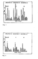
- Nach der Herstellung des ELISA-Protokolls unter der Verwendung des Testsystems gemäß Beispiel 1 wurden Humanseren auf Autoantikörper gegen die verschiedenen Kollagene I bis VI getestet. Die Serumverdünnung betrug hierbei 1:50. Die Ergebnisse der Versuche sind in den Fig. 1 und 2 dargestellt. Es wurden Doppelbestimmungen durchgeführt, um den Assay hinsichtlich seiner Reproduzierbarkeit überprüfen zu können. Es zeigten sich signifikante Unterschiede in den Reaktionen der einzelnen Patienten gegen die menschlichen Kollagene I, II und III (Fig.l). Gegen die Kollagene IV, V und VI waren die Reaktionen schwächer (Fig.2). Als positive Kontrolle werden bei diesen Experimenten die Antikörper einzelner Patienten gegen Bakterienmembranen gemessen, als negative Kontrolle die Reaktionen gegen Rinderserum-Albumin und die Reaktionen mit normalem Patientenserum gegen die Kollagene.
- Die Seren wurden gegen die Kollagene titriert. Hierbei erhielt man bei den einzelnen Patienten stark unterschiedliche Serumtiter. Es wurden hierbei logarithmische Serumverdünnungen mit Kollagen I-VI im ELISA getestet. Alle diese Experimente dienten dem Nachweis von Autoantikörpern der IgG-Klasse.
- Die Höhe des Serumtiters kann für diagnostische Zwecke von Bedeutung sein. Eine Beurteilung des Standes der Erkrankung ist hiernach möglcih.
- Der Nachweis von Autoantikörpern ist nicht nur mit Hilfe menschlicher Kollagene möglich. Auch Kalbskollagene erlauben die Darstellung von Autoantikörpern in dem im Beispiel 1 beschriebenen ELISA-Testsystem. Dazu wurden 400 ng Kalbskollagen in Puffer A 12 Stunden bei 37°C in die Kavitäten der unter Beispiel 1 bezeichneten Mikrotiterplatte inkubiert und gemäß Protokoll weiterverfahren. Die so erhaltenen Ergebnisse standen in guter Obereinstimmung zu den in Beispiel 3 aufgeführten.
- Um die Spezifität der im ELISA gemessenen Reaktionen zu überprüfen, wurden Proteinblots nach der von Towbin et al. (PNAS 76, 4350-4354 (1979)) beschriebenen Verfahren durchgeführt. Der Nachweis erfolgte mit Hilfe einer Farbstofflösung (10 % Diethanolamin-HCI; pH 9,8; 0,01 % Nitroblautetrazolium; 0,005 % 5-Bromo-4-Chloro-3-indolylphosphat; 4 mM MgCl2). In der Tat konnten hierbei die Autoantikörper als direkt mit der Kollagenproteinbande reagierend nachgewiesen werden. Dies ist direkter Beweis für die Richtigkeit der im ELISA gemessenen Ergebnisse.
- Es gelang auch ein Autoantikörpernachweis gegen Kollagene in Form eines Proteindotblotassays.Hierzu wurden die Kollagene direkt auf Nitrocellulosepapierstreifen punktförmig aufgetragen. Nach Absättigung der restlichen Proteinbindungsstellen der Nitrocellulose mit Hilfe einer l%-igen Rinderserum-Albumin-Lösung wurden die Teststreifen in Serum für 30 Minuten inkubiert. Nach zweimaliger Wäsche in Puffer E (10 mM Tris-HCl; 0,9 % NaCl) . wurde der Teststreifen in Konjugatlösung inkubiert und abermals gewaschen. Anschließend wurde für 5 Minuten in der in Beispiel 5 beschriebenen Farbstofflösung inkubiert. Ein positives Ergebnis zeigte sich in einer punktförmigen Anfärbung des Nitrocellulosepapiers. Als Kontrolle wurde parallel ein Teststreifen mit Normalserum inkubiert und wie oben beschrieben weiterverfahren.
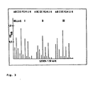
- Alle bis jetzt beschriebenen Anwendungsbeispiele lassen sich problemlos auf den Nachweis von Immunglobulinen, die nicht der IgG-Klasse zugehören, übertragen. Hierzu ist es notwendig, mit einem die entsprechende Immunglobulinklasse erkennenden Antiantikörperkonjugat zu arbeiten. Hier wurden Untersuchungen mit den in Beispiel 1 bis 6 vorgestellten Methoden zum Nachweis von Autoantikörpern der IgM-Klasse durchgeführt. Die entsprechenden Ergebnisse sind in Fig. 3 dargestellt. In der Tat gibt es solche Personen, die keine gegen Kollagen gerichtete Autoantikörper der IgG-Klasse, wohl aber solche der IgM-Klasse haben.
- In der nachfolgenden Tabelle sind die Ergebnisse der in Beispiel 1 bis 7 erläuterten Experimente zusammengefaßt (A-I Patientenseren; J,K negative Kontrollseren)

Claims (9)
Applications Claiming Priority (2)
| Application Number | Priority Date | Filing Date | Title |
|---|---|---|---|
| DE3513915 | 1985-04-17 | ||
| DE19853513915 DE3513915A1 (de) | 1985-04-17 | 1985-04-17 | Immunologischer testkit |
Publications (2)
| Publication Number | Publication Date |
|---|---|
| EP0199261A2 true EP0199261A2 (de) | 1986-10-29 |
| EP0199261A3 EP0199261A3 (de) | 1988-01-07 |
Family
ID=6268398
Family Applications (1)
| Application Number | Title | Priority Date | Filing Date |
|---|---|---|---|
| EP86105184A Withdrawn EP0199261A3 (de) | 1985-04-17 | 1986-04-15 | Immunologischer Testkit |
Country Status (3)
| Country | Link |
|---|---|
| EP (1) | EP0199261A3 (de) |
| JP (1) | JPS61274262A (de) |
| DE (1) | DE3513915A1 (de) |
Cited By (3)
| Publication number | Priority date | Publication date | Assignee | Title |
|---|---|---|---|---|
| WO1994017411A1 (en) * | 1993-01-27 | 1994-08-04 | Duke University | Methods and agents for the diagnosis and treatment of rheumatoid arthritis |
| CN105353122A (zh) * | 2015-11-05 | 2016-02-24 | 厦门敖依生物科技有限公司 | 干燥综合征特异自身抗体免疫印迹试剂盒 |
| US9739773B1 (en) | 2010-08-13 | 2017-08-22 | David Gordon Bermudes | Compositions and methods for determining successful immunization by one or more vaccines |
Families Citing this family (1)
| Publication number | Priority date | Publication date | Assignee | Title |
|---|---|---|---|---|
| FI20050814A0 (fi) * | 2005-08-11 | 2005-08-11 | Procollagen Oy | Menetelmä nivelreumassa muodostuvien kollageeniautovasta-aineiden havaitsemiseksi |
Family Cites Families (4)
| Publication number | Priority date | Publication date | Assignee | Title |
|---|---|---|---|---|
| US4391904A (en) * | 1979-12-26 | 1983-07-05 | Syva Company | Test strip kits in immunoassays and compositions therein |
| EP0058428B1 (de) * | 1981-02-18 | 1985-10-09 | Eisai Co., Ltd. | Enzym-Immunovefahren zur gleichzeitigen Messung einer Mehrheit von Proben und Testgefäss zur Durchführung dieses Verfahrens |
| US4487830A (en) * | 1982-05-14 | 1984-12-11 | American Hoechst Corporation | Enzyme/immunofluorescent assay for autoantibodies |
| DE3346795A1 (de) * | 1983-12-23 | 1985-07-04 | E. Prof. Dr. 3550 Marburg Geyer | Enzymimmunoassay und entsprechendes diagnostikum |
-
1985
- 1985-04-17 DE DE19853513915 patent/DE3513915A1/de not_active Withdrawn
-
1986
- 1986-04-15 EP EP86105184A patent/EP0199261A3/de not_active Withdrawn
- 1986-04-16 JP JP8918286A patent/JPS61274262A/ja active Pending
Cited By (3)
| Publication number | Priority date | Publication date | Assignee | Title |
|---|---|---|---|---|
| WO1994017411A1 (en) * | 1993-01-27 | 1994-08-04 | Duke University | Methods and agents for the diagnosis and treatment of rheumatoid arthritis |
| US9739773B1 (en) | 2010-08-13 | 2017-08-22 | David Gordon Bermudes | Compositions and methods for determining successful immunization by one or more vaccines |
| CN105353122A (zh) * | 2015-11-05 | 2016-02-24 | 厦门敖依生物科技有限公司 | 干燥综合征特异自身抗体免疫印迹试剂盒 |
Also Published As
| Publication number | Publication date |
|---|---|
| JPS61274262A (ja) | 1986-12-04 |
| EP0199261A3 (de) | 1988-01-07 |
| DE3513915A1 (de) | 1986-10-23 |
Similar Documents
| Publication | Publication Date | Title |
|---|---|---|
| DE68922243T2 (de) | Immuntest für HIV-1-Antigene unter Benutzung von F(AB')2-Fragmenten als Sonde. | |
| DE68913086T2 (de) | Qualitativer Enzymtest zur visuellen Auswertung. | |
| DE69115369T2 (de) | Immunoassay mit komplementärem sichtbaren Signal | |
| DE69306804T2 (de) | Immunologischer test auf festphase | |
| DE68918166T2 (de) | Spezifische Bindungszusammensetzung mit einem Protein oder einem Kohlenhydrat mit Nieder-pI und ein diagnostischer Testsatz und Verfahren zur Anwendung. | |
| DE69025940T2 (de) | Durch Silber intensiviertes, auf Goldmarkierung beruhendes Immuntestverfahren | |
| DE69626027T2 (de) | Verfahren zur elimination der interferenz des rheumatoiden faktors in diagnostischen tests | |
| DE2851150A1 (de) | Verfahren zum testen auf das vorhandensein eines analyten | |
| DE69626683T2 (de) | Verfahren zur feststellung von antipolymerantikörper und diagnosetestsatz zur feststellung von siliconabhängigen krankheiten | |
| DE69713498T2 (de) | Methode zum nachweis übertragbarer spongiformer enzephalopathien | |
| DE3834766A1 (de) | Verfahren zur bestimmung einer spezifisch bindefaehigen substanz | |
| EP1630558B1 (de) | Diagnostisches Testsystem für den Nachweis von Antikörpern gegen Erreger von akuten respiratorischen Infektionen und atypischen Pneumonien | |
| EP0782585B1 (de) | RHEUMAFAKTOR-ENTSTÖRUNG MIT ANTI-Fd-ANTIKÖRPERN | |
| EP0174652A2 (de) | Immunchemisches Messverfahren für Haptene und Proteine | |
| DE69919503T2 (de) | Verfahren zur bestimmung eines analyten und kit dafür | |
| DE10250368A1 (de) | Mittel und Verfahren zur Diagnose einer Treponemainfektion | |
| DE68921151T2 (de) | Substrat-Zusammensetzung für Alkali-Phosphatase sowie ihre Verwendung bei Testverfahren. | |
| DE3346795A1 (de) | Enzymimmunoassay und entsprechendes diagnostikum | |
| DE3636540A1 (de) | Verfahren zur bestimmung von anti-hiv, mittel dazu und ihre verwendung in diesem verfahren | |
| DE3688072T2 (de) | Immuntestverfahren und satz. | |
| DE69013730T2 (de) | Für ganze Zellen geeigneter Satz und Verfahren zu enzymatischen Bestimmungen. | |
| DE69606024T2 (de) | Feststellung von antikörperproduktion | |
| EP0199261A2 (de) | Immunologischer Testkit | |
| DE69217038T2 (de) | In vitro verfahren zur bestimmung in allergiediagnosen und weitere entzündungskrankheiten | |
| DE69229960T2 (de) | Agglutierungsassays und kolloide farbstoffe verwendende testsätze |
Legal Events
| Date | Code | Title | Description |
|---|---|---|---|
| PUAI | Public reference made under article 153(3) epc to a published international application that has entered the european phase |
Free format text: ORIGINAL CODE: 0009012 |
|
| AK | Designated contracting states |
Kind code of ref document: A2 Designated state(s): AT BE CH DE FR GB IT LI LU NL SE |
|
| PUAL | Search report despatched |
Free format text: ORIGINAL CODE: 0009013 |
|
| AK | Designated contracting states |
Kind code of ref document: A3 Designated state(s): AT BE CH DE FR GB IT LI LU NL SE |
|
| STAA | Information on the status of an ep patent application or granted ep patent |
Free format text: STATUS: THE APPLICATION IS DEEMED TO BE WITHDRAWN |
|
| 18D | Application deemed to be withdrawn |
Effective date: 19880708 |
|
| RIN1 | Information on inventor provided before grant (corrected) |
Inventor name: BUNTE, THOMAS, DR. Inventor name: RUPRECHT, JOHANN, DR. DIPL.-CHEM. Inventor name: HEYL, EDUARD, DR. MED. Inventor name: PARR, WOLFGANG, DR. DIPL.-CHEM. Inventor name: V. KLEIST, ELKE, DR. Inventor name: SCHMOLKE, BERND, DR. DIPL.-CHEM. Inventor name: VORLAENDER, WOLFGANG, DR. MED. DIPL.-CHEM. |