EP0215772A2 - Procédé et dispositif pour les diagnostics de tumeurs au moyen de sérums - Google Patents
Procédé et dispositif pour les diagnostics de tumeurs au moyen de sérums Download PDFInfo
- Publication number
- EP0215772A2 EP0215772A2 EP86890254A EP86890254A EP0215772A2 EP 0215772 A2 EP0215772 A2 EP 0215772A2 EP 86890254 A EP86890254 A EP 86890254A EP 86890254 A EP86890254 A EP 86890254A EP 0215772 A2 EP0215772 A2 EP 0215772A2
- Authority
- EP
- European Patent Office
- Prior art keywords
- serum
- light
- fluorescence intensity
- measured
- sample
- Prior art date
- Legal status (The legal status is an assumption and is not a legal conclusion. Google has not performed a legal analysis and makes no representation as to the accuracy of the status listed.)
- Granted
Links
Images

Classifications
-
- G—PHYSICS
- G01—MEASURING; TESTING
- G01N—INVESTIGATING OR ANALYSING MATERIALS BY DETERMINING THEIR CHEMICAL OR PHYSICAL PROPERTIES
- G01N21/00—Investigating or analysing materials by the use of optical means, i.e. using sub-millimetre waves, infrared, visible or ultraviolet light
- G01N21/62—Systems in which the material investigated is excited whereby it emits light or causes a change in wavelength of the incident light
- G01N21/63—Systems in which the material investigated is excited whereby it emits light or causes a change in wavelength of the incident light optically excited
- G01N21/64—Fluorescence; Phosphorescence
- G01N21/6486—Measuring fluorescence of biological material, e.g. DNA, RNA, cells
-
- G—PHYSICS
- G01—MEASURING; TESTING
- G01N—INVESTIGATING OR ANALYSING MATERIALS BY DETERMINING THEIR CHEMICAL OR PHYSICAL PROPERTIES
- G01N21/00—Investigating or analysing materials by the use of optical means, i.e. using sub-millimetre waves, infrared, visible or ultraviolet light
- G01N21/01—Arrangements or apparatus for facilitating the optical investigation
- G01N21/03—Cuvette constructions
- G01N2021/0385—Diffusing membrane; Semipermeable membrane
-
- G—PHYSICS
- G01—MEASURING; TESTING
- G01N—INVESTIGATING OR ANALYSING MATERIALS BY DETERMINING THEIR CHEMICAL OR PHYSICAL PROPERTIES
- G01N21/00—Investigating or analysing materials by the use of optical means, i.e. using sub-millimetre waves, infrared, visible or ultraviolet light
- G01N21/62—Systems in which the material investigated is excited whereby it emits light or causes a change in wavelength of the incident light
- G01N21/63—Systems in which the material investigated is excited whereby it emits light or causes a change in wavelength of the incident light optically excited
- G01N21/64—Fluorescence; Phosphorescence
- G01N2021/6417—Spectrofluorimetric devices
- G01N2021/6421—Measuring at two or more wavelengths
-
- G—PHYSICS
- G01—MEASURING; TESTING
- G01N—INVESTIGATING OR ANALYSING MATERIALS BY DETERMINING THEIR CHEMICAL OR PHYSICAL PROPERTIES
- G01N21/00—Investigating or analysing materials by the use of optical means, i.e. using sub-millimetre waves, infrared, visible or ultraviolet light
- G01N21/62—Systems in which the material investigated is excited whereby it emits light or causes a change in wavelength of the incident light
- G01N21/63—Systems in which the material investigated is excited whereby it emits light or causes a change in wavelength of the incident light optically excited
- G01N21/64—Fluorescence; Phosphorescence
- G01N21/6428—Measuring fluorescence of fluorescent products of reactions or of fluorochrome labelled reactive substances, e.g. measuring quenching effects, using measuring "optrodes"
- G01N2021/6432—Quenching
-
- G—PHYSICS
- G01—MEASURING; TESTING
- G01N—INVESTIGATING OR ANALYSING MATERIALS BY DETERMINING THEIR CHEMICAL OR PHYSICAL PROPERTIES
- G01N21/00—Investigating or analysing materials by the use of optical means, i.e. using sub-millimetre waves, infrared, visible or ultraviolet light
- G01N21/62—Systems in which the material investigated is excited whereby it emits light or causes a change in wavelength of the incident light
- G01N21/63—Systems in which the material investigated is excited whereby it emits light or causes a change in wavelength of the incident light optically excited
- G01N21/64—Fluorescence; Phosphorescence
- G01N21/645—Specially adapted constructive features of fluorimeters
- G01N2021/6484—Optical fibres
Definitions
- the invention relates to a method for tumor diagnosis using Sera and a device for performing the method.
- tumor markers play the most important role in the diagnosis of malignancies via serum tests. These are parameters that can give an indication of a neoplastic process.
- Methods of tumor diagnosis are mainly used immunological methods.
- the object of the invention is to provide a method for tumor diagnosis with which the differentiation between normal sera and tumor sera is possible in a simple manner and with relatively high accuracy, or to propose a device for carrying out this method.
- the serum to be examined is excited with excitation radiation of at least a wavelength between 250 and 300 nm, that the fluorescence intensity of the serum is measured at predetermined emission wavelengths, and that deviations of these measured values from those of a standard or standard serum are determined and it is concluded that there are neoplastic diseases.
- a novel spectroscopic method for tumor diagnosis is thus available, taking advantage of the fact that the emission-optical properties of a normal serum differ slightly from those of a tumor serum.
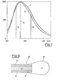
- the fluorescence spectrum of the normal serum shows an emission maximum at approx. 337 nm, which mainly results from protein-bound tryptophan (Trp).
- the fluorescence intensity of the serum to be examined is measured, standardized and compared with the fluorescence intensity of a standard or standard serum at an emission wavelength of more than 340 nm, and that the presence of neoplastic diseases is inferred from a decrease in the standardized fluorescence intensity .
- the spectra can be normalized, for example, to their peak maximum. It can thus be seen that the fluorescence spectra of human serum change in a very characteristic manner when neoplastic diseases occur. These changes occur, for example, on the long-wave flank of the spectrum, where they are particularly pronounced.
- a deviation from the normal spectral distribution determining fluorescence is not enough; only to determine the fluorescence intensity, since its explicitly intensity varies from serum to serum.
- it is expedient to normalize the fluorescence intensity by relating it to the intensity at the emission maximum of the spectrum.
- the fluorescence intensity 1 of the diluted human serum is determined at the emission maximum, i.e. at approx. 337 nm.
- the fluorescence intensity Ix is determined on the long-wave flank of the emission spectrum, i.e. in the range between 350 and 400 nm at an excitation wavelength of 250 up to 300 nm, typically at 287 nm.
- the quotient of the two intensities (I / I) must reach a certain minimum value in a healthy subject. If this value is fallen short of, this is indicative of a tumor.
- This minimum value is determined by calibration with a standard. This can be, for example, an average of several sera from healthy people or a synthetic standard such as N-acetyl-tryptophanamide.
- the implementation of the method by means of the two-wavelength measurement is easier and faster than the acquisition of an entire emission spectrum and subsequent subtraction from a stored standard normal serum, or also the formation of a ratio I x / I o from the data of the entire emission spectrum. This is essential advantage in the context of screening examinations.
- the fluorescence intensity of the serum to be examined is additionally measured, normalized and compared with the fluorescence intensity of a standard serum at an emission wavelength and that the presence of neoplastic diseases is inferred from the increase in the standardized fluorescence intensity .
- the accuracy of the method can be further increased by including the short-wave edge of the spectrum and measuring the intensity changes in this area.
- the hit probability can be increased significantly with the aid of the three-wavelength measurement, by additionally measuring the fluorescence intensity Iy in the short-wave region of the spectrum.
- ratio I x / I o may only reach a certain minimum value in Sera of healthy persons and at the same time the ratio Iy / Io may reach a certain maximum value, these values are undercut or exceeded in Sera by patients with neoplastic diseases.
- the fluorescence intensity of the serum to be examined is measured and normalized both in physiological and in acidic solution with pH values below 4 and that the difference or the ratio of the standardized measured values indicates the presence of neoplastic diseases is closed.
- Another difference in the fluorescence-optical properties of the sera of healthy or tumor-sick people was found when studying the pH dependence of the corresponding fluorescence properties.
- the emission maximum of human serum from healthy people is, with only very slight deviations, between 336.5 and 337.5 nm (measured on an Aminco SPF 500 instrument). In comparison, the emission maximum from Tumorsera is somewhat shorter ren wavelengths shifted. This applies to pH values in the physiological range.
- the Trp emission maximum of the sera from patients with malignancies is red-shifted compared to those of healthy people. All deviations observed in the physiological pH range only change the sign in acidic solution.
- all measurement methods as described above can also be used in the acidic range for measurements in the acidic pH range.
- other quotients of the fluorescence intensities apply.
- the mean value of the individual quotients must be determined in the acidic pH range by arithmetically averaging a certain number of quotas from sera of healthy people or using a standard and the normal range must be determined.
- the measuring method described last has the advantage that when measuring simultaneously at a physiological or strongly acidic pH value - to determine the normal ranges of the quotients - a large number of control sera does not have to be measured. Only in the physiological range are the intensities I o and I x , as well as in the acidic range and x measured. The difference of the quotients / minus xx I x / I o results in a negative value in the long-wave range of healthy people. If this value is positive, this is indicative of a tumor. Knowing the above fact, the measuring method is therefore independent of the exact position of the normal range of the quotients, which means that none Calibration using standard normal serum is required.
- a further variant of the invention presents the possibility that the fluorescence intensity of the serum to be examined is determined at at least one emission wavelength in the presence and in the absence of a dynamic fluorescence quencher and that the difference or the ratio is used to infer the presence of neoplastic diseases.
- the fluorescence quenching usually obeys the Stern-Volmer relationship:
- I o means the fluorescence of the Trp in the absence of an quencher
- I L the intensity in the presence of a concentration (L) of quencher
- K sv the quenching constant
- iodide ions are used as dynamic fluorescence quenchers.
- acrylamide, molecular oxygen and some other molecules are also able to reversibly reduce the fluorescence of the protein-bound tryptophan.
- the serum is diluted with a factor of 200-1000 before the measurement, for example with water.
- albumin or globulin sub-fractions of the serum are used for the measurement. These can be isolated from the serum by known methods, for example by electrophoresis or chromatography and dissolved in water in a concentration of, for example, 0.1-0.3 mg / ml, and fed to the measurement.
- An advantageous embodiment of the invention exists if the measurement is carried out at two different excitation wavelengths, preferably at 287 and 295 nm, and the fluorescence intensity is measured at three different wavelengths, preferably at 325, 337 and 365 nm.
- the greatest accuracy in the three-wavelength measurement can be achieved by using two different excitation wavelengths.
- a measuring cell is required for each excitation wavelength.
- I o or I x are measured in the long-wave range, and in the second case I 'or I' y in the short-wave range.
- the ratio I x / I o may a certain minimum value and at the same time the ratio I '/ I' reach a certain maximum value.
- y 0 These values are undercut or exceeded in Sera by patients with neoplastic diseases.
- This provides a simple device in which the wavelength required for the excitation is realized either by a light source emitting in this area or by a filter.
- One embodiment of the invention provides that two sample spaces are available, to which light, possibly different excitation wavelengths, can be fed to the same light source via a filter.
- This variant of the device is required for all measuring methods in which a sample is excited with different wavelengths, or in which part of the sample is admixed with an additive which influences the fluorescence.
- only one light guide to and to the sample spaces is provided per measuring device, which light is both the excitation and also guides the fluorescence radiation, a dichroic mirror or a beam splitter coupling the latter out of the light guide and feeding it to the measuring device.
- a separate light guide with a typical diameter of 100 to 150 micrometers for each wavelength combination consisting of the excitation wavelength and the emission wavelength to be measured.
- the induced fluorescent light is fed to the corresponding measuring device with the aid of a beam splitter. It is also possible to successively couple several wavelengths into just one optical fiber, for example with the help of a rotating filter wheel.
- the sample space is enclosed by a protein-permeable membrane, preferably a cellulose membrane, attached to the sample-side end of the light guide.
- a protein-permeable membrane preferably a cellulose membrane
- a thin protein-permeable membrane is advantageous to cover the end of the light guide with a thin protein-permeable membrane.
- the penetrating serum is diluted so that the internal filter effects that occur with undiluted sera can be eliminated. Concentration gradients that occur are negligible due to the short measuring time (approx. 2 s). Secondly, this reduces the risk of air bubbles being introduced into the test material (e.g. blood).
- the wavelength in nm is plotted on the abscissa and the intensity on the ordinate.
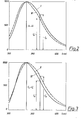
- Fig. 2 shows typical normalized fluorescence spectra of a normal serum, once, labeled I at physiological pH (7.4) and then, labeled III, in 0.1 molar hydrochloric acid. With Normalsera there is a clear blue shift when measuring in the strongly acidic range.
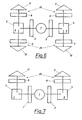
- the simultaneous determination of the fluorescence intensity at two different wavelengths can be carried out in an arrangement as outlined in FIG. 4.
- the light from a light source 1 falls through a light filter 2 onto a sample 4 (for example serum or protein fraction in high dilution) into a sample space 5 that is permeable to both the excitation and the emission radiation. If a requirement of typically 2 ml of sample solution is required, this is calculated a need for serum of 2-10 microliters or 0.2-0.6 mg albumin or globulin fraction).
- the fluorescent light is emitted from the sample space 5 in all directions of the space and is determined simultaneously with the aid of two measuring devices 25 consisting of optical filters 8, 9 and two light detectors 11, 12 at two wavelengths.
- the one optical filter 8 is transparent to the light Wavelength 337 nm and preferably an interference filter.
- the other filter 9 can be an interference filter or a steep edge filter and preferably transmits light of the wavelength above 340 nm.
- the light intensity is measured using the photodetectors 11 and 12, which e.g. can be a SEV-based photo amplifier, a photodiode, or a phototransistor.
- the resulting electrical signals are now fed to an evaluation unit (not shown further) and divided there.
- the ratio of the two light intensities depends on the device and is influenced, among other things, by the spectral properties of the filters 8, 9 and the spectral sensitivity of the photo amplifiers.
- the quotient of the intensities at 365 and 337 nm for the normal serum is 0.714 +/- 0.008 when averaged from 20 samples.
- the simultaneous determination of the fluorescence intensity at three different wavelengths can be carried out in an arrangement as outlined in FIG. 5.
- light from the light source 1 falls through a light filter 3, which is only permeable to light of the wavelength of typically 295 nm, onto the sample 4.
- the intensity of the emission light is now measured at three different wavelengths, namely an intensity I (365 nm) Light filter x 9 and detector 12), an intensity 1 at 337 nm (light filter 8 and detector 11) and an intensity I y at 325 nm (light filter 7 and detector 10).
- the samples 4, 6 are filled into the sample spaces 5.
- the excitation light from the light source 1 falls through the light filter 2, which is permeable to light of the wavelength 287 nm, onto the Sample 4 and excitation light of the wavelength 295 nm through a filter 3 onto the sample 6.
- the intensity of the emission light is now measured at three different emission wavelengths, specifically for each of the samples 4 and 6 at 337 nm with a light filter 8 and a detector 11, the fluorescence intensities I o and Io.
- the light filters 9 and 7 are transmissive to light of the wavelengths 365 and 325 nm, the fluorescence intensities I x and 1 being measured at the detectors 12 and 10, respectively.
- the analytical wavelengths specified are device-specific and are determined by the spectral characteristics of the light source and the photo amplifier.
- the analytical wavelengths specified are device-specific and are determined by the spectral characteristics of the light source and the photo amplifier.
- the excitation filter 3 must be replaced by a filter 2, the emission filter 7 by filter 9 and detector 10 by detector 12.
- the light from the light source 1 then falls through the light filter 2 onto an acidic 6 or a neutral sample 4.
- the fluorescent light is emitted from both sample spaces in all directions and strikes the optical filters 8 and 9.
- the fluorescence intensity is determined simultaneously with two light detectors 11 and 12 at two wavelengths.
- the optical filters 8 are again transparent to light of the wavelength 337 nm and provide the fluorescence intensities I o and I ', the optical filters 9 for light of the wavelength 0 ge 365 nm and provide the fluorescence intensities I x and I' x . x
- a device is shown, the measurement by means of fluorescence quencher method in an analyti wavelength.
- the excitation light falls from the light source 1 through two light filters 2 onto the samples 4, 6 located in the sample rooms 5.
- Sample 4 contains diluted serum
- sample 6 contains the same diluted serum and the quencher additive.
- the fluorescence radiation propagates from the two samples 4, 6 in all directions.
- the fluorescence intensities of the two samples are determined at the emission wavelengths of 337 nm (or longer wavelength) with the aid of a suitable optical filter and correlated in a division unit.
- the quotient I L / I o will be higher in the case of a serum of a person with a tumor than the corresponding quotient, determined on a standard.
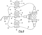
- FIG. 8 shows an arrangement chosen on the basis of FIG. 5, with the aid of which an in vivo measurement is also possible.
- lenses 13 are coupled into light guides 14 from a UV light source 1 after passing through the filters 2.
- the light guides 14 bring the excitation light to the measuring location.
- the induced fluorescent light returns to the measuring devices via light guide 15. It is also possible, as shown in FIG. 8, to guide the excitation light and the fluorescent light for each emission wavelength to be measured in a light guide and the fluorescent light in each case with the aid of a dichroic mirror or a beam splitter 16, 17, 18 onto one of the lenses 19 to steer.
- the filters 7, 8 and 9 set to 325, 337 and 365 nm in the individual measuring devices 25, the respective intensity I is determined and evaluated as described earlier.
- the ends of the light guides 15 are preferably combined into a catheter-shaped bundle 20.
- the light source 1 a reference photodetector can be assigned to compensate for intensity fluctuations.
- Fig. 9 shows the end 21 of a light guide 15 made of a catheter-shaped bundle 20 with a balloon-shaped cellulose membrane. 22, which prevents the penetration of cellular components into the sample space 5, whereas blood serum can diffuse, 23 and 24 are the core and cladding of the light guide 15.
Landscapes
- Health & Medical Sciences (AREA)
- Life Sciences & Earth Sciences (AREA)
- Engineering & Computer Science (AREA)
- Biomedical Technology (AREA)
- Molecular Biology (AREA)
- Nuclear Medicine, Radiotherapy & Molecular Imaging (AREA)
- Physics & Mathematics (AREA)
- Chemical & Material Sciences (AREA)
- Analytical Chemistry (AREA)
- Biochemistry (AREA)
- General Health & Medical Sciences (AREA)
- General Physics & Mathematics (AREA)
- Immunology (AREA)
- Pathology (AREA)
- Investigating, Analyzing Materials By Fluorescence Or Luminescence (AREA)
- Investigating Or Analysing Biological Materials (AREA)
- Investigating Or Analysing Materials By The Use Of Chemical Reactions (AREA)
Applications Claiming Priority (2)
| Application Number | Priority Date | Filing Date | Title |
|---|---|---|---|
| AT0270385A AT387860B (de) | 1985-09-16 | 1985-09-16 | Verfahren und vorrichtung zur tumordiagnose mittels sera |
| AT2703/85 | 1985-09-16 |
Publications (3)
| Publication Number | Publication Date |
|---|---|
| EP0215772A2 true EP0215772A2 (fr) | 1987-03-25 |
| EP0215772A3 EP0215772A3 (en) | 1987-08-19 |
| EP0215772B1 EP0215772B1 (fr) | 1992-07-01 |
Family
ID=3538963
Family Applications (1)
| Application Number | Title | Priority Date | Filing Date |
|---|---|---|---|
| EP86890254A Expired - Lifetime EP0215772B1 (fr) | 1985-09-16 | 1986-09-09 | Procédé et dispositif pour les diagnostics de tumeurs au moyen de sérums |
Country Status (5)
| Country | Link |
|---|---|
| US (1) | US4755684A (fr) |
| EP (1) | EP0215772B1 (fr) |
| JP (1) | JPS62103571A (fr) |
| AT (1) | AT387860B (fr) |
| DE (1) | DE3685866D1 (fr) |
Cited By (16)
| Publication number | Priority date | Publication date | Assignee | Title |
|---|---|---|---|---|
| DE3736027A1 (de) * | 1987-10-24 | 1989-05-03 | Gerhard Dipl Phys Artmann | Verfahren zur ermittlung der zu einem bestimmten zeitpunkt vorliegenden form von zellen und einrichtung zur durchfuehrung des verfahrens |
| WO1990012536A1 (fr) * | 1989-04-14 | 1990-11-01 | Massachusetts Institute Of Technology | Diagnostic spectral des tissus malades |
| EP0512965A1 (fr) * | 1991-05-08 | 1992-11-11 | Xillix Technologies Corporation | Système de visualisation endoscopique pour tissus malade |
| US5590660A (en) * | 1994-03-28 | 1997-01-07 | Xillix Technologies Corp. | Apparatus and method for imaging diseased tissue using integrated autofluorescence |
| US5647368A (en) * | 1996-02-28 | 1997-07-15 | Xillix Technologies Corp. | Imaging system for detecting diseased tissue using native fluorsecence in the gastrointestinal and respiratory tract |
| US5769792A (en) * | 1991-07-03 | 1998-06-23 | Xillix Technologies Corp. | Endoscopic imaging system for diseased tissue |
| US9610021B2 (en) | 2008-01-25 | 2017-04-04 | Novadaq Technologies Inc. | Method for evaluating blush in myocardial tissue |
| US9816930B2 (en) | 2014-09-29 | 2017-11-14 | Novadaq Technologies Inc. | Imaging a target fluorophore in a biological material in the presence of autofluorescence |
| US10041042B2 (en) | 2008-05-02 | 2018-08-07 | Novadaq Technologies ULC | Methods for production and use of substance-loaded erythrocytes (S-IEs) for observation and treatment of microvascular hemodynamics |
| US10219742B2 (en) | 2008-04-14 | 2019-03-05 | Novadaq Technologies ULC | Locating and analyzing perforator flaps for plastic and reconstructive surgery |
| US10265419B2 (en) | 2005-09-02 | 2019-04-23 | Novadaq Technologies ULC | Intraoperative determination of nerve location |
| US10278585B2 (en) | 2012-06-21 | 2019-05-07 | Novadaq Technologies ULC | Quantification and analysis of angiography and perfusion |
| US10434190B2 (en) | 2006-09-07 | 2019-10-08 | Novadaq Technologies ULC | Pre-and-intra-operative localization of penile sentinel nodes |
| US10492671B2 (en) | 2009-05-08 | 2019-12-03 | Novadaq Technologies ULC | Near infra red fluorescence imaging for visualization of blood vessels during endoscopic harvest |
| US10631746B2 (en) | 2014-10-09 | 2020-04-28 | Novadaq Technologies ULC | Quantification of absolute blood flow in tissue using fluorescence-mediated photoplethysmography |
| US10992848B2 (en) | 2017-02-10 | 2021-04-27 | Novadaq Technologies ULC | Open-field handheld fluorescence imaging systems and methods |
Families Citing this family (26)
| Publication number | Priority date | Publication date | Assignee | Title |
|---|---|---|---|---|
| US4978731A (en) * | 1986-07-10 | 1990-12-18 | Minnesota Mining And Manufacturing Company | Process for fluorimetric monitoring of functional coatings and compositions and fluorescent agents therefor |
| US4922113A (en) * | 1986-07-10 | 1990-05-01 | Minnesota Mining And Manufacturing Company | Process for fluorimetric monitoring of functional coatings and compositions and fluorescent agents therefor |
| US5270116A (en) * | 1986-07-10 | 1993-12-14 | Minnesota Mining And Manufacturing Company | Process for fluorimetric monitoring of functional coatings and compositions and fluorescent agents therefor |
| US5087670A (en) * | 1986-07-10 | 1992-02-11 | Minnesota Mining And Manufacturing Company | Process for fluorimetric monitoring of functional coatings and compositions and fluorescent agents therefor |
| IE81170B1 (en) * | 1988-10-21 | 2000-05-31 | Zeneca Ltd | Pyridine derivatives |
| CN1034120C (zh) * | 1988-10-21 | 1997-02-26 | 曾尼卡有限公司 | 制备二噁烷链烯酸衍生物的方法 |
| US5182316A (en) * | 1989-05-31 | 1993-01-26 | Minnesota Mining And Manufacturing Company | Fluorescent degree of cure monitors |
| US5047444A (en) * | 1989-05-31 | 1991-09-10 | Minnesota Mining And Manufacturing Company | Fluorescent degree of cure monitors |
| US5118559A (en) * | 1989-05-31 | 1992-06-02 | Minnesota Mining And Manufacturing Company | Fluorescent degree of cure monitors |
| EP0440195B1 (fr) | 1990-01-31 | 1997-07-30 | Fuji Photo Film Co., Ltd. | Produit photographique couleur à l'halogénure d'argent |
| US5418130A (en) * | 1990-04-16 | 1995-05-23 | Cryopharm Corporation | Method of inactivation of viral and bacterial blood contaminants |
| US6187572B1 (en) | 1990-04-16 | 2001-02-13 | Baxter International Inc. | Method of inactivation of viral and bacterial blood contaminants |
| FR2694193B1 (fr) * | 1992-07-28 | 1994-10-28 | Abraxas Bio Labs Sa | Méthode de cytodiagnostic utilisant l'alstonine comme marqueur sélectif et kit de diagnostic contenant ce marqueur. |
| US5991653A (en) * | 1995-03-14 | 1999-11-23 | Board Of Regents, The University Of Texas System | Near-infrared raman spectroscopy for in vitro and in vivo detection of cervical precancers |
| US6258576B1 (en) | 1996-06-19 | 2001-07-10 | Board Of Regents, The University Of Texas System | Diagnostic method and apparatus for cervical squamous intraepithelial lesions in vitro and in vivo using fluorescence spectroscopy |
| US5697373A (en) * | 1995-03-14 | 1997-12-16 | Board Of Regents, The University Of Texas System | Optical method and apparatus for the diagnosis of cervical precancers using raman and fluorescence spectroscopies |
| US5842995A (en) * | 1996-06-28 | 1998-12-01 | Board Of Regents, The Univerisity Of Texas System | Spectroscopic probe for in vivo measurement of raman signals |
| US6080584A (en) * | 1996-12-02 | 2000-06-27 | The Research Foundation Of City College Of New York | Method and apparatus for detecting the presence of cancerous and precancerous cells in a smear using native fluorescence spectroscopy |
| GB9717021D0 (en) * | 1997-08-12 | 1997-10-15 | Kalibrant Limited | A detector |
| US6055451A (en) * | 1997-12-12 | 2000-04-25 | Spectrx, Inc. | Apparatus and method for determining tissue characteristics |
| US6174291B1 (en) | 1998-03-09 | 2001-01-16 | Spectrascience, Inc. | Optical biopsy system and methods for tissue diagnosis |
| AU6139199A (en) | 1998-09-11 | 2000-04-03 | Spectrx, Inc. | Multi-modal optical tissue diagnostic system |
| US6697666B1 (en) | 1999-06-22 | 2004-02-24 | Board Of Regents, The University Of Texas System | Apparatus for the characterization of tissue of epithelial lined viscus |
| RU2167502C1 (ru) * | 2000-05-22 | 2001-05-20 | Московский энергетический институт (Технический университет) | Способ индукционного нагрева металлических изделий и устройство для его реализации |
| US20040064053A1 (en) * | 2002-09-30 | 2004-04-01 | Chang Sung K. | Diagnostic fluorescence and reflectance |
| CN112384130B (zh) * | 2018-06-28 | 2024-08-30 | 贝克顿·迪金森公司 | 用于标准化血液培养测量系统中的信号的系统和方法 |
Family Cites Families (14)
| Publication number | Priority date | Publication date | Assignee | Title |
|---|---|---|---|---|
| US3327117A (en) * | 1963-08-12 | 1967-06-20 | Ibm | Cancer cell detector using two wavelengths for comparison |
| US3918812A (en) * | 1973-05-07 | 1975-11-11 | Us Energy | Diagnoses of disease states by fluorescent measurements utilizing scanning laser beams |
| US3973129A (en) * | 1975-01-10 | 1976-08-03 | Bell Telephone Laboratories, Incorporated | Fluorimetric apparatus and method for analysis of body fluid |
| IL49776A (en) * | 1976-06-13 | 1979-05-31 | Elscint Ltd | Method and appararus for differentiating of normal and abnormal mammalian cells |
| JPS53122498A (en) * | 1977-03-31 | 1978-10-25 | Laurel Bank Machine Co | Money exchanging and dispensing machine |
| JPS5913697B2 (ja) * | 1977-04-01 | 1984-03-31 | オリンパス光学工業株式会社 | 螢光偏光測光顕微鏡によるガン診断装置 |
| JPS53135660A (en) * | 1977-04-30 | 1978-11-27 | Olympus Optical Co Ltd | Fluorescent photometric microscope using laser light |
| US4215275A (en) * | 1977-12-07 | 1980-07-29 | Luxtron Corporation | Optical temperature measurement technique utilizing phosphors |
| AT362079B (de) * | 1978-12-18 | 1981-04-27 | Helmut Dr Wachter | Verfahren zur diagnostizierung, insbesondere frueherkennung von malignen tumoren |
| US4419583A (en) * | 1980-05-31 | 1983-12-06 | Noeller Hans Guenter | Polarization fluoroimmunoassay with pulsed light source |
| JPS5784339A (en) * | 1980-11-17 | 1982-05-26 | Hitachi Ltd | Fluorescence polarizing apparatus |
| JPS5832145A (ja) * | 1981-08-20 | 1983-02-25 | Kyoto Giken:Kk | 細胞診断装置 |
| US4476870A (en) * | 1982-03-30 | 1984-10-16 | The United States Of America As Represented By The Department Of Health And Human Services | Fiber optic PO.sbsb.2 probe |
| JPS5913697A (ja) * | 1982-07-12 | 1984-01-24 | Fujitsu Ltd | 液相エピタキシヤル成長装置 |
-
1985
- 1985-09-16 AT AT0270385A patent/AT387860B/de not_active IP Right Cessation
-
1986
- 1986-09-09 DE DE8686890254T patent/DE3685866D1/de not_active Expired - Lifetime
- 1986-09-09 EP EP86890254A patent/EP0215772B1/fr not_active Expired - Lifetime
- 1986-09-10 US US06/905,558 patent/US4755684A/en not_active Expired - Lifetime
- 1986-09-16 JP JP61219107A patent/JPS62103571A/ja active Granted
Cited By (27)
| Publication number | Priority date | Publication date | Assignee | Title |
|---|---|---|---|---|
| DE3736027A1 (de) * | 1987-10-24 | 1989-05-03 | Gerhard Dipl Phys Artmann | Verfahren zur ermittlung der zu einem bestimmten zeitpunkt vorliegenden form von zellen und einrichtung zur durchfuehrung des verfahrens |
| WO1990012536A1 (fr) * | 1989-04-14 | 1990-11-01 | Massachusetts Institute Of Technology | Diagnostic spectral des tissus malades |
| EP0512965A1 (fr) * | 1991-05-08 | 1992-11-11 | Xillix Technologies Corporation | Système de visualisation endoscopique pour tissus malade |
| US5507287A (en) * | 1991-05-08 | 1996-04-16 | Xillix Technologies Corporation | Endoscopic imaging system for diseased tissue |
| US5769792A (en) * | 1991-07-03 | 1998-06-23 | Xillix Technologies Corp. | Endoscopic imaging system for diseased tissue |
| US5827190A (en) * | 1994-03-28 | 1998-10-27 | Xillix Technologies Corp. | Endoscope having an integrated CCD sensor |
| US5590660A (en) * | 1994-03-28 | 1997-01-07 | Xillix Technologies Corp. | Apparatus and method for imaging diseased tissue using integrated autofluorescence |
| US5647368A (en) * | 1996-02-28 | 1997-07-15 | Xillix Technologies Corp. | Imaging system for detecting diseased tissue using native fluorsecence in the gastrointestinal and respiratory tract |
| US10265419B2 (en) | 2005-09-02 | 2019-04-23 | Novadaq Technologies ULC | Intraoperative determination of nerve location |
| US10434190B2 (en) | 2006-09-07 | 2019-10-08 | Novadaq Technologies ULC | Pre-and-intra-operative localization of penile sentinel nodes |
| US9610021B2 (en) | 2008-01-25 | 2017-04-04 | Novadaq Technologies Inc. | Method for evaluating blush in myocardial tissue |
| US11564583B2 (en) | 2008-01-25 | 2023-01-31 | Stryker European Operations Limited | Method for evaluating blush in myocardial tissue |
| US9936887B2 (en) | 2008-01-25 | 2018-04-10 | Novadaq Technologies ULC | Method for evaluating blush in myocardial tissue |
| US10835138B2 (en) | 2008-01-25 | 2020-11-17 | Stryker European Operations Limited | Method for evaluating blush in myocardial tissue |
| US10219742B2 (en) | 2008-04-14 | 2019-03-05 | Novadaq Technologies ULC | Locating and analyzing perforator flaps for plastic and reconstructive surgery |
| US10041042B2 (en) | 2008-05-02 | 2018-08-07 | Novadaq Technologies ULC | Methods for production and use of substance-loaded erythrocytes (S-IEs) for observation and treatment of microvascular hemodynamics |
| US10492671B2 (en) | 2009-05-08 | 2019-12-03 | Novadaq Technologies ULC | Near infra red fluorescence imaging for visualization of blood vessels during endoscopic harvest |
| US10278585B2 (en) | 2012-06-21 | 2019-05-07 | Novadaq Technologies ULC | Quantification and analysis of angiography and perfusion |
| US11284801B2 (en) | 2012-06-21 | 2022-03-29 | Stryker European Operations Limited | Quantification and analysis of angiography and perfusion |
| US12186055B2 (en) | 2012-06-21 | 2025-01-07 | Stryker Corporation | Quantification and analysis of angiography and perfusion |
| US10488340B2 (en) | 2014-09-29 | 2019-11-26 | Novadaq Technologies ULC | Imaging a target fluorophore in a biological material in the presence of autofluorescence |
| US9816930B2 (en) | 2014-09-29 | 2017-11-14 | Novadaq Technologies Inc. | Imaging a target fluorophore in a biological material in the presence of autofluorescence |
| US10631746B2 (en) | 2014-10-09 | 2020-04-28 | Novadaq Technologies ULC | Quantification of absolute blood flow in tissue using fluorescence-mediated photoplethysmography |
| US10992848B2 (en) | 2017-02-10 | 2021-04-27 | Novadaq Technologies ULC | Open-field handheld fluorescence imaging systems and methods |
| US11140305B2 (en) | 2017-02-10 | 2021-10-05 | Stryker European Operations Limited | Open-field handheld fluorescence imaging systems and methods |
| US12028600B2 (en) | 2017-02-10 | 2024-07-02 | Stryker Corporation | Open-field handheld fluorescence imaging systems and methods |
| US12464215B2 (en) | 2017-02-10 | 2025-11-04 | Stryker Corporation | Open-field handheld fluorescence imaging systems and methods |
Also Published As
| Publication number | Publication date |
|---|---|
| US4755684A (en) | 1988-07-05 |
| DE3685866D1 (de) | 1992-08-06 |
| EP0215772B1 (fr) | 1992-07-01 |
| JPH0558506B2 (fr) | 1993-08-26 |
| JPS62103571A (ja) | 1987-05-14 |
| AT387860B (de) | 1989-03-28 |
| EP0215772A3 (en) | 1987-08-19 |
| ATA270385A (de) | 1988-08-15 |
Similar Documents
| Publication | Publication Date | Title |
|---|---|---|
| EP0215772B1 (fr) | Procédé et dispositif pour les diagnostics de tumeurs au moyen de sérums | |
| DE3855242T2 (de) | Verfahren zur Messung der Kohlenstoffdioxidkonzentration in einem Medium | |
| EP1028318B1 (fr) | Dispositif pour la détermination optique de la concentration de l'hémoglobine totale | |
| EP1130998B1 (fr) | Dispositif pour la determination non invasive du metabolisme de l'oxygene dans des tissus | |
| US4930516A (en) | Method for detecting cancerous tissue using visible native luminescence | |
| DE10133451B4 (de) | Vorrichtung zum Erkennen von Karies, Plaque, Konkrementen oder bakteriellem Befall an Zähnen | |
| DE3031249C2 (de) | Vorrichtung zum Entdecken von Karies und Anwendung dieser Vorrichtung | |
| EP2710348B1 (fr) | Procédé et système de détermination de la concentration de substances dans des liquides corporels | |
| DE2944113C2 (fr) | ||
| CH665345A5 (de) | Sonde zum bestimmen des sauerstoffpartialdruckes. | |
| EP0034156A1 (fr) | Procede et dispositif pour la determination de glucose dans serum ou l'urine. | |
| DE2422016A1 (de) | Diagnose von krankheitszustaenden durch fluoreszenzmessungen unter verwendung von abtastlaserstrahlen | |
| EP1987765A1 (fr) | Oxymetre | |
| EP0247103B1 (fr) | Agencement de mesure de l'opacite de cristallin de l'oeil | |
| CH640350A5 (en) | Instrument for the quantitative determination of optically active substances | |
| DE4446390C1 (de) | Verfahren und Vorrichtung zur Messung der Konzentration eines in einer Probe enthaltenen Analyten | |
| DE2603069A1 (de) | Verfahren und vorrichtung zur bestimmung des gesamtproteingehaltes oder einzelner aminosaeuren | |
| DE3825352A1 (de) | Verfahren und vorrichtung zur bestimmung von lokalen farbstoff-konzentrationen und von streuparametern in tierischen und menschlichen geweben | |
| DE3702210A1 (de) | Verfahren zur bestimmung der konzentration von in einer substanz enthaltenen stoffen, insbesondere von sauerstoff | |
| EP3591378B1 (fr) | Procédé de détermination des lipides, de hémoglobine et de bilirubine dans des échantillons de liquide corporel | |
| DE4325529C2 (de) | Vorrichtung zum Bestimmen der Konzentration von Stoffen im Blut | |
| DE3806556A1 (de) | Verfahren und vorrichtung zur schnellen bestimmung der spermienanzahl in spermaproben und/oder des anteiles der lebenden spermien sowie verwendung von propidiumjodid | |
| DE10129754A1 (de) | Verfahren und Vorrichtung zur Erfassung von Stoffen in vitalem Gewebe | |
| DE2721676A1 (de) | Lichtblitzphotometer | |
| DE102004051830B4 (de) | Multifunktionales Referenzsystem bei Analytbestimmungen durch Fluoreszenz |
Legal Events
| Date | Code | Title | Description |
|---|---|---|---|
| PUAI | Public reference made under article 153(3) epc to a published international application that has entered the european phase |
Free format text: ORIGINAL CODE: 0009012 |
|
| AK | Designated contracting states |
Kind code of ref document: A2 Designated state(s): DE FR GB IT |
|
| ITCL | It: translation for ep claims filed |
Representative=s name: MODIANO & ASSOCIATI S.R.L. |
|
| EL | Fr: translation of claims filed | ||
| PUAL | Search report despatched |
Free format text: ORIGINAL CODE: 0009013 |
|
| AK | Designated contracting states |
Kind code of ref document: A3 Designated state(s): DE FR GB IT |
|
| 17P | Request for examination filed |
Effective date: 19870930 |
|
| 17Q | First examination report despatched |
Effective date: 19890829 |
|
| RAP1 | Party data changed (applicant data changed or rights of an application transferred) |
Owner name: AVL MEDICAL INSTRUMENTS AG |
|
| GRAA | (expected) grant |
Free format text: ORIGINAL CODE: 0009210 |
|
| AK | Designated contracting states |
Kind code of ref document: B1 Designated state(s): DE FR GB IT |
|
| REF | Corresponds to: |
Ref document number: 3685866 Country of ref document: DE Date of ref document: 19920806 |
|
| ET | Fr: translation filed | ||
| GBT | Gb: translation of ep patent filed (gb section 77(6)(a)/1977) | ||
| ITF | It: translation for a ep patent filed | ||
| PLBE | No opposition filed within time limit |
Free format text: ORIGINAL CODE: 0009261 |
|
| STAA | Information on the status of an ep patent application or granted ep patent |
Free format text: STATUS: NO OPPOSITION FILED WITHIN TIME LIMIT |
|
| 26N | No opposition filed | ||
| PGFP | Annual fee paid to national office [announced via postgrant information from national office to epo] |
Ref country code: DE Payment date: 19930816 Year of fee payment: 8 |
|
| PGFP | Annual fee paid to national office [announced via postgrant information from national office to epo] |
Ref country code: GB Payment date: 19930901 Year of fee payment: 8 |
|
| PGFP | Annual fee paid to national office [announced via postgrant information from national office to epo] |
Ref country code: FR Payment date: 19930930 Year of fee payment: 8 |
|
| PG25 | Lapsed in a contracting state [announced via postgrant information from national office to epo] |
Ref country code: GB Effective date: 19940909 |
|
| GBPC | Gb: european patent ceased through non-payment of renewal fee |
Effective date: 19940909 |
|
| PG25 | Lapsed in a contracting state [announced via postgrant information from national office to epo] |
Ref country code: FR Effective date: 19950531 |
|
| PG25 | Lapsed in a contracting state [announced via postgrant information from national office to epo] |
Ref country code: DE Effective date: 19950601 |
|
| REG | Reference to a national code |
Ref country code: FR Ref legal event code: ST |
|
| PG25 | Lapsed in a contracting state [announced via postgrant information from national office to epo] |
Ref country code: IT Free format text: LAPSE BECAUSE OF NON-PAYMENT OF DUE FEES Effective date: 20050909 |