EP0236289A2 - Gewebe-Plasminogenaktivator von normalen menschlichen Dickdarmzellen - Google Patents
Gewebe-Plasminogenaktivator von normalen menschlichen Dickdarmzellen Download PDFInfo
- Publication number
- EP0236289A2 EP0236289A2 EP87870023A EP87870023A EP0236289A2 EP 0236289 A2 EP0236289 A2 EP 0236289A2 EP 87870023 A EP87870023 A EP 87870023A EP 87870023 A EP87870023 A EP 87870023A EP 0236289 A2 EP0236289 A2 EP 0236289A2
- Authority
- EP
- European Patent Office
- Prior art keywords
- fraction
- digestion
- oligosaccharides
- mannosidase
- oligosaccharide
- Prior art date
- Legal status (The legal status is an assumption and is not a legal conclusion. Google has not performed a legal analysis and makes no representation as to the accuracy of the status listed.)
- Ceased
Links
Images


Classifications
-
- C—CHEMISTRY; METALLURGY
- C12—BIOCHEMISTRY; BEER; SPIRITS; WINE; VINEGAR; MICROBIOLOGY; ENZYMOLOGY; MUTATION OR GENETIC ENGINEERING
- C12N—MICROORGANISMS OR ENZYMES; COMPOSITIONS THEREOF; PROPAGATING, PRESERVING, OR MAINTAINING MICROORGANISMS; MUTATION OR GENETIC ENGINEERING; CULTURE MEDIA
- C12N9/00—Enzymes; Proenzymes; Compositions thereof; Processes for preparing, activating, inhibiting, separating or purifying enzymes
- C12N9/14—Hydrolases (3)
- C12N9/48—Hydrolases (3) acting on peptide bonds (3.4)
- C12N9/50—Proteinases, e.g. Endopeptidases (3.4.21-3.4.25)
- C12N9/64—Proteinases, e.g. Endopeptidases (3.4.21-3.4.25) derived from animal tissue
- C12N9/6421—Proteinases, e.g. Endopeptidases (3.4.21-3.4.25) derived from animal tissue from mammals
- C12N9/6424—Serine endopeptidases (3.4.21)
- C12N9/6456—Plasminogen activators
- C12N9/6459—Plasminogen activators t-plasminogen activator (3.4.21.68), i.e. tPA
-
- C—CHEMISTRY; METALLURGY
- C08—ORGANIC MACROMOLECULAR COMPOUNDS; THEIR PREPARATION OR CHEMICAL WORKING-UP; COMPOSITIONS BASED THEREON
- C08B—POLYSACCHARIDES; DERIVATIVES THEREOF
- C08B37/00—Preparation of polysaccharides not provided for in groups C08B1/00 - C08B35/00; Derivatives thereof
-
- C—CHEMISTRY; METALLURGY
- C12—BIOCHEMISTRY; BEER; SPIRITS; WINE; VINEGAR; MICROBIOLOGY; ENZYMOLOGY; MUTATION OR GENETIC ENGINEERING
- C12Y—ENZYMES
- C12Y304/00—Hydrolases acting on peptide bonds, i.e. peptidases (3.4)
- C12Y304/21—Serine endopeptidases (3.4.21)
- C12Y304/21069—Protein C activated (3.4.21.69)
-
- A—HUMAN NECESSITIES
- A61—MEDICAL OR VETERINARY SCIENCE; HYGIENE
- A61K—PREPARATIONS FOR MEDICAL, DENTAL OR TOILETRY PURPOSES
- A61K38/00—Medicinal preparations containing peptides
Definitions
- This invention relates to plasminogen activators which are useful thrombolytic agents. More particularly, this invention relates to glycosylated tissue plasminogen activator from cultured normal human colon cells.
- plasminogen activators are widely distributed throughout the body and can be purified from tissue extracts. Typical examples of tissue sources are kidney and lung tissues. The best characterized of these plasminogen activators fall into two major groups, urokinase plasminogen activator (u-PA) and tissue plasminogen activator (t-PA). u-PA and t-PA are present in ng/ml concentrations in human plasma but are immunologically unrelated. t-PA has been demonstrated to have higher affinity for fibrin than u-PA. u-PA products isolated and purified from human urine and from mammalian kidney cells are pharmaceutically available as thrombolytic agents.
- u-PA urokinase plasminogen activator
- t-PA tissue plasminogen activator
- One method of producing t-PA on a large scale comprises isolating the protein from the culture fluid of human melanoma cells grown under in vitro cell culture conditions.
- An established human melanoma cell line (Bowes) has been used for this purpose. See, for example, European Patent Application 41,766, published Dec. 16, 1981; Rijken and Collen, J. Biol. Chem. 256(13), 7035-7041 (1981); and K Kunststoff et al., Adv. Biotech. Proc. 2, Alan R. Liss, Inc., 1983, pp. 97-110.
- the Bowes melanoma t-PA is a glycoprotein which has a molecular weight of about 68,000-70,000 daltons and a 527 amino acid structure with serine as the N-terminal.
- the melanoma t-PA exists as two chains, an A-chain and a B-chain. It also separates into two variants in the A-chain, known as types I and II, which differ by about M 2000-3000. See Ramby et al., FEBS Lett. 146 (2), 289-292 (1982), and Wallen et al., Eur. J. Biochem. 132, 681-686 (1983).
- Type I is glycosylated at Asn-117, Asn-184 and Asn-448 whereas Type II is glycosylated only at Asn-117 and Asn-448 according to Pohl et al., Biochemistry 23, 3701-3707 (1984).
- a high mannose structure has been assigned to Asn-117 whereas two complex carbohydrate structures are assigned to Asn-184 and Asn-448 by Pohl et al., "EMBO Workshop on Plasminogen Activators," Amalfi, Italy, Oct. 14-18, 1985.
- cancer cells can produce human transforming growth factors. See, for example, Delarco and Todaro, Proc. Natl. Acad. Sci. USA 75, 4001-4005 (1978), and Todaro et al., Ibid., 77, 5258-5261 (1980). Even the smallest amount of residual DNA from the cancer cells can be integrated into and expressed in the E. coli or genetically engineered mammalian cells, thereby raising the possibility of harmful effects if t-PA from such source is administered to the patient.
- the recombinant-derived t-PA produced in E. coli is non-glycosylated and contains only the protein moiety of t-PA.
- the specific function of the carbohydrate moiety on t-PA has not been determined, it is known, in general, that glycosylation can cause certain differences in the protein of which the following are of biological interest: antigenicity, stability, solubility and tertiary structure.
- the carbohydrate side-chains also can affect the protein's half-life and target it to receptors on the appropriate cells. See, for example, Delente, Trends in Biotech. 3 (9), 218 (1985), and Van Brunt, Biotechnol. 4, 835-839 (1986).
- glycosylation is suggested in various of the foregoing European Patent Application disclosures, the glycosylation patterns are not described and sugar molecules are not identified.
- hCG human chorionic gonadotropin
- glycosylated t-PA is obtained from cultured normal human colon cells which are adaptable to large scale production.
- a purified colon t-PA is provided which has a unique, heterogeneous glycosylation pattern that differs significantly from the t-PA of Bowes melanoma although the protein moieties are substantially similar.
- glycosylated colon t-PA of this invention has been isolated in a highly purified form which did not exist in the human colon cells from which it was obtained. That is, it has been prepared in a form which is essentially free of other glycoproteins, and free from other cellular components and tissue matter.
- the glycosylated colon t-PA of this invention is isolated from the normal human colon fibroblast cell line CCD-18Co. Both u-PA and t-PA are obtainable from the culture fluids of this cell line in large scale production lots.
- the colon u-PA has a molecular weight of about 54,000 daltons whereas the colon t-PA has a molecular weight of about 67,000 daltons.
- the CCD-18Co cell line is on deposit without restriction in the permanent collection of the American Type Culture Collection, Rockville, Maryland, under accession number ATCC CRL-1459. Samples of the cell line can be obtained by the public upon request to that depository.
- Determination of the structure of the protein moiety of the colon t-PA can be carried out by well-known methods described for melanoma t-PA by Pennica et al., Nature 301, 214-221 (1983), and UK Patent Application 2,119,804, published Nov. 23, 1983.
- biochemical synthetic methods employ enzymes and subcellular components of the protein synthesizing systems of living cells, either in vitro in cell-free systems, or in vivo in microorganisms.
- the principal element is provision of a deoxyribonucleic acid (DNA) of specific sequence which contains the information required to specify the desired amino acid sequence.
- DNA deoxyribonucleic acid
- Such a specific DNA sequence is termed a gene.
- the coding relationships whereby a DNA sequence is used to specify the amino acid sequence of a protein is well-known and operates according to a fundamental set of principles. See, for example, Watson, Molecular Biology of the Gene, 3d ed., Benjamin-Cummings, Menlo Park, Calif., 1976.
- a cloned gene may be used to specify the amino acid sequence of proteins synthesized by in vitro systems.
- DNA-directed protein synthesizing systems are well established in the art. Single-stranded DNA can be induced to act as messenger RNA (mRNA) in vitro, thereby resulting in high fidelity translation of the DNA sequence.
- mRNA messenger RNA
- the mature t-PA protein from human melanoma cells has a 527 amino acid sequence as disclosed by Pennica and in UK Pat. Appln. 2,119,804, supra. It has also been determined that the t-PA protein molecule from human melanoma cells is glycosylated at asparagine positions 117, 184 and 448. See Vehar et al., Biotech., December 1984, pp. 1051-1057. Said disclosures on amino acid sequence and glycosylation positions are incorporated herein by reference.
- the amino acid sequence of the colon t-PA of this invention is substantially similar to the corresponding protein from melanoma t-PA and also is glycosylated at asparagine positions 117, 184 and 448, the glycosylation pattern differs significantly as described hereinbelow.
- Determination of the structure of the oligosaccharides from the colon t-PA employs adaptation of the method used for Immunoglobulin G-derived asparagine-linked oligosaccharides as described by Rademacher and Dwek, Prog. Immunol. 5, 95-112 (1983) and Parekh et al., Nature 316, 452-457 (1985).
- the glycoprotein sample is subjected to controlled hydrazinolysis to release intact its associated oligosaccharide moieties as described by Takahasi et al., Meth. Enzymol. 83, 263-268 (1982).
- Reduction of the reducing terminal N-acetylglucosamine residues using NaB 3 H 4 is then performed to label radioactively each carbohydrate chain.
- Each labeled oligosaccharide mixture is then subjected to exhaustive neuraminidase digestion in order to analyze the distribution of neutral structures.
- the resulting 'asialo' oligosaccharide mixtures are then fractionated by Bio-Gel@ P-4 ( ⁇ 400 mesh) gel filtration chromatography, which separates neutral oligosaccharides on the basis of the effective hydrodynamic volumes as described by Yamashita et al., Meth. Enzymol. 83, 105-126 (1982).
- Bio-Gel P-4 is a gel filtration material of choice for analysis of reduced oligosaccharides by high voltage gel permeation chromatography due to the polyacrylamide structure.
- Bio-Gel P is prepared by copolymerization of acrylamide with N,N'-methylene bis-acrylamide.
- P-4 has an exclusion limit and fractionation range of about 800-4000 daltons. This well-known gel filtration material is commercially available from Bio-Rad Laboratories, Richmond, California.
- the oligosaccharides also can be initially isolated from the t-PA glycoprotein by the method described by Rademacher and Dwek in co-pending application Ser. No. 772,988, filed Sept. 6, 1985, and assigned to a common assignee.
- Said method employs hydrazinolysis of the glycoprotein under reaction conditions to cause cleavage at the N-linked sites, producing a mixture having as a major component a de-N-acetylated hydrazone derivative of the oligosaccharides, followed by N-acylation of the hydrazone derivative, acid-catalysis of the hydrazone derivative to produce unreduced oligosaccharides, and subjecting the resulting unreduced oligosaccharides to cellulose column chromatography to remove contaminants and to recover the unreduced oligosaccharides.
- the latter materials being essentially pure, can be used for attachment to various peptide or protein chains for further study.
- t-PA unique glycosylation pattern of t-PA derived from cultured normal human colon cells, including the structure, monosaccharide sequence and site location of the individual oligosaccharides on the polypeptide backbone. It facilitates the further study and use of oligosaccharide structural variations and alterations by deletion and/or attachment of specific sugar moieties on the t-PA protein molecule. It enables the preparation of homogeneous glycosylated t-PA from the protein backbone and any of these oligosaccharides. Modified t-PA glycoproteins can thus be prepared with varying stability, solubility, activity and other such properties. The serum half-life of the t-PA also can be extended by attaching certain terminal moieties to the oligosaccharide chain, for example, sialic acid which is a charged sugar molecule that prevents rapid filtration through the kidneys.
- Antibodies to the individual oligosaccharides of the colon t-PA can be prepared and used in diagnostic assays.
- Biotin- and fluorescent-labeled probes can be prepared from the individual oligosaccharides of the colon t-PA and then used to identify the oligosaccharide receptors.
- Glyconic acids glycuronic acids, 2-amino-2-deoxy- saccharides, and their N-acetyl derivatives are designated by modified symbols. For example:
- the CCD-18Co cell line used in a preferred embodiment of this invention was originally cultured in CRCM medium with 20% fetal bovine serum and antibiotics.
- CRCM is a nutrient medium devloped by the American Type Culture Collection. During passage, the medium was changed to minimum essential medium (Eagle) with non-essential amino acids in Earle's BSS (balanced salt solution) supplemented with 10% fetal bovine serum.
- Eagle minimum essential medium
- DMEM Dulbecco's modified Eagle medium
- medium 199 fetal bovine serum
- RPMI 1640 medium fetal bovine serum
- These conventional culture media contain known amino acids, mineral salts, vitamins, hormones and carbohydrates. They are also frequently fortified with mammalian sera such as fetal bovine serum.
- Other components which are desirably used in the media are protein hydrolysates such as lactalbumin hydrolysate, tryptone, tryptose, peptone and the like materials.
- normal human colon fibroblast cell lines also can be used in accordance with the invention.
- another suitable normal human colon fibroblast cell line is the cell line designated CCD-112CoN which is available without restriction from the American Type Culture Collection under accession number ATCC CRL-1541.
- Patents 4,166,768; 4,289,854; 4,335,215; and 4,537,860 disclose particularly useful methods and apparatus for the large scale growth and maintenance of cells for the production of plasminogen activators.
- the disclosures in said patents are incorporate herein by reference.
- the methods and apparatus disclosed therein can be used for the culture of the colon cells defined herein.
- the cells are preferably cultured in nutrient medium at 37°C in agitated microcarrier suspension culture as described in U.S. Patent 4,335,215 and, after a suitable growth period, are maintained in the static maintenance reactor described in U.S. Patent 4,537,860 in which the medium is supplemented with 0.5% lactalbumin hydrolysate.
- Purification of the t-PA from the spent culture media can employ various known procedures for the separation of proteins such as, for example, salt and solvent fractionation, adsorption with colloidal materials, gel filtration, ion exchange chromatography, affinity chromatography, immunoaffinity chromatography, electrophoresis and high performance liquid chromatography (HPLC).
- Procedures found to be particularly useful are affinity chromatography with zinc chelate-agarose, p-aminobenzamidine-agarose, concanavalin A-agarose (Con A-Sepharose®), fibrin-Celite® and fibrin-agarose (fibrin-Sepharose); HPLC with a TSK 3000 SW size exclusion column; and immunoaffinity chromatography with monoclonal antibodies.
- a preferred process for producing human plasminogen activator according to the present invention comprises culturing in vitro normal human colon cells in nutrient culture medium, subjecting the resulting conditioned medium to a first affinity chromatography with zinc chelate-agarose and then a second affinity chromatography with material selected from the group consisting of (a) concanavalin A-agarose, (b) fibrin-Celite, and (c) fibrin-agarose, followed by TSK 3000 SW size exclusion high performance liquid chromatography and p-aminobenzamidine-agarose affinity chromatography and recovering the resulting plasminogen activator fractions.
- the zinc chelate-agarose can be prepared essentially as described by Rijken and Collen, J. Biol. chem. 256(13), 7035-7041 (1981) by coupling iminodiacetic acid to Sepharose 4B and saturating this material with zinc chloride (7.3 mM), regenerating with 0.05 M EDTA, pH 8.0, 0.05 M NH 4 HCO 3 , pH 10.5, and water, and resaturating with zinc chloride.
- Sepharose 6B an agarose gel in bead form, 60-140p wet bead diameter, available from Pharmacia Fine Chemicals, Inc., Piscataway, N.J. can be used in place of the Sepharose 4B.
- Con A-Sepharose is similarly available from Pharmacia Fine Chemicals, Inc., and is prepared by coupling concanavalin A to Sepharose 4B by the cyanogen bromide method.
- Para-aminobenzamidine-agarose is commercially available from Pierce Chemical Co., Rockford, Ill. and Sigma Chemical Co., St. Louis, Mo.
- the TSK 3000 SW size exclusion HPLC employs a column of hydrophilic, spherical silica. It is commercially available from Toyo Soda Manufacturing Co., Ltd., Yamaguchi, Japan, and Beckman Laboratories, Fullerton, California.
- a preferred TSK 3000 SW column is Spherogel-TSK 3000 SWG which has a 0 pore size of 250 A ⁇ 5%, a particle size of 13 ⁇ 2p and a molecular weight cutoff of 15,000 - 150,000.
- the fibrin-Celite is a fibrin affinity matrix prepared from Celite filter-aid which is a diatomaceous earth (diatomite) commercially available from Manville Filtration & Minerals, Denver, Colorado.
- This matrix can be prepared as described by Husain et al., Proc. Natl. Acad. Sci. USA 78, 4265-4269 (1981). According to this procedure, the Celite matrix surface is exposed to excess fibrinogen in a buffer and then to thrombin in a buffer to convert the fibrinogen to fibrin whereby the adsorptive surface is fully occupied by fibrin. Affinity chromatography on fibrin-Celite is used to remove non-fibrin binding proteins such as the urokinase plasminogen activator.
- a matrix of fibrin is formed on Sepharose (agrose) instead of the Celite (diatomite).
- monoclonal antibodies having an affinity for plasminogen activator are attached to polysaccharide beads which are then used as the chromatographic column.
- Monoclonal antibodies of Bowes t-PA and colon t-PA immobilized on Sepharose 4B have been found useful in this chromatographic procedure.
- Monoclonal antibodies can be made by adaptation of conventional procedures such as originally described by Kohler and Milstein, Nature 256, 495-497 (1975); Eur. J. Immunol. 6, 511-519 (1976). According to this method, tissue-culture adapted mouse myeloma cells are fused to spleen cells from immunized mice to obtain the hybrid cells that produce large amounts of a single antibody molecule.
- the first example illustrates the production of t-PA from cultured normal human colon fibroblast cells on a large scale in sufficient quantities for initial animal testing.
- Example 2 illustrates the significant differences between the colon t-PA of this invention and the Bowes melanoma t-PA of the prior art in their respective glycosylation patterns.
- CCD-18Co cells obtained from the American Type Culture Collection (ATCC CRL-1459) were grown at 37°C in attached culture in 75 cm 2 T-flasks using Dulbecco's MEM high glucose medium supplemented with 10% fetal bovine serum. The resulting cells were then cultured at 37°C in the same medium in large scale microcarrier suspension culture by the method of U.S. Patent 4,335,215 using Corning Geli-Bead microcarriers. The cells, after a suitable growth period to provide 1.5 x 10 11 cells attached to microcarriers, were then maintained at 37°C in serum-free conditioned media supplemented with 0.25 - 0.5% lactalbumin hydrolysate (LAH) in the static maintenance reactor (SMR) system of U.S. Patent 4,537,860. About 1673 liters of crude serum-free conditioned media was recovered for product purification during a 4 month run of the SMR system.
- SMR static maintenance reactor
- t-PA was recovered from the above conditioned media by successive purification steps consisting of: (1) affinity chromatography with zinc chelate-agarose by adsorption to and elution from zinc chelate-Sepharose, (2) affinity chromatography with Con A-agarose by adsorption to and gradient elution from Con A-Sepharose, (3) TSK 3000 SW size exclusion HPLC and (4) affinity chromatography with p-aminobenzamidine-agarose by adsorption to and elution from p-aminobenzamidine-agarose.
- affinity chromatography with zinc chelate-agarose by adsorption to and elution from zinc chelate-Sepharose affinity chromatography with Con A-agarose by adsorption to and gradient elution from Con A-Sepharose
- TSK 3000 SW size exclusion HPLC affinity chromatography with p-aminobenzamidine-agarose by adsorption to and elution from p-a
- Sterile, pyrogen-free injectible grade water and saline (0.9% NaCl) was used for the preparation of all dialysis solutions and diluents.
- Isotonic solutions were obtained in two steps: first, dialysis was made against 0.3 M NaCl, 0.01% Tween@ 80, and second, the sample was diluted with an equal volume of 0.01% Tween 80 (polysorbate 80).
- the t-PA sample was administered to a 20 Kg dog over a 60 minute period with an infusion rate of 1980 t-PA IU/min/kg with a total infusion of 2.38 x 10 6 t-PA IU.
- 3 t-PA samples were administered consecutively to a 25 kg dog over a 58 minute period with infusion rates of 1770, 1700 and 2380 t-PA IU/min/kg, respectively, with a total infusion of 2.91 x 10 6 t-PA IU.
- the p-aminobenzamidine agarose affinity chromatography step was omitted.
- an artificial thrombus was induced by advancing a 1x3 mm copper coil into the left anterior descending coronary artery distal to the first main diagonal branch. This procedure was carried out as described by Van de Werf et al., Circulation 69(3), 605-613 (1984). An occlusive thrombus formed at the site of the coil. Presence and stability of the clot was confirmed by angiography. A 50 ml syringe pump was used to infuse t-PA solution via a midbody, intraveneous catheter over a 60-minute period. Blood samples for assay of fibrinogen, t-PA antigen, and t-PA fibrinolytic activity were taken before t-PA infusion and at 5-minute intervals for 30 minutes following infusion.
- the normal human colon t-PA was shown to be an effective thrombolytic agent in vivo without causing a systemic lytic state.
- the t-PA used in this trial was acid adjusted to pH 4 to improve its solubility and this may have caused the t-PA to be more susceptible to hepatic clearances.
- the half life of the t-PA following infusion was about 50% longer for the second dog trial than for the first dog trial.
- the asparagine-linked sugar chains of t-PA derived from human colon fibroblast CCD-18Co cells (C-tPA) and Bowes melanoma cells (B-tPA) were quantitatively released as oligosaccharides by hydrazinolysis and labeled by NaB 3 H 4 reduction.
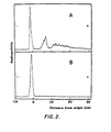
- High-voltage electrophoresis of the hydrazinolysates fractionated the oligosaccharides into four acidic components (A-I, A-2, A-3 and A-4) and a neutral component (FIG. 1).
- Neuraminidase digestion confirmed that the acidic components of C-tPA were exclusively sialic acid (FIG. 2).
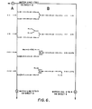
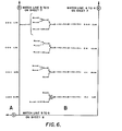
- oligosaccharides from C-tPA were analyzed further in order to determine the monosaccharide sequences present.
- sixteen oligosaccharides (FIG. 6 and Table 2) were isolated from the C-tPA fractions, with high-mannose (C-G-3, C-H, C-I and C-J), hybrid (C-B and C-C-1) and complex type including biantennary (C-D-2, C-E and C-C-2), triantennary (C-C-1, C-C-2, C-C-3 and C-C-4), tetraantennary (C-B) and pentaantennary (C-A) represented.
- oligosaccharides include four isomeric forms of the triantennary complex type (i.e. additional outer chains at the C-4 or C-6 positions of the Man ⁇ 1 ⁇ 3 and the Man ⁇ 1 ⁇ 6 residues of the core) and the presence of a pentaantennary complex type.
- the oligosaccharide structures present on melanoma-derived t-PA (B-tPA) are predominantly of the high mannose type as seen from the elution positions (FIG. 5B).
- the Bowes melanoma 2-chain t-PA (product #110, lot 16-01) was purchased from American Diagnostica Inc., Greenwich, Connecticut and further purified by p-aminobenzamidine-agarose affinity chromatography to remove low molecular weight contaminants visible on sodium dodecylsulfate polyacrylamide gel electrophoresis gels. According to the manufacturer, the Bowes melanoma t-PA is purified from serum-free conditioned medium by adsorption to and elution from PAM-2 Sepharos® immobilized Bowes melanoma t-PA monoclonal antibody, followed by gel filtration on Sephadex ? G-150.
- the colon t-PA was purified from serum-free conditioned medium obtained by the culture of CCD-18Co cells as described in Example 1, above.
- the purification steps consisted of: (1) adsorption to and elution from zinc chelate-Sepharose, (2) adsorption to and gradient elution from Con A-Sepharose, (3) TSK 3000 SW HPLC gel filtration, and (4) adsorption to and elution from p-aminobenzamidine agarose.
- the purified t-PA sample Prior to use, the purified t-PA sample was adjusted to pH 4 and stored sterile in the refrigerator (sample A). Another sample was stored frozen at neutral pH before use (sample B).
- a third colon t-PA sample (sample C) was purified from the serum-free conditioned media obtained by the culture of CCD-18co cells substantially as described in Example 1, above, except that the culture medium was not supplemented with the lactalbumin hydrolysate. A different purification sequence also was employed for sample C.
- the purification steps consisted of: (1) adsorption to and elution from zinc chelate-Sepharose, (2) adsorption to and elution from p-aminobenzamidine-agarose, (3) batch adsorption to and elution from PAM-2 Sepharose, an immobilized Bowes melanoma t-PA monoclonal antibody purchased from American Diagnostica Inc., and (4) TSK 3000 SW HPLC gel filtration.
- SDS polyacrylamide gel electrophoresis was conducted according to the method of Laemmli, Nature 227, 660-665 (1970), using 5-15% gradient gels.
- the sample buffer contained 25 mM dithiothreitol for reduced samples.
- Molecular weight standards were obtained from Pharmacia. Gels were stained with silver nitrate using reagents from Bio-Rad. Activity of final preparations were measured using the amidolytic substrate S-2288 (H-D-Ile-Pro-Arg-paranitroanalide, Kabi) and are given in international units relative to the WHO t-PA standard.
- the assay mixture (200 ⁇ l) contained 50 mM Tris HC1, 0.01% sodium azide, 0.001% Tween 80, 1 mM S-2288, pH 8.7, and t-PA sample or standard as appropriate.
- an amidolytic assay employing the substrate S-2322 H-D-Val-Gly-Arg- paranitroanilide, Kabi was used.
- the assay mixture (100 ⁇ l) contained 20 mM Tris HCl, pH 7.6, 100 mM NaCl, 0.01% Tween 80, 100 pg/ml bovine serum albumin, and enzyme.
- One-hundred forty liters of serum-free medium containing 0.25% lactabumin hydrolysate were conditioned by CCD-18Co cells in the static maintenance reactor. Following addition of 100 units/ml penicillin and 100 pl/ml streptomycin, the conditioned medium was batch adsorbed in two portions (40 and 100 liters) by stirring for three hours with a total of 4.4 liters of Zinc-charged chelating Sepharose-6B (Pharmacia) previously equilibrated with 1M NaCl, 20 mM Tris HCl, 0.01% Tween 80, pH 7.5.
- the resin was transferred to a 10 cm diameter column, washed with about 7 liters of equilibration buffer at 2.7 liters/hr flow rate and the plasminogen activator eluted by gravity flow using about 2 liters of equilibration buffer containing 50 mM imidazole. NaN 3 (0.02%) was added to the eluted fraction.
- the pooled plasminogen activator-containing Zinc chelate-Sepharose eluate (3.6 liters) was applied to a 4.4 x 12.8 cm column of Concanavalin A-Sepaharose-4B (Pharmacia) previously equilibrated with 1 M NaCl, 10 mM potassium phosphate, 0.01% Tween 80, pH 7.5.
- the lyophilized peak fractions from Con A-Sepharose chromatography were dissolved in a minimum volume of H 2 0 (21 ml final volume) and applied in four portions to a 2.15 x 60 cm Spherogel-TSK 3000 SW HPLC column (Beckman Instruments, Inc.) at 5 ml/min flow rate and at room temperature. The column was equilibrated and eluted with 1.6 M KSCN, 20 mM sodium phosphate buffer, pH 6.8, containing 0.01% Tween 80. Fractions (4 ml) were collected and the peak fractions (75 ml total volume) were pooled and dialyzed against 0.3 M NaCl, 0.01% Tween 80.
- the dialyzed TSK 3000 SW fractions were applied at room temperature and 1 ml/min flow rate to a 1.2 x 10 cm column of p-aminobenzamidine-agarose (Pierce Chemical Company) previously equilibrated with 0.5 M NaCl, 50 mM Tris HCl, 0.01% Tween 80, pH 8.0.
- the column was washed with the equilibration buffer until the absorbance at 280 nm of the effluent reached baseline.
- the plasminogen activator was then eluted in a volume of 12 ml with equilibration buffer containing 2 M KSCN.
- the preparation was then diluted by addition of about 12 ml of 0.15 M NaCl, 0.01% Tween 80, 0.88 mM acetic acid, to give 24 ml of slightly cloudy solution with a pH of 4.2. It was then sterile filtered and stored for about 4 months at 4°C. After this extended period, 15 ml of this preparation was adjusted to neutral pH, solid KSCN was added to give a 1.6 M solution, and the sample concentrated to about 1.4 ml in an Amicon ultrafiltration cell using a YM05 membrane. The concentrate was then applied to a small column of Sephadex G-25 (PD-10 column, Pharmacia) equilibrated with 1 M NH4HCO3 and eluted with this same buffer.
- Sephadex G-25 PD-10 column, Pharmacia
- the Bowes melanoma t-PA was commercially supplied by the manufacturer as a frozen solution in 1 M NH 4 HCO 3 .
- About 1.6 ml (1.5 mg) of melanoma t-PA was diluted with 15 ml of 0.5 M NaCl, 50 mM Tris HCl, 0.01% Tween 80, pH 8.0.
- the t-PA was then adsorbed to and eluted from a 1 x 10 cm column of p-aminobenazmi- dine-agarose as described above for sample A, except that 1 M arginine, instead of 2 M KSCN, was used in the elution buffer.
- sample A expressed in terms of S-2288 amidolytic activity was 299,000 I.U./mg.
- the Bowes melanoma t-PA sample had a corresponding activity of 375,000 I.U./mg.
- the ratio of t-PA antigen (as measured by ELISA) to total protein agreed to within 2% for these two samples. Thus, these preparations were free from major contaminants and possessed similar in vitro activities.
- t-PA Purified t-PA (-150 ⁇ g, colon fibroblast and Bowes melanoma) were dialysed exhaustively against distilled water (4°C) and cryogenically dried over activated charcoal at -196°C ( ⁇ 10 -6 bar).
- the t-PA samples were suspended in 250 ⁇ l of freshly distilled anhydrous hydrazine under an anhydrous argon atmosphere. The temperature was raised 12°C/hour from 30°C to 85°C and then maintained at 85°C for a further 10 hours. The hydrazine was removed by evaporation under reduced pressure ( ⁇ 10 -5 bar) at 25°C followed by repeated (5X) flash evaporation from anhydrous toluene.
- hydrazinolysates were N-acetylated by the addition of excess acetic anhydride in saturated NaHCO 3 at 4°C for 10 min. The temperature was then raised to 25°C and a second aliquot of acetic anhydride was added. The reaction was allowed to proceed for 50 minutes.
- the oligosaccharides so isolated were flash-evaporated to dryness (27°C) and reduced with a five-fold molar excess of NaB 3 H 4 (70 Ci/mmole NEN, New England Nuclear) in 50 mM NaOH adjusted to pH 11.2 with saturated boric acid (30°C, 4 hr). An equivalent volume of 1M NaB 3 H 4 in NaOH/boric acid (pH 11.2) was then added and incubation continued for a further 2 hr.
- the mixture was then acidified (pH 4-5) with 1M acetic acid and applied to a Dowex AG 50 X 12 (H + ) column, eluted with water, evaporated to dryness (27°C) and flash-evaporated (27°C) from methanol (X5).
- the samples were then applied to Whatmann 3 MM paper and subjected to descending paper chromatography for 2 days using solvent I.
- Radiochromatogram scanning was performed with a Berthold radiochromatogram scanner LB230. The radioactivity (first 5 cm) was subsequently eluted with water and counted.
- the radioactivity at the origin were recovered from paper by elution with water, desalted using a layered column of Chelex 100 (Na + )/Dowex Ag50 x 12 (H + )/AG3 x 4A(OH - )/QAE-A25 Sephadex, eluted in water, evaporated to dryness and resuspended in 175 ⁇ l of a 20 mg/ml partial dextran hydrolysate.
- Jack bean ⁇ -N-acetylhexosaminidase and Jack bean a-mannosidase were purchased from the Sigma Chemical Company (Poole, England) and further purified by adaptation of the method of Li and Li, Meth. Enzym. 28, 706 (1972).
- Jack bean p-N-acetylhexos- aminidase cleaves all non-reducing terminal ⁇ -linked GlcNAc residues.
- Jack bean a-mannosidase will liberate one mannose from R-Man ⁇ 1 ⁇ 6(Man ⁇ 3)R' but none from R-Man ⁇ 1 ⁇ 3(Man ⁇ 1 ⁇ 6)R' [R ⁇ H] as described by Yamashita et al., J. Biol. Chem.
- Jack bean p-galactosidase was purified from Jack bean meal and cleaves all non-reducing terminal galactose residues linked via a ⁇ 1 ⁇ 4 glycosidic bond.
- ⁇ -N-acetylhexosaminidase from Streptococcus pneumonia was purified by adaptation of the procedure of Glasgow et al, J. Biol. Chem. 252, 8615 (1977). This enzyme will cleave a non-reducing terminal GlcNAc residue if this is part of the structure.
- R can be either H or GlcNAc ⁇ 1 ⁇ 4 as described by Yamashita, Biochem. Biophys. Res. Commun. 100, 226 (1981).
- exoglycosidases were obtained from commercial sources in a form sufficiently pure for sequence analysis: a-fucosidase from bovine epidydimis (Sigma Chemical Company, Poole, England), ⁇ -mannosidase from snail (Seikagaku Kogyo Company, Tokyo, Japan), and neuraminidase from Arthrobacter ureafaciens (Calbiochem).
- Radioactive oligosaccharides [1-500 x 10 4 cpm] were incubated with one of the following enzyme solutions at 37°C for 18h (one unit of exoglycosidase will hydrolyse 1 ⁇ mole of the synthetic substrate per minute).
- jack bean ⁇ -galactosidase 6 units ml -1 in 0.1 M citrate/phosphate buffer, pH 4.0;
- jack bean ⁇ -N-acetylhexosaminidase 10 units ml -1 in 0.1 M citrate buffer, pH 5.0;
- Streptococcus pneumoniae ⁇ -N-acetylhexosaminidase 0.2 units ml -1 in 0.1 M citrate phosphate buffer, pH 6.0;
- jack bean a-mannosidase 10 units ml -1 in 0.1 M acetate buffer, pH 5.0;
- Aspergillus phoenicis ⁇ 1 ⁇ 2 mannosidase 5 ⁇ g ml -1 in 0.1 M acetate buffer, pH 5.0;
- bovine epidydimal a-fucosidase 2 units ml 1 in 0.2 M citrate/phosphate
- Bio-Gel P-4 ( ⁇ 400 mesh) gel permeation chromatography was performed by using a 1.5 x 200 cm column. The column was maintained at 55°C and water (200 pl/min) was used as the eluent. The eluent was monitored for radioactivity using a Berthold HPLC radioactivity monitor (model ⁇ LB503) and for refractive index using a Perkin-Elmer model LC25 refractometer. Analog signals from the monitors were digitized using Nelson Analytical ADC interfaces. The digital values were collected and analysed using Hewlett Packard 9836C computers.
- the elution position of glucose oligomers (partial dextran hydrolysate) in glucose units was detected simultaneously by the refractive index detector. Radioactive sample elution positions (in glucose units) were calculated by cubic spline enterpolation between the internal standard glucose oligomer positions.
- the acidic oligosaccharides fraction of both samples was separated from neutral components by either paper electrophoresis with pyridine/acetate buffer, pH 5.4, and Whatman 3 MM paper (30 cm), 80 V/cm or QAE-A25 Sephadex column chromatography. In the latter procedure samples were applied in 2 mM ammonium acetate (pH 5.3) to a 6 mm x 10 cm column equilibrated in 2 mM ammonium acetate and eluted with a 2-350 mM linear gradient of ammonium acetate.
- the oligosaccharide fraction eluting at 22.5 g.u. (C-A) was found to be susceptible only to Jack bean (J.B.) ⁇ -galactosidase (a) and ⁇ -fucosidase (g) digestion.
- J.B. ⁇ -galactosidase
- g ⁇ -fucosidase
- J.B. ⁇ -galactosidase
- the oligosaccharide was then digested with Streptococcus pneumoniae (S.P.) ⁇ -hexosaminidase (b) with the loss of one ⁇ 1 ⁇ linked GlcNAc residue.
- This digestion product was found to be sensitive to only J.B. ⁇ -hexosaminidase (c) and bovine epidydimis (B.E.) a- fucosidase( g ).
- J.B. ⁇ -hexosaminidase (c) four additional residues of GlcNAc were removed.
- the digestion product which eluted at 8.2 g.u. was subsequently digested with J.B. ⁇ -mannosidase (d)
- the digestion producted eluted at 6.5 g.u., thereby indicating the loss of two residues of a-linked mannose.
- the oligosaccharide fragment eluting at 6.5 g.u.
- the fraction C-B eluting at 19.5 g.u. was found to be susceptible to digestion initially with only J.B. ⁇ -galactosidase (a) and ⁇ -fucosidase (g) .
- J.B. ⁇ -galactosidase a
- the 19.5 g.u. fraction was converted to a fraction eluting at 15.5 g.u.
- the difference in elution volume indicates the release of four ga1 ⁇ groups from the original oligosaccharide.
- S.P. ⁇ -hexosaminidase b
- ⁇ -mannosidase two residues of Man ⁇ were released.
- the oligosaccharide fragment eluting at 6.5 g.u. was susceptible to digestion only with snail ⁇ -mannosidase (e) and ⁇ -fucosidase (g) .
- snail ⁇ -mannosidase e
- g ⁇ -fucosidase
- the oligosaccharides eluting in fraction C-C centered at 17.2 g.u. were initially susceptible to digestion only with J.B. ⁇ -galactosidase (a) and B.E. ⁇ -fucosidase (h) . After digestion with ⁇ -galactosidase the oligosaccharide eluted at 13.5 g.u. indicating a loss of 3 terminal ⁇ -galactose residues. The oligosaccharide fraction was then subjected to digestion with S.P. ⁇ -hexosaminidase (b) . Three fractions (11.5 g.u.), (10.2 g.u.) and (9.5 g.u.) were found upon subsequent Bio-Gel P-4 chromatography.
- Fraction C-C-1 (11.5 g.u.) was formed by the loss of one residue of GlcNAc ⁇ . Fraction C-C-1 was subsequently found to be susceptible to both J.B. ⁇ -hexosaminidase (d) (loss of 2 GlcNAcp residues) and to J.B. ⁇ -mannosidase (c) indicating the exposure of a mannose a ⁇ residue after S.P. ⁇ -hexosaminidase digestion. After J.B. p-hexosaminidase digestion an additional a-linked mannose residue was susceptible to J.B. a-mannosidase digestion( e ).
- the oligosaccharides eluting in fraction C-C centered at 17.2 g.u. were initially susceptible to digestion only with J.B. ⁇ -galactosidase (a) and B.E. ⁇ -fucosidase (g) .
- After digestion with p-galactosidase the oligosaccharide eluted at 13.5 g.u. indicating a loss of 3 terminal p-galactose residues.
- the oligosaccharide fraction was then subjected to digestion with S.P. ⁇ -hexosaminidase (b) .
- Three fractions (11.5 g.u.), (10.2 g.u.) and (9.5 g.u.) were found upon subsequent Bio-Gel P-4 chromatography.
- the 10.2 g.u. fraction was formed by the loss of two residues of GlcNAc ⁇ .
- a-mannosidase the 10.2 g.u. fraction was found to split into a resistant (C-C-2) and a susceptible 9.2 g.u. (C-C-3) fraction.
- C-C-2 was subjected to J.B. ⁇ -hexosaminidase (d) one additional p-linked GlcNAc was removed. Sequential digestion with J.B. ⁇ -mannosidase (d) resulted in the loss of two residues of mannose ⁇ .
- the oligosaccharide was susceptible only to ⁇ -fucosidase (g) and snail ⁇ -mannosidase (e) .
- Digestion with snail ⁇ -mannosidase (e) resulted in the loss of one mannose ⁇ residue.
- the oligosaccharide which then eluted at 5.5 g.u. was then subjected to digestion with J.B. ⁇ -hexosaminidase (f) with the loss of one GlcNAc ⁇ residue.
- This resultant digestion product which eluted at 3.5 g.u. was found to be susceptible to only ⁇ -fucosidase (g) .
- the oligosaccharides eluting in fraction C-C centered at 17.2 g.u. were initially susceptible to digestion only with J.B. ⁇ -galactosidase (a) and B.E. ⁇ -fucosidase (h) . After digestion with p-galactosidase the oligosaccharide eluted at 13.5 g.u. indicating a loss of 3 terminal galactose ⁇ residues. The oligosaccharide fraction was then subjected to digestion with S.P. ⁇ -hexosaminidase (b) . Three fractions (11.5 g.u), (10.2 g.u.) and (9.5 g.u.) were found upon subsequent Bio-Gel P-4 chromatography.
- the 10.2 g.u. fraction was formed by the loss of two residues of GlcNAc ⁇ .
- the 10.2 g.u. fraction was found to split into a resistant C-C-2 and susceptible 9.2 g.u. (C-C-3) fraction.
- the loss of one residue of a linked mannose indicated that the exposure of an a(1-3) linked mannose after S.P. p-hexosaminidase digestion.
- the resultant oligosaccharide was found to be susceptible to only J.B. ⁇ -hexosaminidase (d) and B.E. ⁇ -fucosidase (h) . Digestion with J.B.
- p-hexosaminidase resulted in the loss of one residue of GlcNac ⁇ .
- Subsequent digestion with J.B. ⁇ -mannosidase (e) resulted in the loss of one residue of mannose ⁇ .
- the oligosaccharide eluted at 6.5 g.u. and was susceptible only to ⁇ -fucosidase (h) and snail ⁇ -mannosidase (f) .
- Digestion with snail ⁇ -mannosidase (f) resulted in the loss of one mannose ⁇ residue.
- the oligosaccharide which then eluted at 5.5 g.u.
- the oligosaccharides eluting in fraction C-C centered at the 17.2 g.u. were initially susceptible to digestion only with J.B. ⁇ -galactosidase (a) and B.E. ⁇ -fucosidase (h) After digestion with ⁇ -galactosidase the oligosaccharide eluted at 13.5 g.u. indicating a loss of 3 terminal galactose ⁇ residues. The oligosaccharide fraction was then subjected to digestion with S.P. ⁇ -hexosaminidase (b) .
- ⁇ -hexosaminidase digestion (a) an additional mannose residue was susceptible to J.B. ⁇ -mannosidase (e) digestion indicating that the al-6 mannose was substituted GlcNAc ⁇ 1-4 (GlcNAcpl-2) Man ⁇ 1-6 ⁇ R. Following the sequential digestion a ⁇ b ⁇ c ⁇ d ⁇ e the structure eluted at 5.5 g.u. units. The digestion product was susceptible only to ⁇ -mannosidase (f) . Digestion with snail ⁇ -mannosidase (f) resulted in the loss of one mannose ⁇ residue. The digestion product which eluted at 4.5 g.u. was found to be susceptible only to J.B. ⁇ -hexosaminidase (g) with the loss of one p-GlcNAc residue.
- the oligosaccharide fraction eluting at 15.2 g. u. was found to be susceptible to J.B. P-galactosidase (a) Upon digestion with J.B. ⁇ -galactosidase (a) the 15.2 fraction was converted to a fraction eluting at 13.5 g.u. The difference in elution volume indicates the release of 2 Ga1 ⁇ groups from the original oligosaccharide. Upon incubation of the 13.5 g.u. fraction with S.P. ⁇ -hexosaminidase (b) one GlcNAc ⁇ group was released. This 11.5 g.u. digestion product was resistant to J.B.
- the oligosaccharide fraction eluting at 14.5 g.u. was found to be initially susceptible to J.B. ⁇ -galactosidase (a) and B.E. ⁇ -fucosidase (f) .
- J.B. ⁇ -galactosidase (a) the 14.5 g.u. fraction was converted to a fraction eluting at 12.3 g.u.
- the difference in elution volume indicates the release of 2 gal 7 groups from the original oligosaccharide.
- J.B. p- hexosaminidase or S.P. ⁇ -hexosaminidase the 12.3 g.u.
- the digestion product which eluted at 8.2 g.u. was subsequently incubated with J.B. ⁇ -mannosidase (c) .
- the digestion product eluted at 6.5 g.u. indicating the loss of two residues of a-linked mannose.
- the oligosaccharide fragment eluting at 6.5 g.u. was susceptible to digestion only with snail ⁇ -mannosidase (d) and ⁇ -fucosidase (f) .
- the oligosaccharide fraction eluting at 13.5 g.u. was found to be initially susceptible to only ⁇ -galactosidase (a) .
- the 13.5 g.u. fraction was converted to a fraction eluting at 11.5 g.u.
- the difference in elution volume indicates the release of 2 gal ⁇ groups from the original oligosaccharide.
- the 11.5 g.u. fraction was converted to a fraction eluting at 8.2 g.u.
- the difference in elution volume indicates the release of 2 GlcNAc ⁇ groups.
- the digestion product which eluted at 7.2 g.u. was subsequently incubated with J.B. ⁇ -mannosidase (c) .
- the digestion product eluted at 5.5 g.u. indicating the loss of two residues of a-linked mannose.
- the oligosaccharide fragment eluting at 5.5. g.u. was susceptible to digestion with only snail ⁇ -mannosidase (d) .
- After digestion with snail ⁇ -mannosidase (d) the product eluted at 4.5 g.u. and was susceptible to digestion only with J.B. ⁇ -hexosaminidase (e) .
- After incubation with J.B. ⁇ -hexosaminidase (e) the radioactivity was found at 2.5 g.u. indicating the loss of one GlcNAc ⁇ residue.
- the 12.2 fraction when incubated with J.B. ⁇ -galactosidase (a) was converted to a fraction 11.2 g.u. indicating the loss of one Ga1 ⁇ 1 ⁇ residue.
- the 11.2 g.u. fraction was converted to a fraction eluting a 9.2 g.u. From the difference in elution volume two Man ⁇ residues were removed.
- the 9.2 g.u. fraction was found to convert upon incubation with S.P. ⁇ -hexosaminidase (c) to a fraction eluting at 7.2 g.u. indicating the loss of one GlcNAc ⁇ 1 ⁇ residue.
- the 7.2 g.u. fraction when subsequently incubated with J.B. ⁇ -mannosidase (d) converted to a fraction eluting at 5.5 g.u. indicating the loss of two Manl ⁇ residues.
- the 5.5 g.u. fraction was resistant to all exoglycosidase except snail ⁇ -mannosidase (e) .
- the 5.5 g.u. fraction was converted to a fraction eluting at 4.5 g.u. indicating the loss of one Man ⁇ residue.
- the 4.5 g.u. fraction was subsequently converted to 2.5 g.u. upon incubation with J.B. ⁇ -hexosaminidase (f) .
- J.B. ⁇ -mannosidase (a) the 11.2 g.u. fraction was converted to two fractions, one eluting at 9.5 g.u. (C-G-1) indicating a loss of two Mana units from the parent structure, and one eluting at 10.5 g.u. (C-G-2) indicating a loss of one Mana unit from the parent structure.
- the 9.5 g.u. fraction was converted to a fraction eluting at 8.5 g.u.
- the 10.5 g.u. fraction obtained from the 11.2 g.u. fraction by J.B. ⁇ -mannosidase (a) digestion was incubated with J.B. ⁇ -galactosidase (b) and converted to a 9.5 g.u. fraction, indicating the loss of one gal ⁇ 1 ⁇ 4 residue.
- this 9.5 g.u. fraction was incubated with S.P. ⁇ -hexosaminidase (c) it was converted to a fraction eluting at 7.5 g.u. indicating the loss of one GlcNAc ⁇ 1 ⁇ .
- ⁇ -mannosidase (d) converted to a fraction eluting at 6.5 g.u. indicating the loss of one Mana residue.
- the 6.5 g.u. fraction was resistant to all exoglycosidases except snail ⁇ -mannosidase (e) and B.E. ⁇ -fucosidase (g) .
- snail p-mannosidase the 6.5 g.u. fraction was converted to a fraction eluting at 5.5 g.u. indicating the loss of one mannose ⁇ 1 ⁇ resude.
- the 5.5 g.u. fraction was subsequently converted to a 3.5 g.u. fraction after incubation with J.B.
- ⁇ -hexosaminidase indicating the loss of one GlcNAc ⁇ 1 ⁇ residue.
- B.E. ⁇ -fucosidase g
- the 3.5 g.u. fraction was converted to a 2.5 g.u. fraction indicating the loss of one a-fucose residue.
- a 10.5 g.u. fraction when incubated with Aspergillus phoenicis (A.P.) ⁇ -mannosidase (a) was converted to a fraction.eluting at 8.9 g.u. units indicating the loss of two Mana residues.
- the 8.9 g.u. fraction was converted to a fraction eluting at 5.5 g.u. indicating the loss of four Mana residues.
- the 5.5 g.u. fraction was resistant to all exoglycosidase except snail ⁇ -mannosidase (c) .
- a 9.8 g.u. fraction when incubated with Aspergillus phoenicis ⁇ -mannosidase (a) was converted to a fraction which eluted at 8.9 g.u. indicating the loss of one Mana residue.
- the 8.9 g.u. fraction was converted to a fraction eluting at 5.5 g.u. indicating the loss of 4 Mana residues.
- the 5.5 g.u. fraction was resistant to all exoglycosidases except snail ⁇ -mannosidase (c) . Then incubated with snail ⁇ -mannosidase (c) the 5.5 g.u.
- the fraction eluting at 8.9 g.u. was resistant to digestion with Aspergillus phoenicis a-mannosidase but upon incubation with J.B. ⁇ -mannosidase (a) was converted to a fraction eluting at 5.5 g.u.
- the 5.5 g.u. fraction was resistant to all exoglycosidases except snail ⁇ -mannosidase (b) .
- the 5.5 g.u. fraction was converted to a fraction eluting at 4.5 g.u. indicating the loss of one Man ⁇ residue.
- the 4.5 g.u. fraction was subsequently converted to a 2.5 g.u. fraction upon incubation with J.B. p-hexosamin- ida se (c).
- the fraction eluting at 7.9 g.u. was resistant to digestion with Aspergillus phoenicis a-mannosidase but upon digestion with J.B. ⁇ -mannosidase (a) was converted to a fraction eluting a 5.5 g.u.
- the 5.5 g.u. fraction was resistant to all exoglycosidases except snail ⁇ -mannosidase (b) .
- the 5.5 g.u. fraction was converted to a fraction eluting at 4.5 g.u. indicating the loss of one Man ⁇ residue.
- the 4.5 g.u. fraction was subsequently converted to a 2.5 g.u. fraction upon incubation with J.B. ⁇ -hexosaminidase (c) .
- t-PA samples B and C as prepared above were similarly subjected to the treatment of sample A, above, up to and including the step of obtaining the Bio-Gel P-4 chromatograms following neuraminidase digestion.
- the Bio-Gel column chromatography profiles of the oligosaccharides from colon t-PA samples B and C were substantially similar to the profile of colon t-PA sample A in Figure 5A.
- oligosaccharides from B-tPA were analyzed further in order to determine the monosaccharide sequences present.
- thirteen oligosaccharides (Table 4) were isolated from the B-tPA fractions, with high-mannose, hybrid and complex type represented.
- the oligosaccharides isolated from B-tPA are designated by shorthand notation B-A to B-J.
- the oligosaccharide eluting at 18.6 g.u. was found to be resistant to J.B. p-galactosidase and S.P. ⁇ -hexosaminidase.
- the fraction was converted to a fraction eluting at 8.2 g.u. indicating the loss of 5-6 GlcNAc ⁇ residues.
- the digestion product which eluted at 8.2 g.u., was subsequently digested with J.B. ⁇ -mannosidase (b) .
- the digestion product eluted at 6.5 g.u., indicating the loss of two residues of a-linked mannose.
- the oligosaccharide fragment eluting at 6.5 g.u. was susceptible to digestion only with snail ⁇ -mannosidase (c) and ⁇ -fucosidase (e) .
- J.B. p-hexosaminidase digestion (d) the oligosaccharide eluted at 3.5 g.u. and was susceptible only to ⁇ -fucosidase (e) digestion.
- the oligosaccharide fraction eluting at 15.98 g.u. (B-B) was found to be resistant to S.P. p-hexosaminidase and J.B. ⁇ -galactosidase and susceptible only to J.B. ⁇ -hexosaminidase (a) and ⁇ -fucosidase (e) .
- J.B. ⁇ -hexosaminidase (a) digestion the oligosaccharide eluted at 8.2 g.u. indicating the loss of five ⁇ linked GlcNAc residues. The digestion product which eluted at 8.2 g.u. was subsequently digested with J.B.
- ⁇ -mannosidase (b) .
- the digestion product eluted at 6.5 g.u., indicating the loss of two residues of a-linked mannose.
- the oligosaccharide fragment eluting at 6.5 g.u. was susceptible to digestion only with snail ⁇ -mannosidase (c) and ⁇ -fucosidase (e) .
- the product eluted at 5.5 g.u. and was susceptible only to J.B. ⁇ -hexosaminidase (d) and ⁇ -fucosidase (e) .
- J.B. ⁇ -hexosaminidase digestion( d ) the oligosaccharide eluted at 3.5 g.u. and was susceptible only to ⁇ -fucosidase (e) digestion.
- the oligosaccharides eluting at 15.24 g.u. were resistant to S.P. p-hexosaminidase.
- After digestion with J.B. p-galactosidase two fractions were found (15.2 g.u. and 14.2 g.u.).
- Fraction B-C-1 (15.2 g.u.) was subsequently found to be susceptible to digestion with J.B. ⁇ -hexosaminidase (a) .
- J.B. ⁇ -hexosaminidase (a) digestion the oligosaccharide eluted at 7.2 g.u., indicating the loss of five ⁇ linked GlcNAc residues.
- the digestion product which eluted at 7.2 g.u., was subsequently incubated with J.B. ⁇ -mannosidase (b) .
- the digestion product eluted at 5.5 g.u., indicating the loss of two residues of a-linked mannose.
- the oligosaccharide fragment eluting at 5.5 g.u. was susceptible to digestion only with snail ⁇ -mannosidase (c) .
- snail ⁇ -mannosidase c
- the product eluted at 4.5 g.u. and was susceptible to digestion only with J.B. ⁇ -hexosaminidase (d) .
- the radioactivity was found at 2.5 g.u. indicating the loss of one GlcNAc ⁇ residue.
- the oligosaccharides eluting at 15.24 g.u. were resistant to S.P. ⁇ -hexosaminidase.
- After digestion with J.B. ⁇ -galactosidase (a) two fractions were found (15.2 g.u. and 14.2 g.u.). The 14.2 g.u. fraction (B-C-2) was formed by the loss of one residue of Gal ⁇ .
- S.P. ⁇ -hexosaminidase Upon treatment with S.P. ⁇ -hexosaminidase (b) one ⁇ linked GlcNAc was removed. Digestion with a-mannosidase had no effect on elution volume. Digestion with J.B.
- ⁇ -hexosaminidase resulted in the loss of two additional ⁇ linked GlcNAc residues.
- the digestion product which eluted at 8.2 g.u. was subsequently digested with J.B. ⁇ -mannosidase (d) .
- the digestion product eluted at 6.5 g.u. indicating the loss of two residues of a-linked mannose.
- the oligosaccharide fragment eluting at 6.5 g.u. was susceptible to digestion only with snail ⁇ -mannosidase (e) and a-fucosidase( g ). After digestion with snail ⁇ -mannosidase (e) the product eluted at 5.5 g.u.
- the oligosaccharides eluting at 14.02 g.u. were treated with S.P. ⁇ -hexosaminidase (b) .
- Two fractions were found (14.04 g.u. and 12.2 g.u.).
- Fraction B-D-1 (14.04 g.u.) was found to be susceptible to digestion with J.B. ⁇ -galactosidase (a) .
- the digestion product eluted at 13.1 g.u., indicating the loss of one terminal Gal ⁇ 1 ⁇ residue.
- the 13.1 g.u. digestion product was now susceptible to S.P. ⁇ -hexosaminidase (b) .
- the product eluted at 11.2 g.u., indicating the loss of one GlcNAc ⁇ 1 ⁇ residue.
- Subsequent digestion with J.B. a-mannosidase has no effect on the elution volume.
- the 11.2 g.u. product was susceptible to J.B. ⁇ -hexosaminidase (c) and now eluted at 7.2 g.u., indicating the loss of two GlcNAc ⁇ 1 ⁇ residues.
- the digestion product which eluted at 7.2 g.u. was subsequently incubated with J.B. a-mannosidase( d ).
- the digestion product eluted at 5.5 g.u.
- the oligosaccharide fragment eluting at 5.5 g.u. was susceptible to digestion only with snail ⁇ -mannosidase (e) .
- the product eluted at 4.5 g.u. and was susceptible to digestion only with J.B. ⁇ -hexosaminidase (f) .
- J.B. ⁇ -hexosaminidase (f) After incubation with J.B. ⁇ -hexosaminidase (f) the radioactivity was found at 2.5 g.u., indicating the loss of one GlcNAc ⁇ residue.
- fraction B-D The oligosaccharides eluting in fraction B-D (centered at 14.02 g.u.) were treated with S.P. ⁇ -hexosaminidase (a) . Two fractions were found (14.04 g.u. and 12.2 g.u.).
- Fraction B-D-2 (12.2 g.u.) lost one GlcNAc ⁇ after treatment with S.P. ⁇ -hexosaminidase (a) .
- Subsequent digestion with J.B. a-mannosidase had no effect on elution volume.
- the 12.2 g.u. product was susceptible to J.B. ⁇ -hexosaminidase (b) and eluted at 8.2 g.u. indicating the loss of two additional ⁇ 1 ⁇ linked GlcNAc residues.
- the digestion product which eluted at 8.2 g.u. was subsequently digested with J.B. ⁇ -mannosidase (c) .
- J.B. P-hexosaminidase digestion( e ) the oligosaccharide eluted at 3.5 g.u. and was susceptible to only ⁇ -fucosidase (f) digestion.
- the 13.0 g.u. fraction was resistant to J.B. ⁇ -galactosidase and S.P. ⁇ -hexosaminidase.
- J.B. ⁇ -hexosaminidase Upon incubation with J.B. ⁇ -hexosaminidase (a) the 13.0 g.u. fraction was converted to a fraction eluting at 8.8 g.u. From this difference in elution volume three GlcNAc ⁇ residues must have been removed.
- the 8.8 g.u. fraction was subsequently incubated with J.B. ⁇ -mannosidase (b) and converted to a fraction eluting at 5.5 g.u., indicating the loss of 4 Mana ⁇ residues.
- the fraction was resistant to all exoglycosidases except snail ⁇ -mannosidase (c) .
- the 5.5 g.u. fraction was converted to a fraction eluting at 4.5 g.u. indicating the loss of one Man ⁇ residue.
- the 4.5 g.u. fraction was subsequently converted to 2.5 g.u. upon incubation with J.B. ⁇ -hexosaminidase (d) .
- Fraction B-F The oligosaccharides eluting in Fraction B-F (centered at 11.75 g.u.) were resistant to J.B. p-galactosidase. After digestion with Asperigillus phoenicis (A.P.) a-mannosidase two fractions (11.75 g.u. and 8.9 g.u.) were found. The fraction eluting at 11.75 g.u. (B-F-1) was susceptible to treatment with S.P. ⁇ -hexosaminidase (a) and converted to a fraction eluting at 8.2 g.u. This difference in elution volume indicates the release of 2 GlcNAcs ⁇ residues.
- the digestion product which eluted at 8.2 g.u. was subsequently incubated with J.B. ⁇ -mannosidase (b) .
- the digestion product eluted at 6.5 g.u. indicating the loss of two residues of a-linked mannose.
- the oligosaccharide fragment eluting at 6.5 g.u. was susceptible to digestion only with snail ⁇ -mannosidase (c) and ⁇ -fucosidase (e) .
- snail ⁇ -mannosidase (c) the product eluted at 5.5 g.u. and was susceptible to digestion only with J.B.
- the 10.5 g.u. fraction when incubated with A.P. ⁇ -mannosidase (a) was converted to a fraction eluting at 8.9 g.u. indicating the loss of two Mana 172 residues.
- the 8.9 g.u. fraction was converted to a fraction eluting at 5.5 g.u. indicating the further loss of 4 Mana residues.
- the 5.5 g.u. fraction was resistant to all exoglycosidases except snail ⁇ -mannosidase (c) .
- snail ⁇ -mannosidase (c) the 5.5 g.u.
- the 9.8 g.u. fraction when incubated with A.P. ⁇ -mannosidase (a) , was converted to a fraction which eluted at 8.9 g.u. indicating the loss of one Mana 1 ⁇ 2 residue.
- the 8.9 g.u. fraction was converted to a fraction eluting at 5.5 g.u. indicating the further loss of 4 Mana residues.
- the 5.5 g.u. fraction was resistant to all exoglycosidases except snail ⁇ -mannosidase (c) .
- snail ⁇ -mannosidase (c) When incubated with snail ⁇ -mannosidase (c) the 5.5 g.u.
- the fraction eluting at 8.9 g.u. was resistant to digestion with A.P. a-mannosidase but upon incubation with J.B. ⁇ -mannosidase (a) was converted to a fraction eluting at 5.5 g.u., indicating the loss of four Mana residues.
- the 5.5 g.u. fraction was resistant to all exoglycosidases except snail ⁇ -mannosidase (b) .
- the 5.5 g.u. fraction was converted to a fraction eluting at 4.5 g.u. indicating the loss of one Man ⁇ residue.
- the 4.5 g.u. fraction was subsequently converted to a 2.5 g.u. fraction upon incubation with J.B. ⁇ -hexosaminidase (c) .
- the fraction eluting at 7.9 g.u. was resistant to digestion with A.P. a-mannosidase but upon digestion with J.B. ⁇ -mannosidase (a) was converted to a fraction eluting a 5.5 g.u., indicating the loss of three Mana residues.
- the 5.5 g.u. fraction was resistant to all exoglycosidases except snail ⁇ -mannosidase (b) .
- snail ⁇ -mannosidase When incubated with snail ⁇ -mannosidase the 5.5 g.u. fraction was converted to a fraction eluting at 4.5 g.u. indicating the loss of one Manp residue.
- the 4.5 g.u. fraction was subsequently converted to a 2.5 g.u. fraction upon incubation with J.B. ⁇ -hexosaminidase (c) .
- the three colon t-PA oligosaccharides C-H, C-I and C-J are similar to the melanoma t-PA oligosaccharides B-H, B-I and B-J, whereas the other 13 oligosaccharides of the colon t-PA are not found on the melanoma t-PA
- the location on the protein backbone was determined for the individual oligosaccharides described in Example 2.
- tryptic peptides containing the glycosylation sites of the t-PA glycoprotein were prepared and analyzed. These glycosylation sites on the 527 amino acid polypeptide molecule are Asn-117, Asn-184 and Asn-448.
- the tryptic peptides were obtained by digestion of the t-PA glycoprotein with trypsin and fractionating the resulting mixture by reverse phase HPLC by conventional methods similar to those as described, for example, by Pohl et al., Biochem. 23, 3701-3707 (1984) and Vehar, Biotechnol., Dec. 1984, pp.-1051-1057.
- t-PA glycoprotein fraction was isolated from serum-free conditioned medium by affinity chromatography on Zn 2+ -charged Chelating Sepharose and Concanavalin-A Sepharose essentially as described by Rijken and Collen, J. Biol. Chem. 256, 7035-7041 (1981), and as disclosed in Example 2, Table 2, of copending application Ser. No. 849,933, filed April 9, 1986, assigned to the common assignee and incorporated herein by reference.
- the t-PA fraction eluted from the Concanavalin-A resin as a broad peak ( Figure 10 of said application).
- the dialyzed Concanavalin-A column eluate was batch adsorbed in several 400-800 ml portions (1600 ml total volume) with about 5 ml of Sepharose-4B to which had been coupled about 2 mg of antibody.
- the adsorbed t-PA was washed with 0.25 M KSCN in phosphate-buffered saline (PBS) containing 0.01% Tween 80 and then eluted with 4 M KSCN in this same buffer.
- PBS phosphate-buffered saline
- the t-PA fraction was then concentrated to a volume of 1.5 ml by ultra-filtration using an Amicon YM05 membrane and subjected to HPLC gel filtration using a TSK-3000 SW column (2.15 x 30 cm).
- the column was equilibrated with 1.6 M KSCN, 20 mM sodium phosphate, pH 6.8, 0.01% Tween 80, and was eluted at 1 ml/min at room temperature.
- the peak fractions were combined and the buffer changed to 1 M NH 4 HC0 3 by gel filtration on Sephadex G-25.
- This preparation of colon-t-PA was homogeneous by SDS polyacrylamide gel electrophoresis and contained about 50% single-chain and 50% two-chain t-PA species.
- C-tPA colon-t-PA
- B-tPA Bowes melanoma t-PA
- the lyophilized C-tPA and B-tPA samples were then dissolved in 1 ml of 0.1 M Tris-HCl, pH 8.15, 6 M guanidine-HC1, 2 mM EDTA, degassed under argon, and reduced by adding 10 ⁇ l of 0.5 M dithiothreitol and incubating 60 min at 37°C.
- the samples were then carboxymethylated by adding 100 ⁇ l of 0.5 M iodoacetate in the above tris-guanidine-EDTA buffer and incubating an additional hour.
- the samples were then desalted by gel filtration over small columns of Sephadex G-25 equilibrated with 1 M NH 4 HC0 3 and lyophilized.
- the carboxymethylated, lyophilized samples were then each dissolved in 2 ml of 0.1 M NH 4 HC0 3 and digested for 10 hr at room temperature by the addition of 17.8 ⁇ g (C-tPA sample) or 15.7 ⁇ g (B-tPA sample) of TPCK-treated trypsin (Sigma Chemical Co.). The trypsinized samples were then lyophilized. The lyophilized glycopeptides were dissolved in 1 ml of 0.1% trifluoroacetic acid (TFA) and subjected to reversed-phase HPLC.
- TFA trifluoroacetic acid
- HPLC conditions were as follows: Nucleosil C-18, 5 micron, 200 A, 4.6 x 250 mm column (Macherey-Nagel, Inc.); room temperature; 1 ml/min flowrate; gradient elution from 0% to 55% acetonitrile from 0 to 165 min. All HPLC elution solvents contained 0.1% TFA.
- the tryptic peptides as prepared above were subjected to procedure for isolation of the individual oligosaccharide fractions similar to that described for the intact t-PA glycoprotein in Example 2.
- the resulting Bio-Gel P-4 chromatograms are shown in FIGS. 7, 8 and 9.
- the results for the C-tPA tryptic peptides are further summarized in Tables 5, 6 and 7; whereas, the results for the B-tPA tryptic peptides are further summarized in Tables 8, 9 and 10.
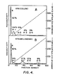
- Tables 5 and 8 show the percentages of the various oligosaccharide fractions (A to J) attached to each of the glycosylation sites Asn-117, Asn-184 and Asn-448.
- Tables 6 and 9 show for each of the glycosylation sites the percentages of the oligosaccharides which are complex, hybrid or oligomannose (high mannose). The data in Tables 6 and 9 are also shown graphically in the form of bar charts in FIG. 10. Tables 7 and 10 show for each of the oligosaccharides the percentages of the oligosaccharides which are neutral or sialylated.
- M, C, H represent the class of oligosaccharides present, i.e., oligomannose, complex or hybrid.
- the order represents their position on the polypeptide, i.e., 117, 184, 448. In type II the order is 117, 448. Glycoforms indicated with an asterisk are found on both Bowes melanoma t-PA and colon t-PA.
- the comparative stabilities of colon t-PA and melanoma t-PA were examined under a variety of temperature conditions and buffer compositions at about normal physiological pH. In all such cases tested, the colon t-PA was found to be substantially more stable than the melanoma t-PA; that is, the colon t-PA ranged from about 1.2 times to about 2.6 times more stable than the melanoma t-PA, depending upon the conditions used. Since the amino acid sequences of the two t-PA products are alike, the differences in stability were deemed to be due to the carbohydrate structure. In another test run under selected assay conditions at a relatively low pH of about 2.8, the colon t-PA was found to be only about 5-10% more stable than the melanoma t-PA.
- the various temperature and buffer conditions used were: (1) 60°C for 10 hours in phosphate buffered saline containing 0.01% Tween 80 and 0.01% sodium azide, pH 7.4, supplemented with either one or ten mg/ml of human serum albumin; (2) 98°C for 60 seconds in the same buffer without the albumin, above, but with or without an equal volume of 0.1 M glycine-HCl, pH 2.5, containing 0.01% Tween 80; and (3) 70-73°C for 40-90 minutes in 20 mM Tris-HC1, pH 7.6, containing 0.1 M NaCl, 0.01% Tween 80 and 100 pg/ml bovine serum albumin.
- amidolytic hydrolysis of the chromogenic peptide substrate S-2322 (H-D-Val-Gly-Arg-p-nitroanilide, KabiVitrum) by t-PA is measured by monitoring the change in absorbance at 410 nm with time.
- parabolic assay plasminogen activation is measured by hydrolysis of the chromogenic plasmin substrate, S-2251 (H-D-Val-Leu-Lys-p-nitroanilide, KabiVitrum), in a reaction mixture containing t-PA, excess plasminogen, and excess S-2251.
- t-PA was isolated from the colon cells and the conditioned culture medium that was either supplemented or unsupplemented with B 2 -tunicamycin (Boehringer-Mannheim) in about microgram amounts per ml.
- B Z -tunicamycin is known to inhibit the N-glycosylation of cell-synthesized proteins.
- a t-PA fraction containing 16% or less of the normal amount of mannose found on the colon t-PA was thus compared with the normally (fully) glycosylated t-PA.
- t-PA stimulator a fibrin-like stimulator
- the untreated normal colon t-PA retained from 69-86% of its activity whereas the tunicamycin-treated t-PA with only partial glycosylation retained only 38-42% of its activity.
- the t-PA stimulator mimics the stimulatory effects of fibrin. See Zamarron et al., J. Biol. Chem. 259, 2080-2083 (1984).
- t-PA fractions were isolated from colon cells and the conditioned culture media supplemented with B 2 -tunicamycin, as above, and compared with colon t-PA isolated from the colon cells and conditioned culture media unsupplemented with B 2 -tunicamycin (control sample).
- t-PA over other plasminogen activators rests in its fibrin-specific action.
- the tunicamycin-treated (less glycosylated) sample was somewhat less active than the control sample.
- the less glycosylated t-PA also was less stable in the presence of the fibrin-like stimulator than the normal (fully) glycosylated t-PA.
- the colon t-PA of the invention can be used for the treatment of thrombolytic conditions by suitable administration to a patient in need of such treatment.
- the amount of the t-PA which would normally be administered is primarily dependent upon the physical characteristics of the recipient and the severity of the thrombolytic condition.
- the amount to be administered must be an effective amount, that is, an amount which is medically beneficial but does not present toxic effects which overweigh the advantages which accompany its use.
- the preferable route of administration is parenteral, especially intravenous. Intravenous administration of the t-PA in solution with normal physiologic saline is illustrative.
- compositions of the active t-PA in pharmaceutically acceptable diluents or carriers in therapeutic dosage form can be prepared by reference to general texts in the pharmaceutical field such as, for example, Remington's Pharmaceutical Sciences, Ed. Arthur Osol, 16th ed., 1980, Mack Publishing Co., Easton, Pennsylvania.
Landscapes
- Chemical & Material Sciences (AREA)
- Health & Medical Sciences (AREA)
- Life Sciences & Earth Sciences (AREA)
- Organic Chemistry (AREA)
- Engineering & Computer Science (AREA)
- Wood Science & Technology (AREA)
- Zoology (AREA)
- General Health & Medical Sciences (AREA)
- Genetics & Genomics (AREA)
- Bioinformatics & Cheminformatics (AREA)
- Biochemistry (AREA)
- Molecular Biology (AREA)
- Biomedical Technology (AREA)
- Medicinal Chemistry (AREA)
- General Engineering & Computer Science (AREA)
- Biotechnology (AREA)
- Microbiology (AREA)
- Materials Engineering (AREA)
- Chemical Kinetics & Catalysis (AREA)
- Polymers & Plastics (AREA)
- Medicines That Contain Protein Lipid Enzymes And Other Medicines (AREA)
- Enzymes And Modification Thereof (AREA)
- Peptides Or Proteins (AREA)
Applications Claiming Priority (6)
| Application Number | Priority Date | Filing Date | Title |
|---|---|---|---|
| US83408086A | 1986-02-26 | 1986-02-26 | |
| US849933 | 1986-04-09 | ||
| US06/849,933 US5132214A (en) | 1986-04-09 | 1986-04-09 | Large scale production of plasminogen activator from normal human colon cells |
| US06/929,950 US4751084A (en) | 1986-02-26 | 1986-11-12 | Tissue plasminogen activator from normal human colon cells |
| US929950 | 1986-11-12 | ||
| US834080 | 1992-02-12 |
Publications (2)
| Publication Number | Publication Date |
|---|---|
| EP0236289A2 true EP0236289A2 (de) | 1987-09-09 |
| EP0236289A3 EP0236289A3 (de) | 1988-12-07 |
Family
ID=27420247
Family Applications (1)
| Application Number | Title | Priority Date | Filing Date |
|---|---|---|---|
| EP87870023A Ceased EP0236289A3 (de) | 1986-02-26 | 1987-02-23 | Gewebe-Plasminogenaktivator von normalen menschlichen Dickdarmzellen |
Country Status (1)
| Country | Link |
|---|---|
| EP (1) | EP0236289A3 (de) |
Cited By (2)
| Publication number | Priority date | Publication date | Assignee | Title |
|---|---|---|---|---|
| EP0302503A3 (en) * | 1987-08-05 | 1990-03-28 | Boehringer Mannheim Gmbh | Method for the separation and purification of t-pa |
| EP0420833A1 (de) * | 1989-09-28 | 1991-04-03 | Monsanto Company | Verfahren zur Erhöhung der spezifischen Aktivität von t-PA |
Family Cites Families (1)
| Publication number | Priority date | Publication date | Assignee | Title |
|---|---|---|---|---|
| DE3015699C2 (de) * | 1979-04-26 | 1982-07-15 | Asahi Kasei Kogyo K.K., Osaka | Herstellung eines Plasminogen-Aktivators |
-
1987
- 1987-02-23 EP EP87870023A patent/EP0236289A3/de not_active Ceased
Cited By (2)
| Publication number | Priority date | Publication date | Assignee | Title |
|---|---|---|---|---|
| EP0302503A3 (en) * | 1987-08-05 | 1990-03-28 | Boehringer Mannheim Gmbh | Method for the separation and purification of t-pa |
| EP0420833A1 (de) * | 1989-09-28 | 1991-04-03 | Monsanto Company | Verfahren zur Erhöhung der spezifischen Aktivität von t-PA |
Also Published As
| Publication number | Publication date |
|---|---|
| EP0236289A3 (de) | 1988-12-07 |
Similar Documents
| Publication | Publication Date | Title |
|---|---|---|
| US4751084A (en) | Tissue plasminogen activator from normal human colon cells | |
| Parekh et al. | Cell-type-specific and site-specific N-glycosylation of type I and type II human tissue plasminogen activator | |
| EP1325113B1 (de) | Glykoformen von faktor vii | |
| Nishimura et al. | Human factor IX has a tetrasaccharide O-glycosidically linked to serine 61 through the fucose residue. | |
| Asada et al. | Structural study of the sugar chains of human leukocyte cell adhesion molecules CD11/CD18 | |
| Fukuda | Purification and characterization of endo-beta-galactosidase from Escherichia freundii induced by hog gastric mucin. | |
| Scudder et al. | Isolation and characterization of an endo-β-galactosidase from Bacteroides fragilis | |
| AU2001291652A1 (en) | Factor VII glycoforms | |
| Pohl et al. | Isolation and characterization of three different carbohydrate chains from melanoma tissue plasminogen activator | |
| US6352852B1 (en) | Method for the purification of human platelet heparanase | |
| TAKASATA et al. | Structural studies of the sugar chains of cold-insoluble globulin isolated from human plasma | |
| Tarentino et al. | Purification and properties of an endo-beta-N-acetylglucosaminidase from hen oviduct. | |
| US4851517A (en) | Tissue plasminogen activator oligosaccharide from normal human colon cells | |
| Bergwerff et al. | Primary structure of N‐linked carbohydrate chains of a human chimeric plasminogen activator K2tu‐PA expressed in Chinese hamster ovary cells | |
| Pfeiffer et al. | Carbohydrate structure of recombinant human uterine tissue plasminogen activator expressed in mouse epithelial cells | |
| EP0236289A2 (de) | Gewebe-Plasminogenaktivator von normalen menschlichen Dickdarmzellen | |
| Ludolph et al. | Degradation of keratan sulphate by β-N-acetylhexosaminidases A and B | |
| Yazawa et al. | α-L-fucosidase from aspergillus niger: Demonstration of a novel α-L-(1→ 6)-fucosidase acting on glycopeptides | |
| US5132214A (en) | Large scale production of plasminogen activator from normal human colon cells | |
| Fukuda | Isolation and characterization of a new endo-. beta.-galactosidase from Diplococcus pneumoniae | |
| EP0565241B1 (de) | Oligosaccharides | |
| US5089409A (en) | Method of increasing specific activity of t-pa | |
| Shiraishi et al. | Cell surface glycoprotein and asparagine-linked sugar chain patterns of rat erythroleukemic cell lines | |
| Bozon et al. | Characterization of cellular oligosaccharides from normal and cystic fibrotic fibroblasts using sequential endoglycosidase digestions | |
| US5070021A (en) | Method of modifying oligosaccharide structure of tissue plasminogen activator |
Legal Events
| Date | Code | Title | Description |
|---|---|---|---|
| PUAI | Public reference made under article 153(3) epc to a published international application that has entered the european phase |
Free format text: ORIGINAL CODE: 0009012 |
|
| AK | Designated contracting states |
Kind code of ref document: A2 Designated state(s): AT BE CH DE ES FR GB GR IT LI LU NL SE |
|
| PUAL | Search report despatched |
Free format text: ORIGINAL CODE: 0009013 |
|
| AK | Designated contracting states |
Kind code of ref document: A3 Designated state(s): AT BE CH DE ES FR GB GR IT LI LU NL SE |
|
| 17P | Request for examination filed |
Effective date: 19890502 |
|
| 17Q | First examination report despatched |
Effective date: 19901213 |
|
| STAA | Information on the status of an ep patent application or granted ep patent |
Free format text: STATUS: THE APPLICATION HAS BEEN REFUSED |
|
| 18R | Application refused |
Effective date: 19930805 |
|
| RIN1 | Information on inventor provided before grant (corrected) |
Inventor name: DWEK, RAYMOND A. Inventor name: CONNOLLY, DANIEL THOMAS Inventor name: RADEMACHER, THOMAS W. Inventor name: TOLBERT, WILLIAM ROBERT Inventor name: PAREKH, RAJ B. Inventor name: FEDER, JOSEPH Inventor name: HARAKAS, NICHOLAOS KONSTANTINOS Inventor name: WITTWER, ARTHUR JOHN Inventor name: SCHAUMANN, JON PETER |