EP0244207B1 - Nachweismethode und Gerät - Google Patents
Nachweismethode und Gerät Download PDFInfo
- Publication number
- EP0244207B1 EP0244207B1 EP19870303763 EP87303763A EP0244207B1 EP 0244207 B1 EP0244207 B1 EP 0244207B1 EP 19870303763 EP19870303763 EP 19870303763 EP 87303763 A EP87303763 A EP 87303763A EP 0244207 B1 EP0244207 B1 EP 0244207B1
- Authority
- EP
- European Patent Office
- Prior art keywords
- substance
- labelled
- gel
- dna
- solution
- Prior art date
- Legal status (The legal status is an assumption and is not a legal conclusion. Google has not performed a legal analysis and makes no representation as to the accuracy of the status listed.)
- Expired
Links
- 238000001514 detection method Methods 0.000 title claims description 20
- 239000000126 substance Substances 0.000 claims description 70
- 238000012360 testing method Methods 0.000 claims description 22
- 238000001962 electrophoresis Methods 0.000 claims description 20
- 238000000034 method Methods 0.000 claims description 13
- 238000000926 separation method Methods 0.000 claims description 8
- 238000000465 moulding Methods 0.000 claims description 6
- 239000011541 reaction mixture Substances 0.000 claims description 4
- 239000000499 gel Substances 0.000 description 50
- 108020004414 DNA Proteins 0.000 description 36
- 239000000243 solution Substances 0.000 description 33
- 239000000203 mixture Substances 0.000 description 15
- 239000003795 chemical substances by application Substances 0.000 description 13
- 238000002372 labelling Methods 0.000 description 13
- 239000000463 material Substances 0.000 description 12
- 239000000427 antigen Substances 0.000 description 9
- 102000036639 antigens Human genes 0.000 description 9
- 108091007433 antigens Proteins 0.000 description 9
- FAPWRFPIFSIZLT-UHFFFAOYSA-M Sodium chloride Chemical compound [Na+].[Cl-] FAPWRFPIFSIZLT-UHFFFAOYSA-M 0.000 description 8
- 238000001179 sorption measurement Methods 0.000 description 8
- 229920000936 Agarose Polymers 0.000 description 7
- 239000012528 membrane Substances 0.000 description 7
- YBJHBAHKTGYVGT-ZKWXMUAHSA-N (+)-Biotin Chemical compound N1C(=O)N[C@@H]2[C@H](CCCCC(=O)O)SC[C@@H]21 YBJHBAHKTGYVGT-ZKWXMUAHSA-N 0.000 description 6
- 102000004190 Enzymes Human genes 0.000 description 6
- 108090000790 Enzymes Proteins 0.000 description 6
- 230000001900 immune effect Effects 0.000 description 6
- 102000004169 proteins and genes Human genes 0.000 description 6
- 108090000623 proteins and genes Proteins 0.000 description 6
- 238000005406 washing Methods 0.000 description 6
- 102000053602 DNA Human genes 0.000 description 5
- 108020004682 Single-Stranded DNA Proteins 0.000 description 5
- 230000000295 complement effect Effects 0.000 description 5
- 239000003298 DNA probe Substances 0.000 description 4
- KCXVZYZYPLLWCC-UHFFFAOYSA-N EDTA Chemical compound OC(=O)CN(CC(O)=O)CCN(CC(O)=O)CC(O)=O KCXVZYZYPLLWCC-UHFFFAOYSA-N 0.000 description 4
- 239000000020 Nitrocellulose Substances 0.000 description 4
- 238000011161 development Methods 0.000 description 4
- 239000007788 liquid Substances 0.000 description 4
- 229920001220 nitrocellulos Polymers 0.000 description 4
- 229920002401 polyacrylamide Polymers 0.000 description 4
- 239000000523 sample Substances 0.000 description 4
- 239000011780 sodium chloride Substances 0.000 description 4
- 239000012085 test solution Substances 0.000 description 4
- KIWIFIXMWIMRBZ-UHFFFAOYSA-L 4-benzamido-2-methoxy-5-methylbenzenediazonium;dichlorozinc;dichloride Chemical compound [Cl-].[Cl-].Cl[Zn]Cl.C1=C([N+]#N)C(OC)=CC(NC(=O)C=2C=CC=CC=2)=C1C.C1=C([N+]#N)C(OC)=CC(NC(=O)C=2C=CC=CC=2)=C1C KIWIFIXMWIMRBZ-UHFFFAOYSA-L 0.000 description 3
- 229960002685 biotin Drugs 0.000 description 3
- 235000020958 biotin Nutrition 0.000 description 3
- 239000011616 biotin Substances 0.000 description 3
- 230000000903 blocking effect Effects 0.000 description 3
- 239000000872 buffer Substances 0.000 description 3
- 239000007853 buffer solution Substances 0.000 description 3
- 239000003153 chemical reaction reagent Substances 0.000 description 3
- 238000001914 filtration Methods 0.000 description 3
- 229920003023 plastic Polymers 0.000 description 3
- 239000004033 plastic Substances 0.000 description 3
- 108090001008 Avidin Proteins 0.000 description 2
- 108091003079 Bovine Serum Albumin Proteins 0.000 description 2
- 108020003215 DNA Probes Proteins 0.000 description 2
- 239000004677 Nylon Substances 0.000 description 2
- 239000004793 Polystyrene Substances 0.000 description 2
- 229920004890 Triton X-100 Polymers 0.000 description 2
- IOMLBTHPCVDRHM-UHFFFAOYSA-N [3-[(2,4-dimethylphenyl)carbamoyl]naphthalen-2-yl] dihydrogen phosphate Chemical compound CC1=CC(C)=CC=C1NC(=O)C1=CC2=CC=CC=C2C=C1OP(O)(O)=O IOMLBTHPCVDRHM-UHFFFAOYSA-N 0.000 description 2
- 239000002390 adhesive tape Substances 0.000 description 2
- 230000000274 adsorptive effect Effects 0.000 description 2
- 239000011543 agarose gel Substances 0.000 description 2
- 229940098773 bovine serum albumin Drugs 0.000 description 2
- -1 dinitrophenyl group Chemical group 0.000 description 2
- 230000005684 electric field Effects 0.000 description 2
- 238000011534 incubation Methods 0.000 description 2
- 230000037230 mobility Effects 0.000 description 2
- 229920001778 nylon Polymers 0.000 description 2
- 229920000642 polymer Polymers 0.000 description 2
- 229920002223 polystyrene Polymers 0.000 description 2
- 239000001509 sodium citrate Substances 0.000 description 2
- NLJMYIDDQXHKNR-UHFFFAOYSA-K sodium citrate Chemical compound O.O.[Na+].[Na+].[Na+].[O-]C(=O)CC(O)(CC([O-])=O)C([O-])=O NLJMYIDDQXHKNR-UHFFFAOYSA-K 0.000 description 2
- 108091032973 (ribonucleotides)n+m Proteins 0.000 description 1
- KJCVRFUGPWSIIH-UHFFFAOYSA-N 1-naphthol Chemical compound C1=CC=C2C(O)=CC=CC2=C1 KJCVRFUGPWSIIH-UHFFFAOYSA-N 0.000 description 1
- 241000972773 Aulopiformes Species 0.000 description 1
- 102000016928 DNA-directed DNA polymerase Human genes 0.000 description 1
- 108010014303 DNA-directed DNA polymerase Proteins 0.000 description 1
- 102000016911 Deoxyribonucleases Human genes 0.000 description 1
- 108010053770 Deoxyribonucleases Proteins 0.000 description 1
- 229910019142 PO4 Inorganic materials 0.000 description 1
- 239000004698 Polyethylene Substances 0.000 description 1
- 229920005654 Sephadex Polymers 0.000 description 1
- 239000012507 Sephadex™ Substances 0.000 description 1
- VMHLLURERBWHNL-UHFFFAOYSA-M Sodium acetate Chemical compound [Na+].CC([O-])=O VMHLLURERBWHNL-UHFFFAOYSA-M 0.000 description 1
- 239000007983 Tris buffer Substances 0.000 description 1
- 241000700605 Viruses Species 0.000 description 1
- AZRNEVJSOSKAOC-VPHBQDTQSA-N [[(2r,3s,5r)-5-[5-[(e)-3-[6-[5-[(3as,4s,6ar)-2-oxo-1,3,3a,4,6,6a-hexahydrothieno[3,4-d]imidazol-4-yl]pentanoylamino]hexanoylamino]prop-1-enyl]-2,4-dioxopyrimidin-1-yl]-3-hydroxyoxolan-2-yl]methoxy-hydroxyphosphoryl] phosphono hydrogen phosphate Chemical compound O1[C@H](COP(O)(=O)OP(O)(=O)OP(O)(O)=O)[C@@H](O)C[C@@H]1N1C(=O)NC(=O)C(\C=C\CNC(=O)CCCCCNC(=O)CCCC[C@H]2[C@H]3NC(=O)N[C@H]3CS2)=C1 AZRNEVJSOSKAOC-VPHBQDTQSA-N 0.000 description 1
- 239000007864 aqueous solution Substances 0.000 description 1
- KGBXLFKZBHKPEV-UHFFFAOYSA-N boric acid Chemical compound OB(O)O KGBXLFKZBHKPEV-UHFFFAOYSA-N 0.000 description 1
- 239000004327 boric acid Substances 0.000 description 1
- 238000004737 colorimetric analysis Methods 0.000 description 1
- 238000007796 conventional method Methods 0.000 description 1
- SUYVUBYJARFZHO-RRKCRQDMSA-N dATP Chemical compound C1=NC=2C(N)=NC=NC=2N1[C@H]1C[C@H](O)[C@@H](COP(O)(=O)OP(O)(=O)OP(O)(O)=O)O1 SUYVUBYJARFZHO-RRKCRQDMSA-N 0.000 description 1
- SUYVUBYJARFZHO-UHFFFAOYSA-N dATP Natural products C1=NC=2C(N)=NC=NC=2N1C1CC(O)C(COP(O)(=O)OP(O)(=O)OP(O)(O)=O)O1 SUYVUBYJARFZHO-UHFFFAOYSA-N 0.000 description 1
- RGWHQCVHVJXOKC-SHYZEUOFSA-J dCTP(4-) Chemical compound O=C1N=C(N)C=CN1[C@@H]1O[C@H](COP([O-])(=O)OP([O-])(=O)OP([O-])([O-])=O)[C@@H](O)C1 RGWHQCVHVJXOKC-SHYZEUOFSA-J 0.000 description 1
- HAAZLUGHYHWQIW-KVQBGUIXSA-N dGTP Chemical compound C1=NC=2C(=O)NC(N)=NC=2N1[C@H]1C[C@H](O)[C@@H](COP(O)(=O)OP(O)(=O)OP(O)(O)=O)O1 HAAZLUGHYHWQIW-KVQBGUIXSA-N 0.000 description 1
- 239000013024 dilution buffer Substances 0.000 description 1
- BNIILDVGGAEEIG-UHFFFAOYSA-L disodium hydrogen phosphate Chemical compound [Na+].[Na+].OP([O-])([O-])=O BNIILDVGGAEEIG-UHFFFAOYSA-L 0.000 description 1
- 229910000397 disodium phosphate Inorganic materials 0.000 description 1
- 235000019800 disodium phosphate Nutrition 0.000 description 1
- 230000000694 effects Effects 0.000 description 1
- 230000002255 enzymatic effect Effects 0.000 description 1
- 238000007429 general method Methods 0.000 description 1
- 239000011521 glass Substances 0.000 description 1
- 238000010438 heat treatment Methods 0.000 description 1
- 230000002401 inhibitory effect Effects 0.000 description 1
- 239000003446 ligand Substances 0.000 description 1
- 235000019799 monosodium phosphate Nutrition 0.000 description 1
- 239000002773 nucleotide Substances 0.000 description 1
- 125000003729 nucleotide group Chemical group 0.000 description 1
- NBIIXXVUZAFLBC-UHFFFAOYSA-K phosphate Chemical compound [O-]P([O-])([O-])=O NBIIXXVUZAFLBC-UHFFFAOYSA-K 0.000 description 1
- 239000010452 phosphate Substances 0.000 description 1
- 229920003229 poly(methyl methacrylate) Polymers 0.000 description 1
- 239000004417 polycarbonate Substances 0.000 description 1
- 229920000515 polycarbonate Polymers 0.000 description 1
- 229920000573 polyethylene Polymers 0.000 description 1
- 239000004926 polymethyl methacrylate Substances 0.000 description 1
- 238000002360 preparation method Methods 0.000 description 1
- 235000019515 salmon Nutrition 0.000 description 1
- 239000001632 sodium acetate Substances 0.000 description 1
- 235000017281 sodium acetate Nutrition 0.000 description 1
- AJPJDKMHJJGVTQ-UHFFFAOYSA-M sodium dihydrogen phosphate Chemical compound [Na+].OP(O)([O-])=O AJPJDKMHJJGVTQ-UHFFFAOYSA-M 0.000 description 1
- 229910000162 sodium phosphate Inorganic materials 0.000 description 1
- 241000894007 species Species 0.000 description 1
- 239000000758 substrate Substances 0.000 description 1
- 125000000472 sulfonyl group Chemical group *S(*)(=O)=O 0.000 description 1
- 238000009210 therapy by ultrasound Methods 0.000 description 1
- 238000013519 translation Methods 0.000 description 1
- LENZDBCJOHFCAS-UHFFFAOYSA-N tris Chemical compound OCC(N)(CO)CO LENZDBCJOHFCAS-UHFFFAOYSA-N 0.000 description 1
- 239000011534 wash buffer Substances 0.000 description 1
Images

Classifications
-
- C—CHEMISTRY; METALLURGY
- C12—BIOCHEMISTRY; BEER; SPIRITS; WINE; VINEGAR; MICROBIOLOGY; ENZYMOLOGY; MUTATION OR GENETIC ENGINEERING
- C12Q—MEASURING OR TESTING PROCESSES INVOLVING ENZYMES, NUCLEIC ACIDS OR MICROORGANISMS; COMPOSITIONS OR TEST PAPERS THEREFOR; PROCESSES OF PREPARING SUCH COMPOSITIONS; CONDITION-RESPONSIVE CONTROL IN MICROBIOLOGICAL OR ENZYMOLOGICAL PROCESSES
- C12Q1/00—Measuring or testing processes involving enzymes, nucleic acids or microorganisms; Compositions therefor; Processes of preparing such compositions
- C12Q1/68—Measuring or testing processes involving enzymes, nucleic acids or microorganisms; Compositions therefor; Processes of preparing such compositions involving nucleic acids
- C12Q1/6813—Hybridisation assays
-
- G—PHYSICS
- G01—MEASURING; TESTING
- G01N—INVESTIGATING OR ANALYSING MATERIALS BY DETERMINING THEIR CHEMICAL OR PHYSICAL PROPERTIES
- G01N27/00—Investigating or analysing materials by the use of electric, electrochemical, or magnetic means
- G01N27/26—Investigating or analysing materials by the use of electric, electrochemical, or magnetic means by investigating electrochemical variables; by using electrolysis or electrophoresis
- G01N27/416—Systems
- G01N27/447—Systems using electrophoresis
-
- G—PHYSICS
- G01—MEASURING; TESTING
- G01N—INVESTIGATING OR ANALYSING MATERIALS BY DETERMINING THEIR CHEMICAL OR PHYSICAL PROPERTIES
- G01N33/00—Investigating or analysing materials by specific methods not covered by groups G01N1/00 - G01N31/00
- G01N33/48—Biological material, e.g. blood, urine; Haemocytometers
- G01N33/50—Chemical analysis of biological material, e.g. blood, urine; Testing involving biospecific ligand binding methods; Immunological testing
- G01N33/53—Immunoassay; Biospecific binding assay; Materials therefor
- G01N33/558—Immunoassay; Biospecific binding assay; Materials therefor using diffusion or migration of antigen or antibody
- G01N33/561—Immunoelectrophoresis
Definitions
- the present invention relates to a method and an apparatus for qualitatively or quantitatively detecting a test substance capable of forming a complex e.g. DNA, RNA or an immunological substance such as an antigen or antibody.
- a test substance capable of forming a complex e.g. DNA, RNA or an immunological substance such as an antigen or antibody.
- Electrophoresis is widely used in separating DNA or RNA or in separating a variety of substances such as proteins, and involves separation by the use of differences in mobility of the substance in an electric field (see, e.g. DE-B2-2250086). This is done by using the fact that there are differences in size and electric charge among DNA, RNA and proteins.
- the separation is feasible in various areas because different mobilities are produced by adjusting concentration of the gel component of a buffer-containing polyacrylamide or agarose gel which is a generally applied medium in the electric field as well as by adjusting pH of the buffer solution. Then, such a separated substance is usually detected as bands on a considerably long area on the gel of plate form.
- DNA probes Several kinds of DNA probe kits are available on the market. In detecting DNA or RNA by utilizing these reagents or kits, materials adsorbing and fixing DNA or RNA such as porous membranes of nitrocellulose or nylon are often employed in practical use.
- DNA or RNA to be detected are adsorbed on the above-mentioned adsorbent-fixer material by spotting a test solution on or passing it through and fixed by heating.
- the adsorbent-fixer material is treated with a DNA not complementary to the DNA or RNA to be detected, for example, salmon sperm DNA.
- a hybrid is formed between the labelled or labelable DNA which is complementary to the DNA or RNA to be detected, namely, DNA probe and the fixed DNA or RNA to be detected.
- the labelled substance is detected (when the probe has been labelled).
- detection is made following treatment with a labeling agent and washing.
- the labeling agent is a protein such as an enzyme
- the residual adsorbing ability of the adsorbent-fixer material is blocked with a protein different from the labeling agent, for example, bovine serum albumin preceding the treatment with a labeling agent.
- the general method for detecting an immunological substance that is, for detecting an antibody in a specimen involves first placing a solution of an antibody against the antigen to be detected in a vessel made of a material highly adsorptive for proteins such as polystyrene to adsorb the antibody on the surface of the vessel at the wall and bottom, blocking the remaining adsorption capability of the vessel surface by treatment with an inactive protein such as bovine serum albumin, and after washing, placing a test solution in the vessel to contact antigen in the test solution with the adsorbed antibody thereby trapping the antigen by means of an antigen-antibody reaction.
- a treatment is made with an antibody labelled with a radioisotope, a fluorescent agent or an enzyme to effect labeling by an antigen-antibody reaction followed by washing and then detection.
- a radioisotope a radioisotope
- a fluorescent agent a fluorescent agent
- an enzyme to effect labeling by an antigen-antibody reaction followed by washing and then detection.
- detection method in general, there is employed as the reaction vessel a plate with 96 wells.
- the objects of the present invention are to overcome such disadvantages of the prior-art methods to provide a detection method which is associated with a lower degree non-specific adsorption, is simpler in procedure and requires a shorter period of time and an apparatus used therefor.
- a method of detecting a test substance which comprises contacting the test substance with a labelled or labelable substance and detecting a complex formed between the test substance and the labelled or labelable substance, characterised in that uncomplexed labelled or labelable substance is separated by electrophoresis on a gel which is chosen such that free labelled or labelable substance, when subjected to electrophoresis, will pass through and that the labelled or labelable substance which forms a complex with the test substance will not, the separation by electrophoresis being carried out prior to detection of said complex.
- the invention also provides apparatus for carrying out the above method which comprises a plurality of tubular passages which are each open at one end thereof and closed with a gel layer at the other end for providing a space to maintain a specimen containing the reaction mixture of the test substance and a labelled or labelable substance, the tubular passages being so arranged as to allow electrophoresis of the specimen through the gel.
- Test substances as used herein include DNA or RNA of virus-, microorganism-, animal- or plant-origin, or immunological substances such as antigen or antibody.
- Labelled substance as used herein refers to DNA or RNA that is complementary to the DNA or RNA of virus-, microorganism-, animal- or plant-origin, and immunological substances to which a labeling agent such as a fluorescent, chemiluminescent, radioisotopic or enzymatic agent is attached.
- the labelled substance of the present invention may be either one to which a labeling agent is directly attached as described above, or one in which the DNA, RNA or immunological substance is attached via a ligand or hapten such as biotin, a dinitrophenyl group or a sulfonyl group to an antibody against a hapten or avidin to which a labeling agent is attached.
- a labeling agent such as biotin, a dinitrophenyl group or a sulfonyl group to an antibody against a hapten or avidin to which a labeling agent is attached.
- One of the characteristic features of the present invention lies in the feasibility of treating the specimen solution containing a test substance with a labelled substance in a homogeneous system within a vessel. Another feature is the use of electrophoresis as a means of detecting the test substance by measuring the labelled substance which forms a complex remaining from separation following removal of the labelled substance which does not form the complex, these two labelled substances having been coexistent in a system.
- An embodiment of the means for separating by electrophoresis the labelled substance that does not form the complex to remove it may be illustrated by the methods set forth below.
- One of these is subjecting to electrophoresis a specimen of the test substance treated with a labelled substance by being applied as a layer on to a gel layer. Thickness of the gel layer may be appropriately chosen, but usually, it is set in a range between 1 and 3 mm in consideration of conditions such as time of electrophoresis and mechanical strength.
- the gel layer is desirably made of a material such as agarose or polyacrylamide fixed at a concentration through which the complex does not, or only to a small extent, passes and through which the labelled substance easily passes.
- the layer is preferably made to gel by the addition of a material such as agarose at such a concentration that the complex and the labelled substance can move. It may be arranged, however, to allow the labelled substance that does not form a complex to move in a direction opposite to the complex by adjusting the pH of the electrophoretic medium.
- An alternative method is one in which the specimen layer is incorporated within the gel layer. It is arranged in such a way that the specimen solution is incorporated into a gel layer of a material such as agarose or polyacrylamide at a concentration through which the complex does not, or only to a small extent, passes, but through which the labelled substance easily passes, to remove the labelled substance that does not form the complex from the gel layer.
- a material such as agarose or polyacrylamide
- the complex is large enough as compared with the labelled substance to facilitate the separation.
- the test substance is e.g. DNA or RNA
- the substance is in nature large enough to allow satisfactory separation by employing labelled DNA, complementary to the former, that is short in nature or has been cut short by such a means as an enzyme or ultrasonic treatment.
- the test substance is e.g. DNA or RNA
- an antigen or antibody which is of approximately the same size as that of the labelled substance separation will become feasible by forming a complex of the labelled substance with the test substance bound to a substance significantly larger than the labelled substance such as, for example, a polymer of antibody against the antigen or antigen against the antibody or a polymer to which the antigen or antibody is bound.
- the apparatus of the present invention which is for detecting a test substance, may include tubular passages which are each open at one end thereof and closed with a gel layer at the other end for providing a space to maintain and migrate a specimen containing the reaction mixture of the test substance and a labelled substance and to be subjected to electrophoresis, the apparatus may further include means for subjecting the specimen to electrophoresis.
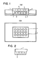
- the apparatus shown in Fig.1 is provided with a plate 1 with a large number of through holes 2 to maintain the specimen or the gel, and a conductive solution layer 5 to subject the specimen layer 4 and the gel layer 3 to electrophoresis.
- the conductive solution layer is composed of two liquid layers each of which contains the mutually different electrode 6 or 6 ⁇ .
- the conductive liquid layer 5 may be arranged in such a way that one of the liquid layer is within each of the holes 2. Alternatively, it may be on top of the holes. Any form may be used provided the two conductive solution layers are arranged on opposite sides of the specimen layer 4 and the gel layer 3 to allow for electrophoresis to occur in both layers.
- the mutually opposed electrodes 6 and 6 ⁇ also may be arranged in such a way that one of the electrodes is set in the liquid layer of each of the holes as with 6, or alternatively, it may be set as with 6 ⁇ .
- the plate 1 is desirably set horizontally. However, a vertical or diagonal arrangement may be used.
- the plate may be made of any material. Examples include plastics such as polystyrene, polycarbonate, polyethylene and polymethyl methacrylate, glass, gels such as agarose and polyacrylamide.
- a plurality of tubular passages which are each open at one end thereof and closed with a gel layer at the other end, is consituted by a plate 1 which has a plurality of through holes 2.
- the shape of the holes 2 is not specially limited, but a circular shape is preferred, and any sectional shape may be adopted.
- those holes may be of such a shape as shown in Fig. 1, or may be tapered as in Fig. 2.
- the holes are 3 to 5 mm in diameter and 1 to 2 cm in length and it is preferred that the holes are arranged regularly.
- plate 1 has a shape similar to the 96-well plate generally used in an immunological detecting method.
- the gel layer may be used in such a mode as shown in Fig.1 in which it is held within the holes and a specimen layer is placed thereon.
- the gel layer may be used in such a mode that it is present outside the holes in close contact with the lower ends of the holes and a specimen layer is placed thereon through the holes. By so doing a tubular passage sealed by the gel layer is formed.
- tubes 7 of a cylindrical or like shape formed of a plastic material for example as shown in Fig. 3.
- the gel layer 3 may be brought into close contact with one ends of the tubes 7 as shown in Fig.3, or one ends of the tubes 7 may be lightly pressed in to the gel layer 3.
- tubular passages and the gel layer may be integrated into a gel molding 9 having a plurality of wells 8, as shown in Fig.4.
- each of the specimens is placed in a reaction vessel to which is then added a labelled substance.
- the mixture reacts to form a complex.
- a gel layer is prepared in the holes of the plate as shown in Fig. 1.
- the gel layer may have been prepared in advance.
- the reaction solution is applied as a layer on to the gel layer, or preferably, the reaction solution containing the gel component added at such concentration as described above is similarly applied as a layer on the gel layer.
- the plate is set in such a way that the lower surface of the plate is in contact with the surface of the conductive solution layer.
- a conductive solution is poured on to the upper surface of the plate to form the upper conductive solution layer, followed by application of electric current at a predetermined voltage for a predetermined period of time. Then, the labelled substance is measured. It is preferable to do this while maintaining the gel layer in the holes.
- the labelling agent is enzyme
- the detection can be done by color development of the labelling enzyme through substrate.
- the labelling agent is a radioisotope
- the use of a scintillation counter or a Geiger counter permits detection, or exposure of film to light is effective.
- biotin or hapten is used as the labelled substance of the complex, a further reaction is allowed to take place between the complex and avidin or antibody with a labelling agent such as enzyme immobilized thereto, and thereafter the labelling agent is measured according to a conventional method, whereby the detection can be effected.
- specimen may be placed in the holes 2 or the tubes 7.
- the gel molding shown in Fig.4 only specimen may be placed in the wells 8 because the bottom of the gel molding 9 corresponds to the gel layer. The following operations are the same as in the case of the plate shown in Fig.1.
- the detection method according to the present invention it is feasible to form a complex of the test substance with a labelled substance by treatment in a homogeneous system in a single vessel.
- the treatment step for inhibiting non-specific adsorption and the washing step required in the prior-art methods may be omitted. Time requirement can also be reduced. Furthermore, there is no clogging of the porous membrane of nitrocellulose or nylon associated with the filtration-adsorption method described above so that treatment of a large amount of test material is feasible.
- the solution was diluted with 1XTBE to five specimens. 1 ⁇ g/ml, 100 ng/ml, 10 ng/ml and 1XTBE only. Each 50 ⁇ l of the specimens was placed in a sample tube, to which 5 ⁇ l of 1:20 diluted solution of a detection complex of Detek-1-acp (ENZO).
- the mixture was incubated at 42°C for 10 minutes. Separately, 50 ⁇ l of a heated solution of 1XTBE containing 3% agarose (BRL.) was introduced into the holes of a multi-hole plate with one side blocked by an adhesive tape of the electrophoresis apparatus shown in Fig.1 and cooled to form a gel. Separately, to the test solution was added each 50 ⁇ l of a heated solution of 1XTBE containing 1.2% agarose, and the mixture was thoroughly stirred. Each 50 ⁇ l of the mixture was applied as a layer on to the gel layer which had been prepared in advance, and allowed to form a gel.
- 1XTBE containing 3% agarose BBL.
- the adhesive tape of the multi-hole plate was removed to allow for such an arrangement that the plate was in contact with the surface of the 1XTBE solution placed in the conductive solution layer.
- electric current was applied at a voltage of 70V for 15 minutes using the electrode of the multi-hole plate as the cathode.
- the gel layer was then soaked in a 100:1 mixture of 1 mM naphthol AS-MX phosphate and predetection buffer solution of Fast Violet B salt 4 mg/ml at room temperature for 15 hours to develop color. The color development was observed on the intersurface of the gel layer and the specimen layer. The lower limit of color development was 1 ng/ml of biotin-labelled ⁇ X-174RF DNA in the solution.
- Each 25 ⁇ l of 1 ⁇ g/ml, 100 ng/ml, 10 ng/ml and 1 ng/ml 1XSSC solution of ⁇ X-174 single-stranded DNA (BRL) was placed in a sample tube, to which was added each 25 ⁇ l of the ultrasonic-treated biotin-labelled ⁇ X-174RF DNA (1 ⁇ g/ml).
- the mixture was boiled for 5 minutes and then incubated at 60°C for 30 minutes and subsequently at 42°C for 10 minutes.
- To the incubated mixture was added 5 ⁇ l of 1:20 diluted solution of a detection complex of ENZO Detek-1-acp followed by incubation at 42°C for 1 minute.
- Example 2 To the incubated mixtures was added each 50 ⁇ l of a heated solution of 1XTBE containing 1.2% agarose at 42°C, and the mixture was thoroughly stirred. As in Example 1, 50 ⁇ l of the mixture was applied as a layer on to a 3% agarose gel layer (prepared in advance) to form a gel. Electric current was applied at 70V for 20 minutes in the same way as in Example 1. The gel layer was soaked in a 100:1 mixture of 1 mM naphthol AS-MS phosphate and a predetection buffer solution of Fast Violet B salt at room temperature for 15 hours. Color development was observed. The detection limit of ⁇ X-174 single-stranded DNA in the solution was 1 ng/ml.
Landscapes
- Health & Medical Sciences (AREA)
- Life Sciences & Earth Sciences (AREA)
- Chemical & Material Sciences (AREA)
- Immunology (AREA)
- Molecular Biology (AREA)
- Engineering & Computer Science (AREA)
- Organic Chemistry (AREA)
- Microbiology (AREA)
- Biochemistry (AREA)
- General Health & Medical Sciences (AREA)
- Analytical Chemistry (AREA)
- Physics & Mathematics (AREA)
- Urology & Nephrology (AREA)
- Proteomics, Peptides & Aminoacids (AREA)
- Electrochemistry (AREA)
- Biomedical Technology (AREA)
- Biotechnology (AREA)
- Pathology (AREA)
- Hematology (AREA)
- General Physics & Mathematics (AREA)
- Zoology (AREA)
- Wood Science & Technology (AREA)
- Chemical Kinetics & Catalysis (AREA)
- Cell Biology (AREA)
- Biophysics (AREA)
- Medicinal Chemistry (AREA)
- Food Science & Technology (AREA)
- Bioinformatics & Cheminformatics (AREA)
- General Engineering & Computer Science (AREA)
- Genetics & Genomics (AREA)
- Measuring Or Testing Involving Enzymes Or Micro-Organisms (AREA)
- Investigating Or Analysing Biological Materials (AREA)
Claims (4)
- Verfahren zum Nachweis einer Testsubstanz, bei dem man die Testsubstanz mit einer markierten oder markierbaren Substanz in Kontakt bringt und einen zwischen der Testsubstanz und der markierten oder markierbaren Substanz gebildeten Komplex nachweist, dadurch gekennzeichnet, daß man die nicht-komplexierte markierte oder markierbare Substanz durch Elektrophorese in einem Gel abtrennt, das so gewählt wird, daß die freie markierte oder markierbare Substanz, wenn sie einer Elektrophorese ausgesetzt wird, es durchläuft und daß die markierte oder markierbare Substanz, die einen Komplex mit der Testsubstanz bildet, es nicht durchläuft, wobei die Trennung durch die Elektrophorese vor dem Nachweis des Komplexes durchgeführt wird.
- Vorrichtung zur Durchführung des Verfahrens nach Anspruch 1 umfassend eine Vielzahl von röhrenförmigen Durchgängen (2), die an einem Ende offen sind und am anderen Ende mit einer Gelschicht (3) verschlossen sind, um einen Raum (4) auszubilden, um eine Probe aufzunehmen, die die Reaktionsmischung der Testsubstanz und einer markierten oder markierbaren Substanz enthält, wobei die röhrenförmigen Durchgänge so angeordnet sind, daß sie die Elektrophorese der Probe durch das Gel erlauben.
- Vorrichtung nach Anspruch 2, in der die röhrenförmigen Durchgänge von einer Platte (1) mit einer Vielzahl von Löchern (2) gebildet werden.
- Vorrichtung nach Anspruch 2, in der die mit einer Gelschicht verschlossenen röhrenförmigen Durchgänge ein Gel-Formteil (9) mit einer Vielzahl von Vertiefungen (8) umfassen.
Applications Claiming Priority (2)
| Application Number | Priority Date | Filing Date | Title |
|---|---|---|---|
| JP97877/86 | 1986-04-30 | ||
| JP9787786 | 1986-04-30 |
Publications (2)
| Publication Number | Publication Date |
|---|---|
| EP0244207A1 EP0244207A1 (de) | 1987-11-04 |
| EP0244207B1 true EP0244207B1 (de) | 1991-07-31 |
Family
ID=14203976
Family Applications (1)
| Application Number | Title | Priority Date | Filing Date |
|---|---|---|---|
| EP19870303763 Expired EP0244207B1 (de) | 1986-04-30 | 1987-04-28 | Nachweismethode und Gerät |
Country Status (3)
| Country | Link |
|---|---|
| EP (1) | EP0244207B1 (de) |
| JP (1) | JPS63228066A (de) |
| DE (1) | DE3771768D1 (de) |
Families Citing this family (8)
| Publication number | Priority date | Publication date | Assignee | Title |
|---|---|---|---|---|
| US5006473A (en) * | 1988-08-09 | 1991-04-09 | Abbott Laboratories | Electrophoresis method using vesicles |
| US5622868A (en) * | 1989-04-27 | 1997-04-22 | Microbiological Research Authority Camr (Centre For Applied Microbiology & Research) | Analytical apparatus utilizing a colorimetric or other optically detectable effect |
| US5482832A (en) * | 1992-07-08 | 1996-01-09 | Akzo Nobel N.V. | Hybridization assays using enzyme-linked probes |
| US6048692A (en) * | 1997-10-07 | 2000-04-11 | Motorola, Inc. | Sensors for electrically sensing binding events for supported molecular receptors |
| US6238909B1 (en) * | 1999-05-04 | 2001-05-29 | Motorola, Inc. | Method and apparatus for obtaining electric field-enhanced bioconjugation |
| FR2795093B1 (fr) * | 1999-06-21 | 2003-08-22 | Inst Rech Developpement Ird | Moyens pour l'identification du locus d'un gene majeur de la resistance au virus de la panachure jaune du riz et leurs applications |
| US9081006B2 (en) * | 2007-02-07 | 2015-07-14 | Lyzer Diagnostics, Inc. | Rapid homogeneous immunoassay using electrophoresis |
| JP2012194015A (ja) * | 2011-03-16 | 2012-10-11 | Graduate School For The Creation Of New Photonics Industries | 電気泳動チップ及び分析方法 |
Family Cites Families (3)
| Publication number | Priority date | Publication date | Assignee | Title |
|---|---|---|---|---|
| GB8306426D0 (en) * | 1983-03-09 | 1983-04-13 | Malcolm A D B | Detecting polynucleotide sequence |
| US4704255A (en) * | 1983-07-15 | 1987-11-03 | Pandex Laboratories, Inc. | Assay cartridge |
| US4849077A (en) * | 1984-08-06 | 1989-07-18 | Akademie Der Wissenschaften Der Ddr | Process for solid phase-sequencing of nucleic acid fragments |
-
1987
- 1987-04-28 JP JP10558187A patent/JPS63228066A/ja active Pending
- 1987-04-28 EP EP19870303763 patent/EP0244207B1/de not_active Expired
- 1987-04-28 DE DE8787303763T patent/DE3771768D1/de not_active Expired - Fee Related
Also Published As
| Publication number | Publication date |
|---|---|
| DE3771768D1 (de) | 1991-09-05 |
| JPS63228066A (ja) | 1988-09-22 |
| EP0244207A1 (de) | 1987-11-04 |
Similar Documents
| Publication | Publication Date | Title |
|---|---|---|
| CA1200761A (en) | Devices and kits for immunological analysis | |
| US6297062B1 (en) | Separation by magnetic particles | |
| EP0612354B1 (de) | Verfahren und Gerät zum Nachweis von Nukleinsäurensequenzen. | |
| EP0087899B1 (de) | Anordnung mit mehreren Bechern für immunologische Untersuchungen | |
| EP0135541B1 (de) | Matrix von mit antikörpern beschichteten flecken für die bestimmung von antigenen | |
| US5098825A (en) | Probe containing a modified nucleic acid recognizable by specific antibodies and use of this probe to detect and characterize a homologous dna sequence | |
| US5415758A (en) | Method and apparatus for electro-elution of biological molecules | |
| US4939098A (en) | Immunoassay and measurement kit used therefor | |
| US5059294A (en) | Method for separating nucleic acids and nucleic acid probes | |
| US5006473A (en) | Electrophoresis method using vesicles | |
| US5723031A (en) | Method for the analytical separation of viruses | |
| KR20010034165A (ko) | 전혈용 크로마토그래피 장치에서의 다가양이온의 중화 | |
| EP0244207B1 (de) | Nachweismethode und Gerät | |
| JPS5972059A (ja) | 抗原検定法及び検定用具 | |
| JPS589070A (ja) | 免疫分析用装置及びキツト | |
| JPH0743376B2 (ja) | 核酸配列の検定方法及び分子遺伝子プローブ | |
| US20080138831A1 (en) | Immunological assay and chip | |
| US5035786A (en) | Fluorescent tag for sugar electrophoresis | |
| EP0402023A1 (de) | Diagnostisches Gerät mit verlängerter Durchflussmembran und Verfahren | |
| KR920004415A (ko) | 알파 1-미소글로불린에 대해 특이적인 항원 결정소를 갖는 펩타이드 | |
| US7276369B2 (en) | Method for detecting a substance and microtiter plate | |
| US4409200A (en) | Reverse transcriptase from human milk, method for its purification, and its use in the detection of breast cancer | |
| US4826771A (en) | Procedure to be performed in conjunction with protein blotting or nucleic acid blotting | |
| Dean | Ultrasensitive Detection of Chemical Substances | |
| JPH06289027A (ja) | 核酸の高感度検出法 |
Legal Events
| Date | Code | Title | Description |
|---|---|---|---|
| PUAI | Public reference made under article 153(3) epc to a published international application that has entered the european phase |
Free format text: ORIGINAL CODE: 0009012 |
|
| AK | Designated contracting states |
Kind code of ref document: A1 Designated state(s): DE FR GB IT NL |
|
| 17P | Request for examination filed |
Effective date: 19880414 |
|
| 17Q | First examination report despatched |
Effective date: 19890906 |
|
| GRAA | (expected) grant |
Free format text: ORIGINAL CODE: 0009210 |
|
| AK | Designated contracting states |
Kind code of ref document: B1 Designated state(s): DE FR GB IT NL |
|
| PG25 | Lapsed in a contracting state [announced via postgrant information from national office to epo] |
Ref country code: IT Free format text: LAPSE BECAUSE OF FAILURE TO SUBMIT A TRANSLATION OF THE DESCRIPTION OR TO PAY THE FEE WITHIN THE PRE;WARNING: LAPSES OF ITALIAN PATENTS WITH EFFECTIVE DATE BEFORE 2007 MAY HAVE OCCURRED AT ANY TIME BEFORE 2007. THE CORRECT EFFECTIVE DATE MAY BE DIFFERENT FROM THE ONE RECORDED.SCRIBED TIME-LIMIT Effective date: 19910731 Ref country code: NL Effective date: 19910731 |
|
| REF | Corresponds to: |
Ref document number: 3771768 Country of ref document: DE Date of ref document: 19910905 |
|
| ET | Fr: translation filed | ||
| NLV1 | Nl: lapsed or annulled due to failure to fulfill the requirements of art. 29p and 29m of the patents act | ||
| PLBE | No opposition filed within time limit |
Free format text: ORIGINAL CODE: 0009261 |
|
| STAA | Information on the status of an ep patent application or granted ep patent |
Free format text: STATUS: NO OPPOSITION FILED WITHIN TIME LIMIT |
|
| 26N | No opposition filed | ||
| PGFP | Annual fee paid to national office [announced via postgrant information from national office to epo] |
Ref country code: FR Payment date: 19930408 Year of fee payment: 7 |
|
| PGFP | Annual fee paid to national office [announced via postgrant information from national office to epo] |
Ref country code: GB Payment date: 19930419 Year of fee payment: 7 |
|
| PGFP | Annual fee paid to national office [announced via postgrant information from national office to epo] |
Ref country code: DE Payment date: 19930506 Year of fee payment: 7 |
|
| PG25 | Lapsed in a contracting state [announced via postgrant information from national office to epo] |
Ref country code: GB Effective date: 19940428 |
|
| PG25 | Lapsed in a contracting state [announced via postgrant information from national office to epo] |
Ref country code: FR Effective date: 19941229 |
|
| PG25 | Lapsed in a contracting state [announced via postgrant information from national office to epo] |
Ref country code: DE Effective date: 19950103 |
|
| GBPC | Gb: european patent ceased through non-payment of renewal fee |
Effective date: 19940428 |
|
| REG | Reference to a national code |
Ref country code: FR Ref legal event code: ST |