EP0245098A2 - Verfahren und Gerät zur Beurteilung einer Wirbelsäuledeformation - Google Patents
Verfahren und Gerät zur Beurteilung einer Wirbelsäuledeformation Download PDFInfo
- Publication number
- EP0245098A2 EP0245098A2 EP87304069A EP87304069A EP0245098A2 EP 0245098 A2 EP0245098 A2 EP 0245098A2 EP 87304069 A EP87304069 A EP 87304069A EP 87304069 A EP87304069 A EP 87304069A EP 0245098 A2 EP0245098 A2 EP 0245098A2
- Authority
- EP
- European Patent Office
- Prior art keywords
- vertebrae
- deformation
- vertebral body
- wedge
- profile
- Prior art date
- Legal status (The legal status is an assumption and is not a legal conclusion. Google has not performed a legal analysis and makes no representation as to the accuracy of the status listed.)
- Granted
Links
Images


Classifications
-
- A—HUMAN NECESSITIES
- A61—MEDICAL OR VETERINARY SCIENCE; HYGIENE
- A61B—DIAGNOSIS; SURGERY; IDENTIFICATION
- A61B5/00—Measuring for diagnostic purposes; Identification of persons
- A61B5/103—Measuring devices for testing the shape, pattern, colour, size or movement of the body or parts thereof, for diagnostic purposes
- A61B5/107—Measuring physical dimensions, e.g. size of the entire body or parts thereof
- A61B5/1079—Measuring physical dimensions, e.g. size of the entire body or parts thereof using optical or photographic means
-
- A—HUMAN NECESSITIES
- A61—MEDICAL OR VETERINARY SCIENCE; HYGIENE
- A61B—DIAGNOSIS; SURGERY; IDENTIFICATION
- A61B5/00—Measuring for diagnostic purposes; Identification of persons
- A61B5/72—Signal processing specially adapted for physiological signals or for diagnostic purposes
- A61B5/7235—Details of waveform analysis
- A61B5/7264—Classification of physiological signals or data, e.g. using neural networks, statistical classifiers, expert systems or fuzzy systems
-
- G—PHYSICS
- G16—INFORMATION AND COMMUNICATION TECHNOLOGY [ICT] SPECIALLY ADAPTED FOR SPECIFIC APPLICATION FIELDS
- G16H—HEALTHCARE INFORMATICS, i.e. INFORMATION AND COMMUNICATION TECHNOLOGY [ICT] SPECIALLY ADAPTED FOR THE HANDLING OR PROCESSING OF MEDICAL OR HEALTHCARE DATA
- G16H15/00—ICT specially adapted for medical reports, e.g. generation or transmission thereof
-
- G—PHYSICS
- G16—INFORMATION AND COMMUNICATION TECHNOLOGY [ICT] SPECIALLY ADAPTED FOR SPECIFIC APPLICATION FIELDS
- G16H—HEALTHCARE INFORMATICS, i.e. INFORMATION AND COMMUNICATION TECHNOLOGY [ICT] SPECIALLY ADAPTED FOR THE HANDLING OR PROCESSING OF MEDICAL OR HEALTHCARE DATA
- G16H50/00—ICT specially adapted for medical diagnosis, medical simulation or medical data mining; ICT specially adapted for detecting, monitoring or modelling epidemics or pandemics
- G16H50/50—ICT specially adapted for medical diagnosis, medical simulation or medical data mining; ICT specially adapted for detecting, monitoring or modelling epidemics or pandemics for simulation or modelling of medical disorders
-
- G—PHYSICS
- G16—INFORMATION AND COMMUNICATION TECHNOLOGY [ICT] SPECIALLY ADAPTED FOR SPECIFIC APPLICATION FIELDS
- G16H—HEALTHCARE INFORMATICS, i.e. INFORMATION AND COMMUNICATION TECHNOLOGY [ICT] SPECIALLY ADAPTED FOR THE HANDLING OR PROCESSING OF MEDICAL OR HEALTHCARE DATA
- G16H50/00—ICT specially adapted for medical diagnosis, medical simulation or medical data mining; ICT specially adapted for detecting, monitoring or modelling epidemics or pandemics
- G16H50/20—ICT specially adapted for medical diagnosis, medical simulation or medical data mining; ICT specially adapted for detecting, monitoring or modelling epidemics or pandemics for computer-aided diagnosis, e.g. based on medical expert systems
Definitions
- the present invention relates to a method and apparatus for judging deformation of a vertebral body. More particularly, the present invention relates to a method and apparatus for judging deformation of a vertebral body which is one of the bones forming the spinal column. A judgment of the presence of vertebral body deformation accompanied by osteoporosis, as well as a classification of the deformation, is very important for grasping the progress of osteoporosis and for confirmation of the effect of therapy.
- the ratio (a/d) of the central length (a) to the front brim length (d) of the third lumbar vertebrae is determined as the index (Lumbar Spine Score) for the degree of bone atrophy, or a change in the longitudinal and lateral bone beams of the third lumbar vertebrae is observed as the index for the degree of bone atrophy (severity classified by Jikei University), or a judgment of the fracture of a vertebral body is made by measurement of the central length (a) , front brim length (d) and rear brim length (c) of a vertebral body [The New England Journal of Medicine, vol. 306, 446 (February 2 5, 1982)] , a method for classifying the type of deformation of a vertebral body has not heretofore been known.
- the objects of the present invention are to eliminate the above mentioned disadvantages of the prior art and to provide a method for objectively judging deformation of a vertebral body.
- a method for judging deformation of a vertebral body which comprises measuring a central length (a), a rear brim length (c), and front brim length (d) from a profile x-ray image of a vertebral body to be judged, determining ratios of c/d, a/c, and a/d, and classifying deformation of the vertebral body to be judged by using indices, c, d, c/d, a/c and a/d.
- a method for judging deformation of a vertebral body which comprises obtaining at least two indices by measuring a central length (a), vertebral body width (b), rear brim length (c) , and front brim length (a) from a profile x-ray image of a vertebral body, analyzing these indices by discriminant function and classifying a deformation of the vertebral body from results of said analysis.
- an apparatus for judging deformation of a vertebral body comprising (i) an input means for inputting at least two values of a central length (a) , vertebral body width (b), rear brim length (c), and front brim length (d) from a profile X-ray image of a vertebral body to be judged, (ii) an arithmetic means for operating the calculation necessary for discriminating "wedge-shaped vertebrae", “inverse wedge-shaped vertebrae”, “fish vertebrae”, and “no deformation” to at least two types by using the above-mentioned input value, (iii) a means for discriminating the at least two types by using the calculation results, (iv) a means for inputting the discriminant function and/or standard necessary for the calculation and discrimination, and (v) an output means for outputting the judgment result.
- the present inventors have made an intensive study of a method of objective evaluation of vertebral body deformation, and found that the presence of a vertebral body deformation can be objectively judged according to the judgment standard in which lengths of the vertebral body and their ratios are combined by accurately measuring the lengths of a front brim, rear brim, central portion from a profile X-ray image (i.e., an image on a film taken from the side by X-ray radiation) of a vertebral body and determining the ratios of the respective portions of the vertebral body, and further found that the type of vertebral body deformation can be judged according to the judgment standard in which lengths of the vertebral body and their ratios are combined, and the progress over a period of time of the vertebral body deformation can be judged from the change in the deformation type as well as a change in the lengths of the vertebral body, and thus accomplished the present invention.
- the profile X-ray image of vertebral body was visually observed by a physician when judging the vertebral body deformation, but according to the present invention, the lengths of the respective portions of the vertebral body are measured, judgment standards are prepared with reference to a judgment by a physician, and the presence of vertebral body deformation as well as a classification of the deformation type are objectively performed following the judgment standards.
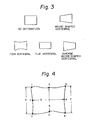
- the ratio c/d of the rear brim length to the front brim length must be between 0.7 and 1.4 as the first condition (a). For, as described below, if c/d becomes 1.4 or more, the front brim portion becomes a deformed wedge-shaped vertebrae, and if c/d becomes 0.7 or less, the rear brim portion becomes a deformed inverse wedge-shaped vertebrae, which in practice is very rare.
- At least one of rear brim length c and front brim length d must be greater than c - 2 ⁇ and d - 1.5a when the average values of the rear brim length c and front brim length d of vertebral body without deformation determined in (i) are made c and d, respectively.
- a, c, d all have a high value, namely a > a - 2 ⁇ a , c ⁇ c - 2 ⁇ c , d > d - 1.5 ⁇ d , but even when the vertebral body is slightly compressed to become a ⁇ a - 2 ⁇ a , provided that at least one of the conditions of c ⁇ c - 2 ⁇ c or d ⁇ d - 1.5 ⁇ d , can be satisfied, no clear vertebral body deformation is recognized and judgment of "no deformation" may be made.
- At least one of a/c and a/d must be more than 0.8. For, if both of a/c and a/d are 0.8 or less, namely the center length a is much smaller, than the rear brim length c and front brim length d, a fish vertebrae condition as described below is determined.
- the front brim length (d) has become much smaller than the rear brim length (c).
- a wedge-shaped vertebral body deformation can be easily recognized, but in some cases, a clear wedge-shaped vertebrae cannot be easily recognized.
- the wedge-shaped vertebrae (type I ) is inclusive of deformations with shortened front brim length, like a wedge-shaped vertebrae, as a result of, for example, an upper brim pressure fracture, upper brim depressed fracture, lower brim pressure fracture, lower brim depressed fracture, and as described below, even in the case of a vertebral body which is judged as a flat vertebrae because a ⁇ a - 2a a , c ⁇ c - 2 ⁇ c , d ⁇ d - 1.5 ⁇ d , if there is a remarkable deformation at the front brim portion satisfying c/d > 1.4, it can be judged to be a wedge-shaped vertebrae.
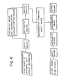
- the inverse wedge-shaped vertebrae may be defined as a vertebral body wherein deformation has occurred at the rear brim portion, and thus the rear brim length (c) has become smaller than the front brim length (d), contrary to the wedge-shaped vertebrae wherein deformation has occurred at the front brim portion, and the front length (d) has become smaller than the rear brim length (c) (see Fig. 3), but in practice, such a vertebral body does not substantially exist.
- the rear brim length (c) and the front brim length (d) are both smaller than c - 2 ⁇ c and d - 1.5 ⁇ d when the average values of the vertebral body without deformation are made c and d; namely, c ⁇ c - 2a c and d ⁇ d - 1.5 ⁇ d .
- the flat vertebrae is a vertebral body having a relatively uniform deformation at the front brim portion, the central portion, and the rear brim portion under pressure (see Fig. 3); namely, all of the front brim length (d), the central length (a) and the rear brim length (c) are smaller.
- the conditions of -2 ⁇ c for c and -1.5 ⁇ d for d are set because c is greater than d ( c > d) as shown in Example 1, and thus c and d become approximately equal values by setting such conditions, to satisfy the conditions for a flat vertebrae.
- the second condition (c) is 0.7 ⁇ c/d ⁇ 1.4.
- deformation at the front brim portion is particularly marked, and where c/d ⁇ 1.4, it can be judged to be a wedge-shaped vertebrae, while in the case of a marked deformation at the rear brim portion when c/d ⁇ 0.7, it is judged to be an inverse wedge-shaped vertebrae.
- “Fish vertebrae” is a vertebrae where a depressed fracture or a pressure fracture has occurred at the central portion, whereby the central length (a) is particularly smaller than the front brim length (d) and rear brim length (c) (see Fig. 3). Accordingly, among the so called “no deformation” classifications, except for wedge-shaped vertebrae, flat vertebrae, and inverse wedge-shaped vertebrae, the central length (a) is particularly smaller.
- the fish vertebrae (type II) is also inclusive of deformations with a shortened central length like fish vertebrae, as a result of an upper brim pressure fracture, upper brim depressed fracture, lower brim pressure fracture, lower brim depressed fracture, etc.
- the upper limit as 1.4 and the lower limit as 0.7 as the judgment standard for c/d as a preferable example, but these values can be selected freely, for example, in the case of the upper limit, from the range of 1.25 to 1.55, preferably from 1.33 to 1.5, more preferably from 1.4 to 1.45.
- the lower limit can be selected freely from the range of 0.8 to 0.6, preferably from 0.75 to 0.65.
- 0.8 was selected in the case of "no deformation” and "fish vertebrae", but this value can be freely selected from the range of 0.65 to 0.9, preferably from 0.7 to 0.85, more preferably from 0.75 to 0.8.
- c and d As the judgment standard for c and d, c - 2 ⁇ c and d - 1.5 ⁇ d were selected in the case of "no deformation", "flat vertebrae', and “fish vertebrae” but the judgment standard for c can be freely selected from the range of c - 1.0 ⁇ c to c - 2.5 ⁇ c , preferably c - 1.25 ⁇ c to c - 2.25 ⁇ c , more preferably c -1.5 ⁇ c to c -2.0 ⁇ c .
- ⁇ 1 to ⁇ 6 may optionally be determined from a discriminating function capable of discriminating two groups with two variants or two groups with one variant by using values obtained by measuring two of a, b, c, d, and e with respect to at least five profile X-ray image in each of two types among four types (i.e., wedge-shaped vertabrae, fish vertabrae flat vertebral, and no deformation).
- ⁇ 1 is z 5 obtained by the following discriminant function using the ratio c/d derived from c and d, which are obtained by measuring at least 5 profile X-ray images in each of two types selected from "wedge-shaped vertebrae" and "no deformation”:
- the present inventors have made an intensive study of a method of objective evaluation of vertebral body deformation, and found that the presence of a vertebral body deformation as well as the deformation type can be judged objectively by use of a discriminant function by accurately measuring the lengths of, for example, the front brim (d), rear brim (c), central portion (a), and the vertebral body width (b) from a profile X-ray image of a vertebral body, and further found that the progress over a period of time of the vertebral body deformation can be judged from the change in the type of said deformation as well as the change in the lengths of the vertebral body, and thus accomplished the present invention.
- a discriminant function by accurately measuring the lengths of, for example, the front brim (d), rear brim (c), central portion (a), and the vertebral body width (b) from a profile X-ray image of a vertebral body, and further found that the progress over a period of time of the verte
- vertebral body deformation has been judged by a physician by visual observation of the profile X-ray image of a vertebral body, but according to the method of the present invention, the lengths of the respective portions of a vertebral body are measured and for the vertebral bodies judged by a physician to be "no deformation", "wedge-shaped vertebrae", “fish vertebrae”, and "flat vertebrae", discrimination is made by discriminant functions to determine discriminant formulae for the respective types of vertebral body deformations, and thereafter, the presence of vertebral body deformation and the type of deformation are objectively judged by this discriminant formulae for a patient whose vertebral body deformation is to be judged.
- deformed vertebral bodies are classified in the present invention generally into wedge-shaped vertebrae, fish vertebrae, and flat vertebrae.
- the wedge-shaped vertebrae (type I) deformation has occurred at the front brim portion as shown in Fig. 4, and the front brim length has become particularly smaller than the rear brim length, including in addition to the so called wedge-shaped vertebrae deformation with a shortened front brim length like wedge-shaped vertebrae as the result of upper brim pressure fracture, an upper brim depressed fracture, lower brim pressure fracture, and lower brim depressed fracture, etc.
- the depressed fracture or pressure fracture has occurred at the central portion, whereby the central length has become particularly smaller than the front brim length and rear brim length (see Fig.
- the front brim portion, central portion, and rear brim portion are deformed relatively uniformly under pressure (see Fig. 4); namely, all of the front rear length, central length, and rear brim length are smaller.
- wedge-shaped vertebrae (type IV). This type is defined as a vertebral body wherein deformation has occurred at the rear brim portion, and thus the rear brim length is smaller than the front brim length, contrary to the wedge-shaped vertebrae where deformation has occurred at the front brim portion, and the front brim portion is smaller than the rear brim length (see Fig. 4). In practice, however, such a vertebrae does not substantially exist. Accordingly, it is sufficient if the four types, wedge-shaped vertebrae, fish vertebrae, flat vertebrae and "no deformation", can be classified.
- the above measurement is used to determine the discriminant formula for discriminating the presence of vertebral body deformation as well as the deformation type.
- the vertebral bodies of these respective types should be preferably vertebral bodies of persons generally 50 to 75 years old, in view of the age of the patient whose vertebral body deformation is to be judged. Also, it is preferable to perform measurements separately for men and women, and it is desirable to perform measurements of about 50 or more vertebral bodies for both men and women.
- the discriminant formula for discrimination of the vertebral body deformation type according to the method of the present invention can be determined if at least 5 each of these deformed vertebral bodies can be measured.
- the size differs for each vertebral body, and therefore, it is necessary to measure each vertebral body and determine each deformation type.
- the thoracic vertebrae from the first thoracic vertebrae to the twelfth thoracic vertebrae, lumbar vertebrae from the first lumbar vertebrae to the fifth lumbar vertebrae should be included, giving a total of 17 vertebral bodies, and therefore, for example, it is advantageous to take the photograph of the profile X-ray image with the eighth thoracic vertebrae as the center separately from the profile X-ray image with the third lumbar vertebrae as the center.
- the first thoracic vertebrae cannot be accurately measured in most cases, and further, the second and the third thoracic vertebrae are unclear in many cases.
- the frequency of vertebral body deformation is not great in these vertebral bodies, it is sufficient if measurement can be made from the fourth thoracic vertebrae or the fifth thoracic vertebrae.
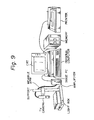
- the center length (a) width (b) , rear brim length (c) , and front brim length (d) of a vertebral body are specifically as shown in Fig. 4.
- the central length (a), width (b), rear brim length (c), and front brim length (d) may be measured for each vertebral body with a scale or slide calipers, but the eight points of 1 to 8 shown in Fig. 4 can be input through a digitizer into a computer to measure the central length (a) (3-4), width (b) (7-8), rear brim length (c) (5-6), and front brim length (d) (1-2).
- the degree of blackening may. be recorded with a TV camera, etc., and the lengths of the respective portions of the vertebral body may be automatically measured by image processing.
- discriminant formula for discriminating "no deformation", wedge-shaped vertebrae, fish vertebrae, and flat vertebrae is determined.
- Such a discriminating analysis is described in, for example, Chuichi Okuno et al "Multivariate Analytical Method” (published by Nikkagiren); Chuichi Okuno et al “Overall Multivariate Analytical Method” (published by Nikkagiren); Koichi Sugiyama “Introduction to Multivariate Data Analysis” (published by Asakura Shoten), and others, and calculation can be made by referring to these literatures.
- z 1 , z 2 , z 3 , and z 4 are discriminant values of "wedge-shaped vertebrae", “fish vertebrae”, “flat vertebrae”, and “no deformation”, respectively
- x 1 , x 2 , x 3 , and x 4 are variables corresponding to a central length, vertebral body width, rear brim length, and front brim length, respectively
- a 10 , a 20 ' a 30 ' and a 40 are constants
- all to a 44 are coefficients.
- This discriminant formula must be determined for each vertebral body from the first thoracic vertebrae to the fifth lumbar vertebrae, generally from the fourth thoracic vertebrae to the fifth lumbar vertebrae.
- the present invention is not limited to the above classification of the four vertebral deformation types of "no deformation", wedge-shaped vertebrae, fish vertebrae and flat vertebrae from the four measured values of central length (a), vertebral body width (b), rear brim length (c), and front brim length (d), but it is also possible to classify the vertebral body deformation types by use of the discriminant function as described below.
- the case occurs wherein classification is required for only the three types of "no deformation", wedge-shaped vertebrae, and flat vertebrae, or the case when sufficient measured values of a, b, c, and d of fish vertebrae cannot be obtained for determining the discriminant formula even if a classification of the four types including fish vertebrae is desired.
- the discriminant formula for classifying the three types of "no deformation", wedge-shaped vertebrae, and flat vertebrae from the four measured values of the central length (a), vertebral body width (b), rear brim length (c), and front brim length (d), or from three of these four measured values, for example, three measured values of a, c, and d.
- the discriminant function is represented by the following formula in which the ratio (c/d) of c and d measured from at least 5 profile X-ray images in each of two types of :wedge-shaped vertebrae" and "no deformation” and the ratio (c/d) of less than z 5 obtained from the vertebral body to be judged is judged as "no deformation” and the ratio (c/d) equal to or larger than z 5 is judged as "wedge-shaped vertebrae".
- ⁇ 1 and ⁇ 2 are averages of c/d relating to the profile X-ray images of "wedge-shaped vertebrae” and “no deformation", respectively, ⁇ 1 and ⁇ 2 are numbers of the X-ray images of "wedge-type vertebrae” and “no deformation”, respectively, ⁇ is an average of ⁇ 1 and ⁇ 2 , and ⁇ is an average of ⁇ 1 and ⁇ 2 ; and (ii) When ⁇ 1 and ⁇ 2 are substantially different,
- ⁇ 1 and ⁇ 2 are averages of c/d relating to the profile X-ray images of "wedge-shaped vertebrae” and “no deformation", respectively, ⁇ 1 and ⁇ 2 are numbers of the X-ray images of "wedge-type vertebrae” and “no deformation”, respectively.
- the apparatus for judging the deformation of vertebral body according to the present invention is used for the practice of the above-mentioned method of the present invention.
- This apparatus is basically composed of an imputting means for at least two indices of a, b, c, and d, an arithmetic means for effecting necessary operations for the descrimination, a discriminating means by using the calculation results, a means for inputting the discriminant function and/or standard necessary for the calculation and discrimination, and an output means for outputting the judgment result.
- a rule means As the inputting means, a rule means, a digitizer means, an image processing means in which an image inputting means such as a TV camera is used.
- c/d, a/c, and a/d are determined by the arithmetic means, and c, d, ⁇ c , o d , and ⁇ 1 - ⁇ 6 are inputted as discriminating standards by the discriminating standard inputting means, and the discriminating means can discriminate the results by these standards.
- the present apparatus can be further provided with a means for obtained the discriminating standards from a number of the profile X-ray images having no substantial deformation.
- the present apparatus can be optionally provided with an arithmetic means capable of discriminating two groups with two variants, which are used for determining the indices ⁇ 1 to ⁇ 6 , or of discriminating two groups with one variant.
- the data of at least two types of the four types of the vertebral body i.e., ,”wedge-shaped vertebrae", “fish vertebrae", “flat vertebrae”, and "no deformation" should be input.
- the arithmetic means is connected to a discriminating standard input means for inputting the discriminant function, whereby the arithmetic means calculates the discriminating values with said discriminant function and the discriminating means determine the type of the vertebral body based on the maximum discriminating value.
- the present apparatus is provided with a function capable of discriminating the four types of the vertebral body, "wedge-shaped vertebrae", “fish vertebrae”, “flat vertebrae”, and "no deformation" from a one-dimensional formula containing four variables a, b, c, and d.
- the apparatus which is capable of judging the two types of the vertebral body with one variant.
- the type of vertebral body deformation can be objectively evaluated, and also the change in the said deformation type and the progress over a period of time of the vertebral body deformation can be judged.
- the present method and apparatus is very useful for determining the progress of bone disease such as osteoporosis, etc., and for confirmation of the effect of therapy.
- the type of vertebral body deformation can be objectively evaluated, and further the change in the type of deformation type and the progress over a period of time of the vertebral body deformation can be judged.
- the present method and apparatus is very useful for determining the progress of bone disease such as osteoporosis, etc., and for confirmation of the effect of therapy.
- the second lumbar vertebrae changed from a "no deformation" classification to a type III (flat vertebrae) within six months.
- the conditions of the front brim and rear brim of the seventh thoracic vertebrae, and the center of the tenth thoracic vertebrae tend to have worsened. Also, the conditions of the centers of the tenth thoracic vertebrae and the third lumbar vertebrae had further worsened.
- vertebral bodies including 6 vertebral bodies judged as wedge-shaped vertebrae, 25 vertebral bodies judged as fish vertebrae, eight vertebral bodies judged as flat vertebrae and 147 vertebral bodies judged as "no deformation" according to visual observation by a physician of a profile X-ray image, with the third lumbar vertebrae as the center, of osteoporosis patients (women), eight points of 1 to 8 shown in Fig.
- the discriminant value for each vertebral body was calculated by substituting the measured values of a, b, c, and d, into the above four discriminant formulae, and the type having the maximum numerical value was judged to be the vertebral body deformation type.
Landscapes
- Health & Medical Sciences (AREA)
- Engineering & Computer Science (AREA)
- Life Sciences & Earth Sciences (AREA)
- Public Health (AREA)
- Medical Informatics (AREA)
- General Health & Medical Sciences (AREA)
- Pathology (AREA)
- Biomedical Technology (AREA)
- Physics & Mathematics (AREA)
- Biophysics (AREA)
- Heart & Thoracic Surgery (AREA)
- Molecular Biology (AREA)
- Surgery (AREA)
- Animal Behavior & Ethology (AREA)
- Primary Health Care (AREA)
- Epidemiology (AREA)
- Veterinary Medicine (AREA)
- Artificial Intelligence (AREA)
- Evolutionary Computation (AREA)
- Fuzzy Systems (AREA)
- Mathematical Physics (AREA)
- Computer Vision & Pattern Recognition (AREA)
- Physiology (AREA)
- Psychiatry (AREA)
- Signal Processing (AREA)
- Dentistry (AREA)
- Oral & Maxillofacial Surgery (AREA)
- Data Mining & Analysis (AREA)
- Databases & Information Systems (AREA)
- Apparatus For Radiation Diagnosis (AREA)
- Measurement Of The Respiration, Hearing Ability, Form, And Blood Characteristics Of Living Organisms (AREA)
Applications Claiming Priority (4)
| Application Number | Priority Date | Filing Date | Title |
|---|---|---|---|
| JP10319886A JPS62261344A (ja) | 1986-05-07 | 1986-05-07 | X線写真像の評価方法 |
| JP103198/86 | 1986-05-07 | ||
| JP61178882A JPS6335233A (ja) | 1986-07-31 | 1986-07-31 | X線写真像の評価方法 |
| JP178882/86 | 1986-07-31 |
Publications (3)
| Publication Number | Publication Date |
|---|---|
| EP0245098A2 true EP0245098A2 (de) | 1987-11-11 |
| EP0245098A3 EP0245098A3 (en) | 1989-03-01 |
| EP0245098B1 EP0245098B1 (de) | 1993-12-01 |
Family
ID=26443849
Family Applications (1)
| Application Number | Title | Priority Date | Filing Date |
|---|---|---|---|
| EP87304069A Expired - Lifetime EP0245098B1 (de) | 1986-05-07 | 1987-05-07 | Verfahren und Gerät zur Beurteilung einer Wirbelsäuledeformation |
Country Status (2)
| Country | Link |
|---|---|
| EP (1) | EP0245098B1 (de) |
| DE (1) | DE3788299T2 (de) |
Cited By (6)
| Publication number | Priority date | Publication date | Assignee | Title |
|---|---|---|---|---|
| US4971069A (en) * | 1987-10-05 | 1990-11-20 | Diagnospine Research Inc. | Method and equipment for evaluating the flexibility of a human spine |
| WO1991011959A1 (en) * | 1990-02-13 | 1991-08-22 | Benn Computer Consultants | Analysing screen-displayed images |
| US5080109A (en) * | 1991-02-15 | 1992-01-14 | Arme Jr Joseph F | Method and apparatus for analysis of postural abnormalities |
| US5088504A (en) * | 1990-10-23 | 1992-02-18 | National Upper Cervical Chiropractic Research Assn. | Machine and method for measuring skeletal misalignments |
| US5099859A (en) * | 1988-12-06 | 1992-03-31 | Bell Gene D | Method and apparatus for comparative analysis of videofluoroscopic joint motion |
| WO1993010709A1 (en) * | 1991-11-29 | 1993-06-10 | Benny Johansson | Medical environment interface |
Family Cites Families (1)
| Publication number | Priority date | Publication date | Assignee | Title |
|---|---|---|---|---|
| US4430749A (en) * | 1981-06-30 | 1984-02-07 | Siemens Gammasonics, Inc. | Medical imaging apparatus and method for furnishing difference images |
-
1987
- 1987-05-07 EP EP87304069A patent/EP0245098B1/de not_active Expired - Lifetime
- 1987-05-07 DE DE87304069T patent/DE3788299T2/de not_active Expired - Fee Related
Cited By (6)
| Publication number | Priority date | Publication date | Assignee | Title |
|---|---|---|---|---|
| US4971069A (en) * | 1987-10-05 | 1990-11-20 | Diagnospine Research Inc. | Method and equipment for evaluating the flexibility of a human spine |
| US5099859A (en) * | 1988-12-06 | 1992-03-31 | Bell Gene D | Method and apparatus for comparative analysis of videofluoroscopic joint motion |
| WO1991011959A1 (en) * | 1990-02-13 | 1991-08-22 | Benn Computer Consultants | Analysing screen-displayed images |
| US5088504A (en) * | 1990-10-23 | 1992-02-18 | National Upper Cervical Chiropractic Research Assn. | Machine and method for measuring skeletal misalignments |
| US5080109A (en) * | 1991-02-15 | 1992-01-14 | Arme Jr Joseph F | Method and apparatus for analysis of postural abnormalities |
| WO1993010709A1 (en) * | 1991-11-29 | 1993-06-10 | Benny Johansson | Medical environment interface |
Also Published As
| Publication number | Publication date |
|---|---|
| DE3788299D1 (de) | 1994-01-13 |
| EP0245098A3 (en) | 1989-03-01 |
| DE3788299T2 (de) | 1994-03-31 |
| EP0245098B1 (de) | 1993-12-01 |
Similar Documents
| Publication | Publication Date | Title |
|---|---|---|
| US5224035A (en) | Method and apparatus for judging deformation of vertebral body | |
| Krishan et al. | A review of sex estimation techniques during examination of skeletal remains in forensic anthropology casework | |
| US6430427B1 (en) | Method for obtaining trabecular index using trabecular pattern and method for estimating bone mineral density using trabecular indices | |
| Frobin et al. | Sagittal plane segmental motion of the cervical spine. A new precision measurement protocol and normal motion data of healthy adults | |
| JP2719444B2 (ja) | 骨の形態を自動的に判断及び分析する方法及び装置 | |
| EP0180482A2 (de) | Verfahren zur Knochenbewertung | |
| US5172695A (en) | Method for improved prediction of bone fracture risk using bone mineral density in structural analysis | |
| CA2130338C (en) | Method for identifying normal biomedical specimens | |
| Battan et al. | Cranio-facial bones evaluation based on clinical CT data for sex determination in Northwest Indian population | |
| Torimitsu et al. | Sexual determination based on multidetector computed tomographic measurements of the second cervical vertebra in a contemporary Japanese population | |
| EP0245098A2 (de) | Verfahren und Gerät zur Beurteilung einer Wirbelsäuledeformation | |
| US20100135549A1 (en) | Vertebral fracture prediction | |
| El-Farouny et al. | Morphometric evaluation of piriform and orbital aperture in sex discrimination by using computed tomography in Egyptian population | |
| Sakaran et al. | Sex estimation on thoracic vertebrae: A systematic review | |
| Tabor et al. | Comparison of trabecular bone architecture in young and old bones | |
| Zdravkovic et al. | A new radiographic method of measuring carpal collapse | |
| RU2727449C1 (ru) | Способ количественной оценки степени изменения ягодичных мышц у больных идиопатическим сколиозом | |
| Wu et al. | Morphological analysis of the human lumbar spine using sagittal magnetic resonance imaging | |
| Kenawy et al. | Foramen Magnum as Dimorphic Tool for Sex Determination in the Egyptian population using Cone Beam Computed Tomography: A Retrospective Study | |
| Naidoo et al. | A novel reconstructive approach of the lumbar vertebral column from 2D MRI to 3D models | |
| Tanrıverdi et al. | The relationship between the ratio of interpedicular distance increase and the ratio of spinal canal compromise in thoracolumbar burst fractures | |
| Uppaluri et al. | Computer-based objective quantitative assessment of pulmonary parenchyma via X-ray CT | |
| Abuelola et al. | Gender-Based Dimorphism of Maxillary and Sphenoid Air Sinuses Via 3D Volumetric Segmentation of CBCT in a Sample of Egyptians | |
| Abdallah et al. | Ethnic Comparison of Inner and Outer Intercanthal Distance among Adult Sudanese | |
| JPH053298B2 (de) |
Legal Events
| Date | Code | Title | Description |
|---|---|---|---|
| PUAI | Public reference made under article 153(3) epc to a published international application that has entered the european phase |
Free format text: ORIGINAL CODE: 0009012 |
|
| AK | Designated contracting states |
Kind code of ref document: A2 Designated state(s): CH DE FR GB IT LI |
|
| PUAL | Search report despatched |
Free format text: ORIGINAL CODE: 0009013 |
|
| AK | Designated contracting states |
Kind code of ref document: A3 Designated state(s): CH DE FR GB IT LI |
|
| 17P | Request for examination filed |
Effective date: 19890427 |
|
| 17Q | First examination report despatched |
Effective date: 19920304 |
|
| GRAA | (expected) grant |
Free format text: ORIGINAL CODE: 0009210 |
|
| AK | Designated contracting states |
Kind code of ref document: B1 Designated state(s): CH DE FR GB IT LI |
|
| REF | Corresponds to: |
Ref document number: 3788299 Country of ref document: DE Date of ref document: 19940113 |
|
| ET | Fr: translation filed | ||
| ITF | It: translation for a ep patent filed | ||
| PLBE | No opposition filed within time limit |
Free format text: ORIGINAL CODE: 0009261 |
|
| STAA | Information on the status of an ep patent application or granted ep patent |
Free format text: STATUS: NO OPPOSITION FILED WITHIN TIME LIMIT |
|
| 26N | No opposition filed | ||
| PGFP | Annual fee paid to national office [announced via postgrant information from national office to epo] |
Ref country code: CH Payment date: 20000327 Year of fee payment: 14 |
|
| PGFP | Annual fee paid to national office [announced via postgrant information from national office to epo] |
Ref country code: FR Payment date: 20000425 Year of fee payment: 14 |
|
| PGFP | Annual fee paid to national office [announced via postgrant information from national office to epo] |
Ref country code: GB Payment date: 20000502 Year of fee payment: 14 |
|
| PGFP | Annual fee paid to national office [announced via postgrant information from national office to epo] |
Ref country code: DE Payment date: 20000630 Year of fee payment: 14 |
|
| PG25 | Lapsed in a contracting state [announced via postgrant information from national office to epo] |
Ref country code: GB Free format text: LAPSE BECAUSE OF NON-PAYMENT OF DUE FEES Effective date: 20010507 |
|
| PG25 | Lapsed in a contracting state [announced via postgrant information from national office to epo] |
Ref country code: LI Free format text: LAPSE BECAUSE OF NON-PAYMENT OF DUE FEES Effective date: 20010606 Ref country code: CH Free format text: LAPSE BECAUSE OF NON-PAYMENT OF DUE FEES Effective date: 20010606 |
|
| GBPC | Gb: european patent ceased through non-payment of renewal fee |
Effective date: 20010507 |
|
| REG | Reference to a national code |
Ref country code: CH Ref legal event code: PL |
|
| PG25 | Lapsed in a contracting state [announced via postgrant information from national office to epo] |
Ref country code: FR Free format text: LAPSE BECAUSE OF NON-PAYMENT OF DUE FEES Effective date: 20020131 |
|
| PG25 | Lapsed in a contracting state [announced via postgrant information from national office to epo] |
Ref country code: DE Free format text: LAPSE BECAUSE OF NON-PAYMENT OF DUE FEES Effective date: 20020301 |
|
| PG25 | Lapsed in a contracting state [announced via postgrant information from national office to epo] |
Ref country code: IT Free format text: LAPSE BECAUSE OF NON-PAYMENT OF DUE FEES;WARNING: LAPSES OF ITALIAN PATENTS WITH EFFECTIVE DATE BEFORE 2007 MAY HAVE OCCURRED AT ANY TIME BEFORE 2007. THE CORRECT EFFECTIVE DATE MAY BE DIFFERENT FROM THE ONE RECORDED. Effective date: 20050507 |