EP0255547A1 - Anticorps monoclonaux spécifiques du récepteur de surface pour IgE, ainsi que des lignes de cellules hybrides produisant ces anticorps et leur utilisation - Google Patents
Anticorps monoclonaux spécifiques du récepteur de surface pour IgE, ainsi que des lignes de cellules hybrides produisant ces anticorps et leur utilisation Download PDFInfo
- Publication number
- EP0255547A1 EP0255547A1 EP86110420A EP86110420A EP0255547A1 EP 0255547 A1 EP0255547 A1 EP 0255547A1 EP 86110420 A EP86110420 A EP 86110420A EP 86110420 A EP86110420 A EP 86110420A EP 0255547 A1 EP0255547 A1 EP 0255547A1
- Authority
- EP
- European Patent Office
- Prior art keywords
- cells
- ige
- monoclonal
- antibodies
- antibody
- Prior art date
- Legal status (The legal status is an assumption and is not a legal conclusion. Google has not performed a legal analysis and makes no representation as to the accuracy of the status listed.)
- Withdrawn
Links
Images





Classifications
-
- C—CHEMISTRY; METALLURGY
- C12—BIOCHEMISTRY; BEER; SPIRITS; WINE; VINEGAR; MICROBIOLOGY; ENZYMOLOGY; MUTATION OR GENETIC ENGINEERING
- C12P—FERMENTATION OR ENZYME-USING PROCESSES TO SYNTHESISE A DESIRED CHEMICAL COMPOUND OR COMPOSITION OR TO SEPARATE OPTICAL ISOMERS FROM A RACEMIC MIXTURE
- C12P21/00—Preparation of peptides or proteins
-
- C—CHEMISTRY; METALLURGY
- C07—ORGANIC CHEMISTRY
- C07K—PEPTIDES
- C07K16/00—Immunoglobulins [IG], e.g. monoclonal or polyclonal antibodies
- C07K16/18—Immunoglobulins [IG], e.g. monoclonal or polyclonal antibodies against material from animals or humans
- C07K16/28—Immunoglobulins [IG], e.g. monoclonal or polyclonal antibodies against material from animals or humans against receptors, cell surface antigens or cell surface determinants
- C07K16/2803—Immunoglobulins [IG], e.g. monoclonal or polyclonal antibodies against material from animals or humans against receptors, cell surface antigens or cell surface determinants against the immunoglobulin superfamily
- C07K16/283—Immunoglobulins [IG], e.g. monoclonal or polyclonal antibodies against material from animals or humans against receptors, cell surface antigens or cell surface determinants against the immunoglobulin superfamily against Fc-receptors, e.g. CD16, CD32, CD64
Definitions
- This invention is concerned with anti-Fc ⁇ R-antibodies specific to surface receptors for IgE, the use thereof and hybrid cells lines producing these antibodies.
- IgE biosynthesis requires the interaction of T and B cells and that the regulation of IgE synthesis has certain peculiarities and involves humoral factors derived from other lymphocytes. It is further known that these factors have affinity to IgE and will bind to precursors of IgE-forming cells, thus enhancing or suppressing their differentiation. In this fashion they play a critical role in IgE-isotype-specific regulation.
- B lymphocytes originate from pluripotent hematopoietic stem cells and differentiate into antibody secreting cells through multistep differentiation stages, such as pre-B cells, immature B cells with surface IgM and mature B cells with surface IgM and IgD.
- multistep differentiation stages such as pre-B cells, immature B cells with surface IgM and mature B cells with surface IgM and IgD.
- monoclonal antibodies 1-10.
- most B cell specific monoclonal antibodies except for antibodies to plasma cells (5) and activated B cells (9,10) are known to recognize cells of the B lineage at a wide range of differentiation stages from pre-B cells to mature B cells. It has therefore been difficult to identify B cells at a specific differentiation stage by employing such B cell-specific monoclonal antibodies.
- An object of the present application is to provide new anti-Fc ⁇ R-antibodies which enable B cells to be identified at a specific differentiation stage as well enabling the different degrees of atopy and Fc ⁇ -receptor expression to be defined as a result by the showing that Fc ⁇ R is a B cell specific phenotype marker, the expression of which is strictly correlated with the differentiation stage of B cells, especially with the isotype switching in B cells. Therefore, Fc ⁇ R positive B cells can be recognized by detection with the new antibodies of the expressed Fc ⁇ R, e.g. the expressed Fc ⁇ R in supernatant having a molecular weight of about 25 Kd.
- the new anti-Fc ⁇ R-antibodies were prepared according to the invention as follows:
- Human IgE myeloma protein was the same preparation as described in reference (6).
- Human IgG1 protein was obtained from Osaka Medical Center and Research Institute for Maternal and Child Health (Dr. A. Shimizu).
- Fluorescein isothiocyanate (FITC)-conjugated goat anti-mouse IgM + IgG antibodies were purchased from Tago, Inc. Burlingame, Ca.; FITC-conjugated anti-Leu 4 was obtained from Becton-Dickinson Monoclonal Center, Inc.
- FITC-anti B1 antibody was obtained from Coulter Immunology, Hialeah, Fl.; the monoclonal anti-B cells antibody, MR11, was prepared as described in reference (12).
- Avidin-FITC and biotinyl-N-hydroxy succinimide ester were obtained from E.Y. Laboratories, Inc. San Mateo, Ca.; human Igs and monoclonal antibodies were conjugated with biotin or FITC as described in references (13) and (14).
- RPMI8866 8866
- Fc ⁇ R positive Epstein-Barr virus-transformed B cell line, which is Fc ⁇ R positive
- CESS CESS
- SKW-CL4 SKW-CL4
- PRMI17808 T cell lines
- the percentage of Fc ⁇ R-bearing cells by rosetting method was 80-95 % in 8866, CESS and SKW-CL4 cells, 50-65 % in RPMI1788 cells, and 0 % in CEM, MOLT 4 and Daudi cells, respectively.
- Somatic cell hybridization and cloning Somatic cell hybridization:
- mice were immunized i.p. with 1 x 10⁷ 8866 cells suspended in Hanks' balanced salt solution. Mice were boosted twice with the same number of cells at 4 weeks intervals. Three hundred million spleen cells were fused with 3 x 10⁷ of hypoxanthine-aminopterine-thymidine (HAT) sensitive P3U1 mouse myeloma cells using polyethylene glycol 4000 (Wako Pure Chemical Inc., Osaka, Japan) as described in reference (15).
- HAT hypoxanthine-aminopterine-thymidine
- the cells were suspended in HAT medium (Dulbecco's modified Eagle's medium with HAT, 20 % FCS, 1 % NEAA, antibiotics and glutamine) at 1 x 10⁵ feeder myeloma cells/ml, and 200 ⁇ l of the cell suspension were dispensed with BALB/c thymocytes into 96 well tissue culture plates (Falcon 3072, Oxnard, Ca). One half of the culture medium was changed with fresh HAT medium every third day. After 14 days, the HAT medium was replaced with HT and the hybrid clones were grown in regular culture medium after 3-4 weeks. Wells with growing hybrid cells were screened for anti-Fc ⁇ R antibodies, and positive cultures were cloned by limiting dilutions. After testing for anti-Fc ⁇ R antibody activity, selected monoclonal clones were transplanted in BALB/c mice pretreated with pristane (Aldrich Chemical Co., Milwaukee, Wis.) and ascitic fluid was obtained.
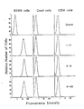
- the initial screening of the B hybridomas secreting anti-Fc ⁇ R antibodies was done on the basis of immunofluorescence with 8866, Daudi, CEM, and Molt 4 cells. A half million of 8866, Daudi, DEM and MOLT 4 cells were incubated with 50 ⁇ l of culture supernatants followed by FITC-anti-mouse IgM + IgG and analyzed by a cell sorter (FACS 440, Becton Dickinson, Mountain View, Ca.).
- Fc R on lymphocytes were detected by an assay employing fixed ox erythrocytes (ORBC) coated with human IgE (11) and the percentage of IgE-rosette-forming cells (RFC) was estimated after subtracting the percentage of non-specific RFC binding with fixed ORBC coated with BSA.
- ORBC fixed ox erythrocytes
- RFC IgE-rosette-forming cells
- IgG rosette inhibition assay was performed essentially in the same manner employing Daudi cells and ORBC sensitized with IgG fraction of rabbit anti-ORBC antiserum.
- Hybridoma clones 3-5 ( ⁇ l), 8-30 ( ⁇ ), and 1-7 ( ⁇ 2b), the first two of which were deposited at the Collection nationale de micro-organisms, Institut Pasteur (C.N.C.M), 25, rue du Dr. Roux, 75724 Paris, Cedex 15 on the 24th July 1986, under the file numbers I-583 and I-584 respectively. were selected as the possible candidates for anti-Fc R antibodies by means of their inhibitory effect on IgE-rosette information.
- the necessary monoclonal antibodies were purified from ascites by 50 % saturated ammonium sulfate precipitation followed by gel filtration using Sepharose 6B (Pharmacia Fine Chemicals, Uppsala, Sweden) for IgM class or by ion exchange chromatography using QAE-Sephadex (Pharmacia Fine Chemicals) for IgG1 and IgG2b classes.
- two monoclonal antibodies, 1-7 and 8-30 inhibited the rosette formation of 8866 cells with IgE-coated ox red cells (ORBC) up to 97 % when used at concentrations of 5 to 10 ⁇ g/ml, while the other antibody, 3-5, showed marginal inhibitory activity only at high concentrations (100 ⁇ g/ml).
- the two monoclonal antibodies (1-7 and 8-30) did not inhibit the rosette formation of Daudi cells with IgG-ORBC, even when 100 ⁇ g/ml of antibodies were employed.
- Peripheral blood mononuclear cells were obtained from normal donors and atopic patients with bronchial asthma or atopic dermatitis but not undergoing systemic corticosteroid therapy. Serum IgE levels of these patients were between 1,000-15,000 IU/ml. Tonsillar mononuclear cells were obtained from patients with chronic tonsillitis. MNC were prepared by Ficoll-Hypaque gradient centrifugation, and B cell-enriched populations (E ⁇ cells) were prepared by rosetting with (2-aminoethyl)-isothiouronium bromide (AET, Aldrich Chemical Co.) -treated sheep red blood cells.
- AET (2-aminoethyl)-isothiouronium bromide
- Propidium iodide (PI, 10 ⁇ g/ml) was included in the last five minutes of the second step to label the dead cells.
- the one-color and two-color immunofluorescence analyses by FACS 440 and VAX computer is described in reference (12).
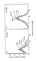
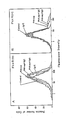
- both 1-7 and 8-30 monoclonal antibodies blocked the IgE binding to 8866 cells in a dose-dependent fashion.
- the monoclonal antibody, 8-30 was much more effective than 1-7. This may be due to the fact that 8-30 is of IgM isotype.
- the monoclonal antibody, 3-5 could display only a slight inhibition of IgE binding at high concentrations.
- the control monoclonal antibody, MR11 of IgM class did not affect the binding of IgE to 8866 cells, indicating that the inhibitory activity of 8-30 is not due essentially to its Fc ⁇ r-portion.
- peripheral MNC precultered for 1 hr and washed three times to remove cytophilic IgG or IgE, were exposed to various concentrations of monoclonal antibodies in 20 ⁇ l of staining buffer in 96 well microtiter plates at 4°C for 30 min. After incubation, cells were washed twice with staining buffer, incubated with FITC-anti B1 and biotinated IgG1 or IgE for 20 min, and washed twice. Subsequently, the cells were incubated with TR-avidin, washed and analyzed by FACS.
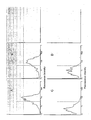
- the binding of IgE to B1-positive cells was inhibited by the presence of either 1 ⁇ g/5 x 105 cells of 1-7 or 8-30 antibody, but not by the monoclonal antibody, 3-5 or the control antibody, MR11.
- the same concentration of the monoclonal antibodies did not inhibit IgG binding to B1-positive cells.
- Tonsillar MNC were stained with FITC-anti-B1 and biotinated 8-30 antibodies and developed with TR-avidin.
- Fc ⁇ R+/B1+ and Fc ⁇ R ⁇ /B1+ cells were sorted by FACS. Sorted cells were incubated with 10 % (V/V) PHA-conditioned medium (PHA-sup) in the presence or the absence of 10 ⁇ g/ml IgE or IgG1 for 3 days. After culturing for 3 days, the cells were stained with anti-B1 and anti-Fc ⁇ R(8-30) antibodies and reanalysed by FACS.
- Intensities of Fc ⁇ R+ B cells were plotted on the histogram by gating B1 positive B cells.
- PHA-sup was prepared as described in reference (12). Briefly, tonsillar T cells were cultured at 1 x 10⁶/ml with 0,1 % PHA for 48 hr and the culture supernatant was collected. The concentrated supernatant was fractionated by HPLC gel filtration and the fraction with a M.W. of approximately 20K was employed as source of crude T cell factor(s), containing BSF-1 (BCGF), B cell differentiation factor (BCDF) and IL-2 activities.
- BCGF BSF-1
- BCDF B cell differentiation factor
- IL-2 activities.
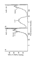
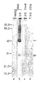
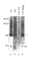
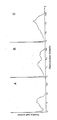
- Fc ⁇ R-positive 8866 and Fc ⁇ R-negative Daudi and CEM cells were surface labelled with 125I and lysed in lysis buffer containing 0,5 % NP-40. After preabsorption with HSA-Sepharose, lysates were immunoprecipitated with the monoclonal antibody, 1-7 or 3-5, or normal mouse Ig (NMIg), and analyzed by SDS-PAGE (Fig 7 and Fig 8).
- a further object of the present invention is the use of the before mentioned antibodies to differentiate bone marrow B cells and circulating B cells showing that Fc ⁇ R is a B cell-specific surface molecule:
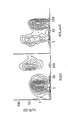
- Tonsillar B cells were stained with anti-Fc ⁇ R and with conjugates directed against different Ig-isotypes. As shown in Fig. 13 (A), Fc ⁇ R was expressed on most ⁇ and ⁇ positive B cells in tonsils. On the other hand, almost all ⁇ - or ⁇ -bearing B cells, which were abundantly observed in tonsils, did not express Fc ⁇ R on their surface. Other experiments were conducted in order to study whether the Fc ⁇ R expression was also correlated with the isotype expression as observed with the Fc ⁇ R.
- biotinated IgE and IgG were employd for the comparison of the expression of Fc ⁇ R and Fc R on ⁇ + ⁇ + B cells and on ⁇ + or ⁇ +B cells.
- the specificity of the binding of biotinated IgE was almost comparable to that of the anti-Fc R antibody, in fact, the IgE-binding was observed on ⁇ + ⁇ + B cells but not on ⁇ + or ⁇ + B cells.
- FIG. 14 shows the histograms of the Fc ⁇ R expression on ⁇ +, ⁇ +, ⁇ + or ⁇ + B cells in PBL. Most ⁇ + (A) or ⁇ + (B) B cells expressed Fc ⁇ R as in the case of tonsillar B cells.
- ⁇ + (C) or ⁇ + (D) B cells did not express Fc ⁇ R, although a small fraction of these cells were found to show a weak expression.
- the results show that Fc ⁇ R is preferentially expressed on circulating ⁇ + and ⁇ + B cells but not on ⁇ + or ⁇ + B cells and that may explain the reason why PBL includes more Fc R+ cells than tonsils.
- FIG. 15 demonstrates the representative results observed in PBL from two patients with hyper IgE-syndrome.
- the Fc ⁇ R expression was augmented in ⁇ + or ⁇ + B cells in these patients compared to that in normal individuals (Fig. 14, A and B).
- ⁇ + or ⁇ + B cells did not express Fc ⁇ R even in PBL from patients with hyper IgE-syndrome (Figs. 2, C and D).
- Circulating ⁇ B cells which are mainly ⁇ + or ⁇ +, do not express Fc ⁇ R.
- ⁇ ⁇ B cells were sorted from tonsillar B1+ cells and incubated with PHA-sup and IgE. As shwon in Fig. 17(A), the stimulation did not induce any expression of Fc ⁇ R on circulating ⁇ B cells, indicating that the loss of the Fc ⁇ R expression was irreversible in the cells which had already completed isotype switching. A small fraction of ⁇ + B cells in tonsil did not express Fc ⁇ R. However, as shown in Fig.
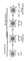
- Fc ⁇ R is a B cell-specific surface molecule, the expression of which is strictly correlated with the differentiation stage of B cells; i) bone marrow B cells with surface IgM and IgD do not express Fc ⁇ R, although its expression could be induced, ii) circulating B cells with surface IgM and IgD express Fc ⁇ R, but iii) circulating B cells with no surface IgD, which have already undergone isotype-switching into IgG, IgA or IgE bearing cells do not express and can not be induced to express Fc ⁇ R. Therefore, according to these observations, the differentiation of B cells can be divided into three stages as described in Fig. 19:
- the Elisa according to the invention comprises usually a solid phase carrying a monoclonal antibody to said surface receptor for IgE, e.g.
- a detectable label such as a radioactive isotope, an enzyme, a fluorescent or chemilu miniscent compound or a second antibody specifically binding to an above mentioned antibody and carrying any one of the afore-mentioned labels, e.g. alkaline phosphatase-conjugated anti-mouse IgM, preferably alkaline phosphatase-
- the monoclonal antibody 3-5 is of ⁇ 1 class, does not significantly inhibit the binding of IgE, and binds to a specific epitope of Fc ⁇ R.
- the monoclonal antibody 8-30 is of ⁇ class and inhibits the binding of IgE and binds to a specific epitope of Fc ⁇ R.
- the above mentioned monoclonal antibodies bind in combination selectively to Fc ⁇ R found in culture supernatants or lysates of RPMI8866 cells, Eb-transformed cells and serum derived from atopic and non-atopic individuals.
- the double antibody sandwich technique utilizing the monoclonal antibodies 3-5 and 8-30 identifies the presence of Fc ⁇ R-receptors in serum from atopic and non-atopic individuals and enables the state of hypersensitivity of an individual to be followed.
- This technique is a potential diagnostic tool in allergy since, as in the case of determining the level of IgE in serum, it allows monitoring and diagnosis of an allergic condition.
- test sample is added to each well and incubated for 2 hours at room temperature (the test sample can be diluted first with diluent buffer to allow a titration).
- the test samples are dumped and the plate washed 4 times with rinse buffer followed by the addi tion of 100 ⁇ l of second monoclonal antibody, 8-30 diluted to 1 ⁇ g/ml in diluent bufer and incubated for 2 hours at room temperature.
- the wells are washed 4 times with rinse buffer followed by the addition of 100 ⁇ l of goat anti-mouse IgM conjugated to alkaline phosphatase (pretitrated) and incubated for 2 hours at room temperature.
- the wells are washed with rinse buffer and 200 ⁇ l of substrate, p-Nitrophenyl Phosphate Disodium added.
- the enzymatic reaction is allowed to proceed and the optical (color) determination measured at 405 and 620 nm.
- IgE-rosette assay 8866 cells were preincubated with varying concentrations of the monoclonal antibodies or control medium and subsequently incubated with fixed ORBC coated with IgE or BSA. IgG-rosette assay was carried out employing Daudi cells and rabbit IgG-coated ORBC.
- 8866 cells were stained with either 3 ⁇ g or 0.2 ⁇ g/5x105 cells of biotin-IgE followed by TR-avidin in the presence or absence of varying concentrations of the monoclonal antibodies (1-7, 3-5 and 8-30) or a B cells-specific monoclonal antibody (MR11) and applied to FACS analysis.
- the mean fluorescence intensity was calculated by VAX-11 computer.
- Peripheral MNC were preincubated with either 1 ⁇ /5x105 cells of the anti-Fc ⁇ R monoclonal antibodies or a B cell specific antibody (MR11). After washing, cells were incubated with FITC-anti-B1 and 3 ⁇ g/5x105 cells of biotinated IgE or IgG1 followed by TR-avidin and analyzed by FACS. Relative number of IgE (panel A) or IgG (panel B) binding cells to B1 positive cells (B cells) was shown in the histograms.
- 8866 cells were stained with 1 ⁇ g/1x106 cells of biotin-3-5(A) or 0.1 ⁇ g/1x106 cells of biotin-8-30(B) followed by TR-avidin in the presence or absence of 9 ⁇ g/1x106 cells of 1-7 and analysed by FACS.
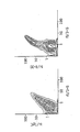
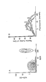
- Tonsillar B cells were stained with 0,5 ⁇ g/1x106 cells of FITC-labelled 3-5 and 0.1 ⁇ g/1x106 cells of biotin-labelled 8-30 or 3 ⁇ g/1x106 cells of biotinated IgE followed by TR-avidin and the two color FACS analysis was carried out.
- 8866 cells were surface-labelled with 125I and cell lysates were immunoprecipitated with 1-7 (lane a) or 3-5 (lane b). After precipitation with 3-5 or NMIg, the supernatant was precipitated with 1-7 (lane c and d) and analysed by SDS-PAGE.
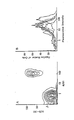
- Fc ⁇ R expression of Fc ⁇ R on B cells from normal, non-atopic individuals. PBL were stained with FITC-anti B1 and biotin-8-30 followed by TR-avidin. A representative result of the two color FACS analysis is shown in panel A and the histograms of Fc ⁇ R positive cells in B+ cells of several donors are shown in panel B.
- Tonsillar MNC were doubly stained with (A) biotinated anti-Fc ⁇ R and FITC-anti-B1, FITC-anti-IgD, PC-anti-IgM, FITC-anti-IgA or PC-anti-IgG, (B) biotinated human IgE and FITC-anti-B1, FITC-anti-IgD, PC-anti-IgM, FITC-anti-IgA and PC-anti-IgG, (C) biotinated human IgG1 and FITC-anti-B1, FITC-anti-IgD, PC-anti-IgM, FITC-anti-IgA. Biotinated reagents were developed with TR-avidin for double staining with FITC antibodies or FITC-avidin for double staining with PC-antibodies.
- Fc ⁇ R expression on various isotype positive B cells from normal PBL PBL from a normal individual were doubly stained with biotinated anti-Fc ⁇ R and FITC-anti-IgD, PC-anti-IgM, FITC-anti-IgA or PC-anti-IgG.
- the fluorescence intensities of Fc ⁇ R staining on IgD+(A), IgM+(B), IgA+(C) or IgG+(D) B cells are shown on the histograms.
- PBL from two hyper IgE patients were doubly stained with biotinated anti-Fc ⁇ R and FITC-anti-IgD, PC-anti-IgM, PC-anti-IgG or FITC-anti-IgA or PC-anti-Fc ⁇ R and Biotinated anti-IgE.
- Biotinated antibodies were developed with TR-avidin or FITC avidin.
- Bone marrow MNC from an immunologically normal adult donor were incubated without (-) or with IgE (10 ⁇ g/ml), PHA-sup (10 % v/v), PHA-sup plus IgG (10 ⁇ g/ml) or PHA-sup plus IgE for 3 days.
- IgE 10 ⁇ g/ml
- PHA-sup 10 % v/v
- PHA-sup plus IgG 10 ⁇ g/ml
- PHA-sup plus IgE for 3 days.
- cells were doubly stained with biotinated anti-Fc ⁇ R and FITC-anti-B1 or FITC-anti-Ig and developed with TR-avidin. Cells were analyzed by FACS. The intensities of Fc ⁇ R staining on B1 + B cells (a) and IgD+ B cells (B) were shown in the histogram.
- Fc R can be induced on + cells but not ⁇ cells in tonsils. Tonsillar MNC were stained with anti-Fc ⁇ R and anti-IgD and ⁇ /Fc R ⁇ (A) and +/Fc ⁇ R ⁇ (B) cells were sorted by FACS. Sorted cells were indubated with ( ? ) or without ( - - - - ) PHA-sup (10 % v/v) and IgE (10 ⁇ g/ml) for 3 days and stained with anti-Fc ⁇ R and anti-B1. The intensities of Fc ⁇ R staining of B1 + B cells were shown in the histograms.
- Fc ⁇ R is inducible on Fc ⁇ R negative PBL from a patient with CVI.
- PBL from a patient with CVI were incubated without (A) or with PHA-sup (10 % v/v) (B) or PHA-sup and IgE (10 ⁇ g/ml) (C) for 3 days. After incubation, cells were stained with anti-Fc ⁇ R and anti-B1 and analyzed by FACS. The intensities of Fc ⁇ R staining of B1 positive B cells are shown in the histograms.
Landscapes
- Chemical & Material Sciences (AREA)
- Health & Medical Sciences (AREA)
- Organic Chemistry (AREA)
- Life Sciences & Earth Sciences (AREA)
- Immunology (AREA)
- Engineering & Computer Science (AREA)
- Biochemistry (AREA)
- Genetics & Genomics (AREA)
- Zoology (AREA)
- Molecular Biology (AREA)
- General Health & Medical Sciences (AREA)
- Wood Science & Technology (AREA)
- Proteomics, Peptides & Aminoacids (AREA)
- Biotechnology (AREA)
- Chemical Kinetics & Catalysis (AREA)
- General Chemical & Material Sciences (AREA)
- Microbiology (AREA)
- Biophysics (AREA)
- Medicinal Chemistry (AREA)
- Bioinformatics & Cheminformatics (AREA)
- General Engineering & Computer Science (AREA)
- Preparation Of Compounds By Using Micro-Organisms (AREA)
- Peptides Or Proteins (AREA)
- Micro-Organisms Or Cultivation Processes Thereof (AREA)
Priority Applications (17)
| Application Number | Priority Date | Filing Date | Title |
|---|---|---|---|
| EP86110420A EP0255547A1 (fr) | 1986-07-29 | 1986-07-29 | Anticorps monoclonaux spécifiques du récepteur de surface pour IgE, ainsi que des lignes de cellules hybrides produisant ces anticorps et leur utilisation |
| EP86111488A EP0254767A1 (fr) | 1986-07-29 | 1986-08-19 | Anticorps monoclonaux spécifiques contre le récepteur de surface pour IgE ainsi que des lignes de cellules hybrides produisant ces anticorps et leur utilisation |
| EP87110658A EP0259585B1 (fr) | 1986-07-29 | 1987-07-23 | Anticorps monoclonaux spécifiques, contre le récepteur de surface pour IgE ainsi que des lignées cellulaires hybrides produisant de tels anticorps et leur utilisation |
| AT87110658T ATE90391T1 (de) | 1986-07-29 | 1987-07-23 | Monoklonale antikoerper gegen oberflaechlichen ige- rezeptor, sowie hybrid-zellinien zu deren herstellung und verwendung. |
| DE8787110658T DE3786130D1 (de) | 1986-07-29 | 1987-07-23 | Monoklonale antikoerper gegen oberflaechlichen ige-rezeptor, sowie hybrid-zellinien zu deren herstellung und verwendung. |
| FI873257A FI873257A7 (fi) | 1986-07-29 | 1987-07-27 | Monoklonaaliset vasta-aineet spesifisiä IgE-pintareseptoreita varten ja näitä vasta-aineita tuottavat hybridisolulinjat ja vasta-aineiden käyttö |
| HU873459A HU204895B (en) | 1986-07-29 | 1987-07-28 | Process for producing 3-5 monoclonal antibodies specific to ige surface receptor, hybrid cell lines producing such antibodies as well as for detecting the receptors |
| JP62188714A JPS63258494A (ja) | 1986-07-29 | 1987-07-28 | IgEの表面レセプタ−に特異的なモノクロ−ナル抗体及びこれら抗体を産生するハイブッド細胞系及びこれらの使用 |
| NO873154A NO873154L (no) | 1986-07-29 | 1987-07-28 | Spesifikke monoklonale antistoffer mot overflatereseptor for ige og hybridcellelinjer som produserer disse antistoffer og anvendelse derav. |
| NZ221235A NZ221235A (en) | 1986-07-29 | 1987-07-28 | Antibodies specific to the ige receptor of b cells, hybridomas and immunoassays |
| ZA875530A ZA875530B (en) | 1986-07-29 | 1987-07-28 | Monoclonal antibodies specific to surface receptor for ige and hybrid cell lines producing these antibodies and use thereof |
| DK392987A DK392987A (da) | 1986-07-29 | 1987-07-28 | Monoklonale antistoffer, der er specifikke over for overfladereceptor for ige, og hybridcellelinier, der producerer disse antistoffer, og anvendelse deraf |
| IL83362A IL83362A (en) | 1986-07-29 | 1987-07-28 | Monoclonal antibodies specific to surface receptor for ige,hybridoma cell lines producing them and their use |
| DD87305383A DD273856A5 (de) | 1986-07-29 | 1987-07-28 | Prozess zur praeparierung neuer monoclonaler antikoerper |
| PT85422A PT85422B (pt) | 1986-07-29 | 1987-07-28 | Processo para a preparacao de anticorpos monoclonais especificos em relacao a receptores de superficie para ige bem como de linhas de celulas hibridas que os produzem e de agentes de analise que os contem |
| AU76191/87A AU618720B2 (en) | 1986-07-29 | 1987-07-28 | Monoclonal antibodies |
| KR1019870008225A KR880001807A (ko) | 1986-07-29 | 1987-07-29 | IgE에 대한 표면수용체에 특이성이 있는 단일군항체와 이들 항체를 생성시키는 하이브리드 세포주 및 이들의 용도 |
Applications Claiming Priority (1)
| Application Number | Priority Date | Filing Date | Title |
|---|---|---|---|
| EP86110420A EP0255547A1 (fr) | 1986-07-29 | 1986-07-29 | Anticorps monoclonaux spécifiques du récepteur de surface pour IgE, ainsi que des lignes de cellules hybrides produisant ces anticorps et leur utilisation |
Publications (1)
| Publication Number | Publication Date |
|---|---|
| EP0255547A1 true EP0255547A1 (fr) | 1988-02-10 |
Family
ID=8195302
Family Applications (2)
| Application Number | Title | Priority Date | Filing Date |
|---|---|---|---|
| EP86110420A Withdrawn EP0255547A1 (fr) | 1986-07-29 | 1986-07-29 | Anticorps monoclonaux spécifiques du récepteur de surface pour IgE, ainsi que des lignes de cellules hybrides produisant ces anticorps et leur utilisation |
| EP86111488A Withdrawn EP0254767A1 (fr) | 1986-07-29 | 1986-08-19 | Anticorps monoclonaux spécifiques contre le récepteur de surface pour IgE ainsi que des lignes de cellules hybrides produisant ces anticorps et leur utilisation |
Family Applications After (1)
| Application Number | Title | Priority Date | Filing Date |
|---|---|---|---|
| EP86111488A Withdrawn EP0254767A1 (fr) | 1986-07-29 | 1986-08-19 | Anticorps monoclonaux spécifiques contre le récepteur de surface pour IgE ainsi que des lignes de cellules hybrides produisant ces anticorps et leur utilisation |
Country Status (5)
| Country | Link |
|---|---|
| EP (2) | EP0255547A1 (fr) |
| KR (1) | KR880001807A (fr) |
| AT (1) | ATE90391T1 (fr) |
| DD (1) | DD273856A5 (fr) |
| ZA (1) | ZA875530B (fr) |
Cited By (1)
| Publication number | Priority date | Publication date | Assignee | Title |
|---|---|---|---|---|
| EP0327283A1 (fr) * | 1988-02-02 | 1989-08-09 | Schering Biotech Corporation | Méthode visant à réduire la réponse immunitaire à l'immunoglobuline E |
Families Citing this family (1)
| Publication number | Priority date | Publication date | Assignee | Title |
|---|---|---|---|---|
| IL122233A (en) * | 1996-12-06 | 2001-04-30 | Akzo Nobel Nv | Method of generating monoclonal antibodies to cell wall and pharmaceutical preparations and diagnostic agents containing them |
Citations (1)
| Publication number | Priority date | Publication date | Assignee | Title |
|---|---|---|---|---|
| JPS60255734A (ja) * | 1984-05-30 | 1985-12-17 | Nichirei:Kk | クラス特異的Fcεレセプタ−に対する単クロ−ン性抗体とそれを産生するハイブリド−マ |
-
1986
- 1986-07-29 EP EP86110420A patent/EP0255547A1/fr not_active Withdrawn
- 1986-08-19 EP EP86111488A patent/EP0254767A1/fr not_active Withdrawn
-
1987
- 1987-07-23 AT AT87110658T patent/ATE90391T1/de not_active IP Right Cessation
- 1987-07-28 ZA ZA875530A patent/ZA875530B/xx unknown
- 1987-07-28 DD DD87305383A patent/DD273856A5/de not_active IP Right Cessation
- 1987-07-29 KR KR1019870008225A patent/KR880001807A/ko not_active Ceased
Patent Citations (1)
| Publication number | Priority date | Publication date | Assignee | Title |
|---|---|---|---|---|
| JPS60255734A (ja) * | 1984-05-30 | 1985-12-17 | Nichirei:Kk | クラス特異的Fcεレセプタ−に対する単クロ−ン性抗体とそれを産生するハイブリド−マ |
Non-Patent Citations (7)
| Title |
|---|
| BIOLOGICAL ABSTRACTS, vol. 80, no. 11, November 1985, abstract no. 95522, Biological Abstracts Inc. PA, US; E.T. NAKAJIMA et al.: "Detection characterization of monoclonal antibodies specific to immunoglobulin E. receptors on human lymphocytes by flow cytometry" * |
| CHEMICAL ABSTRACTS, vol. 103, no. 11, 16th September 1985, page 162, abstract no. 86103r, Columbus, Ohio, US; E. RECTOR et al.: "Detection and characterization of monoclonal antibodies specific to IgE receptors on human lymphocytes by flow cytometry", & IMMUNOLOGY 1985, 55(3), 481-8 * |
| CHEMICAL ABSTRACTS, vol. 104, no. 15, 14th April 1986, page 556, abstract no. 128114y, Columbus, Ohio, US; & JP-A-60 255 734 (NICHIREI CORP.) 17-12-1985 * |
| CHEMICAL ABSTRACTS, vol. 104, no. 3, January 1986, page 379, abstract no. 18371x, Columbus, Ohio, US; S.A. HUDAK et al.: "A microtiter plate assay for detecting IgE-binding molecules and cells bearing Fc receptors for IgE", & J. IMMUNOL. METHODS 1985, 84(1-2), 11-24 * |
| CHEMICAL ABSTRACTS, vol. 105, no. 12, September 1986, page 487, abstract no. 95741r, Columbus, Ohio, US; T. NAKAJIMA et al.: "IgE receptors on human lymphocytes. I. Identification of the molecules binding to monoclonal anti-Fc receptor antibodies", & EUR. J. IMMUNOL. 1986, 16(7), 809-14 * |
| CHEMICAL ABSTRACTS, vol. 105, no. 7, 18th August 1986, page 490, abstract no. 59181z, Columbus, Ohio, US; M. CAPRON et al.: "Functional study of a monoclonal antibody to IgE Fc receptor (FceR2) of eosinophils, platelets, and macrophages", & J. EXP. MED. 1986, 164(1), 72-89 * |
| CHEMICAL ABSTRACTS, vol. 106, no. 1, January 1987, page 351, abstract no. 3603s, Columbus, Ohio, US; H. KIKUTANI et al.: "Per receptor, a specific differentiation marker translently expressed on mature B cells before isotype switching", & J. EXP. MED. 1986, 164(5), 1455-69 * |
Cited By (2)
| Publication number | Priority date | Publication date | Assignee | Title |
|---|---|---|---|---|
| EP0327283A1 (fr) * | 1988-02-02 | 1989-08-09 | Schering Biotech Corporation | Méthode visant à réduire la réponse immunitaire à l'immunoglobuline E |
| WO1989006975A1 (fr) * | 1988-02-02 | 1989-08-10 | Schering Biotech Corporation | Procede de reduction des reactions dues a l'immunoglobuline e |
Also Published As
| Publication number | Publication date |
|---|---|
| KR880001807A (ko) | 1988-04-27 |
| ATE90391T1 (de) | 1993-06-15 |
| ZA875530B (en) | 1989-04-26 |
| EP0254767A1 (fr) | 1988-02-03 |
| DD273856A5 (de) | 1989-11-29 |
Similar Documents
| Publication | Publication Date | Title |
|---|---|---|
| Anderson et al. | Antigens on human plasma cells identified by monoclonal antibodies. | |
| US4434156A (en) | Monoclonal antibodies specific for the human transferrin receptor glycoprotein | |
| Carrel et al. | Subsets of human Ia-like molecules defined by monoclonal antibodies | |
| Isacke et al. | Identification and characterization of the human Pgp-1 glycoprotein | |
| Birkeland et al. | Epitopes on CD45R [T200] molecules define differentiation antigens on murine B and T lymphocytes | |
| IE50725B1 (en) | Hybrid cell line for producing monoclonal antibody to a human prothymocyte antigen,antibody,method of preparation of this antibody,diagnostic and therapeutic uses,and pharmaceutical compositions comprising this antibody | |
| JPS649998B2 (fr) | ||
| JPH0223156B2 (fr) | ||
| JPH0159871B2 (fr) | ||
| JPS6410000B2 (fr) | ||
| US5876917A (en) | Dendritic cell-specific antibodies and method for their preparation | |
| EP0766736B1 (fr) | Anticorps monoclonaux humains specifiques vis-a-vis d'antigenes de surface de gliomes, independants du cycle cellulaire | |
| JP3631750B2 (ja) | ヒトmdr1マルチドラッグ耐性遺伝子産物に対するモノクローナル抗体およびその使用 | |
| CA1219231A (fr) | Anticorps monoclonal regroupant les lymphocytes t "helper" et "killer" et methode d'utilisation | |
| US4818689A (en) | Late differentiation antigen associated with helper T lymphocyte function | |
| US5747036A (en) | Methods and compositions for detecting and treating a subset of human patients having an autoimmune disease | |
| US4816404A (en) | Late differentiation antigens associated with helper T lymphocyte function | |
| Hellström et al. | Monoclonal antibodies to tumor antigens | |
| EP0255547A1 (fr) | Anticorps monoclonaux spécifiques du récepteur de surface pour IgE, ainsi que des lignes de cellules hybrides produisant ces anticorps et leur utilisation | |
| EP0259585B1 (fr) | Anticorps monoclonaux spécifiques, contre le récepteur de surface pour IgE ainsi que des lignées cellulaires hybrides produisant de tels anticorps et leur utilisation | |
| US5028424A (en) | Antibodies to receptor and antigen for natural killer and non-specific cytotoxic cells | |
| JP4563573B2 (ja) | 抗原およびこの抗原を識別するモノクローナル抗体 | |
| US5185430A (en) | Antigen recognized by natural killer and non-specific cytotoxic cells | |
| US5162224A (en) | Monoclonal antibodies specific for b cells and htlv-i transformed t cells | |
| EP0393062A1 (fr) | Anticorps monoclonal reconnaissant un epitope de la chaine gamma de recepteurs de surface des lymphocytes t de l'homme |
Legal Events
| Date | Code | Title | Description |
|---|---|---|---|
| PUAI | Public reference made under article 153(3) epc to a published international application that has entered the european phase |
Free format text: ORIGINAL CODE: 0009012 |
|
| AK | Designated contracting states |
Kind code of ref document: A1 Designated state(s): GB |
|
| RAP3 | Party data changed (applicant data changed or rights of an application transferred) |
Owner name: KISHIMOTO, TADAMITSU, PROF. |
|
| STAA | Information on the status of an ep patent application or granted ep patent |
Free format text: STATUS: THE APPLICATION IS DEEMED TO BE WITHDRAWN |
|
| 18D | Application deemed to be withdrawn |
Effective date: 19881011 |
|
| RIN1 | Information on inventor provided before grant (corrected) |
Inventor name: KISHIMOTO, TADAMITSU, PROF. DR. Inventor name: BARSUMIAN, EDWARD L., DR. Inventor name: KIKUTANI, HITOSHI, DR. Inventor name: SUEMURA, MASAKI, DR. |