EP0260708A2 - Tumoraustrittsfaktor und Verfahren für seine Herstellung - Google Patents
Tumoraustrittsfaktor und Verfahren für seine Herstellung Download PDFInfo
- Publication number
- EP0260708A2 EP0260708A2 EP87113683A EP87113683A EP0260708A2 EP 0260708 A2 EP0260708 A2 EP 0260708A2 EP 87113683 A EP87113683 A EP 87113683A EP 87113683 A EP87113683 A EP 87113683A EP 0260708 A2 EP0260708 A2 EP 0260708A2
- Authority
- EP
- European Patent Office
- Prior art keywords
- egressin
- activity
- isolating
- carrying
- separation
- Prior art date
- Legal status (The legal status is an assumption and is not a legal conclusion. Google has not performed a legal analysis and makes no representation as to the accuracy of the status listed.)
- Withdrawn
Links
- 238000000034 method Methods 0.000 title claims abstract description 21
- 230000008569 process Effects 0.000 title claims abstract description 18
- 206010028980 Neoplasm Diseases 0.000 title abstract description 7
- IVFPIHZOFZLQJG-UHFFFAOYSA-N (5-methyl-2-propan-2-ylphenyl) n-(3-methylbutyl)carbamate Chemical compound CC(C)CCNC(=O)OC1=CC(C)=CC=C1C(C)C IVFPIHZOFZLQJG-UHFFFAOYSA-N 0.000 claims abstract description 196
- 230000006799 invasive growth in response to glucose limitation Effects 0.000 claims abstract description 15
- 206010027480 Metastatic malignant melanoma Diseases 0.000 claims abstract description 9
- 208000021039 metastatic melanoma Diseases 0.000 claims abstract description 9
- 210000004027 cell Anatomy 0.000 claims description 115
- 230000000694 effects Effects 0.000 claims description 102
- QTBSBXVTEAMEQO-UHFFFAOYSA-N Acetic acid Chemical compound CC(O)=O QTBSBXVTEAMEQO-UHFFFAOYSA-N 0.000 claims description 66
- 238000000926 separation method Methods 0.000 claims description 63
- 239000002356 single layer Substances 0.000 claims description 25
- 230000002209 hydrophobic effect Effects 0.000 claims description 22
- 238000005341 cation exchange Methods 0.000 claims description 21
- 210000003734 kidney Anatomy 0.000 claims description 18
- 210000002950 fibroblast Anatomy 0.000 claims description 17
- 230000001939 inductive effect Effects 0.000 claims description 16
- 239000003636 conditioned culture medium Substances 0.000 claims description 14
- 239000004017 serum-free culture medium Substances 0.000 claims description 14
- 230000005012 migration Effects 0.000 claims description 13
- 238000013508 migration Methods 0.000 claims description 13
- 238000004587 chromatography analysis Methods 0.000 claims description 11
- 230000004069 differentiation Effects 0.000 claims description 11
- 239000003795 chemical substances by application Substances 0.000 claims description 10
- 238000012258 culturing Methods 0.000 claims description 10
- 238000002415 sodium dodecyl sulfate polyacrylamide gel electrophoresis Methods 0.000 claims description 10
- 239000003226 mitogen Substances 0.000 claims description 8
- 101710093543 Probable non-specific lipid-transfer protein Proteins 0.000 claims description 7
- FXEDIXLHKQINFP-UHFFFAOYSA-N 12-O-tetradecanoylphorbol-13-acetate Natural products CCCCCCCCCCCCCC(=O)OC1CC2(O)C(C=C(CO)CC3(O)C2C=C(C)C3=O)C4C(C)(C)C14OC(=O)C FXEDIXLHKQINFP-UHFFFAOYSA-N 0.000 claims description 3
- PHEDXBVPIONUQT-RGYGYFBISA-N phorbol 13-acetate 12-myristate Chemical compound C([C@]1(O)C(=O)C(C)=C[C@H]1[C@@]1(O)[C@H](C)[C@H]2OC(=O)CCCCCCCCCCCCC)C(CO)=C[C@H]1[C@H]1[C@]2(OC(C)=O)C1(C)C PHEDXBVPIONUQT-RGYGYFBISA-N 0.000 claims description 3
- 206010061289 metastatic neoplasm Diseases 0.000 abstract description 12
- 230000001394 metastastic effect Effects 0.000 abstract description 11
- 229940079593 drug Drugs 0.000 abstract description 9
- 239000003814 drug Substances 0.000 abstract description 9
- 230000003902 lesion Effects 0.000 abstract description 9
- 239000003153 chemical reaction reagent Substances 0.000 abstract description 8
- 230000008499 blood brain barrier function Effects 0.000 abstract description 7
- 210000001218 blood-brain barrier Anatomy 0.000 abstract description 7
- 238000001514 detection method Methods 0.000 abstract description 7
- 230000001900 immune effect Effects 0.000 abstract description 7
- 230000028709 inflammatory response Effects 0.000 abstract description 7
- 238000004519 manufacturing process Methods 0.000 abstract description 7
- FAPWRFPIFSIZLT-UHFFFAOYSA-M Sodium chloride Chemical compound [Na+].[Cl-] FAPWRFPIFSIZLT-UHFFFAOYSA-M 0.000 description 42
- 238000003556 assay Methods 0.000 description 32
- 210000002966 serum Anatomy 0.000 description 29
- WEVYAHXRMPXWCK-UHFFFAOYSA-N Acetonitrile Chemical compound CC#N WEVYAHXRMPXWCK-UHFFFAOYSA-N 0.000 description 24
- 244000309466 calf Species 0.000 description 23
- 239000011780 sodium chloride Substances 0.000 description 21
- 239000006144 Dulbecco’s modified Eagle's medium Substances 0.000 description 20
- 102000004169 proteins and genes Human genes 0.000 description 17
- 108090000623 proteins and genes Proteins 0.000 description 17
- 239000000499 gel Substances 0.000 description 16
- 238000002835 absorbance Methods 0.000 description 14
- 239000007974 sodium acetate buffer Substances 0.000 description 13
- DTQVDTLACAAQTR-UHFFFAOYSA-N Trifluoroacetic acid Chemical compound OC(=O)C(F)(F)F DTQVDTLACAAQTR-UHFFFAOYSA-N 0.000 description 10
- 239000000463 material Substances 0.000 description 10
- 102000009024 Epidermal Growth Factor Human genes 0.000 description 9
- 230000012010 growth Effects 0.000 description 9
- 238000002360 preparation method Methods 0.000 description 9
- 230000004044 response Effects 0.000 description 9
- IQFYYKKMVGJFEH-XLPZGREQSA-N Thymidine Chemical compound O=C1NC(=O)C(C)=CN1[C@@H]1O[C@H](CO)[C@@H](O)C1 IQFYYKKMVGJFEH-XLPZGREQSA-N 0.000 description 8
- 238000000746 purification Methods 0.000 description 8
- 239000012064 sodium phosphate buffer Substances 0.000 description 8
- 102000004887 Transforming Growth Factor beta Human genes 0.000 description 7
- 108090001012 Transforming Growth Factor beta Proteins 0.000 description 7
- 238000004128 high performance liquid chromatography Methods 0.000 description 7
- 230000002297 mitogenic effect Effects 0.000 description 7
- ZRKFYGHZFMAOKI-QMGMOQQFSA-N tgfbeta Chemical compound C([C@H](NC(=O)[C@H](C(C)C)NC(=O)CNC(=O)[C@H](CCC(O)=O)NC(=O)[C@H](CCCNC(N)=N)NC(=O)[C@H](CC(N)=O)NC(=O)[C@H](CC(C)C)NC(=O)[C@H]([C@@H](C)O)NC(=O)[C@H](CCC(O)=O)NC(=O)[C@H]([C@@H](C)O)NC(=O)[C@H](CC(C)C)NC(=O)CNC(=O)[C@H](C)NC(=O)[C@H](CO)NC(=O)[C@H](CCC(N)=O)NC(=O)[C@@H](NC(=O)[C@H](C)NC(=O)[C@H](C)NC(=O)[C@@H](NC(=O)[C@H](CC(C)C)NC(=O)[C@@H](N)CCSC)C(C)C)[C@@H](C)CC)C(=O)N[C@@H]([C@@H](C)O)C(=O)N[C@@H](C(C)C)C(=O)N[C@@H](CC=1C=CC=CC=1)C(=O)N[C@@H](C)C(=O)N1[C@@H](CCC1)C(=O)N[C@@H]([C@@H](C)O)C(=O)N[C@@H](CC(N)=O)C(=O)N[C@@H](CCC(O)=O)C(=O)N[C@@H](C)C(=O)N[C@@H](CC=1C=CC=CC=1)C(=O)N[C@@H](CCCNC(N)=N)C(=O)N[C@@H](C)C(=O)N[C@@H](CC(C)C)C(=O)N1[C@@H](CCC1)C(=O)N1[C@@H](CCC1)C(=O)N[C@@H](CCCNC(N)=N)C(=O)N[C@@H](CCC(O)=O)C(=O)N[C@@H](CCCNC(N)=N)C(=O)N[C@@H](CO)C(=O)N[C@@H](CCCNC(N)=N)C(=O)N[C@@H](CC(C)C)C(=O)N[C@@H](CC(C)C)C(O)=O)C1=CC=C(O)C=C1 ZRKFYGHZFMAOKI-QMGMOQQFSA-N 0.000 description 7
- XLYOFNOQVPJJNP-UHFFFAOYSA-N water Chemical compound O XLYOFNOQVPJJNP-UHFFFAOYSA-N 0.000 description 7
- HEMHJVSKTPXQMS-UHFFFAOYSA-M Sodium hydroxide Chemical compound [OH-].[Na+] HEMHJVSKTPXQMS-UHFFFAOYSA-M 0.000 description 6
- 239000003480 eluent Substances 0.000 description 6
- 239000000243 solution Substances 0.000 description 6
- 239000006146 Roswell Park Memorial Institute medium Substances 0.000 description 5
- 210000003169 central nervous system Anatomy 0.000 description 5
- 210000002889 endothelial cell Anatomy 0.000 description 5
- 230000001965 increasing effect Effects 0.000 description 5
- 239000011347 resin Substances 0.000 description 5
- 229920005989 resin Polymers 0.000 description 5
- 230000032258 transport Effects 0.000 description 5
- 102000006747 Transforming Growth Factor alpha Human genes 0.000 description 4
- 101800004564 Transforming growth factor alpha Proteins 0.000 description 4
- 238000005277 cation exchange chromatography Methods 0.000 description 4
- 238000010828 elution Methods 0.000 description 4
- 238000001641 gel filtration chromatography Methods 0.000 description 4
- 239000003102 growth factor Substances 0.000 description 4
- 210000004969 inflammatory cell Anatomy 0.000 description 4
- 201000001441 melanoma Diseases 0.000 description 4
- 210000001616 monocyte Anatomy 0.000 description 4
- 239000006228 supernatant Substances 0.000 description 4
- 210000001519 tissue Anatomy 0.000 description 4
- YNJBWRMUSHSURL-UHFFFAOYSA-N trichloroacetic acid Chemical compound OC(=O)C(Cl)(Cl)Cl YNJBWRMUSHSURL-UHFFFAOYSA-N 0.000 description 4
- 210000004881 tumor cell Anatomy 0.000 description 4
- 108091003079 Bovine Serum Albumin Proteins 0.000 description 3
- 229920005654 Sephadex Polymers 0.000 description 3
- 239000012507 Sephadex™ Substances 0.000 description 3
- 208000027418 Wounds and injury Diseases 0.000 description 3
- 239000011324 bead Substances 0.000 description 3
- 238000005119 centrifugation Methods 0.000 description 3
- 230000001143 conditioned effect Effects 0.000 description 3
- 230000006378 damage Effects 0.000 description 3
- 238000010494 dissociation reaction Methods 0.000 description 3
- 230000005593 dissociations Effects 0.000 description 3
- 210000003714 granulocyte Anatomy 0.000 description 3
- 208000015181 infectious disease Diseases 0.000 description 3
- 208000014674 injury Diseases 0.000 description 3
- 238000000464 low-speed centrifugation Methods 0.000 description 3
- NWUYHJFMYQTDRP-UHFFFAOYSA-N 1,2-bis(ethenyl)benzene;1-ethenyl-2-ethylbenzene;styrene Chemical compound C=CC1=CC=CC=C1.CCC1=CC=CC=C1C=C.C=CC1=CC=CC=C1C=C NWUYHJFMYQTDRP-UHFFFAOYSA-N 0.000 description 2
- 229920001817 Agar Polymers 0.000 description 2
- DWRXFEITVBNRMK-UHFFFAOYSA-N Beta-D-1-Arabinofuranosylthymine Natural products O=C1NC(=O)C(C)=CN1C1C(O)C(O)C(CO)O1 DWRXFEITVBNRMK-UHFFFAOYSA-N 0.000 description 2
- 241000283690 Bos taurus Species 0.000 description 2
- 101100118545 Holotrichia diomphalia EGF-like gene Proteins 0.000 description 2
- VYPSYNLAJGMNEJ-UHFFFAOYSA-N Silicium dioxide Chemical compound O=[Si]=O VYPSYNLAJGMNEJ-UHFFFAOYSA-N 0.000 description 2
- 108010009583 Transforming Growth Factors Proteins 0.000 description 2
- 102000009618 Transforming Growth Factors Human genes 0.000 description 2
- 239000002253 acid Substances 0.000 description 2
- 230000004913 activation Effects 0.000 description 2
- 239000008272 agar Substances 0.000 description 2
- 150000001413 amino acids Chemical class 0.000 description 2
- 238000004458 analytical method Methods 0.000 description 2
- IQFYYKKMVGJFEH-UHFFFAOYSA-N beta-L-thymidine Natural products O=C1NC(=O)C(C)=CN1C1OC(CO)C(O)C1 IQFYYKKMVGJFEH-UHFFFAOYSA-N 0.000 description 2
- 210000004204 blood vessel Anatomy 0.000 description 2
- 239000003729 cation exchange resin Substances 0.000 description 2
- 230000009087 cell motility Effects 0.000 description 2
- 239000001913 cellulose Substances 0.000 description 2
- 229920002678 cellulose Polymers 0.000 description 2
- 238000011161 development Methods 0.000 description 2
- 229940042399 direct acting antivirals protease inhibitors Drugs 0.000 description 2
- 210000002919 epithelial cell Anatomy 0.000 description 2
- 238000002474 experimental method Methods 0.000 description 2
- 239000012894 fetal calf serum Substances 0.000 description 2
- 238000002523 gelfiltration Methods 0.000 description 2
- 238000000703 high-speed centrifugation Methods 0.000 description 2
- 210000000987 immune system Anatomy 0.000 description 2
- 230000006698 induction Effects 0.000 description 2
- 239000007788 liquid Substances 0.000 description 2
- 210000002540 macrophage Anatomy 0.000 description 2
- 230000007246 mechanism Effects 0.000 description 2
- 239000002609 medium Substances 0.000 description 2
- 230000004899 motility Effects 0.000 description 2
- 210000000933 neural crest Anatomy 0.000 description 2
- 239000002245 particle Substances 0.000 description 2
- 230000037361 pathway Effects 0.000 description 2
- 239000000137 peptide hydrolase inhibitor Substances 0.000 description 2
- YBYRMVIVWMBXKQ-UHFFFAOYSA-N phenylmethanesulfonyl fluoride Chemical compound FS(=O)(=O)CC1=CC=CC=C1 YBYRMVIVWMBXKQ-UHFFFAOYSA-N 0.000 description 2
- 229920002401 polyacrylamide Polymers 0.000 description 2
- 239000002243 precursor Substances 0.000 description 2
- 239000000047 product Substances 0.000 description 2
- 238000011160 research Methods 0.000 description 2
- 239000000523 sample Substances 0.000 description 2
- 230000028327 secretion Effects 0.000 description 2
- 229940104230 thymidine Drugs 0.000 description 2
- 238000005406 washing Methods 0.000 description 2
- QFVHZQCOUORWEI-UHFFFAOYSA-N 4-[(4-anilino-5-sulfonaphthalen-1-yl)diazenyl]-5-hydroxynaphthalene-2,7-disulfonic acid Chemical compound C=12C(O)=CC(S(O)(=O)=O)=CC2=CC(S(O)(=O)=O)=CC=1N=NC(C1=CC=CC(=C11)S(O)(=O)=O)=CC=C1NC1=CC=CC=C1 QFVHZQCOUORWEI-UHFFFAOYSA-N 0.000 description 1
- HRPVXLWXLXDGHG-UHFFFAOYSA-N Acrylamide Chemical compound NC(=O)C=C HRPVXLWXLXDGHG-UHFFFAOYSA-N 0.000 description 1
- USFZMSVCRYTOJT-UHFFFAOYSA-N Ammonium acetate Chemical compound N.CC(O)=O USFZMSVCRYTOJT-UHFFFAOYSA-N 0.000 description 1
- 239000005695 Ammonium acetate Substances 0.000 description 1
- 238000002965 ELISA Methods 0.000 description 1
- 101800003838 Epidermal growth factor Proteins 0.000 description 1
- WQZGKKKJIJFFOK-GASJEMHNSA-N Glucose Natural products OC[C@H]1OC(O)[C@H](O)[C@@H](O)[C@@H]1O WQZGKKKJIJFFOK-GASJEMHNSA-N 0.000 description 1
- 235000010469 Glycine max Nutrition 0.000 description 1
- 244000068988 Glycine max Species 0.000 description 1
- 206010061218 Inflammation Diseases 0.000 description 1
- 108010002352 Interleukin-1 Proteins 0.000 description 1
- 206010024305 Leukaemia monocytic Diseases 0.000 description 1
- KDXKERNSBIXSRK-UHFFFAOYSA-N Lysine Natural products NCCCCC(N)C(O)=O KDXKERNSBIXSRK-UHFFFAOYSA-N 0.000 description 1
- 239000004472 Lysine Substances 0.000 description 1
- 108091005804 Peptidases Proteins 0.000 description 1
- 102000035195 Peptidases Human genes 0.000 description 1
- 108010001014 Plasminogen Activators Proteins 0.000 description 1
- 102000001938 Plasminogen Activators Human genes 0.000 description 1
- 239000004365 Protease Substances 0.000 description 1
- 108010022999 Serine Proteases Proteins 0.000 description 1
- 102000012479 Serine Proteases Human genes 0.000 description 1
- VMHLLURERBWHNL-UHFFFAOYSA-M Sodium acetate Chemical compound [Na+].CC([O-])=O VMHLLURERBWHNL-UHFFFAOYSA-M 0.000 description 1
- 108010023197 Streptokinase Proteins 0.000 description 1
- 229930006000 Sucrose Natural products 0.000 description 1
- CZMRCDWAGMRECN-UGDNZRGBSA-N Sucrose Chemical compound O[C@H]1[C@H](O)[C@@H](CO)O[C@@]1(CO)O[C@@H]1[C@H](O)[C@@H](O)[C@H](O)[C@@H](CO)O1 CZMRCDWAGMRECN-UGDNZRGBSA-N 0.000 description 1
- 102000004142 Trypsin Human genes 0.000 description 1
- 108090000631 Trypsin Proteins 0.000 description 1
- 229940122618 Trypsin inhibitor Drugs 0.000 description 1
- 101710162629 Trypsin inhibitor Proteins 0.000 description 1
- 108090000435 Urokinase-type plasminogen activator Proteins 0.000 description 1
- 102000003990 Urokinase-type plasminogen activator Human genes 0.000 description 1
- 230000010398 acute inflammatory response Effects 0.000 description 1
- 239000000654 additive Substances 0.000 description 1
- 229940043376 ammonium acetate Drugs 0.000 description 1
- 235000019257 ammonium acetate Nutrition 0.000 description 1
- 239000003242 anti bacterial agent Substances 0.000 description 1
- 229940088710 antibiotic agent Drugs 0.000 description 1
- 239000000427 antigen Substances 0.000 description 1
- 102000036639 antigens Human genes 0.000 description 1
- 108091007433 antigens Proteins 0.000 description 1
- 210000000709 aorta Anatomy 0.000 description 1
- 210000002403 aortic endothelial cell Anatomy 0.000 description 1
- JXHYCCGOZUGBFD-UHFFFAOYSA-N benzoic acid;hydrochloride Chemical compound Cl.OC(=O)C1=CC=CC=C1 JXHYCCGOZUGBFD-UHFFFAOYSA-N 0.000 description 1
- 230000015572 biosynthetic process Effects 0.000 description 1
- 210000004369 blood Anatomy 0.000 description 1
- 239000008280 blood Substances 0.000 description 1
- 210000001185 bone marrow Anatomy 0.000 description 1
- 229940098773 bovine serum albumin Drugs 0.000 description 1
- 150000001768 cations Chemical class 0.000 description 1
- 230000035605 chemotaxis Effects 0.000 description 1
- 230000002860 competitive effect Effects 0.000 description 1
- 150000001875 compounds Chemical class 0.000 description 1
- 238000007796 conventional method Methods 0.000 description 1
- 238000007334 copolymerization reaction Methods 0.000 description 1
- 210000001047 desmosome Anatomy 0.000 description 1
- 230000001627 detrimental effect Effects 0.000 description 1
- 239000012153 distilled water Substances 0.000 description 1
- VHJLVAABSRFDPM-QWWZWVQMSA-N dithiothreitol Chemical compound SC[C@@H](O)[C@H](O)CS VHJLVAABSRFDPM-QWWZWVQMSA-N 0.000 description 1
- 238000001962 electrophoresis Methods 0.000 description 1
- 210000003038 endothelium Anatomy 0.000 description 1
- 229940116977 epidermal growth factor Drugs 0.000 description 1
- 210000002615 epidermis Anatomy 0.000 description 1
- 230000001747 exhibiting effect Effects 0.000 description 1
- 238000000855 fermentation Methods 0.000 description 1
- 230000004151 fermentation Effects 0.000 description 1
- 210000001035 gastrointestinal tract Anatomy 0.000 description 1
- 238000001502 gel electrophoresis Methods 0.000 description 1
- 239000008103 glucose Substances 0.000 description 1
- 238000010438 heat treatment Methods 0.000 description 1
- 210000004251 human milk Anatomy 0.000 description 1
- 235000020256 human milk Nutrition 0.000 description 1
- 239000012535 impurity Substances 0.000 description 1
- 230000000415 inactivating effect Effects 0.000 description 1
- 238000010348 incorporation Methods 0.000 description 1
- 238000011534 incubation Methods 0.000 description 1
- 230000004054 inflammatory process Effects 0.000 description 1
- 230000008611 intercellular interaction Effects 0.000 description 1
- 210000004692 intercellular junction Anatomy 0.000 description 1
- 210000001985 kidney epithelial cell Anatomy 0.000 description 1
- 210000000265 leukocyte Anatomy 0.000 description 1
- 230000033001 locomotion Effects 0.000 description 1
- 210000004072 lung Anatomy 0.000 description 1
- 210000001161 mammalian embryo Anatomy 0.000 description 1
- 210000002752 melanocyte Anatomy 0.000 description 1
- 238000005374 membrane filtration Methods 0.000 description 1
- 210000003632 microfilament Anatomy 0.000 description 1
- 239000000203 mixture Substances 0.000 description 1
- 238000012986 modification Methods 0.000 description 1
- 230000004048 modification Effects 0.000 description 1
- 201000006894 monocytic leukemia Diseases 0.000 description 1
- 230000004660 morphological change Effects 0.000 description 1
- 125000006501 nitrophenyl group Chemical group 0.000 description 1
- 239000008188 pellet Substances 0.000 description 1
- 229950000964 pepstatin Drugs 0.000 description 1
- 108010091212 pepstatin Proteins 0.000 description 1
- FAXGPCHRFPCXOO-LXTPJMTPSA-N pepstatin A Chemical compound OC(=O)C[C@H](O)[C@H](CC(C)C)NC(=O)[C@H](C)NC(=O)C[C@H](O)[C@H](CC(C)C)NC(=O)[C@H](C(C)C)NC(=O)[C@H](C(C)C)NC(=O)CC(C)C FAXGPCHRFPCXOO-LXTPJMTPSA-N 0.000 description 1
- 230000035699 permeability Effects 0.000 description 1
- 229940127126 plasminogen activator Drugs 0.000 description 1
- 238000002264 polyacrylamide gel electrophoresis Methods 0.000 description 1
- 108090000765 processed proteins & peptides Proteins 0.000 description 1
- 238000003127 radioimmunoassay Methods 0.000 description 1
- 230000008521 reorganization Effects 0.000 description 1
- 239000012723 sample buffer Substances 0.000 description 1
- 238000004062 sedimentation Methods 0.000 description 1
- 239000000377 silicon dioxide Substances 0.000 description 1
- 229910052709 silver Inorganic materials 0.000 description 1
- 239000004332 silver Substances 0.000 description 1
- 238000004513 sizing Methods 0.000 description 1
- 210000003491 skin Anatomy 0.000 description 1
- 239000001632 sodium acetate Substances 0.000 description 1
- 235000017281 sodium acetate Nutrition 0.000 description 1
- 229960005202 streptokinase Drugs 0.000 description 1
- 239000005720 sucrose Substances 0.000 description 1
- -1 sulfoethyl Chemical group 0.000 description 1
- 210000001578 tight junction Anatomy 0.000 description 1
- 239000012588 trypsin Substances 0.000 description 1
- 239000002753 trypsin inhibitor Substances 0.000 description 1
- VBEQCZHXXJYVRD-GACYYNSASA-N uroanthelone Chemical compound C([C@@H](C(=O)N[C@H](C(=O)N[C@@H](CS)C(=O)N[C@@H](CC(N)=O)C(=O)N[C@@H](CS)C(=O)N[C@H](C(=O)N[C@@H]([C@@H](C)CC)C(=O)NCC(=O)N[C@@H](CC=1C=CC(O)=CC=1)C(=O)N[C@@H](CO)C(=O)NCC(=O)N[C@@H](CC(O)=O)C(=O)N[C@@H](CCCNC(N)=N)C(=O)N[C@@H](CS)C(=O)N[C@@H](CCC(N)=O)C(=O)N[C@@H]([C@@H](C)O)C(=O)N[C@@H](CCCNC(N)=N)C(=O)N[C@@H](CC(O)=O)C(=O)N[C@@H](CC(C)C)C(=O)N[C@@H](CCCNC(N)=N)C(=O)N[C@@H](CC=1C2=CC=CC=C2NC=1)C(=O)N[C@@H](CC=1C2=CC=CC=C2NC=1)C(=O)N[C@@H](CCC(O)=O)C(=O)N[C@@H](CC(C)C)C(=O)N[C@@H](CCCNC(N)=N)C(O)=O)C(C)C)[C@@H](C)O)NC(=O)[C@H](CO)NC(=O)[C@H](CC(O)=O)NC(=O)[C@H](CC(C)C)NC(=O)[C@H](CO)NC(=O)[C@H](CCC(O)=O)NC(=O)[C@@H](NC(=O)[C@H](CC=1NC=NC=1)NC(=O)[C@H](CCSC)NC(=O)[C@H](CS)NC(=O)[C@@H](NC(=O)CNC(=O)CNC(=O)[C@H](CC(N)=O)NC(=O)[C@H](CC(C)C)NC(=O)[C@H](CS)NC(=O)[C@H](CC=1C=CC(O)=CC=1)NC(=O)CNC(=O)[C@H](CC(O)=O)NC(=O)[C@H](CC=1C=CC(O)=CC=1)NC(=O)[C@H](CO)NC(=O)[C@H](CO)NC(=O)[C@H]1N(CCC1)C(=O)[C@H](CS)NC(=O)CNC(=O)[C@H]1N(CCC1)C(=O)[C@H](CC=1C=CC(O)=CC=1)NC(=O)[C@H](CO)NC(=O)[C@@H](N)CC(N)=O)C(C)C)[C@@H](C)CC)C1=CC=C(O)C=C1 VBEQCZHXXJYVRD-GACYYNSASA-N 0.000 description 1
- 229960005356 urokinase Drugs 0.000 description 1
- 230000002792 vascular Effects 0.000 description 1
Images


Classifications
-
- G—PHYSICS
- G01—MEASURING; TESTING
- G01N—INVESTIGATING OR ANALYSING MATERIALS BY DETERMINING THEIR CHEMICAL OR PHYSICAL PROPERTIES
- G01N33/00—Investigating or analysing materials by specific methods not covered by groups G01N1/00 - G01N31/00
- G01N33/48—Biological material, e.g. blood, urine; Haemocytometers
- G01N33/50—Chemical analysis of biological material, e.g. blood, urine; Testing involving biospecific ligand binding methods; Immunological testing
- G01N33/5005—Chemical analysis of biological material, e.g. blood, urine; Testing involving biospecific ligand binding methods; Immunological testing involving human or animal cells
- G01N33/5008—Chemical analysis of biological material, e.g. blood, urine; Testing involving biospecific ligand binding methods; Immunological testing involving human or animal cells for testing or evaluating the effect of chemical or biological compounds, e.g. drugs, cosmetics
- G01N33/5011—Chemical analysis of biological material, e.g. blood, urine; Testing involving biospecific ligand binding methods; Immunological testing involving human or animal cells for testing or evaluating the effect of chemical or biological compounds, e.g. drugs, cosmetics for testing antineoplastic activity
-
- C—CHEMISTRY; METALLURGY
- C07—ORGANIC CHEMISTRY
- C07K—PEPTIDES
- C07K14/00—Peptides having more than 20 amino acids; Gastrins; Somatostatins; Melanotropins; Derivatives thereof
- C07K14/435—Peptides having more than 20 amino acids; Gastrins; Somatostatins; Melanotropins; Derivatives thereof from animals; from humans
- C07K14/46—Peptides having more than 20 amino acids; Gastrins; Somatostatins; Melanotropins; Derivatives thereof from animals; from humans from vertebrates
- C07K14/47—Peptides having more than 20 amino acids; Gastrins; Somatostatins; Melanotropins; Derivatives thereof from animals; from humans from vertebrates from mammals
-
- A—HUMAN NECESSITIES
- A61—MEDICAL OR VETERINARY SCIENCE; HYGIENE
- A61K—PREPARATIONS FOR MEDICAL, DENTAL OR TOILETRY PURPOSES
- A61K38/00—Medicinal preparations containing peptides
Definitions
- the present invention is a Continuation-in-Part Application of U.S. Patent Application Serial No. 909,356, filed September 19, 1986.
- the present invention relates to a novel cell scattering factor, i.e., tumor egress factor (hereinafter "egressin”) isolated from a clone derived from a human metastatic melanoma (M3827) which possesses a loose colony morphology and from a human monocytic cell line (U937) and processes for producing the same.
- egressin tumor egress factor
- M3827 human metastatic melanoma
- U937 human monocytic cell line
- a cell scattering factor has been isolated from the conditioned media of human embryo lung fibroblasts. This factor is assayed using either human mammary epithelial cells obtained from human milk samples or using a dog kidney epithelial cell line (MDCK). This assay measures the disassociation of the desmosomes between epithelial cells grown as islands in monolayers. This factor has a molecular weight of approximately 55,000 daltons and is a heat and acid labile protein (see Stoker, M., J. Cell. Physiol. , 121 :174-183 (1984); Stoker, M. et al, J. Cell Sci. , 77 :209-233 (1985) and Stoker, M. et al, Nature , 327 :239-242 (1987)).
- a cell scattering factor has also been isolated from a human melanoma cell line (A2058). This factor is assayed using a human melanoma cell line (A2058) as indicator cells for the presence of the factor. This assay measures chemotaxis of the human melanoma cell line (A2058) (see Liotta, L.A. et al, Proc. Natl. Acad. Sci. , USA , 83 :3302-3306 (1986)).
- This cell scattering factor is though to be the same factor as that described by Stoker, M., J. Cell. Physiol. , 121 :174-183 (1984); Stoker, M. et al, J. Cell Sci. , 77 :209-233 (1985) and Stoker, M. et al, Nature , 327 :239-242 (1987).
- transformed cells grow as loosely associated clumps of cells and the normal rat kidney (NRK) colonies induced by the combination of epidermal growth factor (hereinafter “EGF”) and tumor growth factor (hereinafter “TGF”)- ⁇ consist of tightly associated aggregates of cells, suggested that there might be other ectopic factors released by transformed cells that were able to confer a loose colony morphology and that such factors might play a role in determining the metastatic potential of transformed cells.
- EGF epidermal growth factor
- TGF tumor growth factor
- M3827 clone 3 (ATCC No. CRL 9193) released a factor that was able to induce a loose colony morphology in a normal rat kidney fibroblast clone (NRK-49F) (ATCC No. CRL 1570).
- This factor has been designated tumor egress factor or egressin and has been found to coelute with a factor possessing EGF-like activity (TGF- ⁇ ) having an apparent molecular weight of 25,000 daltons in 1.0 M acetic acid.
- TGF- ⁇ EGF-like activity
- Inflammatory cells such as monocytes and granulocytes alaos are believed to move between cell compartments, dusrupting cell junctions and exhibiting increased motility.
- a supernatant from a human monocyte cell line (U937) (ATCC No. CRL 1593), activated into the microphage differentiation pathway, was examined in the present invention for egressin-like activity. It has been ascertained in the present invention that this cell line also produces egressin.
- the present invention was developed, in part, in order to obtain a method of purifying egressin for M3827 clone 3 (ATCC No. CRL 9193) so that it is free of the factor possessing EGF-like activity and from other impurities, thereby providing egressin in a useful form for the production of immunological reagents for the detection and treatment of metastatic lesions, for aiding in the transport of drugs across the blood-brain barrier and for aiding in the control of the inflammatory response.
- the present invention was also developed, in part, in order to obtain a method for purifying egressin from the U937 (ATCC No. 1593) human monocyte cell line thereby providing egressin in a useful form for the production of immunological reagents for the detection and treatment of metastatic lesions, for aiding in the transport of drugs across the blood-brain barrier and for aiding in the control of the inflammatory response.
- an object of the present invention is to provide a novel cell scattering factor useful fot he production of immunological reagents for the detection and treatment of metastatic lesions.
- Another object of the present invention is to provide a novel cell scattering factor useful for aiding in the transport of drugs across the blood-brain barrier.
- Still another object of the present invention is to provide a novel cell scattering factor useful for aiding in the control of the inflammatory response.
- cells from the neural crest must migrate from this site (compartment) to other locations (compartment) within the organism.
- Cells of neural crest origin give rise to the melanocytes (in the skin) and the gastrointestinal tract.
- a given population of differentiated functional cells is often derived from a pool of precursor cells found in a different tissue or compartment, e.g., the Langerhan's cells of the epidermis (final compartment) are derived from a precursor or pool of mesenchymal cells in the bone marrow (compartment of origin). Egressin is believed to facilitate the migration of cells from one compartment to another during development.
- egressin is produced by tumor cells, as an ectopic peptide, is believed to allow the tumor cells to migrate away from the primary tumor to distant sites, thus allowing the establishment of secondary metastatic sites.
- egressin is believed to have a role in normal inflammatory response, migration and motility of macrophages and granulocytes.
- Egressin can be purified from the serum-free conditioned media of human metastatic melanoma M3827 clone 3 (ATCC No. CRL 9193) which possesses a loose colony morphology.
- the human monocytic leukemia cell line U937 (ATCC No. CRL 1593), when pre-treated with a differentiation-inducing agent, possess a loose colony morphology.
- the serum-free conditioned medium of treated cells has also been found to be a source of egressin.
- TPA 12-O-tetradecanoyl-phorbol 13-acetate
- phytohemaglutinin examples include 12-O-tetradecanoyl-phorbol 13-acetate (hereinafter "TPA") and phytohemaglutinin.
- TPA can be employed in an amount of about 10 to 250 ng/ml, preferably about 100 ng/ml.
- Phytohemoglutinin can be employed in an amount of about 10 to 200 ⁇ g/ml, preferably about 100 ⁇ g/ml.
- the purification of egressin from M3827 clone 3 is carried out by a series of unique separation steps.
- One separation step is based on molecular weight
- another separation step is based on cation-exchange
- still another separation step is based on the unknown mechanism of hydroxyapitite chromatography
- yet still another separation step is based on hydrophobicity.
- the above-described separation steps may be carried out in any sequence although it is preferred that they are carried out in the sequence in which they are listed in order to provide maximum resolution.
- the purification of egressin from U937 is also carried out by a series of unique separation steps. Two separation steps are based on cation-exchange, one separation step is based on molecular weight and another separation step is based on hydrophobicity. In addition, it is also preferable to employ a hydroxyapitite separation step in the purification of egressin from U937.
- the above-described separation steps may be carried out in any sequence.
- Molecular weight separation can be carried out using for example, gel-filtration chromatography, sucrose gradient sedimentation, or polyacrylamide gel electrophoresis.
- Gel-filtration can be carried out using well known sizing columns such as Bio-Gel P-30, Bio-Gel P-60, Sephadex G-25, Sephadex G-50, Sephadex G-75 or TSK-HPLC.
- gel-filtration chromatography is preferred, particularly using Bio-Gel P-60, because it is mild and does not denature the protein, it is highly specific and gives good resolution in the molecular weight range of egressin, and it can be scaled up to purify relatively large quantities of protein.
- Cation-exchange separation can be carried out using, for example, a Pharmacia Mono-S cation-exchange resin, Whatman CM-52 cellulose, Whatman sulfoethyl cellulose or a Zeta-SP preparation cartridge.
- a Pharmacia Mono-S cation-exchange resin Whatman CM-52 cellulose, Whatman sulfoethyl cellulose or a Zeta-SP preparation cartridge.
- cation-exchange chromatography using a Pharmacia Mono-S cation-exchange resin or a Zeta-SP preparation cartridge is preferred because of their large capacity.
- Hydroxyapitite chromatography can be carried out using, for example, Bio-Rad or other hydroxyapitite resins or Bio-Rad “high performance hydroxyapitite” (hereinafter "HPHT").
- Bio-Rad HPHT is preferred because it allows rapid separation.
- Hydrophobic separation can be carried out using, for example, a number of reverse phase resins such as C2, C4, C6, C8, C18, etc.
- C18 HPLC chromatography is preferred because it gives excellent resolution in the separation of egressin from other activities released by M3827 clone 3, i.e., TGF- ⁇ and TGF- ⁇ . This method also allows rapid separation, i.e., in a matter of minutes, rather than hours.
- C4 HPLC chromatography is preferred because of its large capacity.
- Egressin activity can be measured by its ability to induce a loose colony morphology in an anchorage independent growth (hereinafter "AIG") assay of cells from the normal rat kidney fibroblast clone (NRK-49F) (ATCC No. CRL 1570). Egressin activity can also be measured by its ability to induce morphological changes, i.e., migration of cells out of a monolayer colony of the NRK-49F cells or dissociation of tight junctions in the normal rat kidney epithelial clone (NRK-52E) (ATCC No. CRL 1571) cultured as a monolayer on tissue culture dishes.
- AIG anchorage independent growth
- NRK-49F cells (ATCC No. CRL 1570) are plated at about 2.5 x 10³ cells per 35 mm dish in 1.25 ml of 0.36% (w/v) agar in Dulbecco's modified Eagles medium (hereinafter "DMEM") containing 10% (v/v) calf serum and 1.0 ng/ml of each of TGF- ⁇ and TGF- ⁇ . Egressin induced a loose colony morphology in the soft agar colonies induced under these conditions.
- DMEM Dulbecco's modified Eagles medium
- NRK-52E cells (ATCC No. CRL 1571) or NRk-49F cells (ATCC No. CRL 1570) are plated at about 1 x 10² cells/ml in 35 mm culture dishes in DMEM containing 10% (v/v) calf serum.
- the cells from small tightly associated colonies after five days in culture. Egressin disrupts the cell-cell interactions, induces migration of cells out from these colonies and changes cell morphology. That is, NRK-49F cells become elongated in the presence of egressin. The elongation is believed to be due to egressin-induced reorganization of actin filaments. Further, NRK-52E cells show ruffled ridges and extended pseudopods in the presence of egressin.
- the monolayer colony assay is the preferred assay because it is more sensitive than the AIG assay and because it can be conducted more rapidly, i.e., on the order of 6-24 hours as compared to 3-4 days with the AIG assay.
- all of the cells are cultured in a humidified incubator at 37°C in 5% CO2/95% air.
- Egressin of the present invention has been found to be (1) heat stable, i.e., egressin activity persists after treatment at 56°C for 30 minutes (although it is inactivated by heat treatment at 100°C for 10 minutes); (2) acid stable, i.e., egressin activity persists after treatment in 1.0 M acetic acid (pH 3.0) for several months when maintained at 4°C (although it is sensitive at pH values lower than about 3.0);(3) egressin activity persists up to about pH 10.0; and (4) egressin is inactivated by 50% (v/v) acetonitrile:0.1% (v/v) trifluoroacetic acid (hereinafter "TFA").
- TFA trifluoroacetic acid
- Egressin activity is not inhibited by the protease inhibitors pepstatin and nitrophenyl guaidine benzoate-HCl.
- egressin activity is enhanced by the protease inhibitors soy bean trypsin inhibitor (Sigma Chemical Co.) and phenyl-methyl sulfonyl-fluoride.
- egressin activity is not affected by either urokinase nor streptokinase. All of these results suggest that egressin activity is not due to serine protease activity nor plasminogen activator activity, although other types of proteases can not be ruled out.
- Egressin activity is also destroyed by treatment with dithiothreitol and trypsin. These results suggest that egressin is a protein stabilized by disulfide bonds.
- immunological reagents mean antibodies.
- the antibodies include polyclonal or monoclonal antibodies which can be prepared by conventional techniques using egressin as the antigen (see Handbook of Experimental Immunology , Ed Weir, D.M., Vol. III (Blackwell Scientific Publications, 1978, Oxford, England).
- the antibodies can be used for either (1) the detection (diagnostic) of a potentially metastatic lesion, for example, by a conventional competitive enzyme-linked immunoassay or radioimmunoassay which measures the presence of egressin in a serum sample or (2) treatment of an already existing metastatic lesion, for example, by administering intravenously the antibody in a therapeutically effective amount such that it will bind with egressin present in the serum thereby inactivating such so as to prevent the formation of additional metastatic sites or to effect regression of the present tumors.
- egressin disperses endothelial cells in a monolayer colony assay, it should also act on the cell-cell junctions of the endothelial cells lining the blood vessels. The dissociation of these junctions would increase the permeability of the vessels and decrease the effectiveness of the blood-brain barrier. Thus, this property of egressin is believed to be useful for aiding in the transport of drugs across the blood-brain barrier for the treatment of certain conditions effecting the central nervous system. Examples of such conditions include infections or tumors of the central nervous system. That is, egressin is believed to be able to increase the entry of drugs such as antibiotics, in addition to antibodies and cells of the immune system, into the central nervous system to combat these infections.
- an amount of egressin effective for the dissociation of the cell-cell junction of endothelial cells is administered intravenously alone (in the case of increasing the entry of antibodies or cells of the immune system into the central nervous system) or along with a therapeutically effective amount of the drug of choice (in the case of increasing the entry of drugs into the central nervous system).
- Egressin can also be employed for aiding in control of the inflammatory response, i.e., migration of leukocytes from blook vessels into lesions.
- granulocytes and monocytes circulating in the blood are activated or induced to differentiate. These activated cells adhere to the endothelium and migrate between the endothelial cell junctions and through the extra vascular tissues to the site of injury or infection.
- the activated inflammatory cells may produce and secrete egressin so as to disrupt the endothelial cell junctions and tissue structure.
- egressin can be used to enhance the movement of inflammatory cells from the blood vessels to the site of injury by therapeutically administering egressin at the site of injury.
- an antibody to egressin could be administered intravenously to block migration of inflammatory cells and to reduce the detrimental effects of inflammation.
- Egressin purified in the present invention has a specific activity of greater than 10,000 units/ml, preferably greater than 100,000 units/ml.
- a "unit” is the amount of egressin required to give a half-maximal response.
- Egressin was purified from serum-free media conditioned by the human metastatic melanoma M3827 clone 3. More specifically, the melanoma cells were grown to a high density (2 x 10⁸ to 1 x 10⁹) in roller bottles in the presence of DMEM containing 4.5 grams per liter of glucose and 10% (v/v) calf serum. Serum components were removed by incubating the cells in two changes of serum-free media, then the cells were incubated in fresh serum free media for 48 hours at 37°C.
- the resulting serum-free conditioned media (hereinafter "SF-CM") was subjected to low speed centrifugation (500 x g for 5 minutes at 10°C) to remove cells followed by high speed centrifugation (35,000 x g for 60 minutes at 10°C) to remove readily sedimentary subcellular particles.
- the resulting SF-CM was dialyzed at 4°C against four changes of 5 volumes each of 1.0M acetic acid, pH 3.0 and lyophilized. The lyophilized material was then suspended in 1/100th the volume of 1.0 M acetic acid, pH 3.0 and clarified by centrifugation at 35,000 x g for 90 minutes at 10°C.
- Bio-Gel P-60 is a porous polyacrylamide bead for high resolution gel-filtration.
- the gel is prepared by copolymerization of acrylamide and N,N ⁇ -methylene-bis-acrylamide. The column was calibrated prior to use with molecular weight standards.
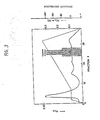
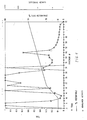
- the bound proteins were eluted with a linear gradient of 0-1.0 M NaCl having a slope of 22 mM/min and run at 1.0 ml/min. 2.0 ml fractions were collected. The flow through was monitored by absorbance at A278 20 ⁇ l aliquots from each fraction were lyophilized in the presence of 100 ⁇ g of BSA, suspended in 500 ⁇ l of DMEM containing 0.5% (v/v) calf serum and assayed for egressin activity using the AIG assay without added growth factors as described above. The results are shown in Figure 2. In Figure 2, "-" indicates no response, i.e., where all of the cells remain as single cells, while "++++" indicates the maximum response, i.e., where colonies of loosely associated cells are formed.
- Bio-Rad HPHT Fractions 20-22 described above containing egressin activity were pooled and loaded onto a Bio-Rad HPHT column which had been equilibrated with 10 mM sodium phosphate buffer, pH 6.8.
- Bio-Rad HPHT is a beaded resin and has an unknown mechanism and separation. The majority of the protein was eluted from the column by washing with 100 mM sodium phosphate buffer, pH 6.8. The tightly bound proteins were eluted with a linear gradient of 100-300 mM sodium phosphate buffer pH 6.8 over 60 minutes with a flow rate of 0.5 ml/min. 2.0 ml fractions were collected. The elution was monitored by absorbance at A215.
- Vydac C18 HPLC consists of a C18(-(CH2)17-CH3) hydrophobic bonded phase on a 300 ⁇ pore-size, 10 ⁇ spherical silica bead.
- the column was equilibrated with 0.1% (v/v) TFA:H2O and run at 1.0 ml/min. 1.0 ml fractions were collected.
- Loosely bound proteins were eluted with a step to 30% (v/v) acetonitrile:0.1% (v/v) TFA:H2O.
- the more tightly bound proteins were eluted with a linear gradient of 30-60% (v/v) acetonitrile:0.1% (v/v) TFA:H2O developed at 0.5% (v/v)/min. and the column was washed with 100% (v/v) acetonitrile:0.1% (v/v) TFA. the elution was monitored by absorbance at A206.
- Egressin was purified from serum-free media conditioned by the human monocytic cell line U937 which was pre-treated with 12-0-tetradecanoyl-phorbol 13-acetate at 37°C for 48 hours. More specifically, the human monocytic cells were grown to a density of about 1 x 10⁶ cells per ml in roller bottles in the presence of RPMI media containing 10% (v/v) calf serum. Serum components were removed by centrifugating the cells (500 x g for 5 min at 25°C). Then the cells were incubated in fresh serum-free RPMI media for 94 hours at 37°C in the presence of about 100 ng/ml of TPA.
- the cells were then centrifuged as above and resuspended in serum-free RPMI media for 24 hours at 37°C in the presence of about 100 ng/ml of TPA.
- the resulting SF-CM was subjected to low speed centrifugation (500 x g for 5 min at 25°C) to remove the cells.
- the resulting SF-CM was acidified with 6.0 N HCl to pH 5.5 and clarified by centrifugation (35,000 x g for 90 min at 10°C).
- Preparative cation-exchange chromatography was performed on the SF-CM described above using a Zeta-SP preparation cartridge which was equilibrated with 0.025 M sodium acetate buffer, pH 5.5. Approximately 20 liters of SF-CM was passed over the Zeta-SP preparation cartridge at a flow rate of approximately 5.0 ml/min. The eluent was monitored by absorbance at A278. The cartridge was washed with 0.025 M sodium acetate buffer, pH 5.5 until absorbance was returned to background levels. The column was then washed with 0.025 M sodium acetate buffer, pH 5.5 containing 1.0 M NaCl and the eluent was collected until the absorbance again returned to background. Approximately 400 ml were collected.
- the cartridge was washed with 0.025 M sodium acetate buffer, pH 5.5 containing 2.0 M NaCl until the background returned to zero. Aliquots of the flow through, 1.0 M NaCl pool and the 2.0 M NaCl were dialyzed against 1.0 M acetic acid, pH 3.0, lyophilized, resuspended in 500 ⁇ l of DMEM containing 0.5% (v/v) calf serum and tested for egressin activity using the monolayer assay as described above. Egressin activity was found to elute in the 1.0 M NaCl pool. At this point, the egressin activity was purified approximately 50 fold over that found in the initial SF-CM.
- the 1.0 M NaCl pool described above was dialyzed at 4°C against four changes at 5 volumes each of 1.0 M acetic acid, pH 3.0 and then lyophilized.
- the resulting material was resuspended in 10 ml of 1.0 M acetic acid, pH 3.0 and subjected to high speed centrifugation (35,000 x g at 10°C for 45 minutes) to remove particulates.
- the clarified supernatant was loaded onto a Bio-gel P-60 column (2.6 cm x 95 cm) which was equilibrated with 1.0 M acetic acid, pH 3.0.
- the chromatogram was developed in 1.0 M acetic acid, pH 3.0 at a flow rate of 15 ml/hr at 25°C.
- 7.5 ml fractions were collected. The eluent was monitored by absorbance at A278. 100 ⁇ l aliquots of the fractions were lyophilized in the presence of 100 ⁇ g of BSA, then resuspended in 500 ⁇ l of DMEM containing 0.5% (v/v) calf serum and assayed for egressin activity using the monolayer assay as described above. Egressin activity was found to elute in fractions with an apparent molecular weight of approximately 25,000 daltons. At this point, the egressin activity was purified approximately 2,000 fold over that found in the initial SF-CM.
- the fractions obtained above having an apparent molecular weight of 25,000 containing egression activity in the Bio-gel P-60 column were pooled, lyophilized and resuspended in 10 ml of 0.025 M sodium acetate buffer, pH 5.0 containing 0.15 M NaCl.
- the sample was centrifuged to remove particulates as described above and then loaded onto a Pharmacia Mono-S column which has been equilibrated with 0.025 M sodium acetate buffer, pH 5.0.
- the column was washed with 0.025 M sodium acetate buffer pH 5.0 containing 0.3 M NaCl until the absorbance of the eluent measured at A278 returned to background.
- the remaining tightly bound proteins were eluted with a linear gradient of 0.3 to 1.0 M NaCl having a slope of 22 mM NaCl/min and run at 1.0 ml/min. 50 ⁇ l aliquots from each fraction were lyophilized in the presence of 100 ⁇ g of BSA and then resuspended in 500 ⁇ l DMEM containing 0.5% (v/v) calf serum and assayed for egressin activity using the monolayer assay described above. Egressin activity was found to elute primarily in fractions between 0.6 M and 0.7 M NaCl. At this point, the egressin activity was purified approximately 12,000 fold over that found in the initial SF-CM.
- the fractions obtained above containing egressin activity were pooled and loaded onto a Bio-Rad HPHT column which had been equilibrated with 10 mM sodium phosphate buffer, pH 6.8. The majority of the protein was eluted from the column by washing with 100 mM sodium phosphate buffer, pH 6.8. The tightly bound proteins were eluted with a linear gradient of 100-300 mM sodium phosphate buffer, pH 6.8 over 60 minutes with a flow rate of 0.5 ml/min. 2.0 ml fractions were collected. The elution was monitored by absorbance at A215.
- the fractions from the HPHT column containing egressin obtained above were pooled and the pH was adjusted to 2.5 with 1.0 M HCl.
- the material was loaded over a Vydac C4 semi-preparative column equilibrated with 0.01% (v/v) TFA:H2O.
- the eluent was monitored by absorbance at A206.
- the column was washed with 0.01% (v/v) TFA:H2O until the absorbance returned to background.
- the more tightly bound proteins were eluted with a linear gradient of 25-35% (v/v) acetonitrile:0.1% (v/v) TGA:H2O developed over 100 minutes.
- Egressin was purified from serum-free media conditioned by the human monocytic cell line U937 which was pre-treated with TPA. More specifically, the human monocytic cells were grown to a density of about 1 x 10⁶ cells per ml in a 75 liter fermentation vessel in the presence of RPMI media containing 7.0% (v/v) fetal calf serum. Prior to induction, serum components were removed by concentrating media containing the cells to approximately 10 liters using a Pro-stalk membrane filtration system then subjecting the cells to low speed centrifugation (500 x g for 5 min at 10°C).
- the cell pellets were resuspended in fresh serum-free RPMI media in the presence of about 100 ng/ml of TPA and inoculated in a 40 liter fermenter. Ater 36 hrs of incubation at 37°C, the cells were removed by passing the media through a conditions flow ⁇ -Lavell centrifugation system. The resulting SF-CM was concentrated using a Pelicon concentration device employing 10,000 daltons cut-off filters. The SF-CM was concentrated approximately 40 fold to a final volume of about 1.0 liter. This material was acidified with 6.0 N HCl to pH 5.5.
- Preparative cation-exchange chromatography was performed on the SF-CM described above using a Zeta-SP preparation cartridge which was equilibrated with 0,025 M sodium acetate buffer, pH 5.5.
- the conductivity of the SF-CM was adjusted with 0.025 M sodium acetate buffer, pH 5.5 to be equal to or lower than that of a 0.025 M sodium acetate buffer, pH 5.5, 0.15 M NaCl solution.
- the resulting material was passed over the Zeta-SP preparation cartridge at a flow rate of approximately 5.0 ml/min. The cartridge was washed and the proteins were eluted as described in Example 2 above.
- the 1.0 M NaCl described above was adjusted with 0.025 M sodium acetate, pH 5.5 so that its conductivity was approximately that of a 0.15 M NaCl solution.
- the material was then loaded into a Pharmacia Mono-S 10/10 semi-preparative cation-exchange column equilibrated with 0.025 M sodium acetate buffer, pH 5.5 containing 0.3 M NaCl.
- the eluent was monitored by absorbance at A278.
- the column was run at 4.0 ml/min and 4.0 ml fractions were collected. The column was washed until the absorbance returned to background level. Then, proteins were eluted with a linear gradient of 0.3 M NaCl to 1.0 M NaCl over 35 minutes.
- the fractions from the Mono-S column containing egressin activity were pooled and dialyzed against 1.0 M acetic acid, pH 3.0.
- the dialyzed fractions were lyophilized and resuspended in 10 ml at 1.0 M acetic acid, pH 3.0 and then subjected to high speed centrifugration as described above to remove particulates.
- the chromatogram was developed using a Bio-Gel P-60 column as described in Example 2 above. Egression activity was assayed by the monolayer assay as described above and found to elute elute with an apparent molecular weight of approximately 25,000 daltons.
- steps (D) and (E) can be interchanged.
- Example 2 The purified egressin obtained in Example 2 above was subjected to amino acid analysis as described in Roth, M., Anal. Chem. , 43:880-882 (1971). The results are set forth in Table II below.
- composition data described above is based on the assumption of a certain number of one residue and then the relative amounts of the others are determined.
- lysine was set to [10] and the remaining residues were calculated on relative amounts, assuming the total molecular weight to be about 10,000 daltons.
- Sodium dodecyl-sulfate polyacrylamide gel electrophoresis was performed as described in Laemnli, U.K. Nature , 227 :680 (1970). The resulting gel was 15% (w/v) polyacrylamide. After electrophoresis, gels were stained for proteins with Coomassie Blue, then with silver using a commercially available reagent obtained from Bio-Rad. Molecular weight markers were used to standardize the gels.
- EGF was employed at a concentration of 5.0 ng/ml in DMEM containing 0.% (v/v) calf serum. 50 ⁇ l of fraction 22 from the chromatograph shown in Figure 2 was lyophilized and suspended in 100 ⁇ l of DMEM containing 0.5% (v/v) calf serum or the 5.0 ng/ml EGF solution.
- the solubilized material was added to 5.0 ml of an aqueous based liquid scintillant (Safety-solve, Research products Institute) and counted in a scintillation counter.
- the mitogenic index expressed as the ratio of cpm incorporated in the presence of added factors/cpm incorporated in cultures receiving no added factors, is shown in Table III below. All experiments were done on duplicate wells.
- Egressin was also tested for its ability to increase cell numbers. More specifically, egressin prepared as described above was added to the above serum deprived cultures and the cell number determined after 5 days. While the cell number increased approximately four fold in cells cultured in 5.0 ng/ml of EGF or in 10% (v/v) fetal calf serum, there was no increase in cell number in cultures treated with egressin. This also demonstrates that egressin is not a growth factor.
- Egressin was also tested for an indirect mitogenic effect, i.e., the ability to increase uptake of 3H-thymidine into trichloroacetic acid precipitatable material in confluent, density inhibited NRK-49F and bovine aorta cells in the presence of EGF, TGF- ⁇ or calf serum.
- egressin is an indirect mitogen, i.e., egressin increased thymidine incorporation in contact-inhibited NRK-49F cells in the presence of 10% (v/v) calf serum, and this effect was enhanced by TGF- ⁇ and EGF.
- Egressin lacked any mitogenic effect on the bovine aortic endothelial cells in the presence or absence of TGF- ⁇ and did not induce growth of serum-deprived NRK-49F cells.
Landscapes
- Health & Medical Sciences (AREA)
- Life Sciences & Earth Sciences (AREA)
- Chemical & Material Sciences (AREA)
- Engineering & Computer Science (AREA)
- Molecular Biology (AREA)
- Biomedical Technology (AREA)
- Immunology (AREA)
- Organic Chemistry (AREA)
- Toxicology (AREA)
- General Health & Medical Sciences (AREA)
- Biochemistry (AREA)
- Medicinal Chemistry (AREA)
- Hematology (AREA)
- Urology & Nephrology (AREA)
- Gastroenterology & Hepatology (AREA)
- Proteomics, Peptides & Aminoacids (AREA)
- Genetics & Genomics (AREA)
- Biophysics (AREA)
- Zoology (AREA)
- Physics & Mathematics (AREA)
- Biotechnology (AREA)
- General Physics & Mathematics (AREA)
- Pathology (AREA)
- Analytical Chemistry (AREA)
- Tropical Medicine & Parasitology (AREA)
- Cell Biology (AREA)
- Microbiology (AREA)
- Food Science & Technology (AREA)
- Bioinformatics & Cheminformatics (AREA)
- Peptides Or Proteins (AREA)
- Preparation Of Compounds By Using Micro-Organisms (AREA)
- Medicines That Contain Protein Lipid Enzymes And Other Medicines (AREA)
Applications Claiming Priority (4)
| Application Number | Priority Date | Filing Date | Title |
|---|---|---|---|
| US90935686A | 1986-09-19 | 1986-09-19 | |
| US909356 | 1986-09-19 | ||
| US96576 | 1987-09-15 | ||
| US07/096,576 US5039794A (en) | 1986-09-19 | 1987-09-15 | Tumor egress factor and processes for producing the same |
Publications (2)
| Publication Number | Publication Date |
|---|---|
| EP0260708A2 true EP0260708A2 (de) | 1988-03-23 |
| EP0260708A3 EP0260708A3 (de) | 1988-11-17 |
Family
ID=26791847
Family Applications (1)
| Application Number | Title | Priority Date | Filing Date |
|---|---|---|---|
| EP87113683A Withdrawn EP0260708A3 (de) | 1986-09-19 | 1987-09-18 | Tumoraustrittsfaktor und Verfahren für seine Herstellung |
Country Status (4)
| Country | Link |
|---|---|
| US (1) | US5039794A (de) |
| EP (1) | EP0260708A3 (de) |
| KR (1) | KR910002703B1 (de) |
| DK (1) | DK491087A (de) |
Families Citing this family (20)
| Publication number | Priority date | Publication date | Assignee | Title |
|---|---|---|---|---|
| US5346988A (en) * | 1990-06-20 | 1994-09-13 | Tampa Bay Research Institute | Process for obtaining cellular protein having anti-HIV activity |
| US5543390A (en) * | 1990-11-01 | 1996-08-06 | State Of Oregon, Acting By And Through The Oregon State Board Of Higher Education, Acting For And On Behalf Of The Oregon Health Sciences University | Covalent microparticle-drug conjugates for biological targeting |
| US5827819A (en) | 1990-11-01 | 1998-10-27 | Oregon Health Sciences University | Covalent polar lipid conjugates with neurologically active compounds for targeting |
| US5643881A (en) * | 1994-08-04 | 1997-07-01 | Tampa Bay Research Institute | Protein having anti-HIV activity and method for obtaining it |
| EP1006794B1 (de) * | 1997-03-12 | 2007-11-28 | Robert W. Esmond | Verfahren zur behandlung oder prävention der alzheimerischen krankheit |
| US20040058873A1 (en) * | 1998-03-12 | 2004-03-25 | Esmond Robert W. | Method for treating or preventing Alzheimer's disease |
| JP2002532416A (ja) * | 1998-12-17 | 2002-10-02 | マインドセット・バイオファーマシューティカルズ・(ユーエスエイ)・インコーポレイテッド | 脳のグルコース利用の増進 |
| US7045543B2 (en) | 2001-11-05 | 2006-05-16 | Enzrel Inc. | Covalent conjugates of biologically-active compounds with amino acids and amino acid derivatives for targeting to physiologically-protected sites |
| US7244764B2 (en) * | 2003-06-23 | 2007-07-17 | Neurochem (International) Limited | Methods and compositions for treating amyloid-related diseases |
| US7414076B2 (en) * | 2003-06-23 | 2008-08-19 | Neurochem (International) Limited | Methods and compositions for treating amyloid-related diseases |
| US20070010573A1 (en) * | 2003-06-23 | 2007-01-11 | Xianqi Kong | Methods and compositions for treating amyloid-related diseases |
| BRPI0517790A (pt) * | 2004-11-12 | 2008-10-21 | Neurochem Int Ltd | métodos e composições fluoradas para tratar doenças relacionadas à amilóide |
| CA2586334A1 (en) * | 2004-11-16 | 2006-06-08 | Neurochem (International) Limited | Compounds for the treatment of cns and amyloid associated diseases |
| WO2006085149A2 (en) | 2004-12-22 | 2006-08-17 | Neurochem (International) Limited | Methods and compositions for treating amyloid-related diseases |
| US20070232556A1 (en) * | 2006-03-31 | 2007-10-04 | Montine Thomas J | Methods and compositions for the treatment of neurological diseases and disorders |
| US20100316626A1 (en) * | 2006-08-11 | 2010-12-16 | The Government Of The United States Of America As Represented By The Secretary | Methods for treatment and diagnosis of psychiatric disorders |
| KR101608445B1 (ko) | 2006-10-12 | 2016-04-01 | 비에이치아이 리미티드 파트너쉽 | 3-아미노-1-프로판설폰산을 전달하기 위한 방법, 화합물, 조성물 및 비히클 |
| EP2342356A4 (de) | 2008-09-29 | 2012-11-21 | Univ Ben Gurion | Amyloid-beta-peptidasen und verwendungsverfahren dafür |
| EP3634978A1 (de) | 2017-06-07 | 2020-04-15 | Adrx, Inc. | Tau-aggregationshemmer |
| EP3668886A2 (de) | 2017-08-18 | 2020-06-24 | Adrx, Inc. | Peptidinhibitoren der tau-aggregation |
-
1987
- 1987-09-15 US US07/096,576 patent/US5039794A/en not_active Expired - Fee Related
- 1987-09-18 EP EP87113683A patent/EP0260708A3/de not_active Withdrawn
- 1987-09-18 DK DK491087A patent/DK491087A/da not_active Application Discontinuation
- 1987-09-19 KR KR1019870010466A patent/KR910002703B1/ko not_active Expired
Non-Patent Citations (4)
| Title |
|---|
| JOURNAL OF CELL SCIENCE, vol. 77, 1985, pages 209-223, The Company of Biologists Ltd, GB; M. STOKER et al.: "An epithelial scatter factor released by embryo fibroblasts" * |
| NATURE, vol. 327, 21st May 1987, pages 239-242; M. STOKER et al.: "Scatter factor is a fibroblast-derived modulator of epithelial cell mobility" * |
| PROC. NATL. ACAD. SCI. USA, vol. 82, August 1985, pages 5015-5019; J.E. DE LARCO et al.: "Ectopic peptides released by a human melanoma cell line that modulate the transformed phenotype" * |
| PROC. NATL. ACAD. SCI. USA, vol. 83, May 1986, pages 3302-3306; L.A. LIOTTA et al.: "Tumor cell autocrine motility factor" * |
Also Published As
| Publication number | Publication date |
|---|---|
| KR880003901A (ko) | 1988-05-31 |
| KR910002703B1 (ko) | 1991-05-03 |
| DK491087A (da) | 1988-03-20 |
| DK491087D0 (da) | 1987-09-18 |
| US5039794A (en) | 1991-08-13 |
| EP0260708A3 (de) | 1988-11-17 |
Similar Documents
| Publication | Publication Date | Title |
|---|---|---|
| US5039794A (en) | Tumor egress factor and processes for producing the same | |
| US4816561A (en) | Biologically active polypeptides | |
| US5240912A (en) | Transforming growth factor (TGF) peptides | |
| AU601144B2 (en) | Cytotoxic protein | |
| US4863899A (en) | Biologically active polypeptides | |
| US5359032A (en) | Interkeukin-1 inhibitor | |
| CA1245153A (en) | Cell matrix receptor system and use in cancer diagnosis and management | |
| JP3390005B2 (ja) | Tgf−ベータ2及びtgf−ベータ3の潜伏性で大きい複合体 | |
| Arend et al. | Absence of induction of IL-1 production in human monocytes by complement fragments. | |
| NABIL et al. | Identification of human complement factor H as a chemotactic protein for monocytes | |
| Ryan et al. | Characterization and purification of mouse macrophage procoagulant-inducing factor. | |
| NZ225940A (en) | Interleukin-1 inhibitors (il-1 inh) and pharmaceutical compositions | |
| JP2559035B2 (ja) | 細胞生長調節因子 | |
| US5219991A (en) | Macrophage stimulating protein | |
| EP0550756A1 (de) | Interleukin 6-zusammensetzung und seine herstellung | |
| Liu et al. | A monoclonal antibody specific for a monocyte-macrophage membrane component blocks the human monocyte response to migration inhibitory factor. | |
| Easter et al. | Immunosuppression by a peptide from the gelatin binding domain of human fibronectin | |
| MacArthur et al. | A peptide secreted by human alveolar macrophages releases neutrophil granule contents. | |
| Newcom et al. | High molecular weight transforming growth factor β is excreted in the urine in active nodular sclerosing Hodgkin's disease | |
| US5071956A (en) | Protein with vascular activity derived from monocytes and its analogues, a process for its extraction and their uses for therapeutic purposes and for the preparation of antibodies | |
| AU630873B2 (en) | Lung growth stimulatory and inhibitory factors for carcinoma tumor cells | |
| RU2076151C1 (ru) | Способ получения фактора некроза опухоли, человеческий фактор некроза опухоли | |
| EP0434836B1 (de) | Verfahren zur reinigung von tenascin | |
| WO1993017045A1 (en) | A novel serum growth factor | |
| Sieber | Chromatography of human urinary erythropoietin and granulocyte colony-stimulating factor on insolubilized phytohaemagglutinin |
Legal Events
| Date | Code | Title | Description |
|---|---|---|---|
| PUAI | Public reference made under article 153(3) epc to a published international application that has entered the european phase |
Free format text: ORIGINAL CODE: 0009012 |
|
| AK | Designated contracting states |
Kind code of ref document: A2 Designated state(s): CH DE ES FR GB IT LI NL SE |
|
| PUAL | Search report despatched |
Free format text: ORIGINAL CODE: 0009013 |
|
| RHK1 | Main classification (correction) |
Ipc: A61K 37/02 |
|
| AK | Designated contracting states |
Kind code of ref document: A3 Designated state(s): CH DE ES FR GB IT LI NL SE |
|
| 17P | Request for examination filed |
Effective date: 19890126 |
|
| STAA | Information on the status of an ep patent application or granted ep patent |
Free format text: STATUS: THE APPLICATION HAS BEEN WITHDRAWN |
|
| 18W | Application withdrawn |
Withdrawal date: 19910724 |
|
| R18W | Application withdrawn (corrected) |
Effective date: 19910724 |
|
| RIN1 | Information on inventor provided before grant (corrected) |
Inventor name: WIER, MARJORIE L. Inventor name: DE LARCO, JOSEPH E. |