EP0265334A2 - Verfahren und System zur Konstruktion von dreidimensionalen Bildern - Google Patents
Verfahren und System zur Konstruktion von dreidimensionalen Bildern Download PDFInfo
- Publication number
- EP0265334A2 EP0265334A2 EP87402329A EP87402329A EP0265334A2 EP 0265334 A2 EP0265334 A2 EP 0265334A2 EP 87402329 A EP87402329 A EP 87402329A EP 87402329 A EP87402329 A EP 87402329A EP 0265334 A2 EP0265334 A2 EP 0265334A2
- Authority
- EP
- European Patent Office
- Prior art keywords
- bit
- spaced apart
- distance
- planes
- values
- Prior art date
- Legal status (The legal status is an assumption and is not a legal conclusion. Google has not performed a legal analysis and makes no representation as to the accuracy of the status listed.)
- Granted
Links
Images


Classifications
-
- G—PHYSICS
- G06—COMPUTING OR CALCULATING; COUNTING
- G06F—ELECTRIC DIGITAL DATA PROCESSING
- G06F17/00—Digital computing or data processing equipment or methods, specially adapted for specific functions
- G06F17/10—Complex mathematical operations
- G06F17/17—Function evaluation by approximation methods, e.g. inter- or extrapolation, smoothing, least mean square method
- G06F17/175—Function evaluation by approximation methods, e.g. inter- or extrapolation, smoothing, least mean square method of multidimensional data
-
- G—PHYSICS
- G06—COMPUTING OR CALCULATING; COUNTING
- G06T—IMAGE DATA PROCESSING OR GENERATION, IN GENERAL
- G06T12/00—Tomographic reconstruction from projections
- G06T12/30—Image post-processing, e.g. metal artefact correction
Definitions
- This invention is concerned with imaging processes and more particularly with unique interpolation processes used to obtain three-dimensional (3D) images with smooth surfaces in short time periods, with accuracy and fidelity when only partial three dimensional information is available in the form of tomographically acquired data.
- Computerized tomography by definition provides two dimensional (2D) images of planes (planar views) in a patient's body. Often times it is important for the viewer (diagnostician and/or doctor) to be able to obtain three-dimensional views rather than planar views of the interior of the patient's body. For example, in surgical prcedures it is extremely helpful if the doctor can see a three-dimensional view of the interior of the body in the area of the operation. Prior to brain surgery which are supplemented by three-dimensional views elaborate measuring methods are used to increase the probability of a successful operation.
- the three-dimensional views are obtained either by special x-ray equipment such as for example disclosed in the U.S. Patent 4,309,615 or by taking a squence of the tomographic views of the portion of the body of interest and subseqeuently processing these views to provide the desired three-dimensional view.
- special x-ray equipment such as for example disclosed in the U.S. Patent 4,309,615
- a squence of the tomographic views of the portion of the body of interest and subseqeuently processing these views to provide the desired three-dimensional view.
- the object of this invention to provide a reliable method of obtaining a full description of the shape of internal organs from a series of planar slices of those organs.
- the result of applying the process described is a three dimensional binary bit matrix, in which the regions of bits having "1" values correspond to regions of space occupied by the organic of interest, while regions of bits having "0" values correspond to unoccupied space.
- This 3-D matrix can then be used to present the structure of the organs to the interested viewer in forms, such as a shaded surface image, well known to those skilled in the art.
- a prior art problem is to locate the edge of the organ of interest.
- thresholding is practiced to convert the originally acquired data into binary maps to determine the edges of the organs. Interpolation is then done between portions of the edges having opposite bit values.
- the functional values (not the binary values) of the given plane image are used for the interpolation between the planes.
- the interpolation acquired values are converted to binary maps using thresholding. Accordingly a prior art problem is the necessity of using thresholding discriminators to convert the interpolated functional values to bit values.
- the prior art is also plagued with a "staircase" artifact in the images of the surface of the organ of interest.
- a unique interpolation process which is used to obtain high fidelity three-dimensional descriptions of volumes of interest in a subject, said process comprises the steps of: detecting signals from at least two spaced apart planes in a subject, processing said detected signals to obtain images of said two spaced apart planes, said images comprising rows and columns or image values, each intersecting row and column corresponding to an integral area of said plane and said image value corresponding to a pixel value of said integral area, processing the image values of the two spaced apart images to obtain a first and a second bit map where for example "1" values correspond to locations occupied by the organ of interest and "0" values correspond to locations not occupied by the organ of interest, processing the bits in said first and second bit maps to obtain a logic exclusive-OR (XOR) bit map, using said XOR bit map and said first and second bit maps to obtain first and second distance maps, and using said first and second distance maps in conjunction with either one of said first or second bit maps to obtain bit values of interpolated planes between
- XOR logic exclusive-
- a feature of the invention includes using available regular 2D front end tomographic systems for acquiring the 3D image data.
- CT computerized tomographic
- the three-dimensional imaging system 11 of Fig. 1 comprises a front end system 12 for detecting signals from specific locations in a subject being examined.
- the signals are obtained using an x-ray CT system, for example, when the oppositely disposed radiation source and detectors rotate about the patient and provide radiation intensity values for a multiplicity of radiation paths in a plane through the patient.
- Well known techniques are used in processing the detected radiation to define elemental area location of radiation absoption values within the patient.
- the original signal from the front end is normally analog; therefore, an analog to digital converter (ADC) 13 is preferably provided to convert the signals to digital form.
- An image processor 14 uses the digital signals and determines the absorption values of the elemental area locations in the plane by back-projection methods, for example.
- An image is provided comprising a matrix of rows and columns in which the values correspond to the values of the elemental area in the plane through the patient and to pixel locations in the actual display image.
- At least two images are obtained by moving the subject relative to the radiation source and detector means to obtain image data on more than one plane.
- a plurality of planes are imaged and interpolation is done between successively imaged planes.
- the planes are spaced apart and it is the volume between the planes that will be imaged in three-dimensional form.
- the multiplicity of images are shown at 16.
- a pixel processor 17, which in a preferred embodiment uses a thresehold discriminator and a specific area discriminator, or manual contour tracing, converts the pixels into bits having values of zero and one.
- the spaced apart planes are thus converted to a lower image bit map 18 and an upper image bit map 19.
- an XOR bit map shown at 21 is provided from the bits of the lower and upper image bits maps.
- both of the upper and lower bit maps have the same binary value at a matrix location, that location is given a "0" value in the XOR bit maps, otherwise, that location is give a "1" value.
- the XOR bit map is then used in combination with the lower image bit map and the upper image bit map to obtain a lower distance map 22 and an upper distance map 23.
- a distance map shows the length of the shortest path lying entirely within the XOR bit map from the center of a bit to the edge of a bit with the opposite value on the corresponding map (upper or lower). The path goes only through centers of neighboring bits on the corresponding map.
- interpolated planes there is no limit on the number of interpolated planes that can be obtained by this method.
- three interpolated planes are obtained. They are the planes located at one quarter of the distance, half the distance and three quarters of the distance beween the two spaced apart image planes.
- the number of interpolated planes is such that the distance between planes is equal to the distance between neighboring pixels within the planes.
- the interpolated planes contain bit values in each of the elemental areas.
- the collection of sequential planes thus obtained forms a volume defining"description" of the imaged "organ” (organ, bone, tissue, body part etc.). This description is then processed using known techniques to obtain images of outer or inner surfaces of the organs or to measure various spatial (3D) properties such as volumes, angles and distances.
- the sample solids 41 and 42 of Fig. 2 are shown as being located between lower and upper bit map planes 18a and 19a respectively.
- the volume 41 is shown as a skewed cylinder while the volume 42 is a cubical volume intersected only by the lower bit map plane 18a.
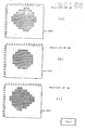
- Fig. 3 the lower bit map 18 and the upper bit map 19 are shown by way of example only, as matrices of fifteen columns and fourteen rows having bit values thereon.
- the bit values "0" indicate no contact by the transverse planes Pu and PL with volumes 41 or 42.
- the bit values '1' indicate contact with volumes 41 or 42.
- Corresponding bit values are used to create the XOR map 21 of Fig. 4. For example, in column 6, row 2 the bit value of the lower bit map is "0" while the bit value in the upper bit map is "1". Thus, the XOR bit map value for that location is "1".
- bit value is "0"; therefore, the corresponding row and column in the XOR bit map is "0".
- bit values of the lower bit map is "1" while the value of the upper bit map is "0”; accordingly,the bit value of the XOR bit map for that loaction is "1".
- bit value of both maps is 1; therefore the bit values of the XOR bit map is "0".
- the bit value of the fourth row, fourth column of the XOR bit map 21 is one.
- the bit value of the corresponding location in the lower bit map is zero.
- the closest bit value on the lower bit map corresponding to the bit value of the XOR map is at a distance of 1 1/2. More particularly the distances are measured from the center of the matrix area to the border of the closest matrix area having the opposite bit value in the corresponding (upper or lower) bit map.
- the shortest distance across each matrix area is one.
- the diagonal distance from the center of the matrix to a corner of the matrix is ⁇ 2/2.
- the distance is measured along a path that goes only through the centers of matrix areas that have a "1" value on the XOR bit map. Looking at the lower bit map, it is easy to see that the distance from the center of the matrix area of the second row, seventh column to a matrix area having the value "1" is the distance to the matrix area located at the second row in column eight. That distance is 1/2. Similarily the shortest distance from the fifth row of the eleventh column in the lower bit map 18 area having an opposite value "0" is 1 plus ⁇ 2/2 or (3/2) ⁇ 2. In a similar manner every area of the XOR bit map that has a value "1' is applied to the lower and upper bit maps 18 and 19 to determine lower and upper distance maps 22 and 23.
- the search looks at the area on the upper and lower bit maps that correspond to areas in the XOR map that have a "1" value. Then, the length of the shortest path, lying entirely within the XOR map 21 from the center of a bit area to the edge of a bit area with the opposite value in the corresponding map, is entered for each such area in the appropriate distance map 22 or 23. In case no such path exists as in the 13th row of the second column in the upper distance map 23 some reasonable constant value is entered. It has been found that the distance between the upper and lower planes in matrix units gives good results.
- the distance values are used to obtain the interpolated bit maps.
- the fourth row, fourth column of the lower bit map has a bit value of "0".
- the corresponding area; that is the fourth row, fourth column of the upper bit map has a bit value of "1".
- the lower distance map corresponding matrix area has a value 3/2.
- the upper distance map at that matrix area has a value of 2/2.
- the change over point from "1" to "0" is [1.5/(1.5+0.70)]Dt or 0.68 Dt. Anything within 0.68 of the distance between the lower bit map and the upper bit has the bit value of "0" and anything at a greater distance has the value "1".
- any number of interpolated planes or maps can be derived from each two spaced apart sequential plane images.
- the three-dimensional binary matrix holds a full description of the body, or bodies, of interest.
- Methods well known to those skilled in the art are used to produce 3D shaded images from the acquired and interpolated data. For example the locations of the occupied ("1") bit areas in the matrix can be multipled by a rotation matrix and projected onto a two-dimensional plane, to create a surface image 28 of the body in a given rotational angle. That image can then be displayed on the display unit 29.
Landscapes
- Engineering & Computer Science (AREA)
- Physics & Mathematics (AREA)
- General Physics & Mathematics (AREA)
- Theoretical Computer Science (AREA)
- Mathematical Physics (AREA)
- Computational Mathematics (AREA)
- Mathematical Analysis (AREA)
- Mathematical Optimization (AREA)
- Pure & Applied Mathematics (AREA)
- Data Mining & Analysis (AREA)
- Algebra (AREA)
- Databases & Information Systems (AREA)
- Software Systems (AREA)
- General Engineering & Computer Science (AREA)
- Image Analysis (AREA)
- Image Processing (AREA)
- Apparatus For Radiation Diagnosis (AREA)
Applications Claiming Priority (2)
| Application Number | Priority Date | Filing Date | Title |
|---|---|---|---|
| IL80364 | 1986-10-20 | ||
| IL80364A IL80364A (en) | 1986-10-20 | 1986-10-20 | Three dimensional image construction using binary space interpolation |
Publications (3)
| Publication Number | Publication Date |
|---|---|
| EP0265334A2 true EP0265334A2 (de) | 1988-04-27 |
| EP0265334A3 EP0265334A3 (en) | 1989-10-25 |
| EP0265334B1 EP0265334B1 (de) | 1993-07-28 |
Family
ID=11057224
Family Applications (1)
| Application Number | Title | Priority Date | Filing Date |
|---|---|---|---|
| EP87402329A Expired - Lifetime EP0265334B1 (de) | 1986-10-20 | 1987-10-19 | Verfahren und System zur Konstruktion von dreidimensionalen Bildern |
Country Status (4)
| Country | Link |
|---|---|
| US (1) | US4894776A (de) |
| EP (1) | EP0265334B1 (de) |
| DE (1) | DE3735519C2 (de) |
| IL (1) | IL80364A (de) |
Cited By (4)
| Publication number | Priority date | Publication date | Assignee | Title |
|---|---|---|---|---|
| GB2246689A (en) * | 1990-08-04 | 1992-02-05 | David John Winter | Creating a 3-D image from a plurality of cross-sections |
| EP0412748A3 (en) * | 1989-08-09 | 1993-01-27 | Picker International, Inc. | Methods and apparatus for generating three-dimensional images |
| ES2048034A2 (es) * | 1991-07-25 | 1994-03-01 | Ibermatica S A | Procedimiento de obtencion de informacion e imagenes de organos vitales. |
| EP0571026A3 (de) * | 1992-05-21 | 1994-11-23 | Koninkl Philips Electronics Nv | Richtungsabhängige Interpolation in der Angiographie. |
Families Citing this family (17)
| Publication number | Priority date | Publication date | Assignee | Title |
|---|---|---|---|---|
| US5157388A (en) * | 1989-02-14 | 1992-10-20 | Intel Corporation | Method and apparatus for graphics data interpolation |
| US5390291A (en) * | 1990-10-05 | 1995-02-14 | Atlantic Richfield Company | Method for interpolating between two regions of a display |
| EP0559978B1 (de) * | 1992-03-12 | 1998-08-05 | International Business Machines Corporation | Verfahren zur Bildverarbeitung |
| US5450098A (en) * | 1992-09-19 | 1995-09-12 | Optibase Advanced Systems (1990) Ltd. | Tri-dimensional visual model |
| DE69331943D1 (de) * | 1992-12-22 | 2002-06-27 | Canon Kk | Verfahren und Einrichtung zur Zeichenherstellung |
| FR2706043B1 (fr) * | 1993-06-02 | 1995-07-07 | Commissariat Energie Atomique | Installation et procédé de reconstruction d'images tridimensionnelles. |
| US5764871A (en) * | 1993-10-21 | 1998-06-09 | Eastman Kodak Company | Method and apparatus for constructing intermediate images for a depth image from stereo images using velocity vector fields |
| US6611575B1 (en) * | 2001-07-27 | 2003-08-26 | General Electric Company | Method and system for high resolution 3D visualization of mammography images |
| JP2005521123A (ja) * | 2001-10-22 | 2005-07-14 | ライカ ミクロジュステムス ヴェツラー ゲーエムベーハー | 光学顕微鏡検出3次元画像の生成方法及び生成装置 |
| WO2005065085A2 (en) * | 2003-12-21 | 2005-07-21 | Kremen Stanley H | System and apparatus for recording, transmitting, and projecting digital three-dimensional images |
| US20070279436A1 (en) * | 2006-06-02 | 2007-12-06 | Hern Ng | Method and system for selective visualization and interaction with 3D image data, in a tunnel viewer |
| US8019661B2 (en) | 2007-11-26 | 2011-09-13 | International Business Machines Corporation | Virtual web store with product images |
| US8065200B2 (en) | 2007-11-26 | 2011-11-22 | International Business Machines Corporation | Virtual web store with product images |
| US8253727B2 (en) | 2008-03-14 | 2012-08-28 | International Business Machines Corporation | Creating a web store using manufacturing data |
| US8396248B2 (en) * | 2008-09-16 | 2013-03-12 | Varian Medical Systems, Inc. | Sequential stereo imaging for estimating trajectory and monitoring target position |
| US20150113372A1 (en) * | 2013-10-18 | 2015-04-23 | Apple Inc. | Text and shape morphing in a presentation application |
| WO2019092167A1 (en) * | 2017-11-09 | 2019-05-16 | Agfa Healthcare Nv | Method of segmenting a 3d object in a medical radiation image |
Family Cites Families (7)
| Publication number | Priority date | Publication date | Assignee | Title |
|---|---|---|---|---|
| US4066903A (en) * | 1974-07-20 | 1978-01-03 | Emi Limited | Radiology |
| CA1159957A (en) * | 1980-07-30 | 1984-01-03 | James M. Gessert | Real time fill circuit |
| US4446529A (en) * | 1981-01-26 | 1984-05-01 | Rca Corporation | Linear interpolation between regularly spaced digital samples |
| JPS58118735A (ja) * | 1982-01-06 | 1983-07-14 | 株式会社東芝 | コンピユ−タ断層撮影装置 |
| JPS60152942A (ja) * | 1984-01-23 | 1985-08-12 | Toshiba Corp | Nmr―ctスキャン計画装置 |
| CA1250064A (en) * | 1985-03-29 | 1989-02-14 | Kenichi Anjyo | Method for constructing three-dimensional polyhedron model |
| JPS62187982A (ja) * | 1985-10-07 | 1987-08-17 | フエアチヤイルド セミコンダクタコ−ポレ−シヨン | ビツトプレ−ン面積相関器 |
-
1986
- 1986-10-20 IL IL80364A patent/IL80364A/xx unknown
-
1987
- 1987-10-19 US US07/109,400 patent/US4894776A/en not_active Expired - Lifetime
- 1987-10-19 EP EP87402329A patent/EP0265334B1/de not_active Expired - Lifetime
- 1987-10-20 DE DE3735519A patent/DE3735519C2/de not_active Expired - Lifetime
Cited By (4)
| Publication number | Priority date | Publication date | Assignee | Title |
|---|---|---|---|---|
| EP0412748A3 (en) * | 1989-08-09 | 1993-01-27 | Picker International, Inc. | Methods and apparatus for generating three-dimensional images |
| GB2246689A (en) * | 1990-08-04 | 1992-02-05 | David John Winter | Creating a 3-D image from a plurality of cross-sections |
| ES2048034A2 (es) * | 1991-07-25 | 1994-03-01 | Ibermatica S A | Procedimiento de obtencion de informacion e imagenes de organos vitales. |
| EP0571026A3 (de) * | 1992-05-21 | 1994-11-23 | Koninkl Philips Electronics Nv | Richtungsabhängige Interpolation in der Angiographie. |
Also Published As
| Publication number | Publication date |
|---|---|
| IL80364A (en) | 1990-03-19 |
| DE3735519C2 (de) | 2003-01-30 |
| US4894776A (en) | 1990-01-16 |
| EP0265334A3 (en) | 1989-10-25 |
| IL80364A0 (en) | 1987-01-30 |
| DE3735519A1 (de) | 1988-04-28 |
| EP0265334B1 (de) | 1993-07-28 |
Similar Documents
| Publication | Publication Date | Title |
|---|---|---|
| US4894776A (en) | Binary space interpolation | |
| US6014419A (en) | CT cone beam scanner with fast and complete data acquistion and accurate and efficient regional reconstruction | |
| EP0492895B1 (de) | 3-D-Bilderrekonstruktion | |
| Kennedy et al. | Super-resolution in PET imaging | |
| EP0964366B1 (de) | Dreidimensionales Bildrekonstruktionsverfahren | |
| CN100573588C (zh) | 使用截短的投影和在先采集的3d ct图像的锥形束ct设备 | |
| US6731283B1 (en) | C-arm calibration method utilizing aplanar transformation for 3D reconstruction in an imaging system | |
| US6915004B2 (en) | Continuous tomography bed motion data processing apparatus and method | |
| EP0707289B1 (de) | System und Verfahren zur Darstellung von Bildern | |
| US5647360A (en) | Digital subtraction angiography for 3D diagnostic imaging | |
| US5287274A (en) | Method for acquisition of radiological data in multiple orthogonal orientations using a 2D detector relating to a body irradiated with x-rays and for reconstruction of structures corresponding to said body using an algebraic algorithm | |
| US5241471A (en) | Method of multi-scale reconstruction of the image of the structure of a body at an increased speed | |
| US4989142A (en) | Three-dimensional images obtained from tomographic slices with gantry tilt | |
| EP0751484A2 (de) | Bildrekonstruierung von spiralabgetasteten Teilkegelstrahldaten | |
| EP0520778B1 (de) | Tomographische Bilderzeugung mittels Schrägstrahlen | |
| EP0928458B1 (de) | Eine computertomographische methode und ein computertomograph | |
| JP3787374B2 (ja) | 断層撮影画像内のアーチファクトを低減するための方法および装置 | |
| US5341460A (en) | Method and apparatus for producing a three-dimensional computerized tomography image of an object with improved conversion of cone beam data to radon data | |
| CN107016672A (zh) | 医学扫描图像的重建方法和装置以及医学成像系统 | |
| JP2001057976A (ja) | 立体画像再構成方法及び装置並びにctスキャナー | |
| JPH08308825A (ja) | 3次元計算機式断層写真作像を実施する方法及びシステム | |
| JPH07143979A (ja) | 断層撮影画像中のアーチファクトを低減する方法および装置 | |
| Brasse et al. | Fast fully 3-D image reconstruction in PET using planograms | |
| EP0519445B1 (de) | Verfahren und Vorrichtung zur Spect-Bildzusammensetzung unter Aufrechterhaltung einer hohen Raumauflösung | |
| CN1936958A (zh) | 用于从二维投影图像中重建三维图像体积的方法和装置 |
Legal Events
| Date | Code | Title | Description |
|---|---|---|---|
| PUAI | Public reference made under article 153(3) epc to a published international application that has entered the european phase |
Free format text: ORIGINAL CODE: 0009012 |
|
| AK | Designated contracting states |
Kind code of ref document: A2 Designated state(s): FR GB NL |
|
| EL | Fr: translation of claims filed | ||
| TCNL | Nl: translation of patent claims filed | ||
| PUAL | Search report despatched |
Free format text: ORIGINAL CODE: 0009013 |
|
| AK | Designated contracting states |
Kind code of ref document: A3 Designated state(s): FR GB NL |
|
| 17P | Request for examination filed |
Effective date: 19900207 |
|
| 17Q | First examination report despatched |
Effective date: 19920213 |
|
| GRAA | (expected) grant |
Free format text: ORIGINAL CODE: 0009210 |
|
| AK | Designated contracting states |
Kind code of ref document: B1 Designated state(s): FR GB NL |
|
| ET | Fr: translation filed | ||
| PLBE | No opposition filed within time limit |
Free format text: ORIGINAL CODE: 0009261 |
|
| STAA | Information on the status of an ep patent application or granted ep patent |
Free format text: STATUS: NO OPPOSITION FILED WITHIN TIME LIMIT |
|
| 26N | No opposition filed | ||
| PGFP | Annual fee paid to national office [announced via postgrant information from national office to epo] |
Ref country code: FR Payment date: 19961011 Year of fee payment: 10 |
|
| PGFP | Annual fee paid to national office [announced via postgrant information from national office to epo] |
Ref country code: GB Payment date: 19961021 Year of fee payment: 10 |
|
| PG25 | Lapsed in a contracting state [announced via postgrant information from national office to epo] |
Ref country code: GB Free format text: LAPSE BECAUSE OF NON-PAYMENT OF DUE FEES Effective date: 19971019 |
|
| PG25 | Lapsed in a contracting state [announced via postgrant information from national office to epo] |
Ref country code: FR Free format text: THE PATENT HAS BEEN ANNULLED BY A DECISION OF A NATIONAL AUTHORITY Effective date: 19971031 |
|
| GBPC | Gb: european patent ceased through non-payment of renewal fee |
Effective date: 19971019 |
|
| REG | Reference to a national code |
Ref country code: FR Ref legal event code: ST |
|
| PGFP | Annual fee paid to national office [announced via postgrant information from national office to epo] |
Ref country code: NL Payment date: 20021031 Year of fee payment: 16 |
|
| PG25 | Lapsed in a contracting state [announced via postgrant information from national office to epo] |
Ref country code: NL Free format text: LAPSE BECAUSE OF NON-PAYMENT OF DUE FEES Effective date: 20040501 |
|
| NLV4 | Nl: lapsed or anulled due to non-payment of the annual fee |
Effective date: 20040501 |