EP0268636B1 - Polynucleotidsequenzen, die für den humanen FC-REZEPTOR FÜR IMMUNOGLOBULIN codieren - Google Patents
Polynucleotidsequenzen, die für den humanen FC-REZEPTOR FÜR IMMUNOGLOBULIN codieren Download PDFInfo
- Publication number
- EP0268636B1 EP0268636B1 EP87903479A EP87903479A EP0268636B1 EP 0268636 B1 EP0268636 B1 EP 0268636B1 EP 87903479 A EP87903479 A EP 87903479A EP 87903479 A EP87903479 A EP 87903479A EP 0268636 B1 EP0268636 B1 EP 0268636B1
- Authority
- EP
- European Patent Office
- Prior art keywords
- human
- fcr
- receptor
- amino acid
- sequence
- Prior art date
- Legal status (The legal status is an assumption and is not a legal conclusion. Google has not performed a legal analysis and makes no representation as to the accuracy of the status listed.)
- Expired - Lifetime
Links
Images








Classifications
-
- C—CHEMISTRY; METALLURGY
- C07—ORGANIC CHEMISTRY
- C07K—PEPTIDES
- C07K14/00—Peptides having more than 20 amino acids; Gastrins; Somatostatins; Melanotropins; Derivatives thereof
- C07K14/435—Peptides having more than 20 amino acids; Gastrins; Somatostatins; Melanotropins; Derivatives thereof from animals; from humans
- C07K14/705—Receptors; Cell surface antigens; Cell surface determinants
- C07K14/70503—Immunoglobulin superfamily
- C07K14/70535—Fc-receptors, e.g. CD16, CD32, CD64 (CD2314/705F)
-
- A—HUMAN NECESSITIES
- A61—MEDICAL OR VETERINARY SCIENCE; HYGIENE
- A61P—SPECIFIC THERAPEUTIC ACTIVITY OF CHEMICAL COMPOUNDS OR MEDICINAL PREPARATIONS
- A61P37/00—Drugs for immunological or allergic disorders
Definitions
- This invention relates to a Fc receptor for immunoglobulin.
- this invention relates to a polynucleotide encoding a human Fc receptor for immunoglobulin or a fragment thereof capable of binding the Fc region of human immunoglobulin selected from the extracelluar domain of the human Fc receptor and human Fc receptor binding sites, said polynucleotide encoding part or all of the amino acid sequences of Figures 8, 10 and 11 and conservative amino acid variants thereof and allelic variants thereof capable of binding the Fc region of human immunoglobulin.
- Murine clones were sequenced and expression of mRNA and the cell surface Fc receptor investigated.
- the murine Fc receptor is a 301 amino acid transmembrane glycoprotein with two homologous extracelluar domains that are also homologous to members of the Ig superfamily.
- the Fc receptor has 4 sites of N linked glycosylation and a long 94 amino acid cytoplasmic tail.
- Northern analysis of mRNA, immune complex binding and serological studies of cell lines and transfectants demonstrated that the receptor encoded by the cDNA clone binds murine IgG2b and possibly other Ig.
- Human Fc ⁇ R cDNA clones were also isolated and found to encode a glycosylated transmembrane molecule that bears striking homology to both the beta 1 and alpha mouse FcR at both the nucleic and amino acid levels.
- the present application describes a receptor for the Fc portion of immunoglobulin, the receptor exhibiting the ability to bind to the Fc portion of mouse and human immunoglobulin.
- the receptor preferably exhibits an ability to bind to antibodies and immune complexes, has
- the receptor includes the amino acid sequences:-
- the receptor includes at least one of the amino acid sequences:-
- the present invention also provides a vector adapted to encode or to produce a material adapted to encode for the receptor above or a fragment thereof.
- the present invention also provides a vector adapted to encode or produce a material adapted to encode for the leader sequence, extracelluar region, transmembrane region or intracellular region of the receptor above.
- a preferred nucleotide sequence, cDNA clone, gene or vector containing same and capable of encoding for a receptor for the Fc portion of immunoglobulin includes the nucleotide sequences:-
- nucleotide sequence, cDNA clone, gene or vector containing same includes at least one of the nucleotide sequences:-
- Fc receptors form a major group of cell membrane glycoproteins involved in homeostasis of the immunological system.
- Specific receptors for all immunoglobulin (Ig) classes have been defined and are found on a wide variety of immune cells - B cells and some T cells as well as myeloid cells and non-haemopoietic cells (Hubscher et al, 1971; Dickler, 1976; Tsay et al, 1980; Unkless et al, 1981).
- the principal role of these receptors is to bind (Ig) via the Fc region of the Ig molecule and it is through this interaction that a wide range of biological effects are mediated.
- Monoclonal Antibodies The monoclonal anti-Ly-17.2 and Ly-2.1 antibodies have been previously described (Hibbs et al 1985, Hogarth et al 1982). Preparation of F(ab') 2 fragments from purified antibody was performed as described (16). The 2.4 G2 antibody has also been described before (Unkless 1979).
- J774 macrophage cells (2x10 10 ) were harvested from ascites fluid obtained from (CBAxBALB/c)F1 mice and immediately lysed in cold phosphate buffered saline (PBS) / 0.5% Nonidet P-40 (PBS/0.5% NP40) pH 7.4 containing 1% aprotinin (Sigma, St. Louis, MO) and 1 mM phenylmethylsulfonyl fluoride (Sigma, St. Louis, MO). An additional 2x10 7 cells were surface labeled with 125 I, and pooled with the cold lysate.
- the lysates were clarified and incubated for 1 hour at 4°C with anti-Ly-17.2 antibody conjugated Sepharose 4B (Pharmacia, Uppsala, Sweden).
- the immunoabsorbent was washed three times in 0.6M NaCl, 0.0125M KH 2 PO4, pH 7.4, three times in PBS/0.5% NP40 pH 7.4 and a further three times in 0.5% deoxycholate in 100mM Tris-HCl pH 8.0.
- the immunoabsorbent was packed into a column and the bound material eluted with 0.5% sodium dodecyl sulfate (NaDodSO 4 ) in 0.1M triethylamine pH 11.5 and freeze dried.
- the purity of the Fc gamma R preparations was assessed by NaDodSO 4 /polyacrylamide gel electrophoresis (NaDodSO 4 /PAGE) and proteins were detected by Coomassie blue staining or by autoradiography.
- Protein Sequence Analysis Protein samples were carboxamidomethylated and ethanol precipitated prior to sequencing. Briefly, samples were dissolved in 50mM boric acid, 0.1% (w/v) SDS and 10mM dithiothreitol pH 8.0 (NaOH) and heated to 60°C for 1 hour. Iodoacetamide was added to a final concentration of 22mM and the samples incubated for 15 minutes at ambient temperature in the dark. Ice cold ethanol containing 50mM HCl was added and the protein allowed to precipitate at -20°C for 2 hours.
- FcgammaR peptides were obtained by digesting samples of affinity purified, carboxamidomethylated Fc gamma R with either S. aureus V8 protease (Miles Laboratories) (Hibbs et al 1986) lysine-C proteinase (according to manufacturers instructions, Boehringer Mannheim, Mannheim, FRG) or cyanogen bromide (CNBr). Peptides were purified by reverse phase chromatography using a Pharmacia fast protein liquid chromatography (FPLC).
- FPLC Pharmacia fast protein liquid chromatography
- the proteinase digested material was applied to a Pep RPC HR5/5 (C2/C18) reverse phase column (Pharmacia) while a Pro RPC HR 5/10 (C1/C8) reverse phase column (Pharmacia) was used to purify CNBr peptides.
- Peptides were eluted with ascending linear acetonitrile gradients using 20mM ammonium formate as a buffer and an absorbance trace at 214nm to detect peptide peaks. Selected peak fractions were then rechromatographed on the same column again using linear gradients of acetonitrile but in this case containing 0.1% (v/v) unbuffered CF 3 COOH to promote ion suppression.
- Oligonucleotide probes were synthesized by the phosphoramidite method using an Applied Biosystems 380A DNA Synthesizer. The oligonucleotide probes were purified from crude mixtures by reverse phase HPLC and were radiolabeled with [gamma 32 P]ATP and T4 polynucleotide kinase to 2x10 8 dpm/ug (Maniatis et al 1982).
- the cDNA library used in the murine study was kindly provided by Drs N. Gough and A. Dunn (Ludwig Institute for Cancer Research, Melbourne, Australia) and was constructed using poly A + mRNA from the FcR + myelomonocytic cell line WEHI 3B (Gough et al, 1985).
- the cDNA was GC tailed into the SacI site of the pJL3 vector, with the result that all cDNA inserts are flanked by EcoRI sites.
- Oligodeoxynucleotide probes (Tables 2,3) were constructed using the phosphoramidite method (Winnaker and Dorper, 1982) and were complementary to mRNA for use in Northern analysis.
- the cDNA library was screened with end labeled oligonucleotide probes using T4 polynucleotide kinase to >10 9 dpm/ug DNA. Hybridisation and washing conditions were as previously described (Hibbs et al., 1986).
- PolyA + mRNA (5ug of polyA + or variable amounts of total mRNA) was denatured and electrophoresed in 1% agarose gels formaldehyde, transferred to nylon membrane and hybridised overnight (Maniatis et al, 1982).
- Hybridisations with the cDNA probe were performed in 5xSSPE, 0.1% SDS, 50% formamide, 0.125% skim milk powder and 1ug/ml degraded DNA, at 42°C for 16 hrs. The filters were then washed twice in 1xSSPE at room temperature, then twice in 0.2xSSPE at 50°C for 16 hrs.
- Hybridisations with the oligodeoxynucleotide probe were performed in 5xSSPE, 6% SDS, 10x Denhardts and 1ug/ml degraded salmon sperm DNA at 42°C. Filters were washed twice in 2xSSPE at room temperature then twice in 1xSSPE at 42°C.
- the cDNA probe was labelled by nick translation and unincorporated label removed on a spinning sepharose G50 column (Maniatis et al, 1982).
- the oligodeoxynucleotide probe was end labelled with 32 P- ATP for 1 hour at 37°C using T4 polynucleotide kinase then added directly to the hybridisation mix (Maniatis et al, 1982).
- Filters containing HFc3.1 HFc3.0 or HFc3.47 were probed with nick translated cDNA or end labelled oligonucleotides by hybridisation at 35° in 20% formamide, 5xSSC, 0.1% SDS, 0.125% skim milk powder for 16 hours, then washed in 1xSSC at 35°C.
- Detection of FcR by EA rosetting Sheep erythrocytes were washed four times in normal saline. Packed cells (50ul) were added to 4ml of trinitrobenzene sulphonate (12.5 mg/ml in phosphate buffered saline (PBS)) and the pH adjusted to pH 7.2. The mixture was incubated at room temperature for 20' then washed three times.
- PBS phosphate buffered saline
- the cells were resuspended in 20ml of anti-TNP antibody (K1, IgG2b or A3 IgG1; Lopez et al., 1983) or rabbit IgG anti sheep erythrocytes at a subhaemagglutinating dilution to give IgG2b-EA, IgG1-EA or rabbit IgG-EA After a 20' incubation at room temperature, the cells were washed twice and resuspended to 1% in PBS + 5% BSA and 10mM azide. EA rosetting for detection of FcR was then performed using the sensitized erythrocytes (Parish and Haywood, 1974).
- the cell lines used in this study were the BALB/c macrophage tumour J774, BALB/c myelomonocytic cell line WEHI 3B, and the AKR thymoma K36. Cell line were maintained in vitro in Dulbecco's modified Eagle's medium or RPMI 1640 both supplemented with 10% fetal calf serum.
- Serological Detection of Ly-17 (FcR) expression Cell lines were tested for FcR expression using a monoclonal anti-Ly-17.2 antibody and rosetting with sheep anti-mouse Ig coated erythrocytes (Hibbs et al., 1985; Parish and McKenzie, 1978).
- DNA sequencing The cDNA inserts of pFc24, pFc113 were sequenced by either chemical cleavage described by Maxam and Gilbert (1980) or Sanger di-deoxy sequencing (Sanger et al., 1977) after subcloning into M13mp8, M13mp9, or M13mp18 according to the sequencing strategy outlined in Fig. 3A.
- the cDNA inserts of the human FcR clones were also sequenced by the dideoxy nucleotide method after subcloning fragments into M13 vectors.
- a human monocyte library (from the human THP-1 cell line) constructed in the lamdagt10 vector was used to isolate human Fc(gamma)R cDNA. Dual screenings were performed using both the mouse beta1 cDNA clone and a pool of oligonucleotide probes constructed from the nucleotide sequence of the mouse beta1 Fc(gamma)R cDNA. This pool consisted of three unique probes which corresponded to nucleotides 137-185 in the mouse (N-terminus), nucleotides 545-574 (second extracellular domain) and nucleotide 956-1000 (C-terminus).
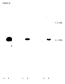
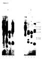
- Figure 1 One dimensional NaDodSO4/PAGE analysis of the IgG FcR of J774 macrophage cells. Sizes are given in kDa.
- A Immunoprecipitation of Fc gamma R from surface iodinated J774 cells. Immunoprecipitations were performed with anti-Ly-2.1 F(ab')2 (Lane 1) or with anti-Ly-17.2 F(ab')2 (Lane 2).
- B NaDodSO4/PAGE and Coomassie staining of a typical preparation of affinity purified FcR used for protein sequencing and peptide generation. Lanes 1-6 represent successive fractions of eluted material from the affinity column (C). Autoradiogram of NaDodSO4/PAGE of affinity purified 125 I-labelled FcR. Lanes 1-6 are as for B, above.
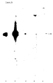
- FIG. 1 Southern blot analysis of pFc24 and pJL3.
- One microgram of pFc24 (lanes B,D,F) or pJL3 (lanes A,C,E) was digested with EcoRI and electrophoresed in 1% agarose gel.
- the DNA was transferred onto nitrocellulose and hybridised with [32P] labelled probe 1 (lanes A,B), [32P] probe 2 (lanes G,D) and [32P] probe 3 (lanes E,F).
- Hinf1 fragments of pBR322 DNA were used as markers to estimate the size of the cDNA insert of pFc24 (shown in kb).
- the relative position of pJL3 is also indicated (5.49kb).
- FIG. 3 (A) Partial restriction map and sequencing strategy for cDNA inserts of clones pFc24 and pFc113. Restriction enzymes sites shown are A, AluI; B, BamHI; E, EcoRI; S, Sau3A1; and V, PvuII. The EcoRI sites shown are present in the polycloning site of the pJL3 vector. The shaded area represents sequence encoding the signal sequence and coding sequence for the mature protein is located within the open box. Sequence from 5' and 3' untranslated regions is indicated by the solid line while unsequenced untranslated regions are represented by the dashed line.
- sequences underlined with a solid line correspond to sequences identical to the amino terminal sequence (NH2) and to amino acid sequence of peptides (L9, V17, 2V16, 2V8, CNBR, L5, L3, L4, V10, V11) isolated from the immunopurified FcR (Hibbs et al., 1986). Other notation is as follows: o, first Cys in each domain; 0 second Cys in each domain; N-linked glycosylation sites.
- the nucleotide sequence of pFc24 is embodied within the cDNA insert of pFc113 from nucleotides 61 to 1023.
- FIG. 4 Amino acid sequence comparison of Fc(gamma)R domains (amino acids 5-116 for domain 1 and amino acids 118-175 for domain 2) to each other and to Ig related molecules. Identical residues are boxed and a "-" indicates a break in sequence for alignment purposes.
- A The FcR domains were aligned with each other to give the optimal score using the ALIGN program with a matrix bias of +6 and break penalty of 6 (see text).
- V 2 MOPC 104E (Apella, 1977); Vk, MOPC 149 (Nishioka and Leder, 1980); T4, L3T4 (Classon et al, 1986; Tourvieille et al, 1986; G. Clark, N. Deacon Personal Communication); N-CAM, Neural Cell Adhesion Molecule (Hemperly et al, 1986); Poly IgR, Poly Ig Receptor (Mostov et al, 1984).
- FIG. 5 Analysis of FcR mRNA transcripts in mouse cell lines.
- A Northern blot of RNA from FcR-erythroleukaemia cells F4N (a) or FcR+ WEHI 3B cells (b) probed with 840 base pair Bam HI - Eco RI fragment of pFc24 (Eco RI site of the vector polycloning site).
- B Northern blot of poly A+ RNA from FcR+ cell lines, WEHI 3B (a); J774 macrophage cells (b); K36 T lymphoma (c); and FcR- cells F4N (d), probed with an oligodeoxynucleotide corresponding to nucleotides 542-674 in pFc113. The position of 28S and 18S rRNA is indicated.
- FIG. 6 Southern analysis of the FcR gene. Spleen DNA from C57BL/6 mice was digested with (a) HindIII, (b) EcoRI or (c) PstI and probed with the 840bp BamHI-EcoRI fragment of pFc24 (see Legend Fig. 3). Molecular size-markers in Kb(Hind III digested lamdaphage DNA) are indicated.
- FIG. 7 Southern blot analysis of human FcR clones HFc3.1 (a) and HFc3.47 (b).
- HFc3.1 or HFc3.47 DNA was digested with EcoRI and electrophoresed in 1% agarose gel, transferred to nylon membranes as per the materials and methods and hybridised with A, pFc24 cDNA; B, probe 1.5; C, probe 1.10; D, probe alpha or E, probe 1.6.
- the position of the 1.5kb cDNA insert of HFc3.1 and of the 1.9kb insert of HFc3.47 is indicated on the right hand side of each figure.
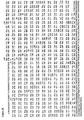
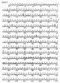
- Figure 8 Nucleotide and predicted amino acid sequence of the human FcR encoded by HFc3.1 Nucleotides are numbered every decade and the translated sequence is found above the nucleotide sequence. Amino acids are numbered above the line and number 1 indicates the N-terminal residue. The incomplete signal sequence is underlined by a broken line to residue -1. The two glycosylation sites are marked by stars and the single hydrophobic transmembrane region is underlined by a solid line. Cysteine residues involved in disulphide bonding are circled. The "intronlike" sequence is located between the vertical arrows.
- Figure 9 Homology of the human FcR encoded by HFc3.1 cDNA with the mouse alpha and beta1 Fc(gamma)R. Alignment of the nucleotides of the human FcR with both mouse alpha (A) and beta1 (B) Fc(gamma)R. Breaks (indicated by dashes) in the sequence have been introduced to optimize the alignment. Amino acid residues common to the human FcR encoded by HFc3.1 and the alpha FcR (A) or beta1 (B) are shown by asterisks.
- the human (HFc3.1), mouse alpha or beta FcR leader sequence, extracellular domain, transmembrane domain and cytoplasmic domains are indicated by the sequences between the vertical arrows.
- the cysteine (C) residues within the extracellular domains are identified by the solid circles.
- FIG. 10 Partial nucleotide sequence and predicted amino acid sequence for HFc3.47 cDNA derived from two non-overlapping HFc3.47 fragments and identification of homologous regions in the beta (1 or 2) FcR of the mouse.
- the HFc3.47 nucleotide and amino acid sequences are shown as the upper sequences in A and the lower sequences in B.
- Diamonds indicate nucleotide identities where as asterisks indicate amino acid identities.
- An "X" in the sequence indicates an unknown residue.
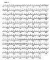
- Figure 11 Nucleotide and predicted amino acid sequence of the human FcR encoded by HFc3.0. Nucleotides are numbered every decade and the translated sequence is found above the nucleotide sequence. Amino acids are numbered above the line and number 1 indicates the N-terminal residue. The incomplete signal sequence is underlined by a broken line to residue -1. The two glycosylation sites are marked by stars and the single hydrophobic transmembrane region is underlined by a solid line. Cysteine residues involved in disulphide bonding are circled.
- Figure 12 Homology of the human FcR encoded by HFc3.0 cDNA with the mouse alpha and beta1 FcR. Alignment of the nucleotides of the human FcR with both mouse alpha (A) and beta1 (B) FcR. Amino acid residues common to the human FcR encoded by HFc3.0 and the alpha FcR (A) or beta 1 (B) are shown by asteriks.
- the human (HFc3.0) mouse alpha or beta FcR leader sequence, extracellular domain, transmembrane domain and cytoplasmic domains are indicated by the sequences between the vertical arrows.
- the cysteine (C) residues within the extracellular domains are identified by the solid circles.
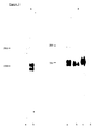
- FIG. 13 Northern blot analysis of total RNA isolated from normal human spleen and probed with the cDNA insert from HFc3.1. The 28s and 18s ribosomal RNAs are indicated. A, 20ug RNA loaded; B, 5ug RNA loaded; C No RNA.
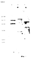
- FIG. 14 Southern blot analysis of the human FcR gene.
- Human thymus DNA (A) and human peripheral leukocyte DNA (B) was digested with 1, Hind III; 2, PvuII; 3, EcoRI and 4, PstI and probed with the cDNA insert from HFc3.1.
- Molecular size markers HindIII digested RNA are indicated.
- pFc113 (Fig. 3) was isolated by reprobing the WEHI 3B library with oligodeoxynucleotide probes constructed from the nucleotide sequence of the 5' and 3' ends of pFc24. These probes corresponded to nucleotides 140-187 (probe 1.5) or 959-1000 (probe 1.6) (Fig. 3B, Table 3) and only clones to which both probes hybridised were subsequently isolated.
- the cDNA insert of pFc113 contains approximately 2.0 kilobase pairs whereas that of pFc24 has only 962bp but is entirely embodied in pFc113 between nucleotides 61 and 1023.
- Nucleotide and amino acid sequence The amino acid sequence predicted from the nucleotide sequence of pFc113 (Fig. 3B) indicates that mature FcR is a membrane molecule composed of 301 amino acids and is synthesised with a 29 amino acid leader sequence.
- the amino acid sequences of 10 of 11 peptides we had previously sequenced from immunopurified FcR were found encoded by pFc113 cDNA (Fig. 3B, Hibbs et al, 1986). Miscalling of several amino acid residues during sequencing of the peptides accounts for the differences between the sequence of peptides L5 and CNBR and the predicted sequence of these regions from the cDNA clone.
- the CNBR peptide is preceded by a Trp not the predicted Met but it is known that CNBR will cleave on the C terminal side of Trp residues in acid solutions (Ozols et al, 1977).
- Murine FcR Comparison of the amino acid sequences within the FcR molecule (assessed using the Dayoff ALIGN programme, Dayhoff et al, 1983) indicated that there was a significant degree of internal homology within the extracellular domains (Fig. 4A). Using the mutation data matrix, scoring is based on the extent of mutation required for amino acid substitution in established protein families. The score for the optimal alignment is then represented as the number of standard deviations by which the maximum score for the alignment of two sequences exceeds the score for a large number of alignments after randomization of the original sequences (Dayhoff et al, 1983).
- FcR domain 1 was most homologous to murine Class II antigens in particular to the Ig like beta2 domain of I-Ebeta and gave a highly significant ALIGN score of 8.3SD.
- other features of the FcR domains indicate their relatedness to Ig superfamily members.
- the homology to members of the Ig supergene family is seen principally around the Cys residues.
- the sequences surrounding cysteine residues 70 and 153 were shown to be highly representative of the consensus sequence (Gly-X-Tyr-X-Cys) around the disulphide-bonded Cys residue in IgV-region domains.
- the receptor for immunoglobulin shows a common evolutionary ancestry with its ligand but comparison of entire Ig and FcR domains shows a low overall homology indicating that they must have diverged relatively early in evolution.
- the high degree of homology with MHC Class II molecules is interesting in the light of the physical association of some FcR with Class II molecules on the cell surface (Dickler and Sachs, 1974).
- the Fc(gamma)R receptor described herein is highly homologous to other beta1 Fc(gamma)R except for a stretch of 9 amino acids in the cytoplasmic tail from Gly241 to Pro249.
- the discrepancies between the nucleotide/amino acid sequences shown in Fig. 3B may have arisen by the omission of three nucleotides.
- the nucleotide and predicted amino acid sequence of the beta1 form of the Fc(gamma)R therefore is that shown in Fig. 3B.
- Fc(gamma)R is highly homologous to the FcR described here -almost certainly being a splice variant.
- the splice site occurs in the region encoding the intracellular domain indicating that the beta1 and beta2 forms of the FcR have identical extracellular domains.
- a third form of the FcR has also been identified by cDNA cloning studies and designated the alpha FcR The mature alpha protein shows a little variation from the receptor described herein with approximately 7% variation in amino acid content of the ligand binding domains. All major structural features in the domains of the three variants (alpha, beta1, beta2) are conserved, i.e. (4 N-linked carbohydrate side chains, 2 pairs of cysteine residues).
- Fc(gamma)R clearly indicates that murine FcR are a family of highly homologous proteins. It should also be noted that the Ig binding proteins - protein A and the FcR described herein all have repeated ligand binding domains which may be necessary for stability and specificity of ligand binding.
- Murine FcR We have demonstrated that adjacent pairs of cysteine residues in purified Fc(gamma)R are involved in disulphide bonding. Protein was purified by affinity chromatography then digested with proteolytic enzymes either before or after reduction of disulphide bonds with dithiothreitol.
- Digests were then fractionated by reversed-phase chromatography. Peptides present in the digests of unreduced Fc(gamma)R but absent from the digested reduced Fc(gamma)R were sequenced and shown to correspond to disulphide-bonded peptides. The results obtained indicated that Cys 28 is disulphide-bonded to Cys-70 and that Cys-109 and 153 are involved in disulphide bonding.
- Extensive peptide sequencing has also determined that four possible N-linked glcyosylation sites in mouse Fc(gamma)R are in fact authentic sites of carbohydrate addition. This was judged by the absence of an asparagine in the expected position in the peptide sequence.
- mRNA transcripts and surface FcR To establish the relationship between mRNA transcription and surface Fc(gamma)R expression, we investigated the presence of mRNA transcripts by Northern analysis using oligonucleotide probes specific for the beta1, beta2 and alpha transcripts (Table 3) and compared this with immune complex binding (using rabbit IgG and various IgG isotypes) and whether the cells were Ly-17.2+ or 2.4G2+ at the surface (Table 4).
- beta1 Fc(gamma)R is the receptor for IgG1/2b, heterologous IgG and possibly other Ig since cells which express only the beta1 receptor only can bind IgG1/IgG2b as well as rabbit IgG coated erythrocytes (Table 4).
- K36 cells have the Ly-17.2+ and 2.4G2+ molecules which have been shown to be identical and are epitopes on the Fc(gamma)R molecule (Unkless, 1979; Hibbs et al, 1985; Holmes et al, 1985).
- antibodies to these molecules completely inhibit the binding of IgG1/2b and rabbit IgG complexes to the cell surfaces.
- the beta1 variant must code for molecules which have Fc binding ability and the epitopes detected by the 2.4G2 and Ly-17.2 antibodies. Both the Ly-17.2 and 2.4G2 epitopes are clearly present on the beta1 and beta2 molecules since both antibodies were used to purify these molecules for amino acid sequencing in this study.
- the pFc113'cDNA (in the pKC3 vector) described herein was transfected into FcR negative LTA-5 cells resulting in expression of FcR (Ly-17.2) on the cell surface (Table 4) indicating that this clone encodes an immunoglobulin binding FcR. The interactions of the alpha and beta2 receptors with antibody and immune complexes remains to be precisely determined.
- mouse FcDNA pFc24 and a combination of oligonucleotides, probes 1.5, 1.6, 1.10 (Table 3) to screen a library constructed from mRNA isolated from human acute monocytic leukaemia cells THP-1. Several clones were isolated including HFc3.0, HFc3.1 and HFc3.47. The results (Fig.
- the complete amino acid sequence of the human FcR, aligned with the sequences of mouse alpha and beta1 FcR is shown (Fig. 9A,B). Breaks have been introduced to optimize the alignment.
- the incomplete leader sequence of the human FcR encoded by HFc 3.1 is highly homologous to the leader sequence of the mouse alpha FcR (56% conservation of amino acids) (Fig. 9A) but bears no homology to the mouse beta1 Fc(gamma)R leader sequence (Fig. 9B).
- the N-terminus of the human Fc(gamma)R has been predicted on the basis of homology to the mouse alpha Fc(gamma)R N-terminal sequence.
- the region between the N-terminus and the first cysteine residue is the most highly conserved region between the two species, showing 71 and 73 percent amino acid homology with the murine beta1 and alpha Fc(gamma)R respectively (Fig. 9A,B).
- Fig. 9A,B There is also a high level of amino acid conservation in the remaining extracellular portion between FcR of mouse and man.
- the extracellular region is divided into two disulphide-bonded domains: the first pair of cysteine residues being separated by 41 amino acids and the second pair by 43 amino acids. Both disulphide bonded domains bear striking homology to the mouse beta1 and alpha Fc(gamma)R. Amino acid sequence comparison of the first domain shows approx.
- the human Fc(gamma)R has a transmembrane region of 28 amino acids extending from residue 219 to the hydrophilic stop transfer sequence Arg (245) - Lys (246) - Lys (247) - Arg (248) (Fig. 8).
- This transmembrane sequence is highly homologous to the mouse beta1 Fc(gamma)R (50 percent amino acid homology),but shows no homology to the transmembrane sequence of the mouse alpha Fc(gamma)R receptor (Fig. 9A,B).
- the striking homology of this human Fc(gamma)R to both alpha and beta1 murine Fc(gamma)Rs in the extracellular domain may reflect the fact that human FcR bind mouse IgG and mouse FcR bind human Ig.
- the cytoplasmic domain of the human HFc3.1 FcR is totally unique and bears no resemblance to either mouse alpha, beta1 or beta2 intracellular domains. This difference could mean the presence of a homologous product in the mouse which has yet to be detected or may reflect the evolutionary divergence of the human and mouse proteins in a region where there may be a high rate of mutation.
- Partial nucleotide sequence of HFc3.47 has also revealed homology with the mouse FcRs (Fig. 10). Two non-overlapping fragments were sequenced and the first fragment (Fig. 10A) revealed 80% nucleotide homology with the corresponding sequence in beta1 FcR (nucleotides 629-733). This sequence is present in beta2 and a similar sequence in alpha. The second fragment shared 72% nucleic acid homology with nucleotides 959-1015 of the beta1FcR, this sequence also being present in the beta2FcR but not alphaFcR.
- HFc 3.0 encodes a protein highly homologous to the mouse alpha and beta FcR receptors (Fig. 12A,B).
- the nucleotide sequence HFc 3.0 is identical to HFc 3.1 with the exception of a large segment (nucleotides 338-455 in HFc 3.1) between the two disulphide bonded domains which has been deleted in HFc 3.0.
- the protein encoded by HFc 3.0 has an extracellular ligand binding region consisting of two disulphide bonded domains each with a site of attachment for N-linked carbohydrate.
- the encoded protein has a 28 amino acid transmembrane region and 75 amino acid cytoplasmic tail.
- the extracellular region shows an overall homology of 67% identical amino acid residues and the transmembrane region has 14 of 28 amino acid identities with the mouse beta1 and beta2 transmembrane region (Fig. 12A,B).
- mouse FcR can bind mouse and human immunoglobulin and have many similarities with mouse FcR, (Dickler, 1976; Anderson and Abraham, 1980, Kulczycki et al, 1981; Perussia et al, 1983; Dorrington and Klein, 1983), it is likely that there is a high degree of structural and functional homology which conserved at the nucleic acid and amino acid levels.
- mouse cDNA pFc24 and a combination of oligonucleotides, probes 1.5, 1.6, 1.10 (Table 3) to screen a library constructed from mRNA isolated from human acute monocytic leukaemia cells THP-1. Several clones were isolated including HFc3.1, 3.0 and 3.47.
- the leader sequence of 29 amino acids in the beta1 FcR allows translocation of the nascent protein across the endoslasmic reticulum.
- the key point of this sequence is its hydrophobic nature (required for membrane insertion) and substitution, addition or deletion of one or more hydrophobic (non-polar) amino acids would not substantially alter this function (see list later).
- the extracellular region (FCR) consists of 179 amino acids; their function is to act as the receptor for the Fc piece of Ig molecules. This binding site has not yet been identified but comments on the nature of the sequence are relevant.
- the FcR is divided into two domains by cysteine residues, Domain 1: Cys 28 - Cys 70; Domain 2: Cys 109 - Cys 153. Both of these domains may have FcR activity as they are highly homologous. Within each domain all or most of the amino acids are likely to be involved in Ig binding as they are conserved - the two domains are conserved in man and mouse.
- domain 1 and domain 2 are very similar.
- amino acids substituted belong to the same groups and would therefore not substantially alter the tertiary structure of the molecule and the groupings of amino acid are:-
- ala alanine
- E glutamic acid
- P pro - proline
- This consists of 28 amino acids and extends from Leu 180 - Lys 209 in mouse beta1 FcR.
- the key feature of this is the hydrophobic nature of the sequence is required as this portion interacts with the hydrophobic cell membrane and anchors the molecule in the cell membrane. Because of the hydrophobic nature, it follows that minor alterations of sequence - substitution of one non-polar amino acid by another, would not alter the essential function of the transmembrane region.
- hydrophobic (non-polar) amino acids are alanine (A); valine (V); leucine (L); isoleucine (I); proline (P); phenylalanine (F); methionine (M) and tryptophan (W).
- the intracellular region is involved in signal transmission - obtained when the Fc of Ig binds to the FcR.
- the mode of signal transmission is unknown.
- a useful sequence of the FcR is described; it is likely that variations of this sequence, obtained by deletion of parts of the whole sequence, could also function in signal transmission.
- the region of the beta1 receptor that is spliced out in beta2 is important in cells expressing these molecules.
- the nucleotide sequences encoding the receptor can be variable:-
Landscapes
- Health & Medical Sciences (AREA)
- Chemical & Material Sciences (AREA)
- Life Sciences & Earth Sciences (AREA)
- Immunology (AREA)
- Organic Chemistry (AREA)
- Medicinal Chemistry (AREA)
- General Health & Medical Sciences (AREA)
- Public Health (AREA)
- Zoology (AREA)
- General Chemical & Material Sciences (AREA)
- Pharmacology & Pharmacy (AREA)
- Chemical Kinetics & Catalysis (AREA)
- Animal Behavior & Ethology (AREA)
- Bioinformatics & Cheminformatics (AREA)
- Engineering & Computer Science (AREA)
- Veterinary Medicine (AREA)
- Cell Biology (AREA)
- Toxicology (AREA)
- Nuclear Medicine, Radiotherapy & Molecular Imaging (AREA)
- Gastroenterology & Hepatology (AREA)
- Biochemistry (AREA)
- Biophysics (AREA)
- Genetics & Genomics (AREA)
- Molecular Biology (AREA)
- Proteomics, Peptides & Aminoacids (AREA)
- Peptides Or Proteins (AREA)
- Preparation Of Compounds By Using Micro-Organisms (AREA)
- Medicines That Contain Protein Lipid Enzymes And Other Medicines (AREA)
- Micro-Organisms Or Cultivation Processes Thereof (AREA)
- Medicines Containing Antibodies Or Antigens For Use As Internal Diagnostic Agents (AREA)
Claims (6)
- Ein nicht natürlich vorkommendes Polynukleotid, welches für einen humanen Fc-Rezeptor für Immunglobulin oder für ein Fragment desselben kodiert, welches in der Lage ist, die Fc-Region von humanem Immunglobulin zu binden, ausgewählt aus der extrazellulären Domäne des humanen Fc-Rezeptors und humanen Fc-Rezeptorbindungsstellen, wobei das besagte Polynukleotid für einen Teil oder alle der Aminosäuresequenzen der Figuren 8, 10 und 11 und konservative Aminosäurevarianten derselben und allelische Varianten derselben kodiert, welche in der Lage sind, die Fc-Region von humanem Immunglobulin zu binden.
- Ein nicht natürlich vorkommendes Polynukleotid nach Anspruch 1, umfassend die Nukleotide 84 bis 924 aus Figur 11 oder Varianten derselben, welche für die Aminosäuresequenz, die durch die Nukleotide 84 bis 924 der Figur 11 kodiert wird, und konservative Aminosäurevarianten derselben und allelische Varianten derselben kodieren, welche in der Lage sind, die Fc-Region von humanem Immunglobulin zu binden.
- Ein nicht natürlich vorkommendes Polynukleotid, welches für ein Fragment des humanen Fc-Rezeptors für Immunglobulin kodiert nach Anspruch 1, umfassend die Nukleotide 84-601 aus Figur 11 oder deren Varianten, welche für die Aminosäuresequenz, die durch die Nukleotide 84-601 kodiert wird, und konservative Aminosäurevarianten derselben und allelische Varianten derselben kodieren, welche in der Lage sind, die Fc-Region von humanem Immunglobulin zu binden.
- Ein Vektor enthaltend eine Polynukleotidsequenz gemäß einem der Ansprüche 1 bis 3.
- Eine Wirtszelle enthaltend einen Vektor gemäß Anspruch 4.
- Verfahren zur Herstellung eines humanen Fc-Rezeptors für Immunglobulin oder eines Fragmentes desselben, welcher in der Lage ist, die Fc-Region von humanem Immunglobulin zu binden, ausgewählt aus der extrazellulären Domäne des humanen Fc-Rezeptors und der humanen Fc-Rezeptorbindungsstelle, umfassend die Bereitstellung der Wirtszelle gemäß Anspruch 5 und die Expression des humanen Fc-Rezeptors für Immunglobulin oder eines Fragmentes desselben in dem besagten Wirt.
Applications Claiming Priority (3)
| Application Number | Priority Date | Filing Date | Title |
|---|---|---|---|
| AU6166/86 | 1986-05-29 | ||
| AUPH616686 | 1986-05-29 | ||
| PCT/AU1987/000159 WO1987007277A1 (en) | 1986-05-29 | 1987-05-29 | Fc receptor for immunoglobulin |
Publications (3)
| Publication Number | Publication Date |
|---|---|
| EP0268636A1 EP0268636A1 (de) | 1988-06-01 |
| EP0268636A4 EP0268636A4 (de) | 1988-09-28 |
| EP0268636B1 true EP0268636B1 (de) | 1997-01-08 |
Family
ID=3771638
Family Applications (1)
| Application Number | Title | Priority Date | Filing Date |
|---|---|---|---|
| EP87903479A Expired - Lifetime EP0268636B1 (de) | 1986-05-29 | 1987-05-29 | Polynucleotidsequenzen, die für den humanen FC-REZEPTOR FÜR IMMUNOGLOBULIN codieren |
Country Status (8)
| Country | Link |
|---|---|
| EP (1) | EP0268636B1 (de) |
| JP (1) | JP2713410B2 (de) |
| AT (1) | ATE147432T1 (de) |
| AU (1) | AU595623B2 (de) |
| CA (1) | CA1340935C (de) |
| DE (1) | DE3751996T2 (de) |
| NZ (1) | NZ220504A (de) |
| WO (1) | WO1987007277A1 (de) |
Cited By (23)
| Publication number | Priority date | Publication date | Assignee | Title |
|---|---|---|---|---|
| US7276585B2 (en) | 2004-03-24 | 2007-10-02 | Xencor, Inc. | Immunoglobulin variants outside the Fc region |
| US7317091B2 (en) | 2002-03-01 | 2008-01-08 | Xencor, Inc. | Optimized Fc variants |
| US7662925B2 (en) | 2002-03-01 | 2010-02-16 | Xencor, Inc. | Optimized Fc variants and methods for their generation |
| US7973136B2 (en) | 2005-10-06 | 2011-07-05 | Xencor, Inc. | Optimized anti-CD30 antibodies |
| US8039592B2 (en) | 2002-09-27 | 2011-10-18 | Xencor, Inc. | Optimized Fc variants and methods for their generation |
| US8084582B2 (en) | 2003-03-03 | 2011-12-27 | Xencor, Inc. | Optimized anti-CD20 monoclonal antibodies having Fc variants |
| US8101720B2 (en) | 2004-10-21 | 2012-01-24 | Xencor, Inc. | Immunoglobulin insertions, deletions and substitutions |
| US8188231B2 (en) | 2002-09-27 | 2012-05-29 | Xencor, Inc. | Optimized FC variants |
| US8318907B2 (en) | 2004-11-12 | 2012-11-27 | Xencor, Inc. | Fc variants with altered binding to FcRn |
| US8388955B2 (en) | 2003-03-03 | 2013-03-05 | Xencor, Inc. | Fc variants |
| US8394374B2 (en) | 2006-09-18 | 2013-03-12 | Xencor, Inc. | Optimized antibodies that target HM1.24 |
| US8524867B2 (en) | 2006-08-14 | 2013-09-03 | Xencor, Inc. | Optimized antibodies that target CD19 |
| US8546543B2 (en) | 2004-11-12 | 2013-10-01 | Xencor, Inc. | Fc variants that extend antibody half-life |
| US8735547B2 (en) | 2002-09-27 | 2014-05-27 | Xencor, Inc. | Optimized Fc Variants |
| US8802820B2 (en) | 2004-11-12 | 2014-08-12 | Xencor, Inc. | Fc variants with altered binding to FcRn |
| US9040041B2 (en) | 2005-10-03 | 2015-05-26 | Xencor, Inc. | Modified FC molecules |
| US9051373B2 (en) | 2003-05-02 | 2015-06-09 | Xencor, Inc. | Optimized Fc variants |
| US9200079B2 (en) | 2004-11-12 | 2015-12-01 | Xencor, Inc. | Fc variants with altered binding to FcRn |
| US9475881B2 (en) | 2010-01-19 | 2016-10-25 | Xencor, Inc. | Antibody variants with enhanced complement activity |
| US9714282B2 (en) | 2003-09-26 | 2017-07-25 | Xencor, Inc. | Optimized Fc variants and methods for their generation |
| US11401348B2 (en) | 2009-09-02 | 2022-08-02 | Xencor, Inc. | Heterodimeric Fc variants |
| US11932685B2 (en) | 2007-10-31 | 2024-03-19 | Xencor, Inc. | Fc variants with altered binding to FcRn |
| US12492253B1 (en) | 2008-02-25 | 2025-12-09 | Xencor, Inc. | Anti-human C5 antibodies |
Families Citing this family (6)
| Publication number | Priority date | Publication date | Assignee | Title |
|---|---|---|---|---|
| US5985599A (en) * | 1986-05-29 | 1999-11-16 | The Austin Research Institute | FC receptor for immunoglobulin |
| WO1989005351A2 (en) * | 1987-12-04 | 1989-06-15 | Schering Biotech Corporation | Human fc-gamma receptor |
| US5767077A (en) * | 1988-05-27 | 1998-06-16 | Applied Research Systems Ars Holding N.V. | Human Fc-γ receptor III |
| WO1989011490A1 (en) * | 1988-05-27 | 1989-11-30 | Schering Biotech Corporation | HUMAN Fcgamma RECEPTOR III |
| WO1991006570A1 (en) * | 1989-10-25 | 1991-05-16 | The University Of Melbourne | HYBRID Fc RECEPTOR MOLECULES |
| US20150010550A1 (en) | 2004-07-15 | 2015-01-08 | Xencor, Inc. | OPTIMIZED Fc VARIANTS |
Family Cites Families (1)
| Publication number | Priority date | Publication date | Assignee | Title |
|---|---|---|---|---|
| SE451596B (sv) * | 1983-06-22 | 1987-10-19 | Excorim Kb | Forfarande for utvinning av protein g |
-
1987
- 1987-05-29 AT AT87903479T patent/ATE147432T1/de not_active IP Right Cessation
- 1987-05-29 AU AU74881/87A patent/AU595623B2/en not_active Expired
- 1987-05-29 WO PCT/AU1987/000159 patent/WO1987007277A1/en not_active Ceased
- 1987-05-29 DE DE3751996T patent/DE3751996T2/de not_active Expired - Lifetime
- 1987-05-29 NZ NZ220504A patent/NZ220504A/xx unknown
- 1987-05-29 JP JP62503462A patent/JP2713410B2/ja not_active Expired - Lifetime
- 1987-05-29 CA CA000538363A patent/CA1340935C/en not_active Expired - Lifetime
- 1987-05-29 EP EP87903479A patent/EP0268636B1/de not_active Expired - Lifetime
Non-Patent Citations (5)
| Title |
|---|
| CHEMICAL ABSTRACTS, vol. 104, no. 19, 12 May 1986, Columbus, OH (US); G. GORINI et al., p. 507, no. 166585k * |
| CHEMICAL ABSTRACTS, vol. 104, no. 3, 20 January 1986, Columbus, OH (US); M. VAUGHN et al., p. 384, no. 18431s * |
| JOURNAL OF BIOLOGICAL CHEMISTRY, vol. 260, no.17, 15 August 1985, American Society of Biological Chemists Inc., US; S.A. GREEN et al., pp. 9867-9874 * |
| NATURE, vol. 324, no. 6095, 27 November 1986, London (GB); V.A. LEWIS et al., pp. 372-375 * |
| SCIENCE, vol. 234, no. 4777, 07 November 1986, Washington, DC (US); J.V. RAVETCH et al., pp. 718-725 * |
Cited By (52)
| Publication number | Priority date | Publication date | Assignee | Title |
|---|---|---|---|---|
| US8093357B2 (en) | 2002-03-01 | 2012-01-10 | Xencor, Inc. | Optimized Fc variants and methods for their generation |
| US7317091B2 (en) | 2002-03-01 | 2008-01-08 | Xencor, Inc. | Optimized Fc variants |
| US7662925B2 (en) | 2002-03-01 | 2010-02-16 | Xencor, Inc. | Optimized Fc variants and methods for their generation |
| US8734791B2 (en) | 2002-03-01 | 2014-05-27 | Xencor, Inc. | Optimized fc variants and methods for their generation |
| US8124731B2 (en) | 2002-03-01 | 2012-02-28 | Xencor, Inc. | Optimized Fc variants and methods for their generation |
| US8753629B2 (en) | 2002-09-27 | 2014-06-17 | Xencor, Inc. | Optimized Fc variants |
| US10184000B2 (en) | 2002-09-27 | 2019-01-22 | Xencor, Inc. | Optimized Fc variants and methods for their generation |
| US8093359B2 (en) | 2002-09-27 | 2012-01-10 | Xencor, Inc. | Optimized Fc variants and methods for their generation |
| US9193798B2 (en) | 2002-09-27 | 2015-11-24 | Xencor, Inc. | Optimized Fc variants and methods for their generation |
| US8039592B2 (en) | 2002-09-27 | 2011-10-18 | Xencor, Inc. | Optimized Fc variants and methods for their generation |
| US8188231B2 (en) | 2002-09-27 | 2012-05-29 | Xencor, Inc. | Optimized FC variants |
| US10183999B2 (en) | 2002-09-27 | 2019-01-22 | Xencor, Inc. | Optimized Fc variants and methods for their generation |
| US8802823B2 (en) | 2002-09-27 | 2014-08-12 | Xencor, Inc. | Optimized Fc variants |
| US8753628B2 (en) | 2002-09-27 | 2014-06-17 | Xencor, Inc. | Optimized Fc variants |
| US8858937B2 (en) | 2002-09-27 | 2014-10-14 | Xencor, Inc. | Optimized Fc variants and methods for their generation |
| US8383109B2 (en) | 2002-09-27 | 2013-02-26 | Xencor, Inc. | Optimized Fc variants and methods for their generation |
| US8809503B2 (en) | 2002-09-27 | 2014-08-19 | Xencor, Inc. | Optimized Fc variants and methods for their generation |
| US8735547B2 (en) | 2002-09-27 | 2014-05-27 | Xencor, Inc. | Optimized Fc Variants |
| US9353187B2 (en) | 2002-09-27 | 2016-05-31 | Xencor, Inc. | Optimized FC variants and methods for their generation |
| US10113001B2 (en) | 2003-03-03 | 2018-10-30 | Xencor, Inc. | Fc variants with increased affinity for FcyRIIc |
| US8735545B2 (en) | 2003-03-03 | 2014-05-27 | Xencor, Inc. | Fc variants having increased affinity for fcyrllc |
| US8388955B2 (en) | 2003-03-03 | 2013-03-05 | Xencor, Inc. | Fc variants |
| US8084582B2 (en) | 2003-03-03 | 2011-12-27 | Xencor, Inc. | Optimized anti-CD20 monoclonal antibodies having Fc variants |
| US10584176B2 (en) | 2003-03-03 | 2020-03-10 | Xencor, Inc. | Fc variants with increased affinity for FcγRIIc |
| US9051373B2 (en) | 2003-05-02 | 2015-06-09 | Xencor, Inc. | Optimized Fc variants |
| US9714282B2 (en) | 2003-09-26 | 2017-07-25 | Xencor, Inc. | Optimized Fc variants and methods for their generation |
| US7276585B2 (en) | 2004-03-24 | 2007-10-02 | Xencor, Inc. | Immunoglobulin variants outside the Fc region |
| US8101720B2 (en) | 2004-10-21 | 2012-01-24 | Xencor, Inc. | Immunoglobulin insertions, deletions and substitutions |
| US8338574B2 (en) | 2004-11-12 | 2012-12-25 | Xencor, Inc. | FC variants with altered binding to FCRN |
| US10336818B2 (en) | 2004-11-12 | 2019-07-02 | Xencor, Inc. | Fc variants with altered binding to FcRn |
| US8883973B2 (en) | 2004-11-12 | 2014-11-11 | Xencor, Inc. | Fc variants with altered binding to FcRn |
| US12215165B2 (en) | 2004-11-12 | 2025-02-04 | Xencor, Inc. | Fc variants with altered binding to FcRn |
| US11198739B2 (en) | 2004-11-12 | 2021-12-14 | Xencor, Inc. | Fc variants with altered binding to FcRn |
| US8802820B2 (en) | 2004-11-12 | 2014-08-12 | Xencor, Inc. | Fc variants with altered binding to FcRn |
| US8546543B2 (en) | 2004-11-12 | 2013-10-01 | Xencor, Inc. | Fc variants that extend antibody half-life |
| US9200079B2 (en) | 2004-11-12 | 2015-12-01 | Xencor, Inc. | Fc variants with altered binding to FcRn |
| US8852586B2 (en) | 2004-11-12 | 2014-10-07 | Xencor, Inc. | Fc variants with altered binding to FcRn |
| US8318907B2 (en) | 2004-11-12 | 2012-11-27 | Xencor, Inc. | Fc variants with altered binding to FcRn |
| US8324351B2 (en) | 2004-11-12 | 2012-12-04 | Xencor, Inc. | Fc variants with altered binding to FcRn |
| US8367805B2 (en) | 2004-11-12 | 2013-02-05 | Xencor, Inc. | Fc variants with altered binding to FcRn |
| US9803023B2 (en) | 2004-11-12 | 2017-10-31 | Xencor, Inc. | Fc variants with altered binding to FcRn |
| US9040041B2 (en) | 2005-10-03 | 2015-05-26 | Xencor, Inc. | Modified FC molecules |
| US9574006B2 (en) | 2005-10-06 | 2017-02-21 | Xencor, Inc. | Optimized anti-CD30 antibodies |
| US7973136B2 (en) | 2005-10-06 | 2011-07-05 | Xencor, Inc. | Optimized anti-CD30 antibodies |
| US8524867B2 (en) | 2006-08-14 | 2013-09-03 | Xencor, Inc. | Optimized antibodies that target CD19 |
| US11618788B2 (en) | 2006-08-14 | 2023-04-04 | Xencor, Inc. | Optimized antibodies that target CD19 |
| US8394374B2 (en) | 2006-09-18 | 2013-03-12 | Xencor, Inc. | Optimized antibodies that target HM1.24 |
| US9040042B2 (en) | 2006-09-18 | 2015-05-26 | Xencor, Inc. | Optimized antibodies that target HM1.24 |
| US11932685B2 (en) | 2007-10-31 | 2024-03-19 | Xencor, Inc. | Fc variants with altered binding to FcRn |
| US12492253B1 (en) | 2008-02-25 | 2025-12-09 | Xencor, Inc. | Anti-human C5 antibodies |
| US11401348B2 (en) | 2009-09-02 | 2022-08-02 | Xencor, Inc. | Heterodimeric Fc variants |
| US9475881B2 (en) | 2010-01-19 | 2016-10-25 | Xencor, Inc. | Antibody variants with enhanced complement activity |
Also Published As
| Publication number | Publication date |
|---|---|
| WO1987007277A1 (en) | 1987-12-03 |
| CA1340935C (en) | 2000-03-28 |
| AU7488187A (en) | 1987-12-22 |
| ATE147432T1 (de) | 1997-01-15 |
| EP0268636A4 (de) | 1988-09-28 |
| AU595623B2 (en) | 1990-04-05 |
| JPS63503386A (ja) | 1988-12-08 |
| DE3751996T2 (de) | 1997-07-03 |
| HK1006977A1 (en) | 1999-03-26 |
| EP0268636A1 (de) | 1988-06-01 |
| NZ220504A (en) | 1991-09-25 |
| DE3751996D1 (de) | 1997-02-20 |
| JP2713410B2 (ja) | 1998-02-16 |
Similar Documents
| Publication | Publication Date | Title |
|---|---|---|
| CA1340935C (en) | Fc gamma receptor | |
| Maliszewski et al. | Expression cloning of a human Fc receptor for IgA. | |
| Bell et al. | DNA sequence and characterization of human class II major histocompatibility complex beta chains from the DR1 haplotype. | |
| Sears et al. | Molecular cloning and expression of the mouse high affinity Fc receptor for IgG. | |
| Norment et al. | A second subunit of CD8 is expressed in human T cells. | |
| Simmons et al. | The Fcγ receptor of natural killer cells is a phospholipid-linked membrane protein | |
| Huang et al. | Cloning and characterization of Lnk, a signal transduction protein that links T-cell receptor activation signal to phospholipase C gamma 1, Grb2, and phosphatidylinositol 3-kinase. | |
| Stengelin et al. | Isolation of cDNAs for two distinct human Fc receptors by ligand affinity cloning. | |
| Ernst et al. | Molecular characterization of six variant Fc | |
| Geraghty et al. | A human major histocompatibility complex class I gene that encodes a protein with a shortened cytoplasmic segment. | |
| Ellis et al. | Sequence and expression of mRNAs encoding the α1 and α2 subunits of a DHP-sensitive calcium channel | |
| Warmerdam et al. | Interaction of a human FcγRIIb1 (CD32) isoform with murine and human IgG subclasses | |
| US5985599A (en) | FC receptor for immunoglobulin | |
| Kopito et al. | Structure and organization of the murine band 3 gene. | |
| De Wit et al. | Structure of the gene for the human myeloid IgA Fc receptor (CD89) | |
| Altruda et al. | Cloning of cDNA for a novel mouse membrane glycoprotein (gp42): shared identity to histocompatibility antigens, immunoglobulins and neural-cell adhesion molecules | |
| Ezumi et al. | Molecular cloning, genomic structure, chromosomal localization, and alternative splice forms of the platelet collagen receptor glycoprotein VI | |
| Ichimiya et al. | Structural analysis of the rat homologue of CD1. Evidence for evolutionary conservation of the CD1D class and widespread transcription by rat cells. | |
| Craig Morton et al. | Immunoglobulin-binding sites of human FcαRI (CD89) and bovine Fcγ2R are located in their membrane-distal extracellular domains | |
| WO1991006570A1 (en) | HYBRID Fc RECEPTOR MOLECULES | |
| Staunton et al. | Molecular cloning of the lymphocyte activation marker Blast‐1. | |
| Hogarth et al. | The mouse Fc receptor for IgG (Ly-17): molecular cloning and specificity | |
| US5770396A (en) | Isolation characterization, and use of the human beta subunit of the high affinity receptor for immunoglobulin E | |
| Moguilevsky et al. | Stable Expression of Human H1‐histamine‐receptor cDNA in Chinese Hamster Ovary Cells: Pharmacological Characterisation of the Protein, Tissue Distribution of Messenger RNA and Chromosomal Localisation of the Gene | |
| Sandrin et al. | Isolation and characterization of cDNA clones for Humly9: the human homologue of mouse Ly9 |
Legal Events
| Date | Code | Title | Description |
|---|---|---|---|
| PUAI | Public reference made under article 153(3) epc to a published international application that has entered the european phase |
Free format text: ORIGINAL CODE: 0009012 |
|
| 17P | Request for examination filed |
Effective date: 19880209 |
|
| AK | Designated contracting states |
Kind code of ref document: A1 Designated state(s): AT BE CH DE FR GB IT LI LU NL SE |
|
| A4 | Supplementary search report drawn up and despatched |
Effective date: 19880928 |
|
| 17Q | First examination report despatched |
Effective date: 19910918 |
|
| GRAG | Despatch of communication of intention to grant |
Free format text: ORIGINAL CODE: EPIDOS AGRA |
|
| RAP1 | Party data changed (applicant data changed or rights of an application transferred) |
Owner name: ILEXUS PTY LTD |
|
| GRAH | Despatch of communication of intention to grant a patent |
Free format text: ORIGINAL CODE: EPIDOS IGRA |
|
| GRAH | Despatch of communication of intention to grant a patent |
Free format text: ORIGINAL CODE: EPIDOS IGRA |
|
| GRAA | (expected) grant |
Free format text: ORIGINAL CODE: 0009210 |
|
| AK | Designated contracting states |
Kind code of ref document: B1 Designated state(s): AT BE CH DE FR GB IT LI LU NL SE |
|
| REF | Corresponds to: |
Ref document number: 147432 Country of ref document: AT Date of ref document: 19970115 Kind code of ref document: T |
|
| REG | Reference to a national code |
Ref country code: CH Ref legal event code: EP |
|
| REG | Reference to a national code |
Ref country code: CH Ref legal event code: NV Representative=s name: E. BLUM & CO. PATENTANWAELTE |
|
| REF | Corresponds to: |
Ref document number: 3751996 Country of ref document: DE Date of ref document: 19970220 |
|
| ET | Fr: translation filed | ||
| ITF | It: translation for a ep patent filed | ||
| PLBE | No opposition filed within time limit |
Free format text: ORIGINAL CODE: 0009261 |
|
| STAA | Information on the status of an ep patent application or granted ep patent |
Free format text: STATUS: NO OPPOSITION FILED WITHIN TIME LIMIT |
|
| 26N | No opposition filed | ||
| REG | Reference to a national code |
Ref country code: CH Ref legal event code: NV Representative=s name: MOINAS SAVOYE & CRONIN |
|
| REG | Reference to a national code |
Ref country code: GB Ref legal event code: IF02 |
|
| REG | Reference to a national code |
Ref country code: GB Ref legal event code: 732E |
|
| PGFP | Annual fee paid to national office [announced via postgrant information from national office to epo] |
Ref country code: LU Payment date: 20060427 Year of fee payment: 20 |
|
| PGFP | Annual fee paid to national office [announced via postgrant information from national office to epo] |
Ref country code: DE Payment date: 20060502 Year of fee payment: 20 |
|
| PGFP | Annual fee paid to national office [announced via postgrant information from national office to epo] |
Ref country code: SE Payment date: 20060512 Year of fee payment: 20 |
|
| PGFP | Annual fee paid to national office [announced via postgrant information from national office to epo] |
Ref country code: AT Payment date: 20060515 Year of fee payment: 20 |
|
| PGFP | Annual fee paid to national office [announced via postgrant information from national office to epo] |
Ref country code: GB Payment date: 20060522 Year of fee payment: 20 |
|
| PGFP | Annual fee paid to national office [announced via postgrant information from national office to epo] |
Ref country code: FR Payment date: 20060529 Year of fee payment: 20 |
|
| PGFP | Annual fee paid to national office [announced via postgrant information from national office to epo] |
Ref country code: NL Payment date: 20060530 Year of fee payment: 20 Ref country code: BE Payment date: 20060530 Year of fee payment: 20 |
|
| PGFP | Annual fee paid to national office [announced via postgrant information from national office to epo] |
Ref country code: IT Payment date: 20060531 Year of fee payment: 20 |
|
| PGFP | Annual fee paid to national office [announced via postgrant information from national office to epo] |
Ref country code: CH Payment date: 20060607 Year of fee payment: 20 |
|
| PG25 | Lapsed in a contracting state [announced via postgrant information from national office to epo] |
Ref country code: NL Free format text: LAPSE BECAUSE OF EXPIRATION OF PROTECTION Effective date: 20070529 |
|
| REG | Reference to a national code |
Ref country code: GB Ref legal event code: PE20 |
|
| REG | Reference to a national code |
Ref country code: CH Ref legal event code: PL |
|
| EUG | Se: european patent has lapsed | ||
| NLV7 | Nl: ceased due to reaching the maximum lifetime of a patent |
Effective date: 20070529 |
|
| PG25 | Lapsed in a contracting state [announced via postgrant information from national office to epo] |
Ref country code: GB Free format text: LAPSE BECAUSE OF EXPIRATION OF PROTECTION Effective date: 20070528 |
|
| BE20 | Be: patent expired |
Owner name: *ILEXUS PTY LTD Effective date: 20070529 |