EP0269147A2 - Encodeur de signaux cardio-respiratoires groupés dans le temps et méthode pour grouper des signaux cardio-respiratoires - Google Patents
Encodeur de signaux cardio-respiratoires groupés dans le temps et méthode pour grouper des signaux cardio-respiratoires Download PDFInfo
- Publication number
- EP0269147A2 EP0269147A2 EP87202021A EP87202021A EP0269147A2 EP 0269147 A2 EP0269147 A2 EP 0269147A2 EP 87202021 A EP87202021 A EP 87202021A EP 87202021 A EP87202021 A EP 87202021A EP 0269147 A2 EP0269147 A2 EP 0269147A2
- Authority
- EP
- European Patent Office
- Prior art keywords
- data
- nmr
- respiratory
- heart
- cardiac
- Prior art date
- Legal status (The legal status is an assumption and is not a legal conclusion. Google has not performed a legal analysis and makes no representation as to the accuracy of the status listed.)
- Granted
Links
Images

Classifications
-
- G—PHYSICS
- G01—MEASURING; TESTING
- G01R—MEASURING ELECTRIC VARIABLES; MEASURING MAGNETIC VARIABLES
- G01R33/00—Arrangements or instruments for measuring magnetic variables
- G01R33/20—Arrangements or instruments for measuring magnetic variables involving magnetic resonance
- G01R33/44—Arrangements or instruments for measuring magnetic variables involving magnetic resonance using nuclear magnetic resonance [NMR]
- G01R33/48—NMR imaging systems
- G01R33/54—Signal processing systems, e.g. using pulse sequences ; Generation or control of pulse sequences; Operator console
- G01R33/56—Image enhancement or correction, e.g. subtraction or averaging techniques, e.g. improvement of signal-to-noise ratio and resolution
- G01R33/567—Image enhancement or correction, e.g. subtraction or averaging techniques, e.g. improvement of signal-to-noise ratio and resolution gated by physiological signals, i.e. synchronization of acquired MR data with periodical motion of an object of interest, e.g. monitoring or triggering system for cardiac or respiratory gating
- G01R33/5673—Gating or triggering based on a physiological signal other than an MR signal, e.g. ECG gating or motion monitoring using optical systems for monitoring the motion of a fiducial marker
-
- A—HUMAN NECESSITIES
- A61—MEDICAL OR VETERINARY SCIENCE; HYGIENE
- A61B—DIAGNOSIS; SURGERY; IDENTIFICATION
- A61B5/00—Measuring for diagnostic purposes; Identification of persons
- A61B5/05—Detecting, measuring or recording for diagnosis by means of electric currents or magnetic fields; Measuring using microwaves or radio waves
- A61B5/055—Detecting, measuring or recording for diagnosis by means of electric currents or magnetic fields; Measuring using microwaves or radio waves involving electronic [EMR] or nuclear [NMR] magnetic resonance, e.g. magnetic resonance imaging
-
- A—HUMAN NECESSITIES
- A61—MEDICAL OR VETERINARY SCIENCE; HYGIENE
- A61B—DIAGNOSIS; SURGERY; IDENTIFICATION
- A61B5/00—Measuring for diagnostic purposes; Identification of persons
- A61B5/72—Signal processing specially adapted for physiological signals or for diagnostic purposes
- A61B5/7271—Specific aspects of physiological measurement analysis
- A61B5/7285—Specific aspects of physiological measurement analysis for synchronizing or triggering a physiological measurement or image acquisition with a physiological event or waveform, e.g. an ECG signal
Definitions
- This invention pertains generally to the field of nuclear magnetic resonance (NMR) tomography and in particular to retrospective clustering and analysis of NMR k-space measurements in the cardiac phase - respiratory phase plane.
- the processing system and method effect the reduction of cardio-vascular and respiratory flow motion artifacts in images and the quantitative characterization of cardiac structure and function both with and without imaging.
- NMR spectroscopy the process of analyzing a small sample in a uniform magnetic field and obtaining radio frequency data resulting from precisely pulsed radio frequency excitation, were invented by Bloch and Purcell.
- NMR analysis by spectroscopy has shifted from physical chemistry to biological chemistry and biological medical applications; i.e., biopsy samples of normal and diseased tissues. Lauterbur and Damadian and others separately invented the utilization of NMR principles to produce and image. (R. Damadian, Science 171,1151, 1971; P.C. Lauterbur, Nature 242, 190, 1973 and P.C. Lauterbur, Pure and Applied Chemistry, 40, 149, 1974).
- the resulting devices, NMR imaging systems produced two deminsional and three dimensional data in which the gray scale represented is a function of a number of parameters, including for example the three parameters nuclide density, T1 (longitudinal relaxation time) and T2 (transverse relaxation time) especially in an anatomical image.
- NMR imaging techniques are disclosed, for example, in "Proton NMR Tomography” P.R. Locher, Philips Technical Review, Vol. 41, 1983/84, No. 3, pages 73-88, the contents of which are incorporated herein by reference as a background of NMR imaging technology.
- NMR nuclear magnetic resonance
- NMR electrocardiogram
- the NMR image is reconstructed from time signals by a complex 2DFT.
- 2DFT two-dimensional Fourier transform
- the signals are obtained in 128 consecutive imaging sequences.
- each sequence is triggered by a pulse derived from the R-wave of the patient's ECG. The delay of this pulse determines the imaging phase in the cardiac cycle.
- Each sequence consists of a series of radiofrequency (RF) and magnetic field gradient pulses, respectively, for evoking a signal and providing spatial information in the signal.
- RF radiofrequency
- a gradient magnetic field applied after an RF excitation pulse (90° pulse) makes the spins at different locations in the excited slice precess at different frequencies.
- the spins start to dephase with respect to the resonant frequency phase.
- the associated phase shift is proportional to the gradient amplitude and the time it is active.
- an extra term is added that is directly proportional to the velocity in the gradient direction.
- the present invention pertains to an improved method and system for correlating NMR data with data from cardiac/respiratory monitors commonly used for conventional cardiac triggering and respiratory gating.
- NMR data collection has been started at fixed delays after the ECG R-wave, with possible rejection of abnormal R-R intervals.
- each data collection is started at some fixed time delay after the R-peak.
- data collection continues close to the next R-peak. If the heart beats regularly, each measurement will take place at the same phase in the cardiac style.
- Respiratory cycle timed NMR k-space sampling has also been utilized. However, discarding abnormal physiological cycles, or physiological cycle correlated sampling of k-space may actually cause abnormalities to be missed, which would defeat the diagnostic purpose of an NMR scan. Because of R-R variation, cardiac triggering is less effective at the end of a heart cycle when coronaries fill and the pre-R-wave shape of the heart may give clues to contraction abnormalities.
- the prior approaches tend to be complicated combinations of slices, phases, triggers, delays, windows and data rejection and reordering imposed on the NMR data collection, causing problems with uncertain heart phasing, limited time resolution, and nonuniform spin saturation.
- the present invention is designed to overcome these problems.
- the time-clustered cardio-respiratory encoder of the present invention is a microcomputer based system which collects cardiac (C) R-wave time, respiratory (R) diaphragm position, and NMR data acquisition (A) timing data in parallel with free running image collection on a standard magnetic resonance imager.
- C-A-R phase timing data are uploaded from a microcomputer to the minicomputer of the scanner for use in clustering the raw data into a new data set, one image equivalent for each cardiorespiratory (C-R) phase combination desired.
- the position of an NMR profile in the normalized C-R phase plane determines the clustered image in which it will fall, i.e. which cardiac phase-respiratory phase combination.
- the clustered raw data are then filled and filtered to compensate for the nonuniform k-space sampling, and, finally, reconstructed with ordinary 2D or 3D Fourier Transform Techniques.
- a time clustered image showing, for example, the first fifth of the cardiac cycle from all ther profiles taken during the first fifth of their respective R-R interval and a time clustered image showing the fourth fifth of the cardiac cycle from the profiles taken during the fourth fifth of their respective R-R interval.
- the system is easily implemented with any NMR system and can be used with any pulse sequence. It requires only readily available output signals from the standard cardiac/respiratory monitors commonly used for conventional cardiac triggering and respiratory gating, and software parameter modifications to accommodate different pulse sequences, along with a timing signal from the scanner.
- the object of the invention is a simple, flexible system for retrospectively clustering and analyzing NMR k-space measurement profiles in the cardiac phase - respiratory phase plane to: 1) reduce cardio-vascular and respiratory flow/motion image artifacts, and 2) to quantitatively characterized cardiac structure/function both with and without imaging.
- the invention pertains to a method and its implementation in an encoder for retrospectively clustering NMR k-space measurement profiles in the cardiac phase respiratory phase plane, which method and encoder function to: 1) reduce cardio-vascular and respiratory flow motion, image artifacts, and 2) to characterize quantitatively cardiac structure/function both with and without imaging.
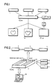
- FIG. 1 presents an overview of the time clustered cardio-respiratory encoder (TCCRE) of the present invention
- FIG. 2 illustrates the hardware used
- Figure 3 emphasizes the functional use of the encoder and the method.
- cardiac (C) time, respiratory (R) time of diaphragm position, and NMR data acquisition (A) cycle marker data are collected by a microcomputer (1) in parallel with free running, preferably steady state, image collection on a standard magnetic resonance imager (2). After collecting the raw data equivalent of multiple images, the C-A-R phase marker data are uploaded from the microcomputer to the minicomputer (3) of the NMR scanner for use in C-R plane clustering of the image raw data.
- the position of the NMR profile in the normalized C-R phase plane is determined and then clustered into a new set of data, one image equivalent for each desired C-R phase combination. These raw data are then filled and filtered to compensate for the nonuniform k-space sampling, and finally reconstructed.
- TCCRE images show less flow/motion artifact and better heart visualization than comparably phased triggered images, especially towards the critical latter part of the cardiac cycle where coronary artery filling occurs.
- the TCCRE receives digital cardiac R-wave signals, analog or digital respiratory input and NMR data acquisition timing data in parallel.
- the time clustered cardiorespiratory encoder integrates all NMR and physiological (PD) data, (e.g. cardiac ECG and respiratory signals) that can be collected during a scan or observation period with mathematical cardiac structure/function models (M) and any data from previous time clustered cardio-respiratory encoder "scans" of the patient's heart.
- PD NMR and physiological
- M mathematical cardiac structure/function models
- M mathematical cardiac structure/function models
- the integrated data is available for improving image quality or computing desired structure/function parameters.
- the output of the system is either a dynamic three dimensional simulation of the heart (D) and/or maximum likelihood fitted cardiac structure-function (CF) characterization parameters ( ) or images (IM) improved by the reduction of flow-motion artifacts.
- D dynamic three dimensional simulation of the heart
- CF maximum likelihood fitted cardiac structure-function
- IM images
- the NMR and physiological timing signals (PD) used are an NMR pulse programmer timing signal (A), the cardiac ECG R-wave (C) and analog diaphragm position respiratory signals (R). These signals are collected throughout the measurement with the microprocessor (1). These data are passed to a minicomputer (3) in which software clusters the NMR k-space profile data in the reference frame of the normalized cardiac and respiratory cycles, the C-R plane.
- a mathematical model (M) can be applied to the data before and during reconstruction and if desired, the model (M) can be modified with preexisting data.
- the data processed can be reconstructed into images (IM), which may be displayed, and/or structure/function parameters ( ) can be computed.
- the structure/function parameters ( ) can be used to drive a simulation of its beating heart. This is schematically illustrated in Figures 1 and 3.
- the system is used for retrospectively clustering continuously collected NMR data with the ECG R-wave signal and digitized respiratory signal in the normalized C-R plane for the purpose of improving image quality.
- FIG. 4A illustrates the collection of two 64 profile images, intended to show the heart at two different phases in its cycle, with no respiratory clustering.
- TCCRE clustered image A corresponding to the first 5th of the cycle, would then be formed from profiles: a2,a7,..., and e58.
- TCCRE clustered image B corresponding to the fourth 5th of the cycle, would be formed from profiles a5, a9, e63, and e64.
- the short R2-R3 interval and long R64-R65 interval illustrate the problem with a fixed triggering delay in a variable heart cycle, profile B2 coming from the end of its R-R interval and B64 coming from the middle.
- each cardaic cluster would be similarly broken down into clusters along the normalized respiratory cycle axis, say a 4 x 3 cardio-respiratory clustering, as shown in Fig. 4B.
- a system to implement the method of the invention includes a M68020 Single Board Computer in VME bus/power supply rack-mount chassis; a 4-8 channel ADC card (ADC); an 8 channel optically isolated digital I/O card (I/O); a digital input LATCH BOX with adjustable latching; a Digital Equipment Corp: VAX/11/750 (3) with array processor (4) based software; Philips' Flexible Reconstruction Package (FLEXIREC) for image reconstruction; and a M68020 based graphics workstation (W) with 3-D analysis and display software.
Landscapes
- Health & Medical Sciences (AREA)
- Life Sciences & Earth Sciences (AREA)
- Physics & Mathematics (AREA)
- Engineering & Computer Science (AREA)
- Biophysics (AREA)
- Nuclear Medicine, Radiotherapy & Molecular Imaging (AREA)
- General Health & Medical Sciences (AREA)
- Physiology (AREA)
- Molecular Biology (AREA)
- Radiology & Medical Imaging (AREA)
- Pathology (AREA)
- Biomedical Technology (AREA)
- Heart & Thoracic Surgery (AREA)
- Medical Informatics (AREA)
- High Energy & Nuclear Physics (AREA)
- Surgery (AREA)
- Animal Behavior & Ethology (AREA)
- Signal Processing (AREA)
- Public Health (AREA)
- Veterinary Medicine (AREA)
- Cardiology (AREA)
- Power Engineering (AREA)
- Pulmonology (AREA)
- Artificial Intelligence (AREA)
- Computer Vision & Pattern Recognition (AREA)
- Psychiatry (AREA)
- Condensed Matter Physics & Semiconductors (AREA)
- General Physics & Mathematics (AREA)
- Magnetic Resonance Imaging Apparatus (AREA)
Applications Claiming Priority (2)
| Application Number | Priority Date | Filing Date | Title |
|---|---|---|---|
| US06/921,980 US4855910A (en) | 1986-10-22 | 1986-10-22 | Time-clustered cardio-respiratory encoder and method for clustering cardio-respiratory signals |
| US921980 | 1997-08-28 |
Publications (3)
| Publication Number | Publication Date |
|---|---|
| EP0269147A2 true EP0269147A2 (fr) | 1988-06-01 |
| EP0269147A3 EP0269147A3 (en) | 1990-12-27 |
| EP0269147B1 EP0269147B1 (fr) | 1996-02-21 |
Family
ID=25446294
Family Applications (1)
| Application Number | Title | Priority Date | Filing Date |
|---|---|---|---|
| EP87202021A Expired - Lifetime EP0269147B1 (fr) | 1986-10-22 | 1987-10-20 | Encodeur de signaux cardio-respiratoires groupés dans le temps et méthode pour grouper des signaux cardio-respiratoires |
Country Status (4)
| Country | Link |
|---|---|
| US (1) | US4855910A (fr) |
| EP (1) | EP0269147B1 (fr) |
| JP (1) | JPH0763458B2 (fr) |
| DE (1) | DE3751715T2 (fr) |
Cited By (6)
| Publication number | Priority date | Publication date | Assignee | Title |
|---|---|---|---|---|
| GB2251491A (en) * | 1990-11-21 | 1992-07-08 | Marconi Gec Ltd | NMR motion artifact reduction |
| WO1994004977A1 (fr) * | 1992-08-11 | 1994-03-03 | Siemens Aktiengesellschaft | Procede d'enregistrement de sequences d'images de reference pour eliminer et filtrer des signaux parasites dans des sequences d'images medicales |
| EP0599456A1 (fr) * | 1992-11-27 | 1994-06-01 | Picker International, Inc. | Procédé et appareil pour l'imagerie cinématographique RMN |
| EP0660252A1 (fr) * | 1991-12-03 | 1995-06-28 | William C. Beavin | Simulation d'un organe vivant par ordinateur |
| WO2005022185A1 (fr) * | 2003-08-27 | 2005-03-10 | Koninklijke Philips Electronics N.V. | Procede d'imagerie cardiaque par resonance magnetique |
| CN101455565B (zh) * | 2007-12-10 | 2011-12-14 | 株式会社东芝 | 磁共振成像装置和磁共振成像方法 |
Families Citing this family (34)
| Publication number | Priority date | Publication date | Assignee | Title |
|---|---|---|---|---|
| FR2616936B1 (fr) * | 1987-06-19 | 1989-10-13 | Thomson Cgr | Procede de prise en compte, dans une image, des mouvements d'un objet |
| US5277182A (en) * | 1988-03-14 | 1994-01-11 | Hitachi, Ltd. | Coronory artery imaging method and apparatus |
| JP2646663B2 (ja) * | 1988-06-07 | 1997-08-27 | 株式会社日立製作所 | 動体イメージング方法およびその装置 |
| JPH02243134A (ja) * | 1989-03-17 | 1990-09-27 | Toshiba Corp | 磁気共鳴イメージング装置 |
| US5315512A (en) * | 1989-09-01 | 1994-05-24 | Montefiore Medical Center | Apparatus and method for generating image representations of a body utilizing an ultrasonic imaging subsystem and a three-dimensional digitizer subsystem |
| US5251128A (en) * | 1990-11-19 | 1993-10-05 | General Electric Company | Motion artifact reduction in projection imaging |
| US5432544A (en) * | 1991-02-11 | 1995-07-11 | Susana Ziarati | Magnet room display of MRI and ultrasound images |
| US5157330A (en) * | 1991-02-22 | 1992-10-20 | The Regents Of The University Of California | Method and apparatus for compensating magnetic field inhomogeneity artifact in MRI |
| JP3382978B2 (ja) * | 1991-10-16 | 2003-03-04 | 東芝医用システムエンジニアリング株式会社 | 医用データ保管システム及びその制御方法 |
| JP3237900B2 (ja) * | 1992-06-19 | 2001-12-10 | 株式会社東芝 | 画像表示システム |
| US5797843A (en) * | 1992-11-03 | 1998-08-25 | Eastman Kodak Comapny | Enhancement of organ wall motion discrimination via use of superimposed organ images |
| US5788640A (en) * | 1995-10-26 | 1998-08-04 | Peters; Robert Mitchell | System and method for performing fuzzy cluster classification of stress tests |
| CA2196118A1 (fr) * | 1997-01-28 | 1998-07-28 | Cheryl Elizabeth Harris | Methode et appareil d'impression d'un trace d'electrocardiogramme sur une carte de voeux et d'autres articles |
| US6198285B1 (en) | 1997-11-28 | 2001-03-06 | Hitachi Medical Corporation | In-room MRI display terminal and remote control system |
| US6032069A (en) * | 1998-05-01 | 2000-02-29 | Uab Research Foundation | Physiological triggering device for high-field magnetic-resonance instrumentation |
| US6937696B1 (en) | 1998-10-23 | 2005-08-30 | Varian Medical Systems Technologies, Inc. | Method and system for predictive physiological gating |
| US6205871B1 (en) * | 1998-12-22 | 2001-03-27 | The Regents Of The University Of California | Vascular phantoms |
| US6501979B1 (en) | 2000-03-09 | 2002-12-31 | Koninklijke Philips Electronics N.V. | Methods and devices for combined ECG and PPU controlled magnetic resonance imaging |
| US6735563B1 (en) * | 2000-07-13 | 2004-05-11 | Qualcomm, Inc. | Method and apparatus for constructing voice templates for a speaker-independent voice recognition system |
| DE10052870B4 (de) * | 2000-10-20 | 2005-06-23 | Siemens Ag | Vorrichtung zur automatischen Sortierung periodischer Datensätze |
| US20040249264A1 (en) * | 2001-07-31 | 2004-12-09 | Salgo Ivan S. | Medical triggering device |
| DE10154799B4 (de) * | 2001-11-08 | 2005-08-18 | Siemens Ag | Bildgebendes medizinisches Diagnosegerät und Verfahren zum Betrieb eines Magnetresonanzgeräts in der Medizin |
| US7620444B2 (en) * | 2002-10-05 | 2009-11-17 | General Electric Company | Systems and methods for improving usability of images for medical applications |
| US20050059880A1 (en) * | 2003-09-11 | 2005-03-17 | Mathias Sanjay George | ECG driven image reconstruction for cardiac imaging |
| US9020575B2 (en) * | 2006-11-10 | 2015-04-28 | Kabushiki Kaisha Toshiba | Magnetic resonance imaging apparatus and magnetic resonance imaging method |
| JP5575356B2 (ja) * | 2006-11-17 | 2014-08-20 | 株式会社東芝 | 画像表示方法及びその装置並びに画像表示プログラム |
| DE102007009182B4 (de) * | 2007-02-26 | 2016-09-22 | Siemens Healthcare Gmbh | Verfahren und Vorrichtung zur Bilddarstellung von sich zyklisch bewegenden Objekten |
| US10667727B2 (en) | 2008-09-05 | 2020-06-02 | Varian Medical Systems, Inc. | Systems and methods for determining a state of a patient |
| US8170307B2 (en) | 2008-09-23 | 2012-05-01 | The Methodist Hospital | Automated wall motion quantification in aortic dissections |
| US8738113B2 (en) * | 2010-09-30 | 2014-05-27 | University Of Utah Research Foundation | Retrospectively correlated turbo spin echo imaging |
| US9161724B2 (en) * | 2012-07-20 | 2015-10-20 | Koninklijke Philips N.V. | Multi-cardiac sound gated imaging and post-processing of imaging data based on cardiac sound |
| DE102016205718A1 (de) | 2016-04-06 | 2017-10-12 | Siemens Healthcare Gmbh | Verfahren zur Darstellung von medizinischen Bilddaten |
| JP7183055B2 (ja) * | 2018-01-31 | 2022-12-05 | キヤノンメディカルシステムズ株式会社 | 画像再構成方法及び再構成装置 |
| CN112649773B (zh) * | 2020-12-22 | 2023-05-26 | 上海联影医疗科技股份有限公司 | 磁共振扫描方法、装置、设备及存储介质 |
Family Cites Families (6)
| Publication number | Priority date | Publication date | Assignee | Title |
|---|---|---|---|---|
| JPS59155239A (ja) * | 1983-02-23 | 1984-09-04 | 株式会社東芝 | 診断用核磁気共鳴装置 |
| JPS61124855A (ja) * | 1984-11-21 | 1986-06-12 | Asahi Chem Ind Co Ltd | 核磁気共鳴を用いた画像情報導出方法 |
| US4564017A (en) * | 1984-11-21 | 1986-01-14 | General Electric Company | Method and apparatus for respiration monitoring with an NMR scanner |
| NL8403627A (nl) * | 1984-11-29 | 1986-06-16 | Philips Nv | Werkwijze en inrichting voor het bepalen van een kernmagnetisatieverdeling in een deel van een lichaam. |
| US4614195A (en) * | 1984-12-18 | 1986-09-30 | General Electric Company | Method for reduction of motion artifacts in Fourier transform NMR imaging techniques |
| US4663591A (en) * | 1985-08-16 | 1987-05-05 | General Electric Company | Method for reducing image artifacts due to periodic signal variations in NMR imaging |
-
1986
- 1986-10-22 US US06/921,980 patent/US4855910A/en not_active Expired - Lifetime
-
1987
- 1987-10-20 EP EP87202021A patent/EP0269147B1/fr not_active Expired - Lifetime
- 1987-10-20 DE DE3751715T patent/DE3751715T2/de not_active Expired - Lifetime
- 1987-10-21 JP JP62264028A patent/JPH0763458B2/ja not_active Expired - Fee Related
Cited By (10)
| Publication number | Priority date | Publication date | Assignee | Title |
|---|---|---|---|---|
| GB2251491A (en) * | 1990-11-21 | 1992-07-08 | Marconi Gec Ltd | NMR motion artifact reduction |
| US5227726A (en) * | 1990-11-21 | 1993-07-13 | Picker International, Inc. | Nuclear magnetic resonance methods and apparatus |
| GB2251491B (en) * | 1990-11-21 | 1994-07-13 | Marconi Gec Ltd | Nuclear magnetic resonance methods and apparatus |
| US5348011A (en) * | 1991-11-14 | 1994-09-20 | Picker International, Inc. | Shared excitation phase encode grouping for improved throughput cardiac gated MRI cine imaging |
| EP0660252A1 (fr) * | 1991-12-03 | 1995-06-28 | William C. Beavin | Simulation d'un organe vivant par ordinateur |
| WO1994004977A1 (fr) * | 1992-08-11 | 1994-03-03 | Siemens Aktiengesellschaft | Procede d'enregistrement de sequences d'images de reference pour eliminer et filtrer des signaux parasites dans des sequences d'images medicales |
| EP0599456A1 (fr) * | 1992-11-27 | 1994-06-01 | Picker International, Inc. | Procédé et appareil pour l'imagerie cinématographique RMN |
| WO2005022185A1 (fr) * | 2003-08-27 | 2005-03-10 | Koninklijke Philips Electronics N.V. | Procede d'imagerie cardiaque par resonance magnetique |
| US7454241B2 (en) | 2003-08-27 | 2008-11-18 | Koninklijke Philips Electronics N.V. | Method for cardiac magnetic resonance imaging |
| CN101455565B (zh) * | 2007-12-10 | 2011-12-14 | 株式会社东芝 | 磁共振成像装置和磁共振成像方法 |
Also Published As
| Publication number | Publication date |
|---|---|
| US4855910A (en) | 1989-08-08 |
| DE3751715D1 (de) | 1996-03-28 |
| JPH0763458B2 (ja) | 1995-07-12 |
| JPS63125250A (ja) | 1988-05-28 |
| EP0269147B1 (fr) | 1996-02-21 |
| EP0269147A3 (en) | 1990-12-27 |
| DE3751715T2 (de) | 1996-09-12 |
Similar Documents
| Publication | Publication Date | Title |
|---|---|---|
| EP0269147B1 (fr) | Encodeur de signaux cardio-respiratoires groupés dans le temps et méthode pour grouper des signaux cardio-respiratoires | |
| EP0082684B1 (fr) | Système de projection d'image de vaisseau sanguin par résonance magnétique nucléaire | |
| EP1182613B1 (fr) | Imagerie diagnostique | |
| Spraggins | Wireless retrospective gating: application to cine cardiac imaging | |
| CN102078196B (zh) | 借助磁共振采集相应于对象的呼吸运动的信号数据的方法 | |
| EP3413075B1 (fr) | Irm impliquant la génération d'un signal de mouvement physiologique utilisant un navigateur de tonalité pilote | |
| US5377680A (en) | MRI cardiac image produced by temporal data sharing | |
| US5435303A (en) | MRA image produced by temporal flow data sharing | |
| JPH10512482A (ja) | 画像アーティファクトを抑制したデジタル消去磁気共鳴血管造影法 | |
| US20100222666A1 (en) | Method and apparatus for breath-held mr data acquisition using interleaved acquisition | |
| EP1221624A2 (fr) | Procédé et appareil pour l'acquisition rapide tridimensionnelle de données de résonance magnétique avec respiration arrêtée et en utilisant un échantillonnage variable | |
| US6144200A (en) | Acquisition of segmented MRI cardiac data using an EPI pulse sequence | |
| EP0256584B1 (fr) | Procédé et appareil d'imagerie par résonance magnétique | |
| Bohning et al. | PC‐based system for retrospective cardiac and respiratory gating of NMR data | |
| US4895157A (en) | Magnetic resonance imaging system | |
| US20040186372A1 (en) | Mr method for the examination of a cyclically changing object | |
| US7383074B2 (en) | System and method for real-time localization for gated MR imaging | |
| JP5738120B2 (ja) | 非造影血管像再構成法及び磁気共鳴イメージング装置 | |
| JP2005080855A (ja) | 磁気共鳴イメージング装置 | |
| US20220390539A1 (en) | Magnetic resonance imaging apparatus and image processing apparatus | |
| Van Geuns et al. | Clinical evaluation of breath-hold MR coronary angiography using targeted volumes | |
| JPH03244437A (ja) | Mri装置 | |
| JP2586030B2 (ja) | Mri装置 | |
| JPH0556972B2 (fr) | ||
| JPH0237770B2 (fr) |
Legal Events
| Date | Code | Title | Description |
|---|---|---|---|
| PUAI | Public reference made under article 153(3) epc to a published international application that has entered the european phase |
Free format text: ORIGINAL CODE: 0009012 |
|
| AK | Designated contracting states |
Kind code of ref document: A2 Designated state(s): DE FR GB |
|
| PUAL | Search report despatched |
Free format text: ORIGINAL CODE: 0009013 |
|
| AK | Designated contracting states |
Kind code of ref document: A3 Designated state(s): DE FR GB |
|
| 17P | Request for examination filed |
Effective date: 19910620 |
|
| 17Q | First examination report despatched |
Effective date: 19930901 |
|
| RAP1 | Party data changed (applicant data changed or rights of an application transferred) |
Owner name: PHILIPS ELECTRONICS NORTH AMERICA CORPORATION |
|
| GRAH | Despatch of communication of intention to grant a patent |
Free format text: ORIGINAL CODE: EPIDOS IGRA |
|
| GRAA | (expected) grant |
Free format text: ORIGINAL CODE: 0009210 |
|
| AK | Designated contracting states |
Kind code of ref document: B1 Designated state(s): DE FR GB |
|
| REF | Corresponds to: |
Ref document number: 3751715 Country of ref document: DE Date of ref document: 19960328 |
|
| ET | Fr: translation filed | ||
| PLBE | No opposition filed within time limit |
Free format text: ORIGINAL CODE: 0009261 |
|
| STAA | Information on the status of an ep patent application or granted ep patent |
Free format text: STATUS: NO OPPOSITION FILED WITHIN TIME LIMIT |
|
| 26N | No opposition filed | ||
| REG | Reference to a national code |
Ref country code: GB Ref legal event code: IF02 |
|
| REG | Reference to a national code |
Ref country code: GB Ref legal event code: 746 Effective date: 20020911 |
|
| REG | Reference to a national code |
Ref country code: FR Ref legal event code: D6 |
|
| PGFP | Annual fee paid to national office [announced via postgrant information from national office to epo] |
Ref country code: FR Payment date: 20061025 Year of fee payment: 20 |
|
| PGFP | Annual fee paid to national office [announced via postgrant information from national office to epo] |
Ref country code: GB Payment date: 20061030 Year of fee payment: 20 |
|
| PGFP | Annual fee paid to national office [announced via postgrant information from national office to epo] |
Ref country code: DE Payment date: 20061227 Year of fee payment: 20 |
|
| REG | Reference to a national code |
Ref country code: GB Ref legal event code: PE20 |
|
| PG25 | Lapsed in a contracting state [announced via postgrant information from national office to epo] |
Ref country code: GB Free format text: LAPSE BECAUSE OF EXPIRATION OF PROTECTION Effective date: 20071019 |