EP0273889A2 - Heteromultimere Proteine und ihre Herstellung - Google Patents
Heteromultimere Proteine und ihre Herstellung Download PDFInfo
- Publication number
- EP0273889A2 EP0273889A2 EP87850379A EP87850379A EP0273889A2 EP 0273889 A2 EP0273889 A2 EP 0273889A2 EP 87850379 A EP87850379 A EP 87850379A EP 87850379 A EP87850379 A EP 87850379A EP 0273889 A2 EP0273889 A2 EP 0273889A2
- Authority
- EP
- European Patent Office
- Prior art keywords
- cell
- lux
- genes
- expression
- luciferase
- Prior art date
- Legal status (The legal status is an assumption and is not a legal conclusion. Google has not performed a legal analysis and makes no representation as to the accuracy of the status listed.)
- Withdrawn
Links
- 108090000623 proteins and genes Proteins 0.000 title claims abstract description 130
- 102000004169 proteins and genes Human genes 0.000 title claims abstract description 39
- 238000004519 manufacturing process Methods 0.000 title claims abstract description 11
- 238000000034 method Methods 0.000 claims abstract description 53
- 230000014509 gene expression Effects 0.000 claims abstract description 34
- 229920001184 polypeptide Polymers 0.000 claims abstract description 33
- 102000004196 processed proteins & peptides Human genes 0.000 claims abstract description 32
- 108090000765 processed proteins & peptides Proteins 0.000 claims abstract description 32
- 239000013604 expression vector Substances 0.000 claims abstract description 30
- 238000010367 cloning Methods 0.000 claims abstract description 17
- 238000001727 in vivo Methods 0.000 claims abstract description 11
- 108010001267 Protein Subunits Proteins 0.000 claims abstract description 7
- 102000002067 Protein Subunits Human genes 0.000 claims abstract description 7
- 239000013600 plasmid vector Substances 0.000 claims abstract description 7
- FFEARJCKVFRZRR-BYPYZUCNSA-N L-methionine Chemical compound CSCC[C@H](N)C(O)=O FFEARJCKVFRZRR-BYPYZUCNSA-N 0.000 claims abstract description 6
- 229930182817 methionine Natural products 0.000 claims abstract description 6
- 108091028043 Nucleic acid sequence Proteins 0.000 claims abstract 2
- 210000004027 cell Anatomy 0.000 claims description 109
- 108060001084 Luciferase Proteins 0.000 claims description 103
- 239000005089 Luciferase Substances 0.000 claims description 89
- 241000196324 Embryophyta Species 0.000 claims description 40
- 239000013598 vector Substances 0.000 claims description 32
- 244000000626 Daucus carota Species 0.000 claims description 28
- 235000002767 Daucus carota Nutrition 0.000 claims description 28
- 241000607618 Vibrio harveyi Species 0.000 claims description 17
- 108090000363 Bacterial Luciferases Proteins 0.000 claims description 12
- 244000061176 Nicotiana tabacum Species 0.000 claims description 12
- 230000001580 bacterial effect Effects 0.000 claims description 12
- 235000002637 Nicotiana tabacum Nutrition 0.000 claims description 11
- 241001465754 Metazoa Species 0.000 claims description 5
- 108091081024 Start codon Proteins 0.000 claims description 5
- 229940070376 protein Drugs 0.000 claims description 4
- 240000004808 Saccharomyces cerevisiae Species 0.000 claims description 2
- 230000002255 enzymatic effect Effects 0.000 claims description 2
- 230000001404 mediated effect Effects 0.000 claims description 2
- 241000233866 Fungi Species 0.000 claims 3
- 210000004102 animal cell Anatomy 0.000 claims 2
- 229960004452 methionine Drugs 0.000 claims 2
- 210000004962 mammalian cell Anatomy 0.000 claims 1
- 230000000694 effects Effects 0.000 abstract description 54
- 101150000296 luxA gene Proteins 0.000 description 57
- 239000013612 plasmid Substances 0.000 description 51
- 101150065800 luxB gene Proteins 0.000 description 44
- 210000001938 protoplast Anatomy 0.000 description 34
- 235000018102 proteins Nutrition 0.000 description 26
- 108020004414 DNA Proteins 0.000 description 19
- 239000000284 extract Substances 0.000 description 18
- 239000012634 fragment Substances 0.000 description 18
- 241000588724 Escherichia coli Species 0.000 description 15
- KSMVZQYAVGTKIV-UHFFFAOYSA-N decanal Chemical compound CCCCCCCCCC=O KSMVZQYAVGTKIV-UHFFFAOYSA-N 0.000 description 14
- 102000004190 Enzymes Human genes 0.000 description 13
- 108090000790 Enzymes Proteins 0.000 description 13
- 238000005415 bioluminescence Methods 0.000 description 13
- 230000029918 bioluminescence Effects 0.000 description 13
- 238000012217 deletion Methods 0.000 description 13
- 230000037430 deletion Effects 0.000 description 13
- 229940088598 enzyme Drugs 0.000 description 13
- 210000001519 tissue Anatomy 0.000 description 13
- YTNIXZGTHTVJBW-SCRDCRAPSA-N FMNH2 Chemical compound OP(=O)(O)OC[C@@H](O)[C@@H](O)[C@@H](O)CN1C=2C=C(C)C(C)=CC=2NC2=C1NC(=O)NC2=O YTNIXZGTHTVJBW-SCRDCRAPSA-N 0.000 description 11
- 239000012131 assay buffer Substances 0.000 description 11
- 238000002474 experimental method Methods 0.000 description 11
- 241000589158 Agrobacterium Species 0.000 description 9
- 108020004705 Codon Proteins 0.000 description 9
- 238000003556 assay Methods 0.000 description 9
- 238000006243 chemical reaction Methods 0.000 description 9
- 238000004520 electroporation Methods 0.000 description 9
- 230000004927 fusion Effects 0.000 description 9
- 150000001413 amino acids Chemical class 0.000 description 8
- 238000010276 construction Methods 0.000 description 8
- 230000029087 digestion Effects 0.000 description 8
- 238000005259 measurement Methods 0.000 description 8
- 239000000419 plant extract Substances 0.000 description 8
- 238000012546 transfer Methods 0.000 description 8
- 235000001014 amino acid Nutrition 0.000 description 7
- 238000003119 immunoblot Methods 0.000 description 7
- 239000000463 material Substances 0.000 description 7
- 230000002103 transcriptional effect Effects 0.000 description 7
- 108091003079 Bovine Serum Albumin Proteins 0.000 description 6
- SEQKRHFRPICQDD-UHFFFAOYSA-N N-tris(hydroxymethyl)methylglycine Chemical compound OCC(CO)(CO)[NH2+]CC([O-])=O SEQKRHFRPICQDD-UHFFFAOYSA-N 0.000 description 6
- 229940098773 bovine serum albumin Drugs 0.000 description 6
- 238000012544 monitoring process Methods 0.000 description 6
- 231100000150 mutagenicity / genotoxicity testing Toxicity 0.000 description 6
- 239000000758 substrate Substances 0.000 description 6
- 230000009466 transformation Effects 0.000 description 6
- 238000013519 translation Methods 0.000 description 6
- 238000011144 upstream manufacturing Methods 0.000 description 6
- 229930006000 Sucrose Natural products 0.000 description 5
- CZMRCDWAGMRECN-UGDNZRGBSA-N Sucrose Chemical compound O[C@H]1[C@H](O)[C@@H](CO)O[C@@]1(CO)O[C@@H]1[C@H](O)[C@@H](O)[C@H](O)[C@@H](CO)O1 CZMRCDWAGMRECN-UGDNZRGBSA-N 0.000 description 5
- 238000007792 addition Methods 0.000 description 5
- 230000012010 growth Effects 0.000 description 5
- 238000003670 luciferase enzyme activity assay Methods 0.000 description 5
- 239000003550 marker Substances 0.000 description 5
- 239000000203 mixture Substances 0.000 description 5
- 239000005720 sucrose Substances 0.000 description 5
- 238000010953 Ames test Methods 0.000 description 4
- 231100000039 Ames test Toxicity 0.000 description 4
- KCXVZYZYPLLWCC-UHFFFAOYSA-N EDTA Chemical compound OC(=O)CN(CC(O)=O)CCN(CC(O)=O)CC(O)=O KCXVZYZYPLLWCC-UHFFFAOYSA-N 0.000 description 4
- 239000007997 Tricine buffer Substances 0.000 description 4
- 150000001299 aldehydes Chemical class 0.000 description 4
- 238000004458 analytical method Methods 0.000 description 4
- 239000000872 buffer Substances 0.000 description 4
- 210000004899 c-terminal region Anatomy 0.000 description 4
- 230000000875 corresponding effect Effects 0.000 description 4
- FVTCRASFADXXNN-SCRDCRAPSA-N flavin mononucleotide Chemical compound OP(=O)(O)OC[C@@H](O)[C@@H](O)[C@@H](O)CN1C=2C=C(C)C(C)=CC=2N=C2C1=NC(=O)NC2=O FVTCRASFADXXNN-SCRDCRAPSA-N 0.000 description 4
- 238000011534 incubation Methods 0.000 description 4
- 239000002609 medium Substances 0.000 description 4
- 231100000707 mutagenic chemical Toxicity 0.000 description 4
- 230000002829 reductive effect Effects 0.000 description 4
- 239000000523 sample Substances 0.000 description 4
- 239000000126 substance Substances 0.000 description 4
- 238000012360 testing method Methods 0.000 description 4
- QKNYBSVHEMOAJP-UHFFFAOYSA-N 2-amino-2-(hydroxymethyl)propane-1,3-diol;hydron;chloride Chemical compound Cl.OCC(N)(CO)CO QKNYBSVHEMOAJP-UHFFFAOYSA-N 0.000 description 3
- 108060002716 Exonuclease Proteins 0.000 description 3
- 108010086093 Mung Bean Nuclease Proteins 0.000 description 3
- AUNGANRZJHBGPY-SCRDCRAPSA-N Riboflavin Chemical compound OC[C@@H](O)[C@@H](O)[C@@H](O)CN1C=2C=C(C)C(C)=CC=2N=C2C1=NC(=O)NC2=O AUNGANRZJHBGPY-SCRDCRAPSA-N 0.000 description 3
- FAPWRFPIFSIZLT-UHFFFAOYSA-M Sodium chloride Chemical compound [Na+].[Cl-] FAPWRFPIFSIZLT-UHFFFAOYSA-M 0.000 description 3
- UZMAPBJVXOGOFT-UHFFFAOYSA-N Syringetin Natural products COC1=C(O)C(OC)=CC(C2=C(C(=O)C3=C(O)C=C(O)C=C3O2)O)=C1 UZMAPBJVXOGOFT-UHFFFAOYSA-N 0.000 description 3
- 239000002246 antineoplastic agent Substances 0.000 description 3
- 229940041181 antineoplastic drug Drugs 0.000 description 3
- QVGXLLKOCUKJST-UHFFFAOYSA-N atomic oxygen Chemical compound [O] QVGXLLKOCUKJST-UHFFFAOYSA-N 0.000 description 3
- 230000033228 biological regulation Effects 0.000 description 3
- 210000002421 cell wall Anatomy 0.000 description 3
- 239000000306 component Substances 0.000 description 3
- 238000001514 detection method Methods 0.000 description 3
- KCFYHBSOLOXZIF-UHFFFAOYSA-N dihydrochrysin Natural products COC1=C(O)C(OC)=CC(C2OC3=CC(O)=CC(O)=C3C(=O)C2)=C1 KCFYHBSOLOXZIF-UHFFFAOYSA-N 0.000 description 3
- 239000003814 drug Substances 0.000 description 3
- 230000009977 dual effect Effects 0.000 description 3
- 102000013165 exonuclease Human genes 0.000 description 3
- 229940013640 flavin mononucleotide Drugs 0.000 description 3
- FVTCRASFADXXNN-UHFFFAOYSA-N flavin mononucleotide Natural products OP(=O)(O)OCC(O)C(O)C(O)CN1C=2C=C(C)C(C)=CC=2N=C2C1=NC(=O)NC2=O FVTCRASFADXXNN-UHFFFAOYSA-N 0.000 description 3
- 239000011768 flavin mononucleotide Substances 0.000 description 3
- 239000001963 growth medium Substances 0.000 description 3
- 230000003834 intracellular effect Effects 0.000 description 3
- 239000003471 mutagenic agent Substances 0.000 description 3
- 230000003505 mutagenic effect Effects 0.000 description 3
- 239000001301 oxygen Substances 0.000 description 3
- 229910052760 oxygen Inorganic materials 0.000 description 3
- 230000008929 regeneration Effects 0.000 description 3
- 238000011069 regeneration method Methods 0.000 description 3
- 230000022532 regulation of transcription, DNA-dependent Effects 0.000 description 3
- 235000019231 riboflavin-5'-phosphate Nutrition 0.000 description 3
- 230000035945 sensitivity Effects 0.000 description 3
- 239000000243 solution Substances 0.000 description 3
- 230000009261 transgenic effect Effects 0.000 description 3
- DGVVWUTYPXICAM-UHFFFAOYSA-N β‐Mercaptoethanol Chemical compound OCCS DGVVWUTYPXICAM-UHFFFAOYSA-N 0.000 description 3
- IJGRMHOSHXDMSA-UHFFFAOYSA-N Atomic nitrogen Chemical compound N#N IJGRMHOSHXDMSA-UHFFFAOYSA-N 0.000 description 2
- 241000894006 Bacteria Species 0.000 description 2
- 108010077805 Bacterial Proteins Proteins 0.000 description 2
- UXVMQQNJUSDDNG-UHFFFAOYSA-L Calcium chloride Chemical compound [Cl-].[Cl-].[Ca+2] UXVMQQNJUSDDNG-UHFFFAOYSA-L 0.000 description 2
- 108010066133 D-octopine dehydrogenase Proteins 0.000 description 2
- 102000004594 DNA Polymerase I Human genes 0.000 description 2
- 108010017826 DNA Polymerase I Proteins 0.000 description 2
- 108010076804 DNA Restriction Enzymes Proteins 0.000 description 2
- 108010014303 DNA-directed DNA polymerase Proteins 0.000 description 2
- 108090000204 Dipeptidase 1 Proteins 0.000 description 2
- LFQSCWFLJHTTHZ-UHFFFAOYSA-N Ethanol Chemical compound CCO LFQSCWFLJHTTHZ-UHFFFAOYSA-N 0.000 description 2
- MHAJPDPJQMAIIY-UHFFFAOYSA-N Hydrogen peroxide Chemical compound OO MHAJPDPJQMAIIY-UHFFFAOYSA-N 0.000 description 2
- 206010035226 Plasma cell myeloma Diseases 0.000 description 2
- 108700008625 Reporter Genes Proteins 0.000 description 2
- 210000003719 b-lymphocyte Anatomy 0.000 description 2
- 230000008901 benefit Effects 0.000 description 2
- 102000005936 beta-Galactosidase Human genes 0.000 description 2
- 108010005774 beta-Galactosidase Proteins 0.000 description 2
- 239000001110 calcium chloride Substances 0.000 description 2
- 235000011148 calcium chloride Nutrition 0.000 description 2
- 229910001628 calcium chloride Inorganic materials 0.000 description 2
- 230000003197 catalytic effect Effects 0.000 description 2
- 230000007910 cell fusion Effects 0.000 description 2
- 238000005119 centrifugation Methods 0.000 description 2
- 230000008859 change Effects 0.000 description 2
- 238000007385 chemical modification Methods 0.000 description 2
- 239000013599 cloning vector Substances 0.000 description 2
- 125000000151 cysteine group Chemical group N[C@@H](CS)C(=O)* 0.000 description 2
- 230000001066 destructive effect Effects 0.000 description 2
- 238000011161 development Methods 0.000 description 2
- 230000018109 developmental process Effects 0.000 description 2
- 238000010790 dilution Methods 0.000 description 2
- 239000012895 dilution Substances 0.000 description 2
- 229940079593 drug Drugs 0.000 description 2
- 150000002192 fatty aldehydes Chemical class 0.000 description 2
- 239000000499 gel Substances 0.000 description 2
- 230000002068 genetic effect Effects 0.000 description 2
- 239000003102 growth factor Substances 0.000 description 2
- 239000000833 heterodimer Substances 0.000 description 2
- 210000005260 human cell Anatomy 0.000 description 2
- 210000004408 hybridoma Anatomy 0.000 description 2
- 108010002685 hygromycin-B kinase Proteins 0.000 description 2
- 208000015181 infectious disease Diseases 0.000 description 2
- 230000004048 modification Effects 0.000 description 2
- 238000012986 modification Methods 0.000 description 2
- 230000035772 mutation Effects 0.000 description 2
- 201000000050 myeloid neoplasm Diseases 0.000 description 2
- 108010058731 nopaline synthase Proteins 0.000 description 2
- 238000007254 oxidation reaction Methods 0.000 description 2
- 230000036961 partial effect Effects 0.000 description 2
- 230000008488 polyadenylation Effects 0.000 description 2
- 238000012545 processing Methods 0.000 description 2
- 230000017854 proteolysis Effects 0.000 description 2
- 230000009467 reduction Effects 0.000 description 2
- 230000001105 regulatory effect Effects 0.000 description 2
- 238000011160 research Methods 0.000 description 2
- 210000003705 ribosome Anatomy 0.000 description 2
- 238000000954 titration curve Methods 0.000 description 2
- 230000014621 translational initiation Effects 0.000 description 2
- 238000011282 treatment Methods 0.000 description 2
- HRPVXLWXLXDGHG-UHFFFAOYSA-N Acrylamide Chemical compound NC(=O)C=C HRPVXLWXLXDGHG-UHFFFAOYSA-N 0.000 description 1
- 102000002260 Alkaline Phosphatase Human genes 0.000 description 1
- 108020004774 Alkaline Phosphatase Proteins 0.000 description 1
- 101150076489 B gene Proteins 0.000 description 1
- 241000283707 Capra Species 0.000 description 1
- 108010059892 Cellulase Proteins 0.000 description 1
- 108091026890 Coding region Proteins 0.000 description 1
- 241000192700 Cyanobacteria Species 0.000 description 1
- 238000001712 DNA sequencing Methods 0.000 description 1
- 102000016928 DNA-directed DNA polymerase Human genes 0.000 description 1
- MYMOFIZGZYHOMD-UHFFFAOYSA-N Dioxygen Chemical compound O=O MYMOFIZGZYHOMD-UHFFFAOYSA-N 0.000 description 1
- 101710202200 Endolysin A Proteins 0.000 description 1
- 241000701867 Enterobacteria phage T7 Species 0.000 description 1
- 241001524679 Escherichia virus M13 Species 0.000 description 1
- 241000206602 Eukaryota Species 0.000 description 1
- WQZGKKKJIJFFOK-GASJEMHNSA-N Glucose Natural products OC[C@H]1OC(O)[C@H](O)[C@@H](O)[C@@H]1O WQZGKKKJIJFFOK-GASJEMHNSA-N 0.000 description 1
- DGAQECJNVWCQMB-PUAWFVPOSA-M Ilexoside XXIX Chemical compound C[C@@H]1CC[C@@]2(CC[C@@]3(C(=CC[C@H]4[C@]3(CC[C@@H]5[C@@]4(CC[C@@H](C5(C)C)OS(=O)(=O)[O-])C)C)[C@@H]2[C@]1(C)O)C)C(=O)O[C@H]6[C@@H]([C@H]([C@@H]([C@H](O6)CO)O)O)O.[Na+] DGAQECJNVWCQMB-PUAWFVPOSA-M 0.000 description 1
- 108020005350 Initiator Codon Proteins 0.000 description 1
- 108010025815 Kanamycin Kinase Proteins 0.000 description 1
- 125000001429 N-terminal alpha-amino-acid group Chemical group 0.000 description 1
- 206010028980 Neoplasm Diseases 0.000 description 1
- 206010029719 Nonspecific reaction Diseases 0.000 description 1
- 108091034117 Oligonucleotide Proteins 0.000 description 1
- 102000035195 Peptidases Human genes 0.000 description 1
- 108091005804 Peptidases Proteins 0.000 description 1
- 102000004160 Phosphoric Monoester Hydrolases Human genes 0.000 description 1
- 108090000608 Phosphoric Monoester Hydrolases Proteins 0.000 description 1
- 108091000080 Phosphotransferase Proteins 0.000 description 1
- 241000254064 Photinus pyralis Species 0.000 description 1
- 239000004365 Protease Substances 0.000 description 1
- QAOWNCQODCNURD-UHFFFAOYSA-L Sulfate Chemical compound [O-]S([O-])(=O)=O QAOWNCQODCNURD-UHFFFAOYSA-L 0.000 description 1
- 241000700605 Viruses Species 0.000 description 1
- 238000009825 accumulation Methods 0.000 description 1
- 230000004075 alteration Effects 0.000 description 1
- 239000012491 analyte Substances 0.000 description 1
- 238000013459 approach Methods 0.000 description 1
- 230000004071 biological effect Effects 0.000 description 1
- 230000015572 biosynthetic process Effects 0.000 description 1
- 201000011510 cancer Diseases 0.000 description 1
- 229940008396 carrot extract Drugs 0.000 description 1
- AZZMGZXNTDTSME-JUZDKLSSSA-M cefotaxime sodium Chemical compound [Na+].N([C@@H]1C(N2C(=C(COC(C)=O)CS[C@@H]21)C([O-])=O)=O)C(=O)\C(=N/OC)C1=CSC(N)=N1 AZZMGZXNTDTSME-JUZDKLSSSA-M 0.000 description 1
- 238000004113 cell culture Methods 0.000 description 1
- 230000010261 cell growth Effects 0.000 description 1
- 230000010307 cell transformation Effects 0.000 description 1
- 229940106157 cellulase Drugs 0.000 description 1
- 239000003795 chemical substances by application Substances 0.000 description 1
- 210000000349 chromosome Anatomy 0.000 description 1
- 229940088530 claforan Drugs 0.000 description 1
- 238000012790 confirmation Methods 0.000 description 1
- 230000008878 coupling Effects 0.000 description 1
- 238000010168 coupling process Methods 0.000 description 1
- 238000005859 coupling reaction Methods 0.000 description 1
- 238000012866 crystallographic experiment Methods 0.000 description 1
- 230000003247 decreasing effect Effects 0.000 description 1
- 230000007812 deficiency Effects 0.000 description 1
- 230000001419 dependent effect Effects 0.000 description 1
- 238000013461 design Methods 0.000 description 1
- 229910001882 dioxygen Inorganic materials 0.000 description 1
- BNIILDVGGAEEIG-UHFFFAOYSA-L disodium hydrogen phosphate Chemical compound [Na+].[Na+].OP([O-])([O-])=O BNIILDVGGAEEIG-UHFFFAOYSA-L 0.000 description 1
- 229910000397 disodium phosphate Inorganic materials 0.000 description 1
- 235000019800 disodium phosphate Nutrition 0.000 description 1
- 238000009826 distribution Methods 0.000 description 1
- 230000000408 embryogenic effect Effects 0.000 description 1
- 239000002158 endotoxin Substances 0.000 description 1
- 230000002708 enhancing effect Effects 0.000 description 1
- 239000000147 enterotoxin Substances 0.000 description 1
- 231100000655 enterotoxin Toxicity 0.000 description 1
- 238000006911 enzymatic reaction Methods 0.000 description 1
- 238000009585 enzyme analysis Methods 0.000 description 1
- 108010052305 exodeoxyribonuclease III Proteins 0.000 description 1
- 238000011049 filling Methods 0.000 description 1
- 238000011010 flushing procedure Methods 0.000 description 1
- 238000001502 gel electrophoresis Methods 0.000 description 1
- 230000004545 gene duplication Effects 0.000 description 1
- 238000003633 gene expression assay Methods 0.000 description 1
- 239000008103 glucose Substances 0.000 description 1
- 238000000227 grinding Methods 0.000 description 1
- 239000012145 high-salt buffer Substances 0.000 description 1
- 230000002209 hydrophobic effect Effects 0.000 description 1
- 230000002163 immunogen Effects 0.000 description 1
- 230000006872 improvement Effects 0.000 description 1
- 238000012623 in vivo measurement Methods 0.000 description 1
- 238000012625 in-situ measurement Methods 0.000 description 1
- 238000011065 in-situ storage Methods 0.000 description 1
- 230000002779 inactivation Effects 0.000 description 1
- 230000003455 independent Effects 0.000 description 1
- 230000002401 inhibitory effect Effects 0.000 description 1
- 230000000977 initiatory effect Effects 0.000 description 1
- 238000002347 injection Methods 0.000 description 1
- 239000007924 injection Substances 0.000 description 1
- 230000003993 interaction Effects 0.000 description 1
- 230000037041 intracellular level Effects 0.000 description 1
- 238000002955 isolation Methods 0.000 description 1
- 229960000318 kanamycin Drugs 0.000 description 1
- 229930027917 kanamycin Natural products 0.000 description 1
- SBUJHOSQTJFQJX-NOAMYHISSA-N kanamycin Chemical compound O[C@@H]1[C@@H](O)[C@H](O)[C@@H](CN)O[C@@H]1O[C@H]1[C@H](O)[C@@H](O[C@@H]2[C@@H]([C@@H](N)[C@H](O)[C@@H](CO)O2)O)[C@H](N)C[C@@H]1N SBUJHOSQTJFQJX-NOAMYHISSA-N 0.000 description 1
- 229930182823 kanamycin A Natural products 0.000 description 1
- 238000002372 labelling Methods 0.000 description 1
- 230000000670 limiting effect Effects 0.000 description 1
- 239000007788 liquid Substances 0.000 description 1
- 210000005265 lung cell Anatomy 0.000 description 1
- 108020004999 messenger RNA Proteins 0.000 description 1
- 231100000219 mutagenic Toxicity 0.000 description 1
- 229960004927 neomycin Drugs 0.000 description 1
- 229910052757 nitrogen Inorganic materials 0.000 description 1
- 239000002773 nucleotide Substances 0.000 description 1
- 125000003729 nucleotide group Chemical group 0.000 description 1
- 230000000888 organogenic effect Effects 0.000 description 1
- 230000003647 oxidation Effects 0.000 description 1
- 230000037361 pathway Effects 0.000 description 1
- 239000008188 pellet Substances 0.000 description 1
- 229920002401 polyacrylamide Polymers 0.000 description 1
- 239000013641 positive control Substances 0.000 description 1
- 230000004481 post-translational protein modification Effects 0.000 description 1
- 230000000063 preceeding effect Effects 0.000 description 1
- 238000002360 preparation method Methods 0.000 description 1
- 230000008569 process Effects 0.000 description 1
- 230000012743 protein tagging Effects 0.000 description 1
- 230000002797 proteolythic effect Effects 0.000 description 1
- 238000000746 purification Methods 0.000 description 1
- 238000012207 quantitative assay Methods 0.000 description 1
- 230000001172 regenerating effect Effects 0.000 description 1
- 108091008146 restriction endonucleases Proteins 0.000 description 1
- 230000000717 retained effect Effects 0.000 description 1
- 230000000979 retarding effect Effects 0.000 description 1
- 239000012723 sample buffer Substances 0.000 description 1
- 238000012163 sequencing technique Methods 0.000 description 1
- 230000002226 simultaneous effect Effects 0.000 description 1
- 229910052708 sodium Inorganic materials 0.000 description 1
- 239000011734 sodium Substances 0.000 description 1
- 239000011780 sodium chloride Substances 0.000 description 1
- 238000000527 sonication Methods 0.000 description 1
- 241000894007 species Species 0.000 description 1
- 239000011550 stock solution Substances 0.000 description 1
- 229910021653 sulphate ion Inorganic materials 0.000 description 1
- 230000001225 therapeutic effect Effects 0.000 description 1
- 230000001052 transient effect Effects 0.000 description 1
- 230000010474 transient expression Effects 0.000 description 1
- IEDVJHCEMCRBQM-UHFFFAOYSA-N trimethoprim Chemical compound COC1=C(OC)C(OC)=CC(CC=2C(=NC(N)=NC=2)N)=C1 IEDVJHCEMCRBQM-UHFFFAOYSA-N 0.000 description 1
- 241000215338 unidentified plant Species 0.000 description 1
- 230000009385 viral infection Effects 0.000 description 1
- 238000005406 washing Methods 0.000 description 1
Images





Classifications
-
- C—CHEMISTRY; METALLURGY
- C12—BIOCHEMISTRY; BEER; SPIRITS; WINE; VINEGAR; MICROBIOLOGY; ENZYMOLOGY; MUTATION OR GENETIC ENGINEERING
- C12N—MICROORGANISMS OR ENZYMES; COMPOSITIONS THEREOF; PROPAGATING, PRESERVING, OR MAINTAINING MICROORGANISMS; MUTATION OR GENETIC ENGINEERING; CULTURE MEDIA
- C12N9/00—Enzymes; Proenzymes; Compositions thereof; Processes for preparing, activating, inhibiting, separating or purifying enzymes
- C12N9/0004—Oxidoreductases (1.)
- C12N9/0071—Oxidoreductases (1.) acting on paired donors with incorporation of molecular oxygen (1.14)
-
- C—CHEMISTRY; METALLURGY
- C12—BIOCHEMISTRY; BEER; SPIRITS; WINE; VINEGAR; MICROBIOLOGY; ENZYMOLOGY; MUTATION OR GENETIC ENGINEERING
- C12N—MICROORGANISMS OR ENZYMES; COMPOSITIONS THEREOF; PROPAGATING, PRESERVING, OR MAINTAINING MICROORGANISMS; MUTATION OR GENETIC ENGINEERING; CULTURE MEDIA
- C12N15/00—Mutation or genetic engineering; DNA or RNA concerning genetic engineering, vectors, e.g. plasmids, or their isolation, preparation or purification; Use of hosts therefor
- C12N15/09—Recombinant DNA-technology
- C12N15/63—Introduction of foreign genetic material using vectors; Vectors; Use of hosts therefor; Regulation of expression
- C12N15/79—Vectors or expression systems specially adapted for eukaryotic hosts
- C12N15/82—Vectors or expression systems specially adapted for eukaryotic hosts for plant cells, e.g. plant artificial chromosomes (PACs)
- C12N15/8201—Methods for introducing genetic material into plant cells, e.g. DNA, RNA, stable or transient incorporation, tissue culture methods adapted for transformation
- C12N15/8209—Selection, visualisation of transformants, reporter constructs, e.g. antibiotic resistance markers
-
- C—CHEMISTRY; METALLURGY
- C12—BIOCHEMISTRY; BEER; SPIRITS; WINE; VINEGAR; MICROBIOLOGY; ENZYMOLOGY; MUTATION OR GENETIC ENGINEERING
- C12Y—ENZYMES
- C12Y114/00—Oxidoreductases acting on paired donors, with incorporation or reduction of molecular oxygen (1.14)
- C12Y114/14—Oxidoreductases acting on paired donors, with incorporation or reduction of molecular oxygen (1.14) with reduced flavin or flavoprotein as one donor, and incorporation of one atom of oxygen (1.14.14)
- C12Y114/14003—Alkanal monooxygenase FMN (1.14.14.3), i.e. bacterial-luciferase
Definitions
- the present invention relates to a method of in vivo production of a heteromultimeric protein, the subunits of which together but not separately are capable of generating a specific bioactivity.
- the invention furthermore provides for a eucaryotic plasmid vector having introduced therein procaryotic DNA-constructs coding for the separate expression of the subunits of such heteromultimeric protein.
- the invention covers also cells harbouring such vector.
- the present invention thus in general deals with the in vivo production of heteromultimeric proteins, such as heterodimeric proteins.
- heteromultimeric proteins such as heterodimeric proteins.
- the invention will be further exemplified with reference to heterodimeric proteins capable of generating bioluminescence, particularly the heterodimer composed of Lux ⁇ and Lux ⁇ polypeptide subunits, it is to be observed that the invention is not in any manner limited to such expression systems but is generally applicable to any expression system capable of producing heteromultimeric proteins showing bioactivity.
- the V. harveyi luciferase is a heterodimer, composed of Lux ⁇ and Lux ⁇ polypeptide subunits (4), which catalyzes the oxidation of long chain fatty aldehydes. the reaction requires reduced flavin mononucleotide and molecular oxygen and results in the emission of blue-green (max 490 nm) light (5).
- Isolated lux A and lux B genes encode the ⁇ and ⁇ subunits of luciferase in E.coli . The expression of these polypeptides is sufficient to produce a functional luciferase enzyme, while the separate polypeptides do not emit light.
- E.coli cultures expressing luciferase enzyme are bioluminescent when the aldehyde substrate n-decanal is supplied, indicating that viable cells take up the aldehyde (1,6), and that reduced flavine mononucleotide and oxygen in adequate concentrations are intracellular avialable.
- bacterial enzymes are expressed and can be used as selectable or detectable markers in transgenic plants, e.g. neomycinphosphotransferase (NTP-II) (7-9), chloroamphenicol-acetyltransferase (7), ⁇ -galactosidase (10) and hygromycin-phosphotransferase (HPT) (11).
- NTP-II neomycinphosphotransferase
- chloroamphenicol-acetyltransferase (7)
- ⁇ -galactosidase 10
- HPT hygromycin-phosphotransferase
- the invention is based on the use of a eucaryotic expression system for the production of heteromultimeric protein, the invention being exemplified in the following by using light emitting bacterial luciferase as a marker system for plant cell transformation.
- luciferase By measuring light emission, luciferase can be easily assayed and luciferase is, therefore, useful as a reporter gene in the transformation of eucaryotic cells.
- the heterodimeric V.harveyi luciferase is a practically useful system to test for the assembly of complex bacterial enzymes in eucaryotic cells, such as plant cells, thus providing a pathway for the expression of multicomponent, heterologous enzyme systems in higher organisms, such as yeasts, plants and animals.
- the invention provides a method of in vivo production of a heteromultimeric protein, the subunits of which together but not separately are capable of generating a specific bioactivity, such as bioluminescence.
- the method of the invention comprises the steps:
- step c) The cloning of the step c) above can be performed either by cloning each gene into a separate eucaryotic expression vector or cloning all genes into one eucaryotic expression vector. In the former case all vectors must, of course, be capable of coexistence in a eucaryotic cell.
- the invention is particularly applicable to the use of genes coding for the expression of protein subunits which together are capable of generating enzymatic activity.
- Said genes may code for the expression of a heterodimeric protein, such as V. harveyi luciferase.
- said genes code for the expres sion of the polypeptide subunits LuxA and LuxB.
- step c) The cloning of step c) is according to the invention performed on eucaryotic expression vectors, such as an animal vector, for example a mammalian expression vector, or a plant vector.
- Said genes are preferably of bacterial origin, but can also originate from other procaryots, such as blue-green algae.
- the 5 ⁇ ends provided under step b) above are suitably selected so as to be recognised by the translational system of the host cell.
- a preferred embodiment of the method of the present invention enabling in vivo production of a functional heterodimeric protein in a eucaryotic cell thus comprises the following steps:
- a eucaryotic plasmid vector wherein there has been introduced procaryotic DNA-constructs coding for the separate expression of the subunits of a heteromultimeric protein capable of generating a specific bioactivity in vivo .
- Such activity may be of the bioluminescence type, such as that produced by V.harveyi luciferase.
- eucaryotic cells harbouring eucaryotic plasmid vectors as defined above.
- the invention also covers DNA-se quences and polypeptides as will be illustrated in the following disclosure.
- the invention makes it possible for the first time to product heteromultimeric proteins of procaryotic origin in eucaryotic cells.
- procaryotic proteins a considerable proportion is heteromultimeric, e.g. a number of very important enzymes.
- the production of procaryotic proteins in eucaryotic cells has many advantages:
- lux A and lux B gene cassettes to produce bacterial luciferase in eucaryotic cells has many analytical applications. These applications are based on the possibility to obtain emission of light (bioluminescence) from cells containing bacterial luciferase. The intensity of the emitted light is related to intracellular levels of bacterial luciferase and other components participating in the bacterial luciferase reaction (certain long chain aldehydes, reduced flavin mononucleotide and oxygen).
- the measurement of light emission is a very convenient and rapid in vivo measurement of the unknown component. Since cell walls don't have to be opened the measurement is non-destructive. Furthermore it is rapid, extremely sensitive, continuous (rapid changes may be monitored) and requires only simple equipment (a luminometer only consists of a light detector, e.g. a photomultiplier, a photodiod or a photographic film, enclosed in a light tight chamber).
- a luminometer only consists of a light detector, e.g. a photomultiplier, a photodiod or a photographic film, enclosed in a light tight chamber.
- analytical conditions may be arranged for measurement of intracellular levels of e.g. certain aldehydes, oxygen or reduced flavin mononucleotide. Measurement of the latter would give a measure of the energy or reduction-oxidation level of the intracellular compartment.
- LuxA and B gene cassettes containing a mutation resulting in a non-luminescent bacterial luciferase can in princip be inserted in the genom of any cell.
- mutagens such cells would change from a non-luminescent to a luminescent form, a change that could easily be continuously and automatically monitored in a suitable luminometer (already available on market).
- mutagenicity testing is done using mutagenized bacteria unable to grow on certain media. Mutation results in bacterial cells able to grow on these media. Growing (mutant cells can be detected as colonies after over-night incubation.
- This so called Ames test involves several disadvanta ges. Firstly, the technique is performed with bacterial cells with genetic processes somewhat different from the eucaryotic cells that are of interest in most mutagenicity testing, i.e. human cells. Secondly, in the human organism mutagenes are metabolized to other substances that often are more mutagenic than the original substance (to some extent this problem is counteracted by inclusion of extracts from mammalian tissues in the incubation of bacterial cells with mutagens). Thirdly, the Ames test is unsuited for automation and requires overnight incubation. The disadvantages of the Ames test make it necessary to confirm results in animal experiments. Also known today is a mutagenicity test involving dark mutants of luminescent bacteria (e.g.
- a mutagenicity testing according to the present invention could not only be performed with human cells but with exactly the cells that are of interest in a particular situation.
- the susceptibility of e.g. a certain type of lung cells to a potential mutagen can be studied in a particular patient.
- Possible products would be mutants of lux A and lux B genes (with and without vectors and promoters for various types of cells).
- the market would be clinical and genetic research institutes and chemical companies involved in mutagenicity testing and, in the future, routine clinical laboratories.
- a drug e.g. an anti-cancer drug
- acts by inhibiting the growth of a certain cell type this effect can be studied by inserting the lux genes into the cells of interest (e.g. cancer cells from a certain patient) followed by measurement of light emission during growth conditions in the presence of various anti-cancer drugs.
- This can be used for finding the drug most efficient in retarding growth, most likely also being the most efficient anti-cancer drug.
- a deficiency for a certain growth factor in a cell can be found by inserting the lux genes into the genom conveniently measuring growth as light emission in the presence of various potential growth factors. The measuring technique in these types of applications can be easily automated and may well develop into routine clinical assays.
- Possible products would be lux A and lux B cassettes (with and without vectors and promoters for various types of cells) and special cell lines containing these genes.
- the market would be clinical research institutes, pharmaceutical companies and, in the future, routine clinical laboratories.
- the gene When transferring a gene between cells the gene is often associated with an indicator gene in the same (or in an identical) vector.
- the indicator gene is used for selection of cells that have accepted the vector.
- the success of a transfer of a gene is usually confirmed by assaying the appearance of product in the recipient cell. This is often a complicated procedure and requires a lot of time. Furthermore, conditions may not be right for gene expression although the gene has actually been transferred.
- the gene that is to be transferred is inserted in a vector containing the lux genes and under control of an identical promoter.
- transfer and expression of gene can be monitored by measuring the light emission. This procedure is convenient, rapid and sensitive. The usefulness of this approach has been shown for lux genes transferred between bacterial strains (J.
- the technique can also be used for monitoring cell fusion resulting in hydridoma cells.
- Monoclonal antibodies are produced by hybridomas between rapidly growing myeloma cells and antibody producing B-cells not suited for growth. Labelling e.g. myeloma cells with lux A gene and B-cells with lux B gene would give a convenient way of monitoring hybridoma formation by measuring onset of light emission.
- lux A and lux B cassettes with and without suitable vectors and promoters for various cell types
- cell lines containing these genes Possible products in these types of applications would be lux A and lux B cassettes (with and without suitable vectors and promoters for various cell types) and cell lines containing these genes.
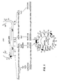
- (P1BLA)-P1 promoter of ⁇ -lactamase gene (P1 ⁇ , P2 ⁇ )-promoters of T R -DNA encoded genes 1 ⁇ and 2 ⁇ , (P NOS ) nopaline synthase promoter, (Pg5)-promoter of T L -DNA) encoded gene 5, (g4pA), (OcspA), (g7pA)-polyadenylation sequences derived from TL-DNA encoded gene 4, the octopine synthase gene and gene 7; (NPT-II)-neomycin phosphotransferase gene, (B L ), (BR)-25 bp left and right border repeats of T-DNA, ( ori T ), ( ori V )-replication and conjugational transfer origin sequences derived from plasmid RK2; ( ori pBR)-replication origin of pBR322; (A)ApaI, (B)BamHI, (Bg)B
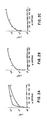
- Fig. A the filled squares indicate light units of luciferase activity in lux assay buffer. Open circles are light units of luciferase activity in lux assay buffer containing 0.1% bovine serum albumin (BSA). The closed circles are light units of luciferase activity in lux assay buffer in the absence of reduced FMN.
- Fig. B Luciferase was mixed with N.tabacum SR-1 leaf extract, prepared by grinding in liquid nitrogen 1.0 g of leaf tissue in 4.0 ml lux assay buffer (50 mM Na2HPO4, pH7 containing 50 mM ⁇ -mercaptoethanol and 0.4 mM sucrose).
- luciferase was mixed with carrot cell extracts (107 cells/ml), prepared as described in Materials and Methods and assayed for luciferase activity (open circles). The closed circles indicate light units of luciferase activity in the carrot extract in the absence of reduced FMN.
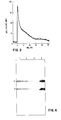
- the substrate n-decanal and reduced FMN were injected into the extract at time zero.
- a similar time course of bioluminescence can be observed with any extracts prepared from pPCV701luxA&B transformed plant tissues by using assay conditions described in Materials and Methods.
- the initial maximum of light intensity is a measure of the initial velocity of luciferase reaction.
- Lanes 1 and 2 are extracts equivalent to 2 and 4x10⁶ carrot protoplasts transformed by electroporation with pPCV701luxA&B DNA.
- Lane 3 protein extract obtained from 4x10⁶ untransformed control carrot protoplasts.
- Lane 4 10 ⁇ g of commercial V . harveyi luciferase. Arrows indicated the position of Lux ⁇ and Lux ⁇ polypeptides in transformed carrot protoplasts, lanes 1 and 2, and in the positive control, lane 4. No bands corresponding to Lux ⁇ and Lux ⁇ were detected in the untransformed carrot protoplast extract, lane 3.
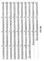
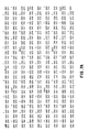
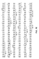
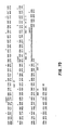
- Figures 5 and 6 show the nucleotide sequence of the translated luxA gene and the corresponding polypeptides, the remaining C-terminal part being known per se (21) (copy of the relevant page being enclosed hereto as Fig. 7)
- Expression vector pPCV701 is an Agrobacterium binary plant cloning vector derived by a series of modifications from the plant vector pPCV002 described previously (15). Part of the vector pPCV701 extending from the ori V and ori T regions to the right 25 bp border sequence (Bp) remained identical to that of pPCV002.
- the plant selectable marker casset te from pPCV002 was modified by coupling the NPT-II coding sequence from the Bc1I-SmaI fragment of plasmid pKm 9 (16) to the promoter sequences of the nopaline synthase gene (8) and by adding the 3 ⁇ -polyadenylation sequence of the T L -DNA gene 4 (17; sequence between position 8840-9240 of the T L -DNA).
- This selectable marker cassette was inserted between the HindIII and Bc1I sites of pPCV002, which destroyed the latter site and resulted in plasmid pPCV002NKMA.
- Plasmid TB7 carrying the lux AB transcriptional unit, was linearized with SalI, and 10 ⁇ g of the plasmid DNA was incubated in high salt buffer (100 mM Tris-HCl pH 8.0, 12 mM CaCl2, 600 mM NaCl, 1 mM EDTA) with increasing amounts (0.018-2.36 U) of Bal31 exonuclease at 30°C for 2-5 minutes, followed by EcoRI digestion. Fragments (1.0-1.2 kb in length) were isolated from gels after Bal31 digestion and subcloned into the DraI-EcoRI sites of pBR322 (20). Plasmid DNA prepared from pooled E .
- high salt buffer 100 mM Tris-HCl pH 8.0, 12 mM CaCl2, 600 mM NaCl, 1 mM EDTA
- Fragments (1.0-1.2 kb in length) were isolated from gels after Bal31 digestion and subcloned into the DraI-
- the DraI-EcoRI fragment pool was cloned into SamI-EcoRI sites of the M13 vector mp18 (22) and the exact endpoints of deletions were determined for 196 independent clones by DNA sequencing (23,24).
- One of the isolated deletion endpoints was located 7 bp upstream from the ATG initiation codon from lux A.
- This plasmid was opened at its BamHI site immediately upstream of the destroyed SmaI/DraI site, further deleted by Bal31 and then religated.
- the ligated samples were derived into three aliquots; one was digested by XbaI and the other by SalI, while the third sample was untreated.
- the samples were transfected into E.coli (24).
- the lux B gene was isolated as an SspI-PvuII DNA fragment from the plasmid pTB7, followed by a BamHI linker addition to the 5 ⁇ and the 3 ⁇ ends, cloned in both orientations into the BamHI site of M13 mp18 and partially sequenced.
- the SalI lux A and the BamHI lux B cassettes were inserted in two steps into single SalI and BamHI sites of pPCV701, respecti vely. This resulted in plasmid pPCV701 lux A&B and rendered lux A under gene 1 ⁇ and lux B under gene 2 ⁇ promoter control.
- pPCV701 lux A&B was transformed into the E.coli strain SM10 and mobilized into Agrobacterium strain GV3101 (pMP90RK) as described (15).
- Agrobacterium strain GV3101 (pMP90RK) carrying plasmid pPCV701 Lux A&B was used in protoplast co-cultivation (25,26) and plant tissue infection experiments (27) to transfer the lux A and lux B genes, as well as the NPT-II selectable marker gene into tobacco and carrot cells.
- Transformants were selected in the presence of 100 ⁇ g/ml kanamycin sulphate, and the Agrobacteria counter-selected by the addition of 500 ⁇ g/ml claforan (26). Conditions of tissue culture and tobacco plant regeneration were as described (15,25,26,28).
- Carrot protoplasts were isolated from cell line WOO1C by using 2.0% cellulase and 1.0% macerozyme according to Dudits (29).
- the protoplasts were resuspended in 30 ml of wash medium containing 0.37 M glucose,1.5mM CaCl2.H2O, and 10mM MES pH 6.5, counted, centrifuged and resuspended in protoplast wash medium at a final concentration of 1x10⁷/ml.
- 1.0 ml aliquots were transferred to a multiwell culture plate (Falcon#3047) and plasmid pPCV701 lux A&B DNA was added to each well to a final concentration of 50 ⁇ g/ml.
- the protoplast/SNA mixture was subjected to electroporation as described by Langridge et al. (30).
- the transformed protoplasts were resuspended in K-3 medium containing 0.4M sucrose as osmoticum and incubated at 26°C in the absence of light (29).
- luciferase activity was measured by a luminometer (Turner TD-20e) as a means of total light produced during the first 10 seconds of enzymatic reaction (31).
- a titration curve showing relationship between light emission and luciferase activity was established by measuring known amounts of commercially available V.harveyi luciferase (Sigma L-1637). Aliquots of 1:100 and 1:1000 dilutions of luciferase enzyme stock solution (1 mg/ml) were diluted with 0.5 ml lux assay buffer (50 mM sodium phopshate (pH7.0), 50 mM 2-mercaptoethanol) and transferred into the luminometer.
- 0.5 ml lux assay buffer 50 mM sodium phopshate (pH7.0), 50 mM 2-mercaptoethanol
- the reaction was started by injection of a mixture of 0.5 ml 100 ⁇ m reduced FMN and 10 ⁇ l n-decanal substrate through a septum in the top of luminometer sample chamber.
- the FMN solution was prepared in 25 mM EDTA (pH 7.0) or in 200 mM tricine buffer (pH 7.0) and kept reduced by light (32).
- the substrate was prepared as a 1:1000 dilution of n-decanal (Sigma D-7384) in lux assay buffer or in H2O and used freshly after sonication (33,34). At low protein concentrations the assay buffer was supplemented with 0.1% BSA.
- Transformed tobacco tissues and carrot protoplasts and cells collected at different time intervals after electroporation were homogenized in 0.5-3.0 ml of lux assay buffer, centrifuged for 5 min in a Eppendorf centrifuge at 4°C. Aliquots of the cleared extracts were diluted in 0.5 ml assay buffer and their luciferase activity was measured as described below.
- the precipitated protein was pelleted by centrifugation at 9.500 x g for 10 minutes at 4°C, resuspended in 100 ⁇ l of 2 times sample buffer, boiled for 2 minutes, and the proteins were separated on a 10% SDS polyacrylamide gel at 50 volts, for 12-14 hours (35).
- the separated proteins were transferred by Tobin et al. (36) and the blot was incubated at 26°C for 12 hours with anti Lux ⁇ and anti Lux ⁇ IgG in a 40 ml volume of 10 mM Tris-HCl pH 7.4 containing 0.9% NaCl and 3.0% BSA.
- the luciferase specific IgG was removed by washing the filter 5 times in the above mentioned buffer, without BSA.
- the immunoblot was then transferred into 40 ml BSA containing buffer and incubated for 6 hours with goat anti-rabbit IgG conjugated to alkaline phosphatase. Following binding, the filter was washed several times to remove the excess amount of the second antibody, and the luciferase ⁇ and ⁇ polypeptides were identified by incubating the blot in 50 ml of 10 mM Tris-HCl, pH7.4, containing 30 ⁇ l of hydrogen peroxide and 30 mg of 4-chloro-1-naphtol.
- the lux AB structural genes are part of a single transcriptional unit, encoding the ⁇ and ⁇ luciferase polypeptides of molecular weights 40 kD and 36 kD, respectively (1).
- ⁇ and ⁇ luciferase polypeptides of molecular weights 40 kD and 36 kD, respectively (1).
- Two separate "transcription-translation cassettes" were therefore constructed as outlined in Figure 1.
- 133 nucleotides, containing three non-essential ATG codons were deleted from the 5 ⁇ leader sequence.
- the final construct resulted in a " lux A cassette" bordered by synthetic SalI sites.
- the SalI site on the 5 ⁇ end originates from the M13 mp 18 polylinker sequence and is separated by 2 bp from the correct initiation codon.
- the added 3 ⁇ SalI linker is located 58 bp downstream from the translational stop codon of the lux A gene.
- a similar construct was also assembled by adding either BamHI or HindIII linkers to both the 5 ⁇ and 3 ⁇ ends of the lux A gene (data not shown). Construction of the " lux B cassette" is also shown in Figure 1.
- lux B cassette has a 5 ⁇ BamHI site separated by 23 bp from the first native ATG triplet and a 3 ⁇ BamHI site 197 bp downstream from the stop codon.
- Alternative " lux B cassette” were also constructed by ligating synthetic SalI of HindIII linkers, at both the 5 ⁇ and 3 ⁇ ends of the lux B gene.
- a plant expression vector was constructed from elements of available expression and binary cloning vectors.
- the constructed vector pPCV701 was derived from binary cloning vector pPCV002 described earlier (15).
- the " lux A cassette” was inserted into the single SalI site and the " lux B cassette” into the BamHI site of the expression vector, and thereby placed under the transcriptional control of the T R -DNA 1 ⁇ and 2 ⁇ promoters, respectively (18).
- the resulting plasmid, designated pPCV701 lux A&B was mobilized from E.coli to Agrobacterium and transferred into tobacco and carrot cells by using protoplast co-cultivation and leaf disc infection methods (25,27). Plasmid pPCV701 lux A&B DNA was also used for transformation of tobacco and carrot protoplasts by electroporation (30).
- luciferase activity could be contributed in various transformation experiments by Agrobacterium strain harbouring plasmid pPCV701- lux A&B, cell culture or sonicated cell extracts of this strain were assayed for luciferase activity (31). Luciferase activity was barely detectable, corresponding to less than 1.0 ng of luciferase/106 cells.
- an E.coli strain carrying plasmid pTB7 (1) in which the lux AB operon is controlled by the P1 promoter of the pBR322 ⁇ -lactamase gene, produces 0.2 to 2.0 ng luciferase/103 cells (data not shown).
- Results summarized in Table 1 demonstrate that luciferase activity can be easily and rapidly detected in transformed plant tissues. Due to the great sensitivity of the luciferase assay, the chimeric lux A&B genes can be used to demonstrate DNA uptake and gene expression in carrot protoplasts, as early as 8-24 hours after introduction of the DNA by electroporation.
- the activity of luciferase in plant extracts is also dependent on the addition of reduced flavin mononucleotide (FMN) and the long chain fatty aldehyde substrate n-decanal.
- FMN reduced flavin mononucleotide
- Stable transformants in the form of carrot calli or tobacco plants emitted from 4.00 to 26.00 light units/gram wet weight of plant tissue, based on the luciferase assay (Table 1).
- the amount of the luciferase protein present in transfored protoplast extracts was estimated by comparison between measured light units and the light units emitted by a known amount of purified V.harveyi luciferase. It was reported that 1.0 mg of purified luciferase emits approximately 1.6 x 10¹⁴ q sec ⁇ 1 light when n-decanal is used as substrate (34). Based on this value, the bioluminescence measured in 107 carrot protoplasts (Table 1, A) was 5.4 x 10⁹ q sec ⁇ 1, which corresponds to 34 ng of luciferase. This amount of luciferase protein agrees well with the estimation from the intensity of the stained bands in the immunoblots.
- Figure 1 shows how a dual promoter expression vector was used to separate the A and B cistrons of the V.harveyi luciferase operon into two separate plant transcription- translation units.
- a SalI/EcoRI fragment from the plasmid pTB7 (1,21) was cloned into the M13 phage mp18.
- a series of N-terminal deletions were constructed by either Exonuclease III treatment followed by Mung bean nuclease digestion, or by phage Bal31 exonuclease digestions. After the exonuclease digestions, the phage DNAs were religated, alternatively the deleted pieces were recloned into mp18. All constructs were thereafter DNA sequenced over the 5 ⁇ region of the gene, to exactly determine were the deletion had occured.
- Fig. 5 shows a representative number of the lux A N- terminal deletions in the mp18 phage, where all upstream restriction sites come from the mp18 polylinker sequence, and the lux A sequence ends by an EcoRI site about 60bp, downstream of the stop codon for translation of the lux A polypeptide.
- the formed plasmids were denoted the prefix pLX followed by a number starting from 101.
- the series continues to pLX120, in which 69 bp were deleted, counting the A of the first methionine (ATG) of the LuxA polypeptide as +1.
- the lux B gene was cloned separately into M13mp18 as a BamHI linkered fragment. This plasmid was denoted pLX150.
- the different constructs were tested in the in vivo T7 polymerase expression system as described (43).
- the plasmid vector pT3/T7-19 was sometimes used (BRL Cat No 5379SA).
- the lux A gene derivatives from the pLX100 plasmids were cloned into pT3/T7-19 as either a SalI/EcoRI or a HindIII/EcoRI fragment. In this way a new series of plasmids were constructed denoted pLX200 etc. (Table III, Fig. 6).
- pAR3038 a vector series denoted pAR3038, pAR3039 and pAR3040 was exploited (42).
- pAR3038 a vector series denoted pAR3038, pAR3039 and pAR3040 was exploited (42).
- vectors carry a BamHI site in all three reading frames situated 12 amino acids downstream of the starting methionine codon of the gene 10 protein of phage T7.
- the pLX200 and pLX300 plasmids were transferred into an E.coli strain carrying the T7 DNA polymerase gene on the chromosome under control of the lac UV5 promoter (43).
- the lux B gene was added in trans on the pLX550:3 plasmid (Table III). This plasmid carries the lux B gene under the T7 promoter on plasmid pACYC184.
- the lux B structural gene was excised from pLX150 as a BamHI fragment, and cloned into BamHI-site of the T7 promoter plasmid pAR2463 (42). From the thus created plasmid pLX350:3 , a BglII/HindIII fragment containing the active lux B gene fused to the T7 promoter was cloned into the BamHI/HindIII sites of plasmid pACYC184, giving rise to pLX550:3.
- the lux B gene was cloned into the EcoRI site of the pLX200 or pLX300 plasmids, giving rise to pLX200150 and pLX300150 lux AB plasmids respectively, and in this way restoring the original lux AB operon.
- lux constructs that were tested for activity was C-terminal protein fusions to the lux A gene. These were also transformed into the T7 polymerase expression strain, and complemented with plasmid pLX550:3 in a similar fashion as above.
- lux AB operon or the lux A, lux B gene system chosen representatives of constructs with or without the ribosomal binding site, with single starting ATG codons, or different deletions were cloned into other vectors, such as pBR322 and pUC18 or pUC19.
- the plasmids were denoted pLX600 and pLX400 respectively (Table III).
- a series of vectors carrying lux A and lux B genes have been constructed. This allows for the possibility to excise the lux A and lux B genes by a number of different restriction enzymes, enhancing the use of these genes as markers in gene regulation studies.
- the two luciferase structural genes cloned under separate promoters have also been expressed in the eucaryotic cell, demonstrating that the rather complex luciferase reaction works also in higher cells. Therefore, these genes can be used as markers both in eucaryotic and procaryotic cells.
- a number of different N-terminal deletion derivatives of the lux A gene have beem characterized. It is shown that the luciferase enzyme remains active both when the lux A N-terminal carries an extra stretch of 17 amino acids (pLX304) or when 11 aminoacids are deleted and four extra are added (pLX 218).
- the lux AB polypeptide complex contains a single active site which resides primarily if not exclusively on the ⁇ subunit.
- the specific role for the ⁇ subunit is unknown, but it is absolutely required for bioluminescence activity.
- Partial amino acid sequence information from regions though to be associated with the active center has been obtained (41).
- chemical modification and partial proteolysis studies it has been shown that a highly reactive sulhydryl group, though to reside in or near the flavin binding site is located close to a region that is highly sensitive to proteases (38,41). In the complete sequence, this position is thought to be at the cysteinyl residue at position 106.
- the ⁇ -subunit might contribute directly to the structure of the active center by binding to this region (40,44,45).
- the subunit also shows a very interesting distribution of charge over the molecule, with a clustring of basic residues surrounding the reactive cysteinyl residue (39).
- lux A and lux B sequences are similar, and the lux B gene is thought to have arisen as a gene duplication of lux A (21,39). Both genes are similar in their N-terminal part, and the most hydrophobic part of both the genes and the polypeptides is in the aminoterminal third. Therefore, it would appear critical to delete or alter too much of these regions.
- our data show that it is possible to add 17 amino acids to the N-terminal part of the ⁇ -peptide with high remaining activity of the ⁇ -complex. Deletions of up to 20 amino acids is possible but this reduces luciferase activity significantly. Also with a deletion of 3 N-terminal amino acids, the luciferase activity is affected. If this is due to reduced, activity of the ⁇ -complex or to a decreased stability of the ⁇ -polypeptide is not known at present.
- E.coli harbouring plasmid pPCV701 has been deposited in accordance with the Budapest Treaty with the Deutsche Sammlung von Mikroorganismen (DSM), Göttingen, BRD, under deposit number 3920.
- A, B and C extracts were prepared from 1 x 10⁷ protoplasts.
- a and B represent luciferase activities in transient expression experiments with 50 ⁇ g pPCV701- lux A&B DNA.
- D-G are 1 g wet weight equivalent tissue extracts.
- the luciferase assays were carried out as described in Materials and Methods. Values obtained by omitting reduced FMN from the assay mix indicate that the level of endogenous reduced FMN in tissue culture cells is not sufficient for in situ detection of luciferase activity followed n-decanal addition alone.
- Carrot W001C protoplasts were prepared and transformed as described in Materials and Methods. Values are expressed in light units/107 protoplasts, extracted 34 hours after electroporation with 50 ⁇ g/ml.
- (C) plasmid pPCV701 lux A&B, containing both lux A and lux B gene cassettes.
Landscapes
- Health & Medical Sciences (AREA)
- Genetics & Genomics (AREA)
- Life Sciences & Earth Sciences (AREA)
- Engineering & Computer Science (AREA)
- Chemical & Material Sciences (AREA)
- Organic Chemistry (AREA)
- Bioinformatics & Cheminformatics (AREA)
- Zoology (AREA)
- Wood Science & Technology (AREA)
- Biomedical Technology (AREA)
- General Engineering & Computer Science (AREA)
- Biotechnology (AREA)
- Biochemistry (AREA)
- General Health & Medical Sciences (AREA)
- Molecular Biology (AREA)
- Microbiology (AREA)
- Physics & Mathematics (AREA)
- Biophysics (AREA)
- Plant Pathology (AREA)
- Preparation Of Compounds By Using Micro-Organisms (AREA)
- Medicinal Chemistry (AREA)
- Cell Biology (AREA)
- Micro-Organisms Or Cultivation Processes Thereof (AREA)
- Peptides Or Proteins (AREA)
- Enzymes And Modification Thereof (AREA)
Applications Claiming Priority (2)
| Application Number | Priority Date | Filing Date | Title |
|---|---|---|---|
| SE8605621A SE8605621D0 (sv) | 1986-12-30 | 1986-12-30 | Heteromultimeric proteins and their manufacture |
| SE8605621 | 1986-12-30 |
Publications (1)
| Publication Number | Publication Date |
|---|---|
| EP0273889A2 true EP0273889A2 (de) | 1988-07-06 |
Family
ID=20366798
Family Applications (1)
| Application Number | Title | Priority Date | Filing Date |
|---|---|---|---|
| EP87850379A Withdrawn EP0273889A2 (de) | 1986-12-30 | 1987-12-07 | Heteromultimere Proteine und ihre Herstellung |
Country Status (3)
| Country | Link |
|---|---|
| EP (1) | EP0273889A2 (de) |
| JP (1) | JPS6413993A (de) |
| SE (1) | SE8605621D0 (de) |
Cited By (11)
| Publication number | Priority date | Publication date | Assignee | Title |
|---|---|---|---|---|
| EP0353464A1 (de) * | 1988-07-01 | 1990-02-07 | Kikkoman Corporation | Luciferase-Gen und neue rekombinante DNA sowie Verfahren zur Herstellung von Luciferase |
| EP0439354A3 (en) * | 1990-01-24 | 1992-06-17 | Amoco Corporation | Signal generating moiety and method for use |
| US5182202A (en) * | 1987-11-30 | 1993-01-26 | Kikkoman Corporation | Purified luciferase from luciola cruciata |
| US5352598A (en) * | 1988-04-12 | 1994-10-04 | Kikkoman Corporation | Purified luciferase from luciola lateralis |
| US5521066A (en) * | 1993-09-13 | 1996-05-28 | Bristol-Myers Squibb Company | Periplasmic membrane-bound system for detecting protein-protein interactions |
| FR2736930A1 (fr) * | 1995-07-17 | 1997-01-24 | Biocem | Procede de production, par des cellules vegetales, de proteines heminiques, proteines ainsi obtenues et produits contenant ces proteines |
| US5916771A (en) * | 1996-10-11 | 1999-06-29 | Abgenix, Inc. | Production of a multimeric protein by cell fusion method |
| EP1048734A3 (de) * | 1989-10-27 | 2001-11-28 | The Scripps Research Institute | Zusammensetzungen, die von Pflanzen produzierte Glykopolypeptidmultimere enthalten und deren Verwendung |
| US6420140B1 (en) | 1996-10-11 | 2002-07-16 | Abgenix, Inc. | Production of multimeric protein by cell fusion method |
| EP1006780A4 (de) * | 1997-01-24 | 2005-03-09 | Dna Plant Techn Corp | Verfahren und 2-komponenten zusammensetzungen, die die letalität von pflanzenzellen einleiten |
| US7101688B2 (en) | 1989-10-27 | 2006-09-05 | The Scripps Research Institute | Method of producing heteromultimeric mammalian proteins in plants |
-
1986
- 1986-12-30 SE SE8605621A patent/SE8605621D0/xx unknown
-
1987
- 1987-12-07 EP EP87850379A patent/EP0273889A2/de not_active Withdrawn
- 1987-12-26 JP JP32846187A patent/JPS6413993A/ja active Pending
Cited By (18)
| Publication number | Priority date | Publication date | Assignee | Title |
|---|---|---|---|---|
| US5182202A (en) * | 1987-11-30 | 1993-01-26 | Kikkoman Corporation | Purified luciferase from luciola cruciata |
| US5352598A (en) * | 1988-04-12 | 1994-10-04 | Kikkoman Corporation | Purified luciferase from luciola lateralis |
| EP0353464A1 (de) * | 1988-07-01 | 1990-02-07 | Kikkoman Corporation | Luciferase-Gen und neue rekombinante DNA sowie Verfahren zur Herstellung von Luciferase |
| US7282625B2 (en) | 1989-10-27 | 2007-10-16 | The Scripps Research Institute | Method of producing single chain protein in plant cells |
| EP1048734A3 (de) * | 1989-10-27 | 2001-11-28 | The Scripps Research Institute | Zusammensetzungen, die von Pflanzen produzierte Glykopolypeptidmultimere enthalten und deren Verwendung |
| US7101688B2 (en) | 1989-10-27 | 2006-09-05 | The Scripps Research Institute | Method of producing heteromultimeric mammalian proteins in plants |
| EP0439354A3 (en) * | 1990-01-24 | 1992-06-17 | Amoco Corporation | Signal generating moiety and method for use |
| US5521066A (en) * | 1993-09-13 | 1996-05-28 | Bristol-Myers Squibb Company | Periplasmic membrane-bound system for detecting protein-protein interactions |
| US6916787B2 (en) | 1995-07-17 | 2005-07-12 | Institut National De La Sante Et De Recherche Medicale | Method for producing hemin proteins using plant cells, resulting proteins and products containing same |
| FR2736930A1 (fr) * | 1995-07-17 | 1997-01-24 | Biocem | Procede de production, par des cellules vegetales, de proteines heminiques, proteines ainsi obtenues et produits contenant ces proteines |
| WO1997004115A3 (fr) * | 1995-07-17 | 1997-02-27 | Biocem | Procede de production, par des cellules vegetales, de proteines heminiques, proteines ainsi obtenues et produits contenant ces proteines |
| US6344600B1 (en) | 1995-07-17 | 2002-02-05 | Meristem Therapeutics | Method for producing human hemoglobin proteins using plant cells |
| US6207418B1 (en) | 1996-10-11 | 2001-03-27 | Abgenix, Inc. | Production of a multimeric protein by cell fusion method |
| US6677138B2 (en) | 1996-10-11 | 2004-01-13 | Abgenix, Inc. | Production of a multimeric protein by cell fusion method |
| US6420140B1 (en) | 1996-10-11 | 2002-07-16 | Abgenix, Inc. | Production of multimeric protein by cell fusion method |
| US5916771A (en) * | 1996-10-11 | 1999-06-29 | Abgenix, Inc. | Production of a multimeric protein by cell fusion method |
| US7429380B2 (en) | 1996-10-11 | 2008-09-30 | Amgen Fremont Inc. | Production of a multimeric protein by cell fusion method |
| EP1006780A4 (de) * | 1997-01-24 | 2005-03-09 | Dna Plant Techn Corp | Verfahren und 2-komponenten zusammensetzungen, die die letalität von pflanzenzellen einleiten |
Also Published As
| Publication number | Publication date |
|---|---|
| JPS6413993A (en) | 1989-01-18 |
| SE8605621D0 (sv) | 1986-12-30 |
Similar Documents
| Publication | Publication Date | Title |
|---|---|---|
| Koncz et al. | Expression and assembly of functional bacterial luciferase in plants | |
| Close et al. | Construction and characterization of the chloramphenicol-resistance gene cartridge: a new approach to the transcriptional mapping of extrachromosomal elements | |
| US5717084A (en) | Chimaeric gene coding for a transit peptide and a heterologous peptide | |
| CA2192849C (en) | Dna molecules which code for a plastid 2-oxoglutarate/malate translocator | |
| AU608219B2 (en) | Expression of wild type and mutant glutamine synthetase in foreign hosts | |
| US5034322A (en) | Chimeric genes suitable for expression in plant cells | |
| Kaulen et al. | Light‐induced expression of the chimeric chalcone synthase‐NPTII gene in tobacco cells | |
| US6271016B1 (en) | Anthranilate synthase gene and method of use thereof for conferring tryptophan overproduction | |
| JPS60137286A (ja) | ス−パ−オキシドジスムタ−ゼ遺伝子 | |
| Olsson et al. | Engineering of monomeric bacterial luciferases by fusion of luxA and luxB genes in Vibrio harveyi | |
| GB2197653A (en) | Gene fusion comprising beta -glucuronidase | |
| Baldwin et al. | Site‐directed mutagenesis of bacterial luciferase: Analysis of the ‘essential’thiol | |
| CA2236267A1 (en) | Plant vde genes and methods related thereto | |
| JP3320064B2 (ja) | 抵抗力を増大させるための植物中のリゾチーム遺伝子構造の使用 | |
| Markgraf et al. | Evidence that carotenoids are required for the accumulation of a functional photosystem II, but not photosystem I in the cotyledons of mustard seedlings | |
| Thöny‐Meyer et al. | Requirements for maturation of Bradyrhizobium japonicum cytochrome c550 in Escherichia coli | |
| Barz et al. | Photosynthetic deficiency of a pufX deletion mutant of Rhodobacter sphaeroides is suppressed by point mutations in the light-harvesting complex genes pufB or pufA | |
| Mayerhofer et al. | Expression of recombinant Renilla luciferase in transgenic plants results in high levels of light emission | |
| Howe et al. | Biosynthesis of cytochrome f in Chiamydomonas reinhardtii: analysis of the pathway in gabaculine-treated cells and in the heme attachment mutant B6 | |
| US5804408A (en) | Expression of human SOD in blue green algae | |
| JP3759628B2 (ja) | ブリーチング除草剤耐性植物の製造 | |
| EP0472658A1 (de) | Fusionsproteine mit in vivo posttranslationaler modifikationsstelle und verfahren zur herstellung und anreicherung | |
| JPH07504820A (ja) | 複数ウイルス耐性を示すトランスジェニック植物およびその生産方法 | |
| KR0163062B1 (ko) | 폴리펩타이드의 형질발현 방법 | |
| US5474929A (en) | Selectable/reporter gene for use during genetic engineering of plants and plant cells |
Legal Events
| Date | Code | Title | Description |
|---|---|---|---|
| PUAI | Public reference made under article 153(3) epc to a published international application that has entered the european phase |
Free format text: ORIGINAL CODE: 0009012 |
|
| AK | Designated contracting states |
Kind code of ref document: A2 Designated state(s): AT BE CH DE FR GB IT LI NL SE |
|
| STAA | Information on the status of an ep patent application or granted ep patent |
Free format text: STATUS: THE APPLICATION IS DEEMED TO BE WITHDRAWN |
|
| 18D | Application deemed to be withdrawn |
Effective date: 19900703 |