EP0274560A2 - Menschlicher, das hämatopoietische Zellwachstum aktivierender Faktor - Google Patents
Menschlicher, das hämatopoietische Zellwachstum aktivierender Faktor Download PDFInfo
- Publication number
- EP0274560A2 EP0274560A2 EP87108782A EP87108782A EP0274560A2 EP 0274560 A2 EP0274560 A2 EP 0274560A2 EP 87108782 A EP87108782 A EP 87108782A EP 87108782 A EP87108782 A EP 87108782A EP 0274560 A2 EP0274560 A2 EP 0274560A2
- Authority
- EP
- European Patent Office
- Prior art keywords
- polypeptide
- sequence
- gene
- hcgpf
- amino acid
- Prior art date
- Legal status (The legal status is an assumption and is not a legal conclusion. Google has not performed a legal analysis and makes no representation as to the accuracy of the status listed.)
- Withdrawn
Links
- TXAYUIRXRADQQA-JLPGSUDCSA-N C=C(C=C(CCCCCCCCCCC(CC1)CC2)C=C3)/C3=C\CC12I Chemical compound C=C(C=C(CCCCCCCCCCC(CC1)CC2)C=C3)/C3=C\CC12I TXAYUIRXRADQQA-JLPGSUDCSA-N 0.000 description 1
- NGPVAIKEOCZACK-OWBHPGMISA-N CCC1/C=C\CCCCCCCCCCCC(CC2)CCC2C1 Chemical compound CCC1/C=C\CCCCCCCCCCCC(CC2)CCC2C1 NGPVAIKEOCZACK-OWBHPGMISA-N 0.000 description 1
Images









Classifications
-
- C—CHEMISTRY; METALLURGY
- C07—ORGANIC CHEMISTRY
- C07K—PEPTIDES
- C07K14/00—Peptides having more than 20 amino acids; Gastrins; Somatostatins; Melanotropins; Derivatives thereof
- C07K14/435—Peptides having more than 20 amino acids; Gastrins; Somatostatins; Melanotropins; Derivatives thereof from animals; from humans
- C07K14/52—Cytokines; Lymphokines; Interferons
-
- A—HUMAN NECESSITIES
- A61—MEDICAL OR VETERINARY SCIENCE; HYGIENE
- A61K—PREPARATIONS FOR MEDICAL, DENTAL OR TOILETRY PURPOSES
- A61K38/00—Medicinal preparations containing peptides
Definitions
- the present invention relates to a gene coding for polypeptide of a factor with growth potentiating activity for hematopoietic cells, particularly bone marrow cells (hereafter HCGPF: Hematopoietic Cell Growth Potentiating Factor), recombinant DNA harboring the gene, living cell lines having the DNA , a method for production of HCGPF using the living cell lines and polypeptide having HCGPF activity.
- HCGPF Hematopoietic Cell Growth Potentiating Factor
- This HCGPF potentiates the growth of bone marrow cells, thymocytes, fetal liver cells even by single use thereof but when used in combination with other hematopoietic cell growth factor HCGF, the growth of these hematopoietic cells is markedly accelerated but the presence of such a factor is unknown hitherto.
- the present inventors have already identified a gene for a factor which induces the growth of mouse IL3-dependent cell line FDC-P cells (Japanese Patent Application No. 2633/86).
- the present inventors have now succeeded in producing this factor as a single cytokine using the gene and in purifying the factor up to a single protein and could identify it as a substance for the first time.
- this factor acts widely on hematopoietic cells such as bone marrow cells, thymocytes, fetal liver cells, etc. and when used in combination with other factors termed HCGFs, this factor induces more remarkable growth of hematopoietic cells and the present invention has thus been come to accomplish.
- This HCGPF exhibits immune control and hematopoietic control functions over a wide range based on the aforesaid activity and its utility has been strongly expected in the fields of immunodeficient diseases, autoimmune diseases, infectious diseases, hepatitis, nephritis, cancers, bone marrow transplantation, etc.
- the factor is effective in potentiating effects when used in combination with other immunoactive substances, immunotherapeutic agents, lymphokines, cytokines, interferon, cell growth factors, chemotherapeutic agents, antibiotics or anti-viral agents as well as alleviating side effects of other drugs.
- This factor is a novel substance having the utility as described above. This factor is not only expected to have utility in the medical field but this factor, the antibody to the factor and the receptor for the factor are also expected to be useful as diagnostic agents.
- hematopoietic cells can be efficiently proliferated artificially using cell culture technique and the provision of the gene of this factor, a method for producting thereof and of this factor as a substance results in great effects. It is particularly important to create a technique capable of providing this substance which is present only in a trace amount in the living human body, in a simple manner in large quantities using microorganisms, etc.
- a colony stimulating factor which directly acts on the growth of hematopoietic cells 3 kinds have recently been reported and their genes have been subjected to cloning (Gouzh, N.M. Et al., Nature, 309 , 763 (1984); Kawasaki, E.S., et al., Science, 230 , 291 (1985) and Nagata, S., et al., Nature, 319 , 415 (1986)).
- These factors are simply referred to as GM-CSF, M-CSF and G-CSF, respectively.
- multi-CSF as a colony stimulating factor which acts widely on hematopoietic cells is known with mice and the factor has already been subjected to genetic cloning (Fung, M.C., et al., Nature, 307 , 233 (1984), Yokota, T., et al., Proc. Nat. Acad. Sci., USA, 81 , 1070 (1984)).
- mouse IL-3 interleukin 3
- IL-3 interleukin 3
- the biological activity of mouse IL-3 is believed to participate not only in differentiation and growth of cells such as neutrophilic granulocytes, megakaryocytes, mast cells, basophilic granulocytes, etc. but also in metabolism of sexual hormones and, its wide biological activity is greatly interesting (Immunological Rev., 63 , 532 (1982)). For these reasons mouse IL-3 is also called multi-CSF.
- this substance is expected to be useful over a wide range of diseases caused by immunological deficiency or abnormality and useful for clinical diagnosis of immune diseases and at the same time, useful as reagents for general investigation of biological and medical science.
- IL-3 is merely clarified on its gene and protein structure with mice but with respect to human IL-3, it is even unclear if human IL-3 is present because there is no appropriate assay system for the activity and suitable IL-3 producing cells are unknown.
- a novel technique that is to be accomplished by the present invention is to identify and provide a substance having an activity of synergestically potentiating the growth of hematopoietic cells by the use in combination with these factors having colony forming action of these hematopoietic cells and by itself showing the action singly.
- this factor has also human IL-3-like activity at the same time.
- HCGPF human hematopoietic cells
- objects of the present invention are to identify the gene for this HCGPF, provide a method for production by introducing the gene in living cell line, produce HCGPF using living cell line containing recombinant DNA and identify this HCGPF as a novel substance.
- the present inventors have prepared human peripheral blood derived mononuclear cells in a conventional manner; their lymphocytes are stimulated for 48 hours in the presence of PHA and TPA,and RNAs are extracted by extraction of the thus activated lymphocytes with phenol in a conventinal manner. After precipitation with ethanol, mRNAs are collected by affinity chromatography using oligo dT-cellulose. In order to improve the purity of the mRNAs and fractionate the mRNAs by size, oligo dT-cellulose chromatography is repeated.
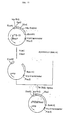
- cDNA expression vector pDE-2 in monkey cells has been constructed (Japanese Patent Application No. 208645 filed October 4, 1984). Namely this pDE-2 is constructed using pML-R11G (Nature, 293 , 79 (1981)) and pKCR (Proc. Natl. Acad. Sci., U.S.A., 78 , 1527 (1981)) as shown in Figure 1 in which cDNA can be inserted in bidirectional SV40 early promoter and pDE-2 can be selected as being capable of replicating in E. coli and by ampicillin resistance.
- This pDE-2 subjected to C-tailing is mixed with the aforesaid double stranded cDNA subjected to G-tailing to perform anealing.
- the anealed DNA is introduced into competent E. coli MC1061 (J. Mol. Biol., 138 , 179-207 (1980)), which is applied to agar plate of L medium to appear colonies.
- the thus obtained colonies are divided into 192 groups of 32 colonies each followed by mix culture. Plasmid DNAs mixed with 32 kinds are purified in a conventional manner.
- This plasmid mixture (32 kinds) is introduced into monkey cells (COS-7) (Cell, 23 , 175-182 (1981)) and treated with chloroquine. After washing the cells, culture is performed for 48 hours. With respect to the culture supernatant, FDC-P cell growth activity is measured and a DNA mixture group having the activity is identified. DNA plasmid in the group containing the DNA mixture in which the activity has been recognized is purified as a single plasmid DNA of 32 and again introduced in COS cells. By assaying for the activity of the culture supernatant in a similar manner, clone p4-15 showing FDC-P cell growth activity, namely, IL-3-like activity, is identified.
- cDNA contained in this clone is analyzed according to the dideoxy chain termination method (Sanger et al., Proc. Natl. Acad. Sci., U.S.A., 74 , 5463-5467 (1977)) and the Maxam-Gilbert method (Meth. Enzym., 65 , 510-580 (1980)), whereby the whole base sequence has been determined. It is clarified that the cDNA comprises base pairs of approximately 900 bp and codes for secretory protein having signal peptide. This cDNA and the structure of polypeptide presumed therefrom have not been found to date and are, therefore, quite novel. It is also assumed that the cDNA would be a novel lymphokine because messenger RNA corresponding to this cDNA expresses specifically on human lymphocytes activated by TPA and PHA.
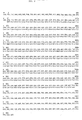
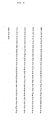
- FIG. 1 A cleavage map of cDNA insert with restriction enzyme endonuclease is shown in Figure 1. As shown in Figure 1, this cDNA has a structure cleaved with restriction enzyme endonuclease of Ava I, Pst I or Ban I. The base sequence of the cDNA is shown in Figure 2.
- the DNA sequence of the cDNA insert holds a large open reading frame therein.
- the first ATG sequence (Kozak, M. Cell., 15 , 1109-1123 (1978)) which is often a reading initiation sequence of eukaryote is present in nucleotide at the 64- to 66-position from the 5 ⁇ -end thereof and 196 codons from the 655- to 657-position in the nucleotide site in which reading termination codon TGA is present are connected with this ATG.
- Linkage of A corresponding to the 3 ⁇ -poly(A) end of mRNA is found at the cDNA end and nucleotide AATAAA (approximately 883 to 888) comprising 6 which is generally found in most of eucaryote mRNA is located frontward (Proudfoot, N.J. & Brownlee C.G., Nature, 263 , 211-214 (1976)).
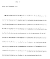
- amino acid sequence III The amino acid sequence which cDNA codes for can be deduced as shown in Figure 5 (amino acid sequence III).
- polypeptide in amino acid sequence III is composed of 197 amino acids and as is reported to be found in most of secretory protein known to date (Blobel G., et al., Sym. Soc. Exp. Med., 33 . 9-36 (1979)), the N-terminal area of the aforesaid deduced HCGPF polypeptide is hydrophobic as well. This region would take a role of signal peptide cleaved upon secretion of mature HCGPF.
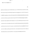
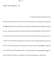
- Example 4 DNA fraction starting with ACC codon at the 118- to 120-positions in the base sequence, namely, code (amino acid sequence I in Figure 3) for polypeptide starting with Thr located at the 19-position expresses polypeptide having HCGPF activity. It has been confirmed that the DNA fraction starting from ATC at the 154- to 156-positions in the base sequence, namely, as shown in amino acid sequence II in Figure 4, the DNA fraction coding for polypeptide starting with Ile located at the 31-position expresses polypeptide having HCGPF activity as shown in Example 5.
- genes of eukaryote show polytypic phenomenon as is known with human intereron genes (Taniguchi et al., Gene, 10 , 11-15 (1980), Ono & Taniguchi, Proc. Natl. Acad. Sci., U.S.A., 77 , 5305-5309 (1981); Gray et al., Nature, 295 , 501-508 (1981)).
- human intereron genes Trigger et al., Gene, 10 , 11-15 (1980), Ono & Taniguchi, Proc. Natl. Acad. Sci., U.S.A., 77 , 5305-5309 (1981); Gray et al., Nature, 295 , 501-508 (1981)).
- the gene of the present invention includes DNAs having the base sequence shown in Figure 2, DNA having a continuous base sequence from ATG sequence at the 64- to 66-positions to at least GCC sequence at the 652- to 654-positions, DNA having a continuous base sequence from ACC sequence at the 118- to 120-positions to GCC sequence, and DNA having a continous base sequence from ATC sequence at the 154- to 156-positions to at least GCC sequence.
- the gene of the present invention also includes DNA ending with GCC at the 652- to 654-positions and starting with G at the 1-position, DNA starting with ATG at the 64- to 66-positions and DNA starting with ATC sequence at the 118- to 120-positions.
- the gene of the present invention includes DNA ending with T at the 909-position and starting with A at the 1-position, DNA starting with ATG at the 64- to 66-positions, DNA starting with ACC at the 118- to 120-positions and DNA starting with ATC sequence at the 154- to 156-positions.
- the gene of the present invention also includes DNA ending with poly(A) and starting with ATG codon at the 64- to 66-positions, DNA starting with ACC sequence at the 118- to 120-positions and DNA starting with ATC sequence at the 154- to 156-positions.
- the present invention also includes the genes having base sequences corresponding to amino acid sequence I, amino acid sequence II and amino acid sequence III.
- polypeptide deficient in one or more amino acids or polypeptide in which one or more amino acids in amino acid sequence III is/are substituted with one or more amino acids may have HCGPF activity and therefore, genes coding for such polypeptides can be used in the present invention.
- genes coding for such polypeptides can be used in the present invention.
- one or more bases capable of expressing one or more amino acids to amino acid sequence I, amino acid sequence II or amino acid sequence III are additionally attached to the genes, such genes are included in the present invention unless the additional amino acids inhibit the expression of HCGPF activity.
- it is a modification region having a supplemental amino acid sequence that inhibits the polypeptide function as HCGPF, it can be utilized as the gene of the present invention if a freshly supplemented region were to be removed.
- Recombinant DNA producing HCGPF in living cells can be prepared by various methods below.
- a sequence coding for HCGPF-cDNA is inserted downstream promoter sequence of expression vector.
- a cDNA fragment having a promoter sequence can be inserted upstream a sequence coding for HCGPF prior to or after the inserting of cDNA of expression vector.
- cDNA is bound to various bacteria promoters and thereafter, hybrid plasmid containing cDNA is formed upstream the promoters.
- the plasmid is transfected to, e.g., Escherichia coli HB101, whereby bacteria biosynthesizing protein having human HCGPF activity are cloned.
- Any bacterial promoter expresses HCGPF-cDNA as far as it is appropriately connected with cDNA. Expression of such cDNA will be later shown in the examples.
- HCGPF-cDNA can be expressed even in yeast if it is incorporated in a suitable expression vector and the vector is introduced in host cells.
- Various shuttle vectors for expressing external gene in yeast have already been reported (Heitzman et al., Nature, 293 , 717-722 (1981); Miyashita et al., Proc. Natl. Acad. Sci. U.S.A., 80 , 1-5 (1983)). These vectors can be grown in both Escherichia coli and yeast. These vectors also include therein the promoter sequence of yeast gene.
- expression vectors can all be utilized for expression of HCGPF-cDNA. When yeast is used, it is possible to enhance the level of HCGPF productivity, as compared to utilization of animal cells or bacteria. It is also possible to produce HCGPF containing sugar regions, which is effective for improving solubility of HCGPF.
- Yeast- Escherichia coli shuttle vectors pAT77 and pAM82 are reported by Miyashita et al. (Proc. Natl. Acad. Sci. U.S.A., 80 , 1-5 (1983)).
- Vector pAM82 is a derivative of pAT77. Both have an ars 1 marker (Stinchcomb, D.T., et al., Nature, 282 , 39-43 (1979), 2 umori (Braoch, J.R., et al., Gene., 8 , 121-133 (1979), leu2 (Ratzkin B. et al., Proc. Natl. Acad. Sci.
- a vector suited for host cells may be bound to cDNA insert cleaved and separated from procaryocyte. Then eucaryocyte is transfected to synthesized vector followed by incubation. The cells having inserted therein recombinant DNA are cultured to amplify the recombinant DNA or produce HCGPF polypeptide.
- This culture is performed in a conventional manner.
- transfected yeast is aerobically cultured at 27 to 37°C at pH of 4 to 7 in medium containing carbon sources, nitrogen sources, inorganic salts and if necessary, organic nutrient sources such as vitamins, amino acids, etc.
- Transformed prokaryote such as Escherichia coli or Bacillus subtilis can also be cultured in a conventional manner.
- HCGPF obtained herein is assumed to be human-derived lymphokine corresponding to mouse IL-3; however, it cannot be concluded that HCGPF would be human derived one corresponding to mouse IL-3 because the presence of human IL-3 is unclear yet and any authorized assay system of human IL-3 activity is unknown. In fact, the structure of this cDNA has no similarity to that of mouse IL-3 cDNA and, it is unlikely that both would be branched from the same ancestral gene.
- FDC-P cells are cell lines conventionally used as IL-3-dependent cell lines in both domestic and foreign countries. It is also confirmed that FDC-P cells do not grow in the presence of other lymphokines. These activities are not found in, for example, interleukin 2, interleukin 1 and interferon.
- the proteins having amino acid sequences which the genetic sequence obtained herein codes for are heretofore unknown.
- the proteins are also different structurally from human G-CSF, M-CSF and GM-CSF which show activity somewhat similar to IL-3 and are substances different from so called selective colony stimulating factor (CSF). Therefore, the activity shown by the polypeptide that is expressed by the gene (cDNA) and assumed to be novel lymphokine obviously takes at least a part of the aforesaid human IL-3 activity.
- human IL-3-like protein can be readily purified, singly or in combination, by salting-out, gel filtration, affinity chromatography, ion exchange chromatography, isoelectric electrophoresis, high speed liquid chromatography and other known methods for separation and purification of polypeptide.
- the IL-3-like protein is expressed in COS cells in large quantities and its activity is assayed; the IL-3-like protein shows even singly growth inducing activity to mouse bone marrow cells, human bone marrow cells, thymocytes or mouse fetal liver cells. Apart from degree of the activity, it has been confirmed that the IL-3-like protein shows multi-CSF-like activity.
- the expression protein induces synergetically remarkable growth in the presence of mouse IL-3.
- this novel lymphokine polypeptide is to designate HCGPF as a factor that has multi-CSF-like activity likewise human IL-3 and acts synergetically with IL-3 to grow hematopoietic cells.
- the thus obtained HCGPF shows the same biochemical and biological behavior as that of HCGPF obtained from mammalian cells and exhibit HCGPF activity.
- the HCGPF activity is stable even after treating with DNase or RNase or after thermally treating at 56°C for 30 minutes.
- the activity is stable at pH of 4 to 9.
- HCGPF of the present invention has immune control function and hematopoietic control function and is thus applicable to the fields of immunodeficient diseases, infectious diseases, cancers, hepatitis, nephritis, bone marrow transplantation, etc. Further HCGPF of the present invention is thought to be effective for potentiationg effects when used in combination with other immunoactive substances, immunotherapeutic agents, lymphokine, cytokine, interferon, cell growth factors, chemotherapeutic agents, antibiotics or anti-viral agents and alleviating side effects of other medicines.
- microorganisms referred to in the examples have been deposited and are available under the following accession numbers: pTHCGPF-19/HB101 ⁇ FERM BP-1246 pTHCGPF-31/HB101 ⁇ FERM BP-1247 pT13S ⁇ HIL2(53)-HCGPF-19/HB101 ⁇ FERM BP-1248 pT13S ⁇ HIL2(53)-HCGPF-31/HB101 ⁇ FERM BP-1251 pT13S ⁇ HIL2(20)-HCGPF-19/HB101 ⁇ FERM BP-1249 pT13S ⁇ HIL2(20)-HCGPF-31/HB101 ⁇ FERM BP-1252 pT13S ⁇ HIL2(11)-HCGPF-19/HB101 ⁇ FERM BP-1250
- RNAs were precipitated with a 2-fold amount of cold ethanol. The precipitates were collected by centrifugation and dissolved in 10 mM Tris-HCl solution having pH of 7.5. The obtained RNAs were 40 mg.
- RNAs Fractionation of mRNAs were carried out by affinity chromatography using oligo dT-cellulose (P.L. Biochemicals, Type 7).
- An adsorbing solution was a solution containing 20 mM Tris-HCl, 0.5 M NaCl, 1 mM EDTA and 0.5% SDS having pH of 7.5.
- buffer (20 mM Tris-HCl, pH 7.5, 0.5 mM NaCl and 1 mM EDTA
- elution was carried out using H2O and 10 nM Tris-HCl having pH of 7.5, alternatively.
- the mRNA obtained by the elution was 0.9 mg based on 33 mg of RNAs applied.
- cDNA library was made from the authentic mRNAs as follows and used for cDNA cloning.
- the cDNA was synthesized fundamentally according to the Land et al method (Nucleic Acids Res., 9 , 2551 (1981)).
- the precipitates were dissolved in 0.3N NaOH solution. The solution was settled at room temperature for 15 hours. Then an equal amount of M Tris-HCl having pH of 7.5 was added thereto to neutralize. Thereafter the mixture was passed through a Sephadex G-50 column to recover cDNA. The recovered single stranded cDNA was approximately 10 ⁇ g.
- reaction mixture was treated with phenol and passed through a Sephadex G-50 column to recover double stranded cDNA fractions. As precipitates from ethanol, approximately 16 ⁇ g of DNA was recovered.
- reaction mixture was treated with phenol and passed through a Sephadex G-50 column to collect DNA fractions. As precipitates from ethanol, approximately 15.5 ⁇ g of DNA was recovered.
- cDNA expression vector pDE-2 in monkey cells was constructed.
- pDE-2 can be inserted into bidirectional SV 40 early promoter, is replicable and can be selected as ampicillin-resistance.
- This pDE-2 was cleaved with EcoRI and cohesive ends were filled up with DNA polymerase I (Klenow). Then approximately 13 dC-tails were added to the aforesaid ds-cDNA at the 3 ⁇ -ends thereof in quite the same manner as in the dG-tails added to the ds-cDNA at the 3 ⁇ -ends thereof.
- E. coli MC 1061 was inoculated on 100 ml of ⁇ medium (2% tryptone, 0.5% yeast extract, 0.5% MgSO4.7H2O, pH 7.6), which was shake cultured at 37°C until absorbance of the culture solution became approxametely 0.3 to 0.5 at 550 nm. After completion of the culture, the culture solution was kept at 0°C for 5 minutes and cells were collected by centrifugation. The cells were suspended in 40 ml of Tfb I (30 mM of potassium acetate, 100 mM of CaCl2, 50 mM of MnCl2, 15% glycerine, pH 5.8) and the suspension wa settled at 0°C for 5 minutes.
- Tfb I 30 mM of potassium acetate, 100 mM of CaCl2, 50 mM of MnCl2, 15% glycerine, pH 5.8
- Tfb II 10 mM of MOPS or PIPES, 75 mM of CaCl2, 10 mM of RbCl, 10% glycerine, pH 6.5
- the suspension was settled at 0°C for 15 minutes.
- the suspension was separately charged and kept at -70°C.
- the culture solution was smeared on agar plate of L medium (1% of tryptone,0.5% of yeast extract, 0.5% of NaCl) containing 25 ⁇ g/ml of ampicillin and 25 ⁇ g/ml of streptomycin. By incubation at 37°C overnight, colonies appeared.
- L medium 1% of tryptone,0.5% of yeast extract, 0.5% of NaCl
- the appeared colonies were divided into 192 groups by 32 colonies each, which were inoculated on 100 ml of ⁇ medium containing 25 ⁇ g/ml of ampicillin and 25 ⁇ g/ml of streptomycin, respectively, followed by shake culture at 37°C for 5 to 7 hours. Next, 100 ml of fresh ⁇ medium containing chloramphenicol in a final concentration of 170 ⁇ g/ml was added to the culture solution followed by shake culture overnight.
- the thus amplified plasmid DNA was purified as follows.
- the culture solution was centrifuged to collect cells only.
- the cells were suspended in 5 ml of 50 mM Tris-HCl showing pH of 7.5. After freezing at -80°C, the system was fused and lysozyme (final concentration, 2 mg/ml) was added thereto and settled at 0°C for 10 minutes. EDTA (final concentration, 0.1 M) was further added thereto and settled at 0°C for 10 minutes. Thereafter Triton X-100 (final concentration, 0.1%) added thereto and settled at 0°C for 60 minutes. Then the ultracentrifugal separation was performed at 30,000 rpm for 30 minutes and the supernatant was treated with an equal amount of water-saturated phenol. The aqueous phase was further treated with an equal amount of chloroform and extracted. Rnase was added to the extract in a final concentration of 20 ⁇ g/ml followed by incubation at 37°C for 60 minutes.
- COS-7 cells were suspended in 1 x 10⁵/ml in 10% fetal bovine serum (FBS)-containing DMEM and 8 ml of the suspension was cultured at 37°C overnight in a Petri's dish in a 5% carbon dioxide gas incubator. Further 5 ml of fresh 10% FBS- containing DMEM was added and culture was continued at 37°C for 4 hours in the 5% carbon dioxide gas incubator. After completion of the culture, the supernatant was removed and the system was washed once with 5 ml of TBS (25 mM of Tris-HCl, pH 7.5, 130 mM of NaCl, 5 mM of KCl and 0.6 mM of Na2PO4).
- FBS fetal bovine serum
- a plasmid mixture (2.5 ml of TBS (+) (TBS added with 0.7 mM of CaCl2 and 0.5 mM of MgCl2)), 5 ⁇ g of plasmid DNA and 128 ⁇ l of 10 mg/ml DEAE-dextran) was added to the system and the mixture was incubated at 37°C for 1 hour in the 5% carbon dioxide gas incubator. After the supernatant was removed, the system was washed with 5 ml of TBS and 5 ml of 150 ⁇ m chloroquine-containing 10% FBS DMEM. After incubating at 37°C for 3 hours in the 5% carbon dioxide gas incubator, the supernatant was removed and the system was washed twice with 5 ml of TBS. Then 8 ml of 10% FBS -containing DMEM was added thereto followed by culturing at 37°C overnight in the 5% carbon dioxide gas incubator.
- the proliferation activity of FDC-P cells was measured in accordance with the following method. In each cell of a 96-well tissue culture plate was charged 100 ⁇ l each of the culture supernatant of the COS cells, FDC-P cell proliferation activity of which was to be assayed and, 2-fold dilution was repeated in RPMI 1640 plus 10% FBS medium.
- each of the FDC-P cells which were IL-3-dependent cell lines were charged in each cell in a cell density of 1 x 10⁴/100 ⁇ l. After incubation at 37°C for 18 hours FDC-P in 5% CO2, 1 ⁇ Ci of tritium thymidine was added to the culture solution. After performing pulsing for 6 hours, cells were harvested on glass fiber filter paper in a manner well known in the art. After a scintillator was added thereto, ⁇ -radiation incoporated into the cells was counted.
- the proliferation activity was assayed with respect to the 192 culture supernatants obtained by transfection of the aforesaid plasmid mixture to the COS cells according to this method and the results shown in Table 1 were obtained, wherein a significant activity was noted with the group designated No. 3-11.
- Group 3-11 was a mixture of 32 clones.
- the 32 clones were separately cultured and plasmid was prepared in quite the same manner as used in Example 1 (4) and transfected to COS cells in the same manner as used in the preceding (1).
- FDC-P cell growth activity was determined, whereby the growth activity was noted in one clone (15th clone, designated p4-15) ( Figure 7). It was identified that this clone had cDNA showing IL-3-like activity.
- HCFPF cDNA was excised from plasmid p4-15 using EcoR I and its base sequence was determined according to the dideoxy chain termination method using M13 and the Maxam-Gilbert method; it was presumed from its open reading frame that this HCFPF cDNA was composed of base pairs of approximately 900 bp and coded for precursor HCGPF protein composed of about 197 amino acids or 83 amino acids.
- this cDNA is composed of 909 bases and the amino acid sequences shown in Figures 8 and 9 are assumed as those that the cDNA codes for.
- cDNA was excised with restriction enzyme EcoR I and a part of them was cleaved with restriction enzyme Bgl I.
- Bgl I-Bgl I fragment of 396 base pairs was made smooth at the both ends thereof using T4 polymerase and then mixed with Hind III linker in a molar ratio of 1:1 followed by ligation using T4 DNA ligase. After the ligated fragment was recovered, they were cleaved with restriction enzymes Hind III and Sph I to give Hind III-Sph I fragment.
- the thus obtained three fragments were mixed in a molar ratio of 1:1:1 and ligated using T4 DNA ligase.
- the thus obtained recombinant DNA was introduced into Escherichia coli HB 101 strains having ampicillin resistance was selected. From the separated strain plasmid was prepared. By cleavage with restriction enzyme and determination of base sequence around the ligated site, bacterial harboring pSP6 4-15 were selected.
- plasmid obtained from the bacteria was cleaved with restriction enzymes Hind III and EcoR I to recover cDNA insert.
- the cDNA was made smooth at the both ends thereof using T4 polymerase, it was mixed with BamH I linker in a molar ratio of 1:1 and the mixture was ligated using T4 ligase.
- the ligated fragment was mixed with pKCR (Glutzman, Y., Cell, 23 , 175-182 (1981)) cleaved with restriction enzyme BamH I and the mixture was cleaved using T4 ligase.
- the thus obtained recombinant DNA was introduced into Escherichia coli HB 101 strains and a strain having ampicillin resistance was selected.
- Plasmid was prepared. By cleavage with restriction enzyme, bacteria harboring plasmid pKCR 4-15 in which cDNA had been inserted in the order of EcoR I-BamH I-SpH I-Bgl I-BamH I were selected (pKCR 4-15).
- the pKCR 4-15/HB 101 was cultured in a conventional manner and plasmid pKCR 4-15 was obtained in a manner similar to the procedure in Example 1, (4).
- DNA Fragments (a) through (l) having the DNA sequence shown in Figure 12 were synthesized by the solid phase phosphoric acid triester method, respectively.
- the DNA fragments other than (a) and (g) were phosphorylated at the 5 ⁇ -end thereof using T4 polynucleotide kinase and ATP.
- Plasmid pT13S(Nco) was cleaved with restriction enzymes Cla I and Pvu II and large DNA fragment was isolated and purified by agarose gel electrophoresis.
- plasmid p4-15 was cleaved with restriction enzyme EcoR I and HCGPF cDNA insert was recovered by agarose gel electrophoresis.
- the EcoRI HCGPF and cDNA insert were fully cleaved with restriction enzymes Bal I and MgiA I and, large DNA fragment was recovered by agarose gel electrophoresis.
- pT13S(Nco) fragment containing tryptophan promoter/operator (trp P/O), HgiA I-Bal I fragment containing HCGPF cDNA and synthetic DNA (I) were mixed and the mixture was ligated using T4 DNA ligase.
- the obtained recombinant DNA was introduced into Escherichia coli HB 101 strains and a strain having ampicillin resistance was selected. From the separated strain plasmid was prepared. By cleavage with restriction enzyme and determination of base sequence around the ligated site, bacteria harboring pTHCGPF-19 were selected (pTHCGPF-19/HB 101).
- pTHCGPF-19/HB 101 was grown at 37°C overnight in 10 ml of LG medium (1% bactotryptone,0.5% yeast extract, 0.5% NaCl and 0.1% glucose, pH 7.5) containing 25 ⁇ g/ml of streptomycin and 25 ⁇ g/ml of ampicillin.
- LG medium 1% bactotryptone,0.5% yeast extract, 0.5% NaCl and 0.1% glucose, pH 7.5
- the cultured cells were centrifuged and washed with 20 mM Tris-hydrochloride (pH 7.5, containing 30 mM NaCl) and then suspended in 8 ml of the same buffer.
- Tris-hydrochloride pH 7.5, containing 30 mM NaCl
- protein produced in the cells was extracted by treatment with 1% sodium dodecyl sulfate (SDS) in the presence of 50 mM of EDTA or digestion with 1 mg/ml of lysozyme and then sonic treatment (50 W, 30 seconds).
- SDS sodium dodecyl sulfate
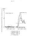
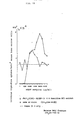
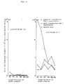
- the culture extract showed the HCGPF activity (Fig. 14).
- pTHCGPF-31 /HB 101 was cultured in a manner similar to Example 4, ii) to give the cell extract. As shown in the figure 14, the culture extract showed the HCGPF activity.
- the obtained recombinant DNA was introduced into Escherichia coli HB 101 strains and a strain having ampicillin resistance was selected. From the separated strain plasmid was obtained. By performing the cleavage test with restriction enzyme and determination of base sequence, bacteria harboring pT13S ⁇ HIL253-HCGPF-19 were selected (pT13S ⁇ HIL253-HCGP-19/HB 101).
- pT13S ⁇ HIL2(53)-HCGPF-19/HB101 was cultured in a manner similar to Example 4 and granules produced in the cells were extracted by the following procedure.
- the cells were collected by centrifugation and 20 mM of Tris-HCl buffer (pH 7.5) containing 30 mM of NaCl was added thereto followed by suspending them. Then 1 mg/ml of lysozyme and 0.05M of EDTA were added thereto. After stirring them, the mixture was allowed to stand in ice water for 1 hour. Then the cells were homogenized by a ultrasonic homogenizer and centrifuged at 10,000 rpm for 5 minutes to recover granules.
- the granules were solubilized with 6M guanidine hydrochloride. As shown in the figure 17, the extract showed the HCGPF activity. Reversed phase HPLC fraction of the extract also showed the HCGPF activity ( Figure 18). Concentrations of the extract were adjusted to give 100 ⁇ g/ml of HIL-2-(53)-HCGPF-protein and 2 M guanidine hydrochloride solution. Then 1 mM of oxidative glutathione and 10 mM of reductive glutathione were added thereto and the mixture was allowed to stand at room temperature for 10 to 16 hours at pH of 8.0.
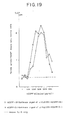
- HCGPF-19 protein After 80 ⁇ g of ⁇ HIL-2-53-HCGPF-19 protein obtained in 50 mM of Tris-HCl buffer (pH 7.8) containing 113 mM of NaCl was reacted with human plasma kallikrein at 37°C for 15 hours, a fraction corresponding to HCGPF-19 was fractionated by reverse phase HPLC. As a result of analysis of amino acid sequence around the N-terminal side thereof with a protein sequencer, it was confirmed that ⁇ HIL-2-53-HCGPF-19 protein was quantitatively converted into HCGPF-19 protein. As shown in the figure 19, HCGPF-19 protein showed the HCGPF activity.
- pT13S ⁇ HIL2(53)-HCGPF-31/HB 101 was cultured in a manner similar to Example 4 and granules produced in the cells were extracted in a manner similar to Example 6.
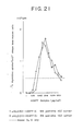
- the granules were solubilized with 6M guanidine hydrochloride. As shown in the figure 21, the extract showed the HCGPF activity. Concentration of the extract was adjusted to give 100 ⁇ g/ml of ⁇ HIL-2-(53)-HCGPF-31 protein and 2 M guanidine hydrochloride solution. Then 1 mM of oxidative glutathione and 10 mM of reductive glutathione were added thereto and the mixture was allowed to stand at room temperature for 10 to 16 hours at pH of 8.0.
- HCGPF-31 protein After 80 ⁇ g of ⁇ HIL2-53-HCGPF-31 protein obtained in 50 mM of Tris-HCl buffer (pH 7.8) containing 113 mM of NaCl was reacted with human plasma kallikrein at 37°C for 15 hours, a fraction corresponding to HCGPF-31 protein was fractionated by reversed phase HPLC. As a result of analysis of amino acid sequence around the N-terminal side thereof with a protein sequencer, it was confirmed that ⁇ HIL2-53-HCGPF-31 protein was quantitatively converted into HCGPF-31 protein. As shown in the figure 19 HCGPF-31 protein showed the HCGPF activity.
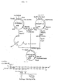
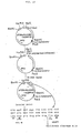
- Plasmid pT13S ⁇ HIL2(20)-HCGPF-19 constructed in Example 6 was cleaved with restriction enzymes Bgl II and Xba I. After treating with DNA polymerase I (Klenow), the system was ligated with T4 DNA ligase ( Figure 22). The obtained recombinant DNA was introduced into Escherichia coli HB 101 strains and a strain having ampicillin resistance was selected. From the separated strain plasmid DNA was obtained.
- bacteria harboring pT13S ⁇ HIL2(20)-HCGPF-19 were selected (pT13S ⁇ HIL2(20)-HCGPF-19/HB 101).
- pT13S ⁇ HIL2(20)-HCGPF-19/HB101 was cultured in a manner similar to Example 4 and granules produced in the cells were extracted by the procedures described in Examples 4 and 6.
- the extract showed the HCGPF activity.
- a molecular weight of the product obtained by SDS polyacrylamide gel electrophoresis was almost identical with one calculated from its amino acid composition.
- As a result of assay for amino acid sequence at the N-terminal side thereof with a protein sequencer it was further confirmed that the product had a sequence of HIL-2.
- Plasmid pT13S ⁇ HIL2(53)-HCGPF-31 constructed in Example 7 was cleaved with restriction enzymes Bgl II and Xba I. After treating with DNA polymerase I (Klenow), the system was ligated using T4 DNA ligase ( Figure 23). The obtained recombinant DNA was introduced into Escherichia coli HB 101 strains and a strain having ampicillin resistance was selected. From the separated strain plasmid DNA was obtained.
- bacteria harboring pT13S ⁇ HIL2(20)-HCGPF-31 were selected (pT13S ⁇ HIL2(20)-HCGPF-31/HB 101).
- pT13S ⁇ HIL2(20)-HCGPF-31/HB101 was cultured and granules produced in the cells were extracted by the procedures described in Example 4 and 6. As shown in the Figure 17, the extract showed the HCGPF activity. Concentrations of the extract were adjusted to give 100 ⁇ g/ml of ⁇ HIL-2(20)-HCGPF-31 protein and 2M guanidine hydrochloride solution. Then 1 mM of oxidative glutathione and 10 mM of reductive glutathione were added thereto and the mixture was allowed to stand at room temperature for 10 to 16 hours at pH of 8.0.
- the obtained recombinant DNA was introduced into Escherichia coli HB 101 strains and a strain having ampicillin resistance was selected. From the separated strain plasmid was obtained. By performing the cleavage test with restriction enzyme and determination of base sequence around the ligated site, bacteria harboring pT13S ⁇ HIL2(11)-HCGPF-19 were selected (pT13S ⁇ HIL2(11)- HCGPF-19/HB 101).
- pT13S ⁇ HIL2(11)-HCGPF-19/HB101 was cultured in a manner similar to Example 4 and granules produced in the cells were extracted by the procedures described in Example 4 and 6. As shown in the figure 17, the extract showed the HCGPF activity. Concentrations of the extract were adjusted to give 100 ⁇ g/ml of ⁇ HIL2(11)-HCGPF-19 protein and 2 M guanidine hydrochloride solution. Then 1 mM of oxidative glutathione and 10 mM of reductive glutathione were added thereto and the mixture was allowed to stand at room temperature for 10 to 16 hours at pH of 8.0.
- ⁇ HIL2(11)-HCGPF-19 protein obtained in 50 mM of Tris-HCl buffer (pH 7.8) containing 113 mM of NaCl was reacted with human plasma kallikrein at 37°C for 15 hours; a fraction corresponding to HCGPF-19 protein was fractionated by reversed phase HPLC.
- ⁇ HIL-2-(11)-HCGPF-19 protein was quantitatively converted into HCGPF-19 protein.
- the HCGPF-19 protein obtained showed the HCGPF activity.
- COS cell culture supernatant containing human HCGPF was prepared as follows. Plasmid pKCR 4-15, 5 ⁇ g, obtained in Example 3 was transfected with monkey COS-7 cells in accordance with the method of Example 2 to give the culture supernatant. The supernatant was concentrated by 5 times using ultrafiltration membrane (YM10) manufactured by Amicon Co., Ltd. to make a sample for measurement of human HCGPF activity.
- YM10 ultrafiltration membrane manufactured by Amicon Co., Ltd.
- mouse bone marrow cell suspension was added to each well by 50 ⁇ l each. After incubation at 37°C for 5 days in the presence of 5% CO2, 1 ⁇ Ci of tritium thymidine was added. After pulsing for 8 hours, ⁇ -radiations incorporated into the cells were counted in a conventional manner.
- this method reveals that the COS supernatant contains the activity of proliferating bone marrow cells singly and the activity of proliferating bone marrow cells in combination with mouse IL 3. None of the activities is recognized in the COS-7 cell supernatant obtained by transfection of cDNA-free plasmid pKCR used for control. Culture extract of Escherichia coli , 6 M guanidine-HCl extract, its fraction purified by reverse phase HPLC and kallikrein cleavage product also showed the HCGPF activity ( Figure 14, 17, 18, 19, 21).
- the HCGPF activity was measured using the same samples as used in Example 11 by the following method.
- the cells were harvested in a conventional manner and then, ⁇ -radiation activity incorporated was determined. As shown in Table 2, significant induction in proliferation was noted although it was weak.
Landscapes
- Chemical & Material Sciences (AREA)
- Health & Medical Sciences (AREA)
- Life Sciences & Earth Sciences (AREA)
- Organic Chemistry (AREA)
- General Health & Medical Sciences (AREA)
- Gastroenterology & Hepatology (AREA)
- Biochemistry (AREA)
- Biophysics (AREA)
- Zoology (AREA)
- Genetics & Genomics (AREA)
- Medicinal Chemistry (AREA)
- Molecular Biology (AREA)
- Proteomics, Peptides & Aminoacids (AREA)
- Toxicology (AREA)
- Micro-Organisms Or Cultivation Processes Thereof (AREA)
- Preparation Of Compounds By Using Micro-Organisms (AREA)
- Peptides Or Proteins (AREA)
Applications Claiming Priority (6)
| Application Number | Priority Date | Filing Date | Title |
|---|---|---|---|
| JP302698/86 | 1986-12-18 | ||
| JP30269886 | 1986-12-18 | ||
| EP87100107 | 1987-01-07 | ||
| EP87100107A EP0232707A1 (de) | 1986-01-09 | 1987-01-07 | Menschlicher Faktor zur Steigerung des hämatopoietischen Zellwachstums |
| JP62002521A JP2569318B2 (ja) | 1986-01-09 | 1987-01-08 | ヒト造血系細胞増殖増強因子をコードする遺伝子 |
| JP2521/87 | 1987-01-08 |
Publications (2)
| Publication Number | Publication Date |
|---|---|
| EP0274560A2 true EP0274560A2 (de) | 1988-07-20 |
| EP0274560A3 EP0274560A3 (de) | 1990-03-21 |
Family
ID=27229962
Family Applications (1)
| Application Number | Title | Priority Date | Filing Date |
|---|---|---|---|
| EP87108782A Withdrawn EP0274560A3 (de) | 1986-12-18 | 1987-06-19 | Menschlicher, das hämatopoietische Zellwachstum aktivierender Faktor |
Country Status (1)
| Country | Link |
|---|---|
| EP (1) | EP0274560A3 (de) |
Cited By (4)
| Publication number | Priority date | Publication date | Assignee | Title |
|---|---|---|---|---|
| US5298603A (en) * | 1985-12-21 | 1994-03-29 | Hoechst Aktiengesellschaft | GM-CSF protein, its derivatives, the preparation of proteins of this type, and their use |
| US5401835A (en) * | 1992-07-31 | 1995-03-28 | Chishti; Athar H. | Human erythroid p55 nucleic acids |
| US5877010A (en) * | 1994-05-02 | 1999-03-02 | University Of Washington | Thymidine kinase mutants |
| US6451571B1 (en) | 1994-05-02 | 2002-09-17 | University Of Washington | Thymidine kinase mutants |
Family Cites Families (1)
| Publication number | Priority date | Publication date | Assignee | Title |
|---|---|---|---|---|
| EP0232707A1 (de) * | 1986-01-09 | 1987-08-19 | Ajinomoto Co., Inc. | Menschlicher Faktor zur Steigerung des hämatopoietischen Zellwachstums |
-
1987
- 1987-06-19 EP EP87108782A patent/EP0274560A3/de not_active Withdrawn
Cited By (4)
| Publication number | Priority date | Publication date | Assignee | Title |
|---|---|---|---|---|
| US5298603A (en) * | 1985-12-21 | 1994-03-29 | Hoechst Aktiengesellschaft | GM-CSF protein, its derivatives, the preparation of proteins of this type, and their use |
| US5401835A (en) * | 1992-07-31 | 1995-03-28 | Chishti; Athar H. | Human erythroid p55 nucleic acids |
| US5877010A (en) * | 1994-05-02 | 1999-03-02 | University Of Washington | Thymidine kinase mutants |
| US6451571B1 (en) | 1994-05-02 | 2002-09-17 | University Of Washington | Thymidine kinase mutants |
Also Published As
| Publication number | Publication date |
|---|---|
| EP0274560A3 (de) | 1990-03-21 |
Similar Documents
| Publication | Publication Date | Title |
|---|---|---|
| EP0118617B1 (de) | Interleukin-2-Polypeptide | |
| EP0091539B1 (de) | Das Polypeptid Interleukin-2 kodierendes Gen, rekombinante, dieses Gen enthaltende DNA, diese rekombinante DNA aufweisende Zelllinien und Verfahren zur Herstellung von Interleukin-2 unter Verwendung der genannten Zellen | |
| JP2515308B2 (ja) | ヒト免疫インタ−フエロン | |
| EP0487512B1 (de) | Herstellung heterodimirer menschlicher Fruchtbarkeitshormone | |
| EP0155549B1 (de) | DNS den menschlichen Tumornekrosisfaktor kodierend und das menschliche Tumornekronisfaktor-Polypeptid | |
| EP0272703B2 (de) | Neues Polypeptid | |
| KR920001377B1 (ko) | 인체 면역 인터페론 단백질의 제조방법 | |
| JP2612149B2 (ja) | α−インターフェロン及びその製造方法 | |
| EP0257406A2 (de) | Rekombinanter B-Zell-Differenzierungsfaktor | |
| JP2677759B2 (ja) | 動物インターフェロンをコードするdna | |
| EP0187991B1 (de) | Substanz mit Antitumorwirkung, Verfahren zu deren Herstellung, diese Substanz enthaltendes Medikament, für diese Substanz kodierendes Gen, dieses Gen enthaltender Vektor und rekombinante Mikroorganismen | |
| JPH06199897A (ja) | リンホカインの生産および精製 | |
| KR960000394B1 (ko) | 백혈구 감소증 치료제의 제조방법 | |
| EP0479071A2 (de) | Polypeptid | |
| US5106733A (en) | Bovine granulocyte-macrophage colony stimulating factor | |
| EP0274560A2 (de) | Menschlicher, das hämatopoietische Zellwachstum aktivierender Faktor | |
| EP0232707A1 (de) | Menschlicher Faktor zur Steigerung des hämatopoietischen Zellwachstums | |
| EP0355720B1 (de) | Verfahren zur Herstellung von motilinähnlichen Polypeptiden und seine Expression | |
| WO1990002183A1 (en) | Production of a novel lymphokine exhibiting differentiation inhibitory activity | |
| EP0315950A2 (de) | Verfahren zur Züchtung von Zellen, welche rekombinantes Protein exprimieren | |
| EP0218431B1 (de) | Polypeptide, deren kodierende Peptide und Methode zur Herstellung dieser Peptide und DNAs | |
| US4851512A (en) | Novel human interleukin-2 polypeptide derivative | |
| EP0211321A2 (de) | Aminosäurezusammensetzung, Verfahren zu deren Herstellung und diese enthaltende medizinische Zusammensetzung | |
| EP0191869A1 (de) | Fisch-kalzitoninabkömmling und dessen herstellung und gensystem dafür | |
| JPH02138224A (ja) | 血小板減少症治療剤 |
Legal Events
| Date | Code | Title | Description |
|---|---|---|---|
| PUAI | Public reference made under article 153(3) epc to a published international application that has entered the european phase |
Free format text: ORIGINAL CODE: 0009012 |
|
| AK | Designated contracting states |
Kind code of ref document: A2 Designated state(s): BE CH DE FR GB IT LI SE |
|
| PUAL | Search report despatched |
Free format text: ORIGINAL CODE: 0009013 |
|
| AK | Designated contracting states |
Kind code of ref document: A3 Designated state(s): BE CH DE FR GB IT LI SE |
|
| STAA | Information on the status of an ep patent application or granted ep patent |
Free format text: STATUS: THE APPLICATION IS DEEMED TO BE WITHDRAWN |
|
| 18D | Application deemed to be withdrawn |
Effective date: 19900922 |
|
| RIN1 | Information on inventor provided before grant (corrected) |
Inventor name: KASHIMA, NOBUKAZUCENTRAL RESEARCH LABORATORIES Inventor name: YAMADA, GEN Inventor name: MATSUI, HIROSHICENTRAL RESEARCH LABORATORIES Inventor name: TANIGUCHI, TADATSUGU Inventor name: HAMURO, JUNJICENTRAL RESEARCH LABORATORIES Inventor name: TAKI, SHINSUKECENTRAL RESEARCH LABORATORIES |