EP0274847A2 - Antigènes-H associes-A, anticorps monoclonaux spécifiques et méthodes les utilisant dans la détermination des groupes sanguins - Google Patents
Antigènes-H associes-A, anticorps monoclonaux spécifiques et méthodes les utilisant dans la détermination des groupes sanguins Download PDFInfo
- Publication number
- EP0274847A2 EP0274847A2 EP87310571A EP87310571A EP0274847A2 EP 0274847 A2 EP0274847 A2 EP 0274847A2 EP 87310571 A EP87310571 A EP 87310571A EP 87310571 A EP87310571 A EP 87310571A EP 0274847 A2 EP0274847 A2 EP 0274847A2
- Authority
- EP
- European Patent Office
- Prior art keywords
- antigens
- see
- chain
- antigen
- type
- Prior art date
- Legal status (The legal status is an assumption and is not a legal conclusion. Google has not performed a legal analysis and makes no representation as to the accuracy of the status listed.)
- Granted
Links
Images

Classifications
-
- C—CHEMISTRY; METALLURGY
- C07—ORGANIC CHEMISTRY
- C07K—PEPTIDES
- C07K16/00—Immunoglobulins [IG], e.g. monoclonal or polyclonal antibodies
- C07K16/18—Immunoglobulins [IG], e.g. monoclonal or polyclonal antibodies against material from animals or humans
- C07K16/34—Immunoglobulins [IG], e.g. monoclonal or polyclonal antibodies against material from animals or humans against blood group antigens
-
- C—CHEMISTRY; METALLURGY
- C07—ORGANIC CHEMISTRY
- C07H—SUGARS; DERIVATIVES THEREOF; NUCLEOSIDES; NUCLEOTIDES; NUCLEIC ACIDS
- C07H3/00—Compounds containing only hydrogen atoms and saccharide radicals having only carbon, hydrogen, and oxygen atoms
- C07H3/06—Oligosaccharides, i.e. having three to five saccharide radicals attached to each other by glycosidic linkages
Definitions
- the present invention relates to antigens present in blood group A and blood group AB erythrocytes but absent in blood group B and blood group O erythrocytes. More specifically, the present invention relates to A-associated H-antigens, monoclonal antibodies specific thereto and methods for employing the same in blood typing.
- Typing of blood is one of the most frequent and important laboratory techniques of medicine. Typing is based on a specific hemagglutination assay using erythrocytes from patients to be typed with human polyclonal antisera, i.e., anti-A and anti-B sera. Due to the variability and limited availability of human reagents, it has been of interest to develop standardized reagents which can be supplied in unlimited quantities. Recent developments in hybridoma antibody technology have enabled the development of various monoclonal antibodies with anti-A properties. However, these monoclonal anti-A antibodies generally fail to agglutinate the minor blood group A subgroups.
- Blood group A has been subgrouped into the major subgroups A1 and A2 and minor subgroups A 3-5 and A x (see Race, R.R. et al. Blood Groups in Man , 6th Ed., pp. 9-13, Blackwell Scientific Publications, Oxford (1975)).
- the chemical difference between the two major phenotypes A1 and A2 has not been clarified, although several studies have indicated both quantitative (see Makela, O. et al. J. Immunol. , 102 :763-771 (1969) and Economidou, J. et al, Vox Sang. , 12 :321-328 (1967)) and qualitative (see Economidou, J. et al, Vox Sang.
- An object of the present invention is to develop antigens which are associated with minor blood group A erythrocytes.
- Another object of the present invention is to use these antigens to generate reagents, i.e., monoclonal antibodies with anti-A properties, useful for blood group A typing.
- Still another object of the present invention is to provide a method of using the monoclonal antibodies for blood group A typing.
- the hapten of the A-associated H-antigen is a combination of an H-antigen linked onto an A-antigen.
- the hapten of the A-associated H-antigen constitutes a unique antigenic entity associated specifically with minor A subgroups,
- the hapten of the antigens of the present invention is present in blood group A and group AB erythrocytes but absent in blood group B and O erythrocytes.
- the present invention is an improvement in the technology disclosed in Clausen, H. et al, J. Biol. Chem. , 261 :1380-1387 (1986) which is incorporated by reference herein in its entirety.
- n is 0 or a positive integer.
- the specific length of the polysaccharide represented by (X) n is not critical to the present invention as long as the hapten of the A-associated H-antigen is accessible for antibody recognition. From an economic and practical standpoint n is no greater than about 30 and preferably n is 0 to 10. Where n is greater than 0, the sugar residues employed may be the same or different. Further, when n is greater than 0, the sugar chain may be branched or unbranched.
- sugar residues which can be employed in the present invention are not critical thereto.
- examples of the sugar residues which can be employed in the present invention include a residue of ⁇ -D-galactose, ⁇ -D-glucose, ⁇ -D-N-acetylglucosamine, ⁇ -D-N-acetylgalactosamine, ⁇ -L fucose, ⁇ -D-galactose, ⁇ -D-acetylgalactosamine, ⁇ -D-mannose and ⁇ -D-mannose.
- the sugar residues can be those found in A-associated H-antigens isolated from A and AB erythrocytes or can be additional sugar residues which are added to the hapten of the A-associated H-antigen by well known means using well known enzymes such as glycosyltransferases.
- polypeptides which can be employed in the present invention are not critical thereto and can be any naturally occurring or synthetic polypeptide.
- examples of the naturally occurring polypeptides which can be employed in the present invention include bovine serum albumin, keyhold limpet hemocyanin and egg ovalbumin.
- the particular primary amino group containing lipids which can be employed in the present invention are not critical thereto.
- Examples of the primary amino group containing lipids which can be employed in the present invention include ceramide and long chain amines such as n-octadecylamine and n-hexadecylamine.
- Ceramides are the preferred primary amino group containing lipids employed in the present invention because they are normally attached to haptens in glycolipids.
- the particular primary amino group containing supports which can be employed in the present invention are not critical thereto.
- Examples of the primary amino group containing supports which can be employed in the present invention include amino derived silica and amino-derived porous glass beads.
- the A-associated H-antigens of the present invention are the immediate precursors to the repetitive A antigen (Type 3 chain A) illustrated below (see Clausen, H. et al, Proc. Natl. Acad. Sci. , USA , 82 :1199-1203 (1985), which has been found to be associated with the major A, subgroup: wherein R1 represents a glycolipid and/or glycopeptide.
- the A-associated H-antigens of the present invention are structurally distinct from other types of H antigens which are distributed in cells irrespective of blood group status as discussed below.
- the major H antigens which are distributed in various human tissues, including erythrocytes, irrespective of blood group ABO status include Type 1 chain H, Type 2 chain H (see Hakomori, S., Semin. Hematol. , 18 :39-62 (1981); Type 3 chain H (O-linked) (see Donald, A.S.R., Eur. J. Biochem. , 120 :243-249 (1981); and Type 4 chain H (Globo-H) (Kannagi, R. et al, J. Biol. Chem. , 258 :8934-8942 (1983)).
- Type 1 chain H, Type 2 chain H and Type 3 chain H, and Type 4 chain H all of which have the external Gal ⁇ 1 component but lack the internal which is distinctive of A antigens are illustrated below: wherein R1 represents a glycolipid and/or glycopeptide.
- Type 1 chain H in secretions is related to the H and Se genes while that of the Type 2 chain H in erythrocytes is dependent only on the H gene (see Kumazaki, T. et al, Proc. Natl. Acad. Sci. USA , 81 :4193-4197 (1984) and Graham, H.A. et al, Human Blood Groups : 5th International Convocation on Immunology , Buffalo, New York, (1976), Eds. Mohn, J.F. et al, pp. 257-267, Karger, Basel).
- the H-antigen in human erythrocytes is carried mainly on the Type 2 chain H variety.
- Type 3 chain H (O-linked) is mainly found on mucin type glycoproteins.
- Type 4 chain H (Globo-H), in contrast, is only found on glycolipids in tissue and erythrocytes (see Kannagi, R. et al, FEBS Lett. , 175 :397-401 (1984)).
- the quantity of H-antigen in various tissues is much higher in blood group O than in blood group A or B individuals.
- Globo-H antigen is absent or present in only a very small quantity of blood group A1 erythrocytes, while it is present in blood group A2 erythrocytes in the same quantity as in blood group O erythrocytes (see Clausen, H. et al, Biochem. Biophys. Res. Commun. , 124 :523-529 (1984)).
- the A-associated H-antigens of the present invention are therefore different from the previously known H-antigens.
- the A-associated H-antigens can be isolated from blood group A or blood group AB erythrocytes or epithelial tissue. In general, this can be carried out by first obtaining a total lipid extract from the whole blood cells or epithelial tissue (see Kannagi, R. et al, J. Biol. Chem. , 257 :14865-14874 (1982)).
- glycolipid fraction is isolated from the total lipid extract (see Hakomori, S. et al, Glycolipid Methodology , pp. 13-47, Ed. Witting, L.A., American Oil Chemists' Society, Champaign, Illinois (1976)).
- HPLC high performance liquid chromatography
- A-associated H-antigens in the fractionated neutral glycolipid fraction can be ascertained by immunostaining with the monoclonal antibody MBr1 (see Clausen, H. et al, Proc. Natl. Acad. Sci. , USA , 82 :1199-1203 (1985) and Menard, S. et al, Cancer Res. , 43 :1295-1300 (1983)).
- A-associated H-antigens can also be obtained by the enzymatic degredation of the Type 3 chain A b having the following structure:
- Type 3 chain A b can be isolated as described in detail in Example 1 hereinafter.
- Hydrolysis of the terminal D-N-acetylgalactosamine can be carried out by incubating 200 ⁇ g of the glycolipid with 0.1 units/ml of ⁇ -galactosaminidase (chicken liver, obtained by Dr. Jack Goldstein of the New York Blood Center) in a reaction mixture containind 100 ⁇ g of sodium deoxytaurocholate in 0.1 M acetic buffer (pH 4.5).
- the A-associated H-antigens obtained by the above-described isolation procedures have the following glycosphingolipid structure.
- the hapten of the A-associated H-antigen can be obtained from the above glycosphingolipid structure and linked to the variable carrier by a variety of well known methods.
- the neutral glycolipid is first selectively exidized at the 3-position of the sphingosine (see Kishimoto, Y. et al, Arch. Bichem. Biophys. , 161 :426-434 (1974); Iwamori, M. et al, Biochem. Biophys. Acta. , 380 :308-319 (1975); and Iwamori, M. et al, J. Lipid Res. , 16 :332-336 (1975)).
- glycosphingolipid is dissolved, at a final concentration of 10 mg/ml, in dioxane or tetrahydrofuran and then reacted with a molar excess of about 20:1 to 50:1 of 2,3-dichloro-5,6-dicyanobenzoquinone to glycosphingolipid at 37 ⁇ 5°C for about 24 hrs.
- the resulting 3-keto-glycosphingolipid is isolated using low pressure chromatography on silica using chloroform/methanol (2:1 (v/v)) as the mobile phase or HPLC on silica using isopropyl alcohol/hexane/water in a gradient of 55:40:5 (v/v/v) to 55:20:25 (v/v/v).
- the isolated 3-keto-glycosphingolipid is treated under very mild basic conditions to release the free reducing oligosaccharide by ⁇ -elimination (chloroform/0.01 N Na2CO3 in methanol (2:1 (v/v)) at 37 ⁇ 5°C for 1 hour (see Iwamori, M.
- the resulting oligosaccharide can then be bonded, via its reducing end, to any suitable primary amino group containing material, e.g., a polypeptide such as bovine serum albumin, a primary amino group containing support such as an amino derived silica or porous glass beads, or a primary amino group containing lipid such as n-octadecylamine or n-hexadecylamine, by reductive amination using 10:1 to 50:1 molar excess of sodium cyanoborohydride in an aqueous solvent.
- a polypeptide such as bovine serum albumin
- a primary amino group containing support such as an amino derived silica or porous glass beads
- a primary amino group containing lipid such as n-octadecylamine or n-hexadecylamine
- the glycosphingolipid is per-acetylated with a greater than 500 molar excess of pyridine/acetic anhydride (2:1 (v/v)) at room temperature overnight or at about 80°C for 1 hr.
- the double bond of the sphingosine is then cleaved using 0.2 M periodic acid in methanol and 5.0% (w/v) osmium tetroxide in diethyl ether.
- 1-3 mg of the acetylated glycolipid is evaporated to dryness under N2 using toluene to azeotrope off the excess pyridine and acetic anhydride.
- the resulting product is taken up in 0.5 ml of methanol and then 0.2 ml of 0.2 M periodic acid in methanol is added, followed by the addition of 25 ml of 5.0% (w/v) of osmium tetroxide in diethyl ether. The mixture is left at 5°C overnight and excess reagent is destroyed with a drop of glycerol. Then, 6.0 ml of chloroform/methanol (2:1 (v/v)) and 1.5 ml water are added, the organic phase is collected and washed four times with 2.5 ml of chloroform/methanol/water (1:10:10 (v/v/v)) and then dried under a N2 stream (see MacDonald, D.L.
- the resulting derivative has the following structure:
- the treatment of this aldehyde using 50 ⁇ l of 0.2 M sodium methoxide in 0.3 ml of methanol de-O-acetylates the oligosaccharide moiety and simultaneously frees it, by a base catalyzed migration of the aldehyde double bond followed by ⁇ -elimination.
- the resulting oligosaccharide can then be coupled, as described above, to a suitable polypeptide, primary amino group containing lipid or primary amino group containing support.
- the aldehyde acetate can be oxidized with performic acid to an acid form. More specifically, the acetylated glycolipid aldehyde is treated with performic acid prepared by mixing 15 ml of 30% (v/v) hydrogen peroxide with 1.0 ml of 80% (v/v) formic acid, which is allowed to stand for two hours. 0.2 ml of this reagent is added to the acetylated glycolipid aldehyde and allowed to stand for about 20 hrs at room temperature (see MacDonald, D.L. et al, J. Lipid Res. , 21 :642-645 (1980)).
- the resulting product can then be coupled to a suitable polypeptide, primary amino group containing lipid or primary amino group containing support with N-hydroxy-succinimide and dicyclohexyl-carbodiimide in dioxane, followed by de-O-acetylation with sodium methoxide in methanol. More specifically, 10 mg of acetylated glycolipid acid is dissolved in 1.0 ml of dioxane.
- the activated ester is formed by adding 5.0 mg of N-hydroxy succinimide dissolved in 0.5 ml of dioxane plus 5.0 mg of dicyclohexylcarbodiimide dissolved in 1.0 ml of dioxane.
- the resulting mixture is incubated at 15 ⁇ 1°C for 30 min. Then, 1.0 g of alkylamine glass beads (Corning CPG-1350) or equivalent material containing 60 ⁇ eq of NH2 groups per gram and 5.0 ml dioxane are added and mixed gently at room temperature for about 72 hours. One drop of glacial acetic acid is then added to derivatize the remaining amino groups on the glass beads and mixing is continued for about 24 hours. After washing the glass with dioxane, the glass immobilized glycolipid is suspended in 10 ml chloroform/methanol (2:1 (v/v)) and 2.0 ml of 0.5% (w/v) sodium methoxide in methanol are added.
- the glass is filtered and washed with chloroform/methanol (2:1 (v/v)), washed with water and washed with phosphate buffer (pH 7.0).
- the glass beads can then be stored in phosphate buffer (pH 7.0) containing sodium azide.
- the resulting complex has the following structure: (see Young, W.W. et al, J. Lipid Res. , 20 :275-278 (1979)).
- the per-o-acetylated glycolipid acid can also be produced directly from the glycosphingolipid, following per-o-acetylation, by treatment with potassium permanganate in acetone at room temperature overnight (see MacDonald, D.L. et al, J. Lipid Res. , 21 :642-645 (1980)).
- the A-associated H-antigens of the present invention can be used to develop monoclonal antibodies for use in blood typing.
- the monoclonal antibodies can be prepared by well known means as described in Kohler, G. et al, Nature , 256 :495-496 (1975) and Clausen, H. et al, Biochem. , 24 :6190-6194 (1985).
- Monoclonal antibodies directed to the A-associated H-antigens can be used alone or in combination with other anti-A antibodies for identification of blood group A erythrocytes including minor A subgroups.
- Blood typing can be carried out according to standard blood banking methods (see Kabat, E.A. et al, Experimental Immunochemistry , 2d Ed. Thomas Publishers, Springfield, Illisois (1967)). This invoves an agglutination test where the antibody is incubated with a patient's erythrocytes for varying times and at different temperatures. Agglutination, i.e., clustering of cells due to specific antigen-antibody reactions, is thereafter observed macroscopically (see Clausen , H et al, Proc. Natl. Acad. Sci. , USA , 82 :1199-1203 (1985)).
- the monoclonal antibodies can also be employed in other immunoassays for identification of blood groups. For example, immunohistology of epithelial tissues where antigens can be localized in relation to epithelial architecture (see Dabelsteen, E. et al, J. Invest. Dermatol. , 82 :13-17 (1984)); immune inhibition of cellular secretions such as saliva, gastric and pancreatic juices (see Kabat, E.A. et al, Experimental Immunochemistry , 2d Ed.
- the A-associated H-antigens of the present invention are relatively abundant in the minor A subgroups and that antibodies directed towards these antigens should preferentially detect minor blood group A erythrocytes. It is therefore desirable to include antibodies directed to other A antigens, i.e., Type 2 chain A-antigen and Type 3 chain A-antigens, which are relatively abundant on the major A subgroups in a test for blood group A typing.
- a antigens i.e., Type 2 chain A-antigen and Type 3 chain A-antigens
- pooled outdated human whole type A blood was lysed in ice-cold tap water containing 0.2% (v/v) of acetic acid and whole blood cell membranes, including those of white cells, were prepared by continuous centrifugation on a Sharpless centrifuge.
- the total lipids were extracted with isopropyl alcohol/hexane/water (55:25:20 (v/v/v)) (see Kannagi, R. et al, J. Biol. Chem. , 257 :14865-14874 (1982)).
- the total lipid extract was evaporated to dryness in a rotary evaporator.
- the upper phase glycolipid fraction was evaporated to dryness and dialyzed through a Spectraper 3 dialysis tube (Spectrum Medical Industries, Los Angeles, CA) against deionized water (see Hakomori, S. et al, Glycolipid Methodology , pp. 13-47, Ed. Witting, L.A., American Oil Chemists' Society, Champaign, Illinois (1976)).
- Neutral glycolipids were separated from the total upper phase glycolipid fraction by DEAE-Sephadex (A-25) chromatography using chloroform/methanol/water (30:60:80 (v/v/v)) (see Yu, R.K. et al, J. Lipid Res. , 13 :680-686 (1972)).
- the neutral glycolipid fraction was further fractionated by HPLC on a 1 x 50-cm column of Iatrobeads 6RS-8060 (60 ⁇ m particles; Iatron Chemical Co., Tokyo, Japan) (see Watanabe, K. et al, Biochem. , 22 :1020-1024 (1981) and Kannagi, R. et al, J. Biol. Chem.
- HPTLC high performance thin-layer chromatography
- MBr1 antibody reacts with Type 3 chain H and Type 4 chain H-antigens including the A-associated H-antigens of the present invention but not with Type 1 chain H or Type 2 chain H-antigens (see Brenner, E.G. et al, J. Biol. Chem. , 259 :14773-14777 (1984), Clausen, H. et al, Proc. Natl. Acad. Sci. , USA , 82 :1199-1203 (1985) and Clausen, H. et al, J. Biol. Chem. , 261 :1380-1387 (1986)).
- HPTLC-immunostaining was performed as described in Magnani, J.L. et al, Anal. Biochem. , 109 :399-402 (1980). More specifically, after the neutral glycolipids had been separated on HPTLC (Baker J.T.-HPFTLC Plate, J.T. Baker Chemical Co., Philipsburg, NJ) as described above, the plates were blocked by immersion in 5% (w/v) bovine serum albumin followed by sequential incubation with MBr1, rabbit anti-mouse Ig and 125I-protein A. The plates were then subjected to autoradiography.
- fractions 54 and 55 contained A-associated H-antigens, as well as Type 2 chain H2 illustrated below, Type 2 chain A b illustrated below and Type 3 chain A b illustrated below.
- Fractions 54 and 55 described above were then rechromatographed on high-pressure HPLC through Iatrobeads 6RS-8010 column using a gradient of isopropyl alcohol/hexane/water from 55:38:7 (v/v/v) to 55:30:15 (v/v/v) (see Watanabe, K. et al, Biochem. , 22 :1020-1024 (1981) and Kannagi, R. et al, J. Biol. Chem. , 258 :8934-8942 (1983)).
- the total volume of the eluent was 200 ml and was collected over 200 fractions for 200 min (1.0 ml/min/fraction).
- Fractions 54 and 55 containing only A-associated H-antigens and Type 2 chain H2 was acetylated and A-associated H-antigens were identified by two-dimensional HPTLC-immunostaining with Mbr1 antibody after deacetylation in situ .
- Acetylation was performed in pyridine/acetic acid (2:1 (v/v)) at room temperature for 18 hours and the acetates (see Saito, T. et al, J. Lipid. Res. , 12 :257-259 (1971)) were separated on HPTLC plates in dichloroethane/acetone/water (50:50:0:1 (v/v/v)) in a first direction and dichloroethane/acetone/water (50:50:1.0 (v/v/v)) in a second direction. Deacetylation was conducted directly on the plates by incubating the plates with 0.2% (w/v) sodium methoxide in methanol. Then, immunostaining was carried out using monoclonal antibody MBr1 as described in Clausen et al, Proc. Natl. Acad. Sci. , USA , 82 :1199-1203 (1985)).
- the A-association H-glycolipids so identified were thereafter purified by preparative HPTLC on Merck HPTLC plates (Silica Gel 60, Merck) in a solvent of dichloroethane/acetone/water (50:50:0.1 (v/v/v)).
- the location of glycolipids on HPTLC was detected by Primulin spray (Aldrich) followed by observation under UV light (see Skipski, V.P., Methods Enzymol. , 34 :396-425 (1975)).
- HPTLC immunostaining with monoclonal antibodies was performed before and after successive enzymatic degradation (see Clausen, H. et al, J. Biol. Chem. , 261 :1380-1387 (1986)).
- the monoclonal antibodies employed for HPTLC immunostaining were IB2 (see Young, W.W. et al, J. Biol. Chem. , 256 :10967-10972 (1981)), anti-T (Chembiomed, Inc., Edmonton, Alberta Canada) and AH16 monoclonal antibodies (see Abe, K. et al, J. Immunol. , 132 :1951-1954 (1984)).
- Monoclonal antibodies IB2, anti-T and AH16 have binding specificity for the following structures, respectively: wherein R1 represents a glycolipid and/or glycopeptide.
- the enzymes used for the enzymatic degradation were bovine liver ⁇ -L-fucosidase (Sigma Co.) and ⁇ -galactosidase of Charonia lampas (Seikagaku Kogyo Co., Ltd., Tokyo, Japan). Hydrolysis was performed by incubation of 10 ⁇ g of glycolipid in 20 ⁇ l of 0.1 unit/ml of enzyme in the presence of 30 ⁇ g of sodium deoxytaurocholate in 0.1 M acetate buffer (pH 4.5) (see Hakomori, S. et al, J. Biol. Chem. , 246 :2271-2277 (1971)).
- the reaction mixture was poured over a "Bond Elut” column, washed with distilled water and eluted with methanol. DEAE-Sephadex and HPLC column chromatography was then carried out as described above.
- the final glycolipid preparation was free of salt, detergent and protein.
- the A-associated H-antigens obtained from A erythrocytes were characterized as having a slightly slower HPTLC mobility that Type 2 chain H2 but faster than Type 2 chain A b .
- the A-associated H-antigens obtained from A erythrocytes were degraded by bovine liver ⁇ -L-fucosidase to a component with the same HPTLC mobility as globo-H antigen (see Kannagi, R. et al, FEBS Lett. , 175 :397-401 (1984)), which was not reactive with 1B2 and anti-T monoclonal antibodies on HPTLC immunostaining, whereas Type 2 chain H2 was stained by 1B2 after defucosylation.
- A-associated H-antigens obtained from A erythrocytes was found to be very difficult and they were only partially degraded by ⁇ -galactosidase of C. lampas , although a complete cleavage was possible by repeated incubation.
- the ⁇ -Gal residue attached to the ⁇ -GalNAc residue of the globoside was also resistant to various ⁇ -galactosidases (see Kannagi, R. et al, J. Biol. Chem. , 258 :8934-8942 (1983)).
- the degradation product after ⁇ -galactosidase showed the same HPTLC mobility as Aa glycolipid, and in addition, was stained by AH16 monoclonal antibody, indicating that A-associated H-antigens obtained from A erythrocytes contain an internal Aa structure.
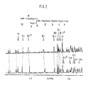
- Proton nuclear magnetic resonance (1H NMR) spectra of the isolated glycolipids were recorded at 500 MHz with a Bruker WM-500 spectrometer equipped with an Aspect 2000 computer and pulse programmer, operating in the Fourier transform mode with quadrature detection.
- the spectra were recorded at 308 ⁇ and 328 ⁇ 2K on deuterium-exchanged samples (200-400 ⁇ g) dissolved in 0.4 ml of dimethyl sulfoxide/d6 containing 2% (v/v) D2O (see Dabrowski, J. et al, Biochem. , 19 :5652-5658 (1980)) and 1% (v/v) tetramethylsilane as a chemical shift reference.
- the spectrum of the defucosylated compound is readily interpreted as the addition of 1 ⁇ -Gal residue to Type 2 chain Aa (see Clausen, H. et al, Biochem. , 24 :3578-3586 (1985)), and the anomeric signals assigned on that basis.
- Table I The large downfield shift of the terminal ⁇ -Gal H-1 resonance on ⁇ 1 ⁇ 2 fucosylation is expected for the Gal ⁇ 1 ⁇ 3HexNAc structure, as seen for Type 1 chain lacto- (see Dabrowski, J. et al, Arch. Biochem. Biophys. , 210 :405-411 (1981)) and globo H-series (see Kannagi, R. et al, J. Biol. Chem.
- this resonance shows a negative temperature-shift coefficient, which is preserved in the H structure, and Type 3 chain A b , helping to confirm the assignment (see Figure 2) (see Clausen, H. et al, Proc. Natl. Acad. Sci , USA , 124 :523-529 (1984)).
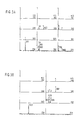
- A-associated H-antigens obtained from A erythrocytes was further supported by methylation analysis with selected ion chromatography of partially O-methylated alditol and hexosaminitol acetates from the hydrolysate of the permethylated glycolipid. More specifically, in Figures 3A and 3B, glycolipids were methylated (see Hakomori, S. J. Biochem. , 55 :205-208 (1964)) and hydrolyzed, partially O-methylated hexitol and hexosaminitol acetate derivatives, prepared as previously described in Bremer, E.G. et al, J. Biol. Chem.
- Peaks 1-7 were identified as follows: Peak 1 - 2,3,4-tri-O-Me-Fuc; Peak 2 - 2,3,6-tri-O-Me-Glc; Peak 3 - 2,4,6-tri-O-Me-Gal; Peak 4 - 3,4,6-tri-O-Me_Gal; Peak 5 - 4,6-di-O-Me-Gal; Peak 6 - 3,6-di-O-Me-GlcNAcMe; and Peak 7 - 4,6-di-O-Me-GalNAcMe.
- the peak identifications were made on the basis of appropriate MH+, (MH-32)+ and (MH-60)+ ions and retention indices, compared with authentic standards
- Example 1 As shown by the (1H NMR) spectras, methylation analysis and enzymatic degradation described in Example 1, the A-associated H-antigens obtained in Example 1 from A erythrocytes have the following structure:
- the A-associated H-antigens obtained in Example 1 above were absorbed onto the surface of Salmonella sp. serotype minnesota (ATCC No. 9700) by lyophilization in order to provide a carrier for the glycolipid hapten (see Hakomori, S., "Monoclonal Antibodies Directed to Cell Surface Carbohydrates" in Monoclonal Antibodies and Functional Cell Lines , Eds. Kennett, R.H. and Beshtol, K.B.. McKearn, T.J., Plenum Publishing Corporation, pages 67-100 (1985)).
- Two monoclonal antibodies directed toward the A-associated H-antigen preparation of Example 2 above were produced by the methods of Kohler, G. et al, Nature , 256 :495-497 (1975) using the modification as described in Clausen, H. et al, Biochem. , 24 :6190-6194 (1985). More specifically, 2 ⁇ g of the A-associated H-antigens coated on acid treated Salmonella sp. serotype minnesota were used for immunization of one BALB/c mouse (3 months old). The mouse received five injections with 5-7 day intervals therebetween. Three days after the last injection, the spleen was removed from the mouse and the spleen cells were found with SP/2-0 as described in Clausen, H.
- Hybridomas were screened by a solid-phase radioimmunoassay using the A-associated H-antigens obtained from A erythrocytes and a panel of control glycolipids.
- the initial glycolipid concentration was 100 ng, with 300 ng of cholesterol and 500 ng of lecithin.
- the monoclonal antibodies were used as an undiluted culture supernatant.
- Hybridomas found to produce the desired monoclonal antibodies were cloned twice by limiting dilution and propagated in culture or in mice. Using this procedure, hybridomas designated HH14 and HH11 were obtained. These hybridomas have been deposited at the Americal Type Culture Collection under ATCC Nos. HB9299 and HB9298, respectively.
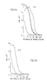
- Monoclonal antibodies from HH11 and HH14 were then harvested from culture medium or ascites fluid and tested for binding specificity using a solid phase radioimmunoassay as described above. The results are shown in Figures 4A and 4B.
- the designations are as follows: ⁇ Type 3 chain A; ⁇ A-associated H-antigen; o Type 4 chain A; and ⁇ Type 4 chain H.
- Figure 4A demonstrates that monoclonal antibodies obtained from HH11 were found to react with the A-associated H-antigen, however, binding was also found to the Type 3 chain A , Type 4 chain A and Type 4 chain H antigens.
- Figure 4B demonstrates that the monoclonal antibody obtained from HH14 was found to be highly specific for A-associated H-antigens, although weak binding was observed to Type 4 chain H. However, no binding was found to any other H antigens (e.g., Type 1 chain H or Type 2 chain H) or any A antigens (e.g., Type 1 chain A, Type 2 chain A, Type 2 chain galactosyl-A, Type 3 chain A or Type 4 chain A) or Fucosyl-GM1 antigens. Therefore, monoclonal antibodies obtained from HH14 are preferred because of their selective binding specificity.
- H antigens e.g., Type 1 chain H or Type 2 chain H
- any A antigens e.g., Type 1 chain A, Type 2 chain A, Type 2 chain galactosyl-A, Type 3 chain A or Type 4 chain A
- Fucosyl-GM1 antigens e.g., Fucosyl-GM1 antigens. Therefore, monoclonal antibodies obtained from HH14
Landscapes
- Chemical & Material Sciences (AREA)
- Health & Medical Sciences (AREA)
- Organic Chemistry (AREA)
- Molecular Biology (AREA)
- Life Sciences & Earth Sciences (AREA)
- Immunology (AREA)
- General Health & Medical Sciences (AREA)
- Genetics & Genomics (AREA)
- Biochemistry (AREA)
- Hematology (AREA)
- Engineering & Computer Science (AREA)
- Biotechnology (AREA)
- Biophysics (AREA)
- Medicinal Chemistry (AREA)
- Proteomics, Peptides & Aminoacids (AREA)
- Medicines Containing Antibodies Or Antigens For Use As Internal Diagnostic Agents (AREA)
- Preparation Of Compounds By Using Micro-Organisms (AREA)
- Peptides Or Proteins (AREA)
Applications Claiming Priority (2)
| Application Number | Priority Date | Filing Date | Title |
|---|---|---|---|
| US07/002,467 US4857639A (en) | 1987-01-12 | 1987-01-12 | A-associated H-antigens, monoclonal antibodies specific thereto and methods for employing the same in blood typing |
| US2467 | 1993-01-08 |
Publications (3)
| Publication Number | Publication Date |
|---|---|
| EP0274847A2 true EP0274847A2 (fr) | 1988-07-20 |
| EP0274847A3 EP0274847A3 (en) | 1989-06-07 |
| EP0274847B1 EP0274847B1 (fr) | 1993-03-31 |
Family
ID=21700908
Family Applications (1)
| Application Number | Title | Priority Date | Filing Date |
|---|---|---|---|
| EP87310571A Expired - Lifetime EP0274847B1 (fr) | 1987-01-12 | 1987-12-01 | Antigènes-H associes-A, anticorps monoclonaux spécifiques et méthodes les utilisant dans la détermination des groupes sanguins |
Country Status (4)
| Country | Link |
|---|---|
| US (1) | US4857639A (fr) |
| EP (1) | EP0274847B1 (fr) |
| DE (1) | DE3785175T2 (fr) |
| DK (1) | DK651787A (fr) |
Cited By (2)
| Publication number | Priority date | Publication date | Assignee | Title |
|---|---|---|---|---|
| WO2007135571A3 (fr) * | 2006-03-23 | 2008-06-19 | Absorber Ab | Antigènes de groupes sanguins de types différents pour applications diagnostiques et thérapeutiques |
| EP4245764A1 (fr) * | 2022-03-18 | 2023-09-20 | RemAb Therapeutics SL | Nouveaux dérivés glucidiques comme mimétiques des antigènes des groupes sanguins a et b |
Families Citing this family (4)
| Publication number | Priority date | Publication date | Assignee | Title |
|---|---|---|---|---|
| US5030723A (en) * | 1988-05-31 | 1991-07-09 | The Biomembrane Institute | Long-chain glycolipid structure |
| US6800468B1 (en) * | 1998-11-13 | 2004-10-05 | Henrik Clausen | UDP-galactose: β-N-acetyl-glucosamine β1,3galactosyltransferases, β3Gal-T5 |
| WO2000029558A1 (fr) * | 1998-11-13 | 2000-05-25 | Henrick Clausen | UDP-GALACTOSE: β-$I(N)-ACETYLE-GLUCOSAMINE β1,3GALACTOSYLE-TRANSFERASES, β3GAL-T5 |
| US6997031B2 (en) | 2003-05-01 | 2006-02-14 | Waters Corporation | Fraction collector for composition analysis |
Family Cites Families (7)
| Publication number | Priority date | Publication date | Assignee | Title |
|---|---|---|---|---|
| US4029762A (en) * | 1971-11-17 | 1977-06-14 | Max-Planck-Gesellschaft Zur Forderung Der Wissenschaften E.V. | Lipid A-preparation |
| SE8001748L (sv) * | 1980-03-05 | 1981-09-06 | Kaellenius Gunilla | Kompositioner for terapeutisk eller diagnostisk anvendning jemte forfarande for terapeutik |
| US4330619A (en) * | 1980-08-14 | 1982-05-18 | New York Blood Center, Inc. | Enzymatic conversion of red cells for transfusion |
| US4343797A (en) * | 1981-01-05 | 1982-08-10 | Ecanow Charles S | Synthetic whole blood and a method of making the same |
| JPS59161320A (ja) * | 1983-03-04 | 1984-09-12 | Maruyama Chisato | リポ多糖体およびその製造法 |
| US4609627A (en) * | 1983-08-01 | 1986-09-02 | New York Blood Center, Inc. | Enzymatic conversion of certain sub-type A and AB erythrocytes |
| JPS6057254A (ja) * | 1983-09-09 | 1985-04-03 | Dainabotsuto Kk | 免疫学的測定法による糖脂質の定量方法 |
-
1987
- 1987-01-12 US US07/002,467 patent/US4857639A/en not_active Expired - Fee Related
- 1987-12-01 EP EP87310571A patent/EP0274847B1/fr not_active Expired - Lifetime
- 1987-12-01 DE DE87310571T patent/DE3785175T2/de not_active Expired - Fee Related
- 1987-12-11 DK DK651787A patent/DK651787A/da not_active Application Discontinuation
Non-Patent Citations (5)
| Title |
|---|
| JOURNAL OF BIOLOGICAL CHEMISTRY, vol. 259, no. 23, 10th December 1984, pages 14773-14777, The American Society of Biological Chemists, Inc., US; E.G. BREMERT et al.: "Characterization of a glycosphingolipid antigen defined by the monoclonal antibody MBr1 expressed in normal and neoplastic epithelial cells of human mammary gland" * |
| JOURNAL OF BIOLOGICAL CHEMISTRY, vol. 261, no. 3, 25th January 1986, pages 1380-1387, The American Society of Biological Chemists, Inc., US; H. CLAUSEN et al.: "Novel blood group H glycolipid antigens exclusively expressed in blood group A and AB erythrocytes (type 3 chain H). I. Isolation and chemicl characterization" * |
| JOURNAL OF BIOLOGICAL CHEMISTRY, vol. 261, no. 3, 25th January 1986, pages 1388-1392, The American Society of Biological Chemists, Inc., US; H. CLAUSEN et al.: "Novel blood group H glycolipid antigens exclusively expressed in blood group A and AB erythrocytes (type 3 chain H). II. Differential conversion of different H substrates by A1 and A2 enzymes, and type 3 chain H expression in relation to secretor status" * |
| JOURNAL OF IMMUNOLOGY, vol. 132, no. 4, April 1984, pages 1951-1954, The American Association of immunologists, US; K. ABE et al.: "The antibody specific to type 1 chain blood group A determinant" * |
| PROCEEDINGS OF THE NATIONAL ACADEMIC OF SCIENCES, USA, vol. 82, February 1985, pages 1199-1203; H. CLAUSEN et al.: "Repetitive A epitope (type 3 chain A) defined by blood group A1- specific monoclonal antibody TH-1: Chemical basis of qualitative A1 and A2 distinction" * |
Cited By (7)
| Publication number | Priority date | Publication date | Assignee | Title |
|---|---|---|---|---|
| WO2007135571A3 (fr) * | 2006-03-23 | 2008-06-19 | Absorber Ab | Antigènes de groupes sanguins de types différents pour applications diagnostiques et thérapeutiques |
| US7897328B2 (en) | 2006-03-23 | 2011-03-01 | Absorber Ab | Blood group antigens of different types for diagnostic and therapeutic applications |
| EP2362223A3 (fr) * | 2006-03-23 | 2012-03-07 | Absorber AB | Antigènes de groupes sanguins de types différents pour applications diagnostiques et thérapeutiques |
| EP2362224A3 (fr) * | 2006-03-23 | 2012-03-14 | Absorber AB | Antigènes de groupes sanguins de types différents pour applications diagnostiques et thérapeutiques |
| US8404456B2 (en) | 2006-03-23 | 2013-03-26 | Absorber Ab | Blood group antigens of different types for diagnostic and therapeutic applications |
| EP4245764A1 (fr) * | 2022-03-18 | 2023-09-20 | RemAb Therapeutics SL | Nouveaux dérivés glucidiques comme mimétiques des antigènes des groupes sanguins a et b |
| WO2023175140A1 (fr) * | 2022-03-18 | 2023-09-21 | Remab Therapeutics Sl | Nouveaux dérivés d'hydrates de carbone utilisés en tant que mimétiques d'antigènes des groupes sanguins a et b |
Also Published As
| Publication number | Publication date |
|---|---|
| DK651787D0 (da) | 1987-12-11 |
| DE3785175D1 (de) | 1993-05-06 |
| DK651787A (da) | 1988-07-13 |
| EP0274847B1 (fr) | 1993-03-31 |
| US4857639A (en) | 1989-08-15 |
| DE3785175T2 (de) | 1993-11-04 |
| EP0274847A3 (en) | 1989-06-07 |
Similar Documents
| Publication | Publication Date | Title |
|---|---|---|
| US4851511A (en) | Monoclonal antibody that specifically binds to disialosyl Lea | |
| Bremer et al. | Characterization of a glycosphingolipid antigen defined by the monoclonal antibody MBr1 expressed in normal and neoplastic epithelial cells of human mammary gland. | |
| US4507391A (en) | Method for detecting the presence of GD3 ganglioside | |
| US5110911A (en) | Human tumor-associated thomsen-friedenreich antigen | |
| Clausen et al. | Novel blood group H glycolipid antigens exclusively expressed in blood group A and AB erythrocytes (type 3 chain H). I. Isolation and chemical characterization. | |
| US5229289A (en) | Monoclonal antibodies and vaccine development directed to human cancer-associated antigens by immunization with animal and human and with synthetic carbohydrate-carrier conjugates | |
| EP0044188B1 (fr) | Synthèse de déterminants antigéniques tumoraux | |
| US5240833A (en) | Method for the production of monoclonal antibodies directed to tumor-associated gangliosides and fucogangliosides | |
| WO1987002777A1 (fr) | Reactif biochimique | |
| EP0381310A1 (fr) | Anticorps monoclonaux dirigés contre des gangliosides et fucogangliosides associés aux tumeurs et méthode pour leur production | |
| Kannagi et al. | Recent studies of glycolipid and glycoprotein profiles and characterization of the major glycolipid antigen in gastric cancer of a patient of blood group genotype pp (Tja-) first studied in 1951 | |
| US5242799A (en) | Lectin-antibody immunoassays for TF epitope-bearing antigens | |
| Miyoshi et al. | Detection of 4-O-acetyl-N-glycolylneuraminyl lactosylceramide as one of tumor-associated antigens in human colon cancer tissues by specific antibody | |
| Okada et al. | Glycolipid antigens with blood group I and i specificities from human adult and umbilical cord erythrocytes. | |
| Coligan et al. | A disaccharide hapten from streptococcal group C carbohydrate that cross-reacts with the Forssman glycolipid | |
| EP0274847B1 (fr) | Antigènes-H associes-A, anticorps monoclonaux spécifiques et méthodes les utilisant dans la détermination des groupes sanguins | |
| EP0173663A2 (fr) | Application d'un antigène spécifique associé aux tumeurs, sialosyllactotétraose, dans des procédures de diagnostic ou de thérapie relatives aux maladies cancéreuses | |
| US5011920A (en) | Disialofucoganglioside immunogen and fucoganglioside monosialosyl Lea II | |
| JPH05500454A (ja) | 結腸癌のムチンエピトープに対するモノクローナル抗体を産生するハイブリドーマct43 | |
| Gahmberg et al. | Organization of glycoprotein and glycolipid in the plasma membrane of normal and transformed cells as revealed by galactose oxidase | |
| US4942224A (en) | A-associated H-antigens, monoclonal antibodies specific thereto and methods for employing the same in blood typing | |
| US5021560A (en) | Immunogenic fraction active against bilharzioses, its preparation, and immunizing compositions containing it | |
| Smorodin et al. | Specificity of human anti-carbohydrate IgG antibodies as probed with polyacrylamide-based glycoconjugates | |
| US5403717A (en) | Diagnosis of premalignant or malignant conditions of human secretory epithelia | |
| Fukuda et al. | Isolation and characterization of poly-N-acetyllactosaminylceramides accumulated in the erythrocytes of congenital dyserythropoietic anemia type II patients |
Legal Events
| Date | Code | Title | Description |
|---|---|---|---|
| PUAI | Public reference made under article 153(3) epc to a published international application that has entered the european phase |
Free format text: ORIGINAL CODE: 0009012 |
|
| AK | Designated contracting states |
Kind code of ref document: A2 Designated state(s): DE FR GB |
|
| PUAL | Search report despatched |
Free format text: ORIGINAL CODE: 0009013 |
|
| AK | Designated contracting states |
Kind code of ref document: A3 Designated state(s): DE FR GB |
|
| 17P | Request for examination filed |
Effective date: 19891102 |
|
| 17Q | First examination report despatched |
Effective date: 19910828 |
|
| GRAA | (expected) grant |
Free format text: ORIGINAL CODE: 0009210 |
|
| AK | Designated contracting states |
Kind code of ref document: B1 Designated state(s): DE FR GB |
|
| REF | Corresponds to: |
Ref document number: 3785175 Country of ref document: DE Date of ref document: 19930506 |
|
| ET | Fr: translation filed | ||
| PLBE | No opposition filed within time limit |
Free format text: ORIGINAL CODE: 0009261 |
|
| STAA | Information on the status of an ep patent application or granted ep patent |
Free format text: STATUS: NO OPPOSITION FILED WITHIN TIME LIMIT |
|
| 26N | No opposition filed | ||
| PGFP | Annual fee paid to national office [announced via postgrant information from national office to epo] |
Ref country code: GB Payment date: 19950525 Year of fee payment: 8 |
|
| PGFP | Annual fee paid to national office [announced via postgrant information from national office to epo] |
Ref country code: DE Payment date: 19950529 Year of fee payment: 8 |
|
| PGFP | Annual fee paid to national office [announced via postgrant information from national office to epo] |
Ref country code: FR Payment date: 19950629 Year of fee payment: 8 |
|
| PG25 | Lapsed in a contracting state [announced via postgrant information from national office to epo] |
Ref country code: GB Effective date: 19951201 |
|
| GBPC | Gb: european patent ceased through non-payment of renewal fee |
Effective date: 19951201 |
|
| PG25 | Lapsed in a contracting state [announced via postgrant information from national office to epo] |
Ref country code: FR Effective date: 19960830 |
|
| PG25 | Lapsed in a contracting state [announced via postgrant information from national office to epo] |
Ref country code: DE Effective date: 19960903 |
|
| REG | Reference to a national code |
Ref country code: FR Ref legal event code: ST |