EP0276836A2 - Implantierbares Material und Verfahren zu seiner Herstellung - Google Patents
Implantierbares Material und Verfahren zu seiner Herstellung Download PDFInfo
- Publication number
- EP0276836A2 EP0276836A2 EP88101184A EP88101184A EP0276836A2 EP 0276836 A2 EP0276836 A2 EP 0276836A2 EP 88101184 A EP88101184 A EP 88101184A EP 88101184 A EP88101184 A EP 88101184A EP 0276836 A2 EP0276836 A2 EP 0276836A2
- Authority
- EP
- European Patent Office
- Prior art keywords
- calcium phosphate
- implant material
- powder
- silicone elastomer
- bone
- Prior art date
- Legal status (The legal status is an assumption and is not a legal conclusion. Google has not performed a legal analysis and makes no representation as to the accuracy of the status listed.)
- Withdrawn
Links
Images

Classifications
-
- A—HUMAN NECESSITIES
- A61—MEDICAL OR VETERINARY SCIENCE; HYGIENE
- A61L—METHODS OR APPARATUS FOR STERILISING MATERIALS OR OBJECTS IN GENERAL; DISINFECTION, STERILISATION OR DEODORISATION OF AIR; CHEMICAL ASPECTS OF BANDAGES, DRESSINGS, ABSORBENT PADS OR SURGICAL ARTICLES; MATERIALS FOR BANDAGES, DRESSINGS, ABSORBENT PADS OR SURGICAL ARTICLES
- A61L27/00—Materials for grafts or prostheses or for coating grafts or prostheses
- A61L27/40—Composite materials, i.e. containing one material dispersed in a matrix of the same or different material
- A61L27/44—Composite materials, i.e. containing one material dispersed in a matrix of the same or different material having a macromolecular matrix
- A61L27/46—Composite materials, i.e. containing one material dispersed in a matrix of the same or different material having a macromolecular matrix with phosphorus-containing inorganic fillers
Definitions
- the present invention relates to an implant material for use, for example, in the field of dental surgery, orthopedics, and the like, for filling a bone defect portion caused by an operation for osteoma, alveolar pyorrhea, or the like, or caused by a fracture, and relates to a method of producing such an implant material.
- hydroxyapatite materials of a calcium phosphate group, represented by hydroxyapatite, as an implant material, which material exhibits osteoconductivity.
- Clinical applications of such implant materials have been developed in the region of the jaw and oral cavity for bone supplementation including the development of an artificial tooth root, artificial jaw bone, and so on.
- the hydroxyapatite material has disadvantages in that the hydroxyapatite is weak in mechanical strength, so that it is inevitably used together with a metal plate or the like in clinical applications, and in that the hydroxyapatite is poor in workability, making it difficult to shape the hydroxyapatite during an operation.
- An object of the present invention is therefore to provide an implant material and a method of producing an implant material which is high in tissue affinity, so that the material can be expected to promote natural bone regrowth when the material is used as a bone-substitute, and which is easy to shape or cut.
- the implant material is characterized in that powder of sintered calcium phosphate is scattered in a continuous phase of silicone elastomer.
- the implant material is characterized in that powder of sintered calcium phosphate is scattered in a continuous phase of silicone elastomer and conduction pores are formed so as to extend from a surface of the silicone elastomer to the inside thereof.
- the method of producing an implant material is characterized by the steps of mixing a powder of sintered calcium phosphate with unhardened silicone elastomer; stirring the powder and the silicone elastomer into a mixture; molding the mixture into a predetermined shape; and thermally hardening the silicone elastomer.
- the method of producing an implant material is characterized by the steps of mixing a powder of sintered calcium phosphate and a water-soluble filler with unhardened silicone elastomer; stirring the silicone elastomer, the powder, and the water-soluble filler to form a mixture; molding the mixture into a predetermined shape; thermally hardening the silicone elastomer; and melting or dissolving the water-soluble filler by means of an extraction liquid to thereby form conduction pores.
- the implant material according to the present invention is made of a complex composed of sintered calcium phosphate and silicone elastomer such that a fine powder of sintered calcium phosphate is surrounded by a continuous phase of silicone. Accordingly the implant material is advantageous in that it can be molded into any given shape owing to the good workability of the silicone elastomer.
- the silicone elastomer has substantial fluidity in the unhardened state thereof, and the implant material can be easily mechanically cut even after being hardened.
- the first implant material according to the present invention is characterized in that a powder of sintered calcium phosphate is scattered in a continuous phase of silicone elastomer.
- the implant material has suitable elasticity, and when the content of the sintered calcium phosphate is selected to be not less than 70% by weight, the implant material exhibits impermeability to X-rays similarly to a natural bone.
- any known compound of a calcium phosphate group can be utilized.
- the calcium phosphate group compound include hydroxyapatite (Ca10(PO4)6(OH)2), fluorapatite (Ca10(PO4)6F2) , chlorapatite (Ca10(PO4)6Cl2), tricalcium phosphate (Ca3(PO4)2), and tetracalcium phosphate (Ca4O(PO4)2), and so on.
- the sintered powder of the above-mentioned compounds can be obtained through the steps of sintering raw-material powder at a suitable high temperature, for example, about 1200 °C, and then crushing the sintered powder so as to adjust the grain size of the powder.
- the grain size of the sintered calcium phosphate is selected preferably to be within a range of from 1 to 300 ⁇ m, more preferably within a range of from 1 to 100 ⁇ m. If the grain size is less than 1 Wm, the sealing force in the silicone elastomer is lowered, so that the calcium phosphate powder becomes apt to come off, especially at the surface of the complex. Such free calcium phosphate powder in a living body may accelerate the appearance of macrophage to thereby cause a biologically undesirable environment. If the grain size is selected to be larger than 300 ⁇ m, the maximum content of the calcium phosphate in the complex is lowered, so that in the implant material, the physical properties become unstable and the biological characteristics such as osteoconductivity and cell adhesion are deteriorated. Further, the grain size of the calcium phosphate need not be uniform, but may rather be made to vary so that particles of different sizes are mixed to increase the content of calcium phosphate in the complex to thereby improve the biological affinity of the implant material.
- the content of the sintered calcium phosphate powder is selected preferably to be within a range of from 30 to 75% by weight. If the content of the powder is selected to be less than 30% by weight, sufficient affinity to living tissue cannot be obtained, while if it is more than 75% by weight, the physical properties of the complex become extremely deteriorated to thereby make clinical use difficult.
- a sintered calcium phosphate powder of substantial porosity as this effectively prevents a capsule reaction (one of the foreign-matter reaction mechanisms of living tissues) from occurring.
- the surface of the molded product is processed so as to expose a part of the sintered calcium phosphate powder on the surface to thereby increase the affinity of the product.
- the above-mentioned first implant material according to the present invention is produced through the steps of mixing sintered calcium phosphate powder into silicone elastomer in a liquid or putty state, preferably under sterilized conditions; sufficiently stirring the powder and the elastomer to form a mixture; molding the mixture into a predetermined shape; and heating the molded mixture at a suitable temperature; for example, in the range of 80 °C to 120 °C, so as to harden the silicone elastomer to obtain an elastic body. Further, in a more preferable embodiment, the surface of the hardened implant material is subjected to mechanical cutting or the like to thereby expose the calcium phosphate powder on the surface of the implant material.
- the unhardened complex of silicone elastomer and sintered calcium phosphate can be molded freely. That is, it is possible to mold the complex into granules, strings, or the like, or into the shape of particular bone structures such as the jaw bone, by filling the complex into a mold. Further, in the case where the shape of the jaw bone is to be given to the complex, a plaster mold may be prepared in advance in accordance with artificial tooth production methods known in general dentistry, and the unhardened complex then filled into the mold and thermally hardened.
- the second implant material according to the present invention is characterized in that a powder of calcium phosphate is scattered in a continuous phase of silicone elastomer and conduction pores are formed so as to extend from a surface of the silicone elastomer to the inside thereof.
- the implant material has sufficient elasticity and is impermeable to X-rays similarly to natural bone when the content of the sintered calcium phosphate powder is selected to be 70% by weight or more. It is possible to give the implant material a variety of shapes within a range of spongy to osteoid, corresponding to the porosity of the material.
- the implant material is superior in affinity when embedded in the tissues of a living body, because the passage of humor through the implant material is good, and even if a phagocyte such as macrophage adheres to the implant material, a foreign matter reaction is unlikely to be caused by macrophage and a capsule reaction is more effectively prevented from occurring, whereby superior affinity to living tissues can be obtained.
- the grain size of calcium phosphate is selected preferably to be within a range of 1 ⁇ m to 300 ⁇ m, more preferably within a range of 1 ⁇ m to 100 ⁇ m.
- the content of the sintered calcium phosphate powder is preferably selected to be within a range of 30 to 75% by weight. Further, it is more preferable that the surface of the molded product be subjected to mechanical cutting so as to expose part of the sintered calcium phosphate powder on the surface. Furthermore, it is much more preferable to use a porous powder of sintered calcium phosphate.
- the porosity of the implant material can be varied in accordance with the desired shape, ranging from spongy to osteoid. Generally, it is desirable to select the porosity to be within a range of from about 10 to 90%, and to select the average diameter of the pores to be within a range of from about 1 m to 500 ⁇ m.
- the second implant material according to the present invention is produced through the steps of mixing a sintered calcium phosphate powder and a water-soluble filler into silicone elastomer in a liquid or putty state, preferably under sterilized conditions; sufficiently stirring the powder, the water-soluble filler and the elastomer to form a mixture; molding the mixture into a predetermined shape; heating the molded mixture at a suitable temperature, for example, in a range of 80 °C to 120 °C, so as to harden the silicone elastomer to obtain an elastic body; and dipping the elastic body in an extraction liquid so as to resolve and remove the water soluble filler.
- the surface of the hardened implant material is subjected to mechanical cutting or the like to thereby expose the calcium phosphate powder on the surface of the implant material.
- the unhardened complex of silicone elastomer and sintered calcium phosphate can be molded freely, similarly to the case of the first implant material. That is, it is possible to mold the complex into granules, strings, and the like, or into special bone shapes by filling the complex in a mold.
- any water soluble material can be utilized so long as it is a water-soluble substance which is inert to living tissues.
- glycine, mannitol, glucose, and the like may be used as the water soluble filler.
- the extraction liquid it is preferable to use water, more preferably distilled water. It is necessary to sufficiently dissolve the water soluble filler so that no filler remains.
- the implant material is embedded into tissues with filler remaining in the implant material, it is considered that the remaining chemicals may change the pH around the complex to thereby change the tissue environment.
- dissolved glycine is discharged from living tissues without changing the tissue environment around the implant complex even if glycine remains in the living body.
- glycine is a more preferable material.
- the first and second implant materials do not have the physical properties of natural bone. However, it is possible to reconstruct a defective bone portion through the use of the implant material together with a metal plate, or the like, when the implant material is used as an artificial bone. In this case, it is easy to perform deletion or formation processes such as trimming during an operation. Further, the implant material may be preferably used for the reconstruction of soft tissues, because the implant material may be formed into various shapes in advance or during an operation.
- Fine particles (having grain size of 1-100 ⁇ m) of porous hydroxyapatite calcium phosphate sintered at 1200 °C were mixed, under sterilized conditions, into silicone elastomer containing polydimethyl siloxane as a main component to obtain four mixtures in which the content of the sintered calcium phosphate was selected to be 10, 30, 50 and 70% by weight, respectively. Each of the mixtures was stirred sufficiently, and heated at 100 °C for 4 hours to thereby be hardened. The surfaces of some of the complexes were cut mechanically so as to expose the particles of the hydroxyapatite.
- the thus obtained complexes were examined for tissue adhesive properties using a colony formation method.
- HeLa S3 cells and L cells were used to examine the surface tissue adhesion, and cell-suspension liquids of those cells were prepared using MEM medium containing 10% calf serum.
- the cell-suspension liquids were poured on complexes applied to respective bottom surfaces of petri dishes.
- the cells were incubated at 37°C in a 5% CO2 incubator, and the state of the cells were observed three days later. 300 Hela S3 cells were incubated on the complex, and the colony forming rate one week later was examined.
- the number of adhering cells increases as the content of apatite in the complex increases, and that if the content of apatite exceeds 30% by weight, the number of colonies increases rapidly.
- the abscissa represents the content of apatite
- the ordinate represents the number of colonies
- line A shows the results with respect to the surface-treated group
- line B shows the result with respect to the non-surface-treated group.
- a selected complex containing hydroxyapatite at 70% by weight, the surface of which was subjected to mechanical cutting was applied onto the leg bone of a rabbit.
- the rabbit was sacrificed one month later and a non-decalcified section was produced after fixing by a common method.
- the non-decalcified section was dyed with toluidine blue and observed. Newborn was seen between the leg bone and the complex, and the newborn was in direct contact with the surface of the complex, proving the osteoconductivity of the complex.
- Fine particles (having a grain size of 10 - 100 ⁇ m) of porous calcium phosphate sintered at 1200 °C were mixed together with glycine (having a grain size of 50 - 200 ⁇ m), under sterilized conditions, into silicone elastomer containing polydimethyl siloxane as a main component to obtain four mixtures in which the content of the sintered calcium phosphate was selected to be 10, 30, 50, and 70% by weight respectively.
- the thus obtained mixtures were stirred sufficiently, heated for four hours at 100 °C so as to be hardened, and then dipped and left in distilled water in which an antibiotic agent was dissolved so that the glycine contained in the complex material could be eluded out.
- the additive rate of glycine was adjusted so that the resultant complexes had a porosity of 50% - 60%.
- Example 2 The cell adhesion properties of the thus obtained complexes were tested by a colony formation method similarly to the case of Example 1. As a result, good cell adhesion properties were recognized in Example 2, similarly to the case of Example 1.
- a hole having a diameter of 5 mm was formed by drilling in a leg bone or femur of a rabbit and the porous complex was embedded in the hole.
- the rabbit was killed one month later, and non-deliming thin cut pieces were produced from the region of the hole.
- the non-deliming thin cut pieces were H-E dyed and bone formation in the surroundings of the complex was observed and examined.
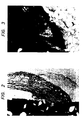
- a newborn was recognized in the surroundings of the complex, and, particularly at a part adjacent to bone cortex, hyperplasia or proliferation of newborn was so remarkable that the newborn was in contact directly with the periphery of the complex as shown in Fig. 2.
- the implant material according to the present invention is advantageous, in that its affinity to living tissues is high and bone addition can be expected in and around the implant material when the implant material is used as a bone-substitute.
- Shape forming or cutting can be made easy in accordance with known dental techniques so that the implant material may be made applicable to numerous uses including corrective surgery for bone deformation or defects, especially in jaw or facial bone reconstruction.
Landscapes
- Health & Medical Sciences (AREA)
- Chemical & Material Sciences (AREA)
- Oral & Maxillofacial Surgery (AREA)
- Transplantation (AREA)
- Materials Engineering (AREA)
- Engineering & Computer Science (AREA)
- Dermatology (AREA)
- Medicinal Chemistry (AREA)
- Inorganic Chemistry (AREA)
- Composite Materials (AREA)
- Epidemiology (AREA)
- Life Sciences & Earth Sciences (AREA)
- Animal Behavior & Ethology (AREA)
- General Health & Medical Sciences (AREA)
- Public Health (AREA)
- Veterinary Medicine (AREA)
- Materials For Medical Uses (AREA)
Applications Claiming Priority (2)
| Application Number | Priority Date | Filing Date | Title |
|---|---|---|---|
| JP62016431A JPS63183069A (ja) | 1987-01-27 | 1987-01-27 | インプラント材料およびその製造方法 |
| JP16431/87 | 1987-01-27 |
Publications (2)
| Publication Number | Publication Date |
|---|---|
| EP0276836A2 true EP0276836A2 (de) | 1988-08-03 |
| EP0276836A3 EP0276836A3 (de) | 1989-07-26 |
Family
ID=11916048
Family Applications (1)
| Application Number | Title | Priority Date | Filing Date |
|---|---|---|---|
| EP88101184A Withdrawn EP0276836A3 (de) | 1987-01-27 | 1988-01-27 | Implantierbares Material und Verfahren zu seiner Herstellung |
Country Status (3)
| Country | Link |
|---|---|
| EP (1) | EP0276836A3 (de) |
| JP (1) | JPS63183069A (de) |
| AU (1) | AU605540B2 (de) |
Cited By (8)
| Publication number | Priority date | Publication date | Assignee | Title |
|---|---|---|---|---|
| WO1990001343A1 (en) * | 1988-08-03 | 1990-02-22 | W.L. Gore & Associates, Inc. | Bone growth matrix and process for making it |
| WO1993025245A1 (de) * | 1992-06-12 | 1993-12-23 | Klaus Draenert | Polymergranulat oder -fasern und herstellungsverfahren |
| EP0684046A4 (de) * | 1993-12-10 | 1996-05-15 | Advance Kk | Implantationsmaterial und verfahren zu seiner herstellung. |
| US5746200A (en) * | 1990-10-19 | 1998-05-05 | Draenert; Klaus | Trabecula nasal filter having both macropores and micropores |
| US5993716A (en) * | 1990-10-19 | 1999-11-30 | Draenert; Klaus | Material and process for its preparation |
| FR2805748A1 (fr) * | 2000-03-01 | 2001-09-07 | Ceravic | Procede de preparation d'un materiau pateux phosphocalcique injectable en vue de former un biomateriau apatitique et application chirurgicale ou dentaire |
| EP1516636A3 (de) * | 2003-09-19 | 2005-08-03 | Broockeville Corporation N.V. | Verwendung eines härtbaren Elastomer-Precusors in einer medizinischen Behandlung |
| EP2392359A3 (de) * | 2002-08-12 | 2012-03-07 | Warsaw Orthopedic, Inc. | Synthese eines Knochen-Polymer-Verbundwerkstoffs |
Families Citing this family (3)
| Publication number | Priority date | Publication date | Assignee | Title |
|---|---|---|---|---|
| US4859712A (en) * | 1988-10-12 | 1989-08-22 | Cox-Uphoff International | Silicone foam and method for making it |
| WO1990015636A1 (en) * | 1989-06-02 | 1990-12-27 | Baxter International Inc. | Porous percutaneous access device |
| CN111892385A (zh) * | 2020-08-18 | 2020-11-06 | 重庆奥福精细陶瓷有限公司 | 一种大尺寸柴油颗粒过滤器的制备方法 |
Family Cites Families (9)
| Publication number | Priority date | Publication date | Assignee | Title |
|---|---|---|---|---|
| DE1216541B (de) * | 1960-06-17 | 1966-05-12 | Union Carbide Corp | Zu Zellkoerpern vulkanisierbare, hitzehaertbare Polysiloxanformmasse |
| SE416443B (sv) * | 1975-12-30 | 1981-01-05 | Sumitomo Chemical Co | Implantat for ben, leder eller tandrotter |
| JPS53144194A (en) * | 1977-05-20 | 1978-12-15 | Kureha Chemical Ind Co Ltd | Compound implanted material and making method thereof |
| DE2850586C2 (de) * | 1978-11-22 | 1982-05-27 | Battelle-Institut E.V., 6000 Frankfurt | Einlage zur Endoprothesenverankerung |
| US4657544A (en) * | 1984-04-18 | 1987-04-14 | Cordis Corporation | Cardiovascular graft and method of forming same |
| EP0192068B1 (de) * | 1985-02-19 | 1990-05-23 | The Dow Chemical Company | Hartgewebeprothese und Verfahren zu ihrer Herstellung |
| JPS6211459A (ja) * | 1985-07-09 | 1987-01-20 | 株式会社アドバンス | 複合インプラント材 |
| JPS62122670A (ja) * | 1985-08-09 | 1987-06-03 | 牧嶋 和見 | 医用形成材料 |
| US4728570A (en) * | 1985-10-29 | 1988-03-01 | United States Surgical Corporation | Calcium-hydroxide-treated polymeric implant matrial |
-
1987
- 1987-01-27 JP JP62016431A patent/JPS63183069A/ja active Granted
-
1988
- 1988-01-27 EP EP88101184A patent/EP0276836A3/de not_active Withdrawn
- 1988-01-27 AU AU10765/88A patent/AU605540B2/en not_active Ceased
Cited By (12)
| Publication number | Priority date | Publication date | Assignee | Title |
|---|---|---|---|---|
| WO1990001343A1 (en) * | 1988-08-03 | 1990-02-22 | W.L. Gore & Associates, Inc. | Bone growth matrix and process for making it |
| US5746200A (en) * | 1990-10-19 | 1998-05-05 | Draenert; Klaus | Trabecula nasal filter having both macropores and micropores |
| US5993716A (en) * | 1990-10-19 | 1999-11-30 | Draenert; Klaus | Material and process for its preparation |
| WO1993025245A1 (de) * | 1992-06-12 | 1993-12-23 | Klaus Draenert | Polymergranulat oder -fasern und herstellungsverfahren |
| EP0684046A4 (de) * | 1993-12-10 | 1996-05-15 | Advance Kk | Implantationsmaterial und verfahren zu seiner herstellung. |
| US5766247A (en) * | 1993-12-10 | 1998-06-16 | Kabushiki Kaisya Advance | Process for producing a bioimbedding material |
| FR2805748A1 (fr) * | 2000-03-01 | 2001-09-07 | Ceravic | Procede de preparation d'un materiau pateux phosphocalcique injectable en vue de former un biomateriau apatitique et application chirurgicale ou dentaire |
| WO2001064260A1 (fr) * | 2000-03-01 | 2001-09-07 | Ceravic | Procede de preparation d'un materiau pateux phosphocalcique injectable |
| US6923989B2 (en) | 2000-03-01 | 2005-08-02 | Ceravic | Method for preparing a calcium phosphate pasty material for injection |
| EP2392359A3 (de) * | 2002-08-12 | 2012-03-07 | Warsaw Orthopedic, Inc. | Synthese eines Knochen-Polymer-Verbundwerkstoffs |
| EP1516636A3 (de) * | 2003-09-19 | 2005-08-03 | Broockeville Corporation N.V. | Verwendung eines härtbaren Elastomer-Precusors in einer medizinischen Behandlung |
| EP2193812A1 (de) * | 2003-09-19 | 2010-06-09 | Broockeville Corporation N.V. | Verwendung eines härtbaren Elastomer-Precusors in einer medizinischen Behandlung |
Also Published As
| Publication number | Publication date |
|---|---|
| AU605540B2 (en) | 1991-01-17 |
| JPS63183069A (ja) | 1988-07-28 |
| JPH0215221B2 (de) | 1990-04-11 |
| AU1076588A (en) | 1988-07-28 |
| EP0276836A3 (de) | 1989-07-26 |
Similar Documents
| Publication | Publication Date | Title |
|---|---|---|
| US4599085A (en) | Bone implant member for prostheses and bone connecting elements and process for the production thereof | |
| Schepers et al. | Bioactive glass particles of narrow size range: a new material for the repair of bone defects | |
| EP0089782B1 (de) | Prothese aus Acrylharz | |
| US6224635B1 (en) | Implantation of surgical implants with calcium sulfate | |
| Costantino et al. | Synthetic bone graft substitutes | |
| Jarcho | Calcium phosphate ceramics as hard tissue prosthetics | |
| Costantino et al. | Hydroxyapatite cement: I. Basic chemistry and histologic properties | |
| US4917702A (en) | Bone replacement material on the basis of carbonate and alkali containing calciumphosphate apatites | |
| US5947893A (en) | Method of making a porous prothesis with biodegradable coatings | |
| CA1062158A (en) | Endodontic composition and method | |
| CA1260391A (en) | Xenogeneic collagen/mineral preparations in bone repair | |
| Homsy et al. | Materials for oral implantation—biological and functional criteria | |
| JPH07206489A (ja) | りん酸カルシウムセメント組成物、その製造方法、及びその使用方法 | |
| Kurashina et al. | Experimental cranioplasty and skeletal augmentation using an α-tricalcium phosphate/dicalcium phosphate dibasic/tetracalcium phosphate monoxide cement: A preliminary short-term experiment in rabbits | |
| EP0386253A1 (de) | Härtendes material für medizinischen und dentalen gebrauch. | |
| AU605540B2 (en) | Implant material and method of producing the same | |
| EP0166263A1 (de) | Füllmaterial für Knochendefekte oder Knochenhohlräume, Kit oder Set zur Herstellung dieses Füllmaterials | |
| AU615771B2 (en) | Implant and method of making it | |
| Propper | A technique for controlled placement of hydroxylapatite over atrophic mandibular ridges | |
| Yoshikawa et al. | Histopathological study of a newly developed root canal sealer containing tetracalcium-dicalcium phosphates and 1.0% chondroitin sulfate | |
| JPH0639372B2 (ja) | 生体活性セメント | |
| EP0196143A2 (de) | Verfahren zur Bereitung eines Implantatmaterials und dafür geeignetes Zweikomponentenkit | |
| JP2883213B2 (ja) | 生体移植材とその製造方法 | |
| WO2023216069A1 (zh) | 抗冲刷能力的可塑型的骨科组合物 | |
| CN113367787A (zh) | 一种防止假体松动及肿瘤术后复发的肿瘤骨切缘填充器的制备方法 |
Legal Events
| Date | Code | Title | Description |
|---|---|---|---|
| PUAI | Public reference made under article 153(3) epc to a published international application that has entered the european phase |
Free format text: ORIGINAL CODE: 0009012 |
|
| AK | Designated contracting states |
Kind code of ref document: A2 Designated state(s): DE FR GB |
|
| PUAL | Search report despatched |
Free format text: ORIGINAL CODE: 0009013 |
|
| RHK1 | Main classification (correction) |
Ipc: A61L 27/00 |
|
| AK | Designated contracting states |
Kind code of ref document: A3 Designated state(s): DE FR GB |
|
| 17P | Request for examination filed |
Effective date: 19890905 |
|
| 17Q | First examination report despatched |
Effective date: 19910523 |
|
| STAA | Information on the status of an ep patent application or granted ep patent |
Free format text: STATUS: THE APPLICATION IS DEEMED TO BE WITHDRAWN |
|
| 18D | Application deemed to be withdrawn |
Effective date: 19911003 |