EP0280468A2 - Méthodes et matériaux de détection et de thérapie du VIH - Google Patents
Méthodes et matériaux de détection et de thérapie du VIH Download PDFInfo
- Publication number
- EP0280468A2 EP0280468A2 EP88301369A EP88301369A EP0280468A2 EP 0280468 A2 EP0280468 A2 EP 0280468A2 EP 88301369 A EP88301369 A EP 88301369A EP 88301369 A EP88301369 A EP 88301369A EP 0280468 A2 EP0280468 A2 EP 0280468A2
- Authority
- EP
- European Patent Office
- Prior art keywords
- hiv
- antibody
- monoclonal
- polypeptide
- protein
- Prior art date
- Legal status (The legal status is an assumption and is not a legal conclusion. Google has not performed a legal analysis and makes no representation as to the accuracy of the status listed.)
- Granted
Links
Images



Classifications
-
- C—CHEMISTRY; METALLURGY
- C07—ORGANIC CHEMISTRY
- C07K—PEPTIDES
- C07K14/00—Peptides having more than 20 amino acids; Gastrins; Somatostatins; Melanotropins; Derivatives thereof
- C07K14/005—Peptides having more than 20 amino acids; Gastrins; Somatostatins; Melanotropins; Derivatives thereof from viruses
-
- C—CHEMISTRY; METALLURGY
- C07—ORGANIC CHEMISTRY
- C07K—PEPTIDES
- C07K16/00—Immunoglobulins [IG], e.g. monoclonal or polyclonal antibodies
- C07K16/42—Immunoglobulins [IG], e.g. monoclonal or polyclonal antibodies against immunoglobulins
- C07K16/4208—Immunoglobulins [IG], e.g. monoclonal or polyclonal antibodies against immunoglobulins against an idiotypic determinant on Ig
- C07K16/4241—Immunoglobulins [IG], e.g. monoclonal or polyclonal antibodies against immunoglobulins against an idiotypic determinant on Ig against anti-human or anti-animal Ig
- C07K16/4258—Immunoglobulins [IG], e.g. monoclonal or polyclonal antibodies against immunoglobulins against an idiotypic determinant on Ig against anti-human or anti-animal Ig against anti-receptor Ig
- C07K16/4275—Immunoglobulins [IG], e.g. monoclonal or polyclonal antibodies against immunoglobulins against an idiotypic determinant on Ig against anti-human or anti-animal Ig against anti-receptor Ig against anti-CD4 Ig
-
- A—HUMAN NECESSITIES
- A61—MEDICAL OR VETERINARY SCIENCE; HYGIENE
- A61K—PREPARATIONS FOR MEDICAL, DENTAL OR TOILETRY PURPOSES
- A61K38/00—Medicinal preparations containing peptides
-
- A—HUMAN NECESSITIES
- A61—MEDICAL OR VETERINARY SCIENCE; HYGIENE
- A61K—PREPARATIONS FOR MEDICAL, DENTAL OR TOILETRY PURPOSES
- A61K39/00—Medicinal preparations containing antigens or antibodies
-
- C—CHEMISTRY; METALLURGY
- C12—BIOCHEMISTRY; BEER; SPIRITS; WINE; VINEGAR; MICROBIOLOGY; ENZYMOLOGY; MUTATION OR GENETIC ENGINEERING
- C12N—MICROORGANISMS OR ENZYMES; COMPOSITIONS THEREOF; PROPAGATING, PRESERVING, OR MAINTAINING MICROORGANISMS; MUTATION OR GENETIC ENGINEERING; CULTURE MEDIA
- C12N2740/00—Reverse transcribing RNA viruses
- C12N2740/00011—Details
- C12N2740/10011—Retroviridae
- C12N2740/16011—Human Immunodeficiency Virus, HIV
- C12N2740/16111—Human Immunodeficiency Virus, HIV concerning HIV env
- C12N2740/16122—New viral proteins or individual genes, new structural or functional aspects of known viral proteins or genes
Definitions
- the present invention relates generally to methods and materials useful in the diagnosis and treatment of infection with Human Immunodeficiency Virus (HIV). More particularly, the invention relates to immunologically active polypeptides, preferably antibodies or antibody fragments, and most preferably monoclonal antibodies, which are reactive with idiotypes of antibodies to human lymphocyte T4 protein and also reactive with the HIV virion in a manner allowing for in vitro and in vivo neutralization of HIV infectivity. Moreover, the invention relates to immunologically active polypeptides useful in vaccine compositions for developing protective responses to HIV infection.
- HIV Human Immunodeficiency Virus
- T4 surface glycoprotein (sometimes referred to as "CD4" protein or determinant) of human T lymphocytes in infection by HIV has been extensively studied as represented by Dalgleish et al., Nature , 312 , pp. 763-767 (1984); Klatzman et al., Science , 312 , 767-768 (1986); Klatzman et al., Science , 225 , pp. 59-62 (1984); McDougal et al., J. Immunol. , 135 , pp. 3151-3162 (1985); and, Maddon et al., Cell , 47 , pp. 333-348 (1986). See also, Marrack et al., "The T Cell and Its Receptor", Scientific American , February 1986, pp. 36-45; and, McDougal et al., Science , 231 , 382-385 (1986).
- CD4 may have therapeutic utility in treatment of HIV infection. See, Fisher et al., Nature , 331 , 76-77 (1988); Hussey et al., Ibid , at pp. 78-81; Deen et al., Ibid , at pp. 82-83; and Traunecker et al., Ibid , at pp. 84-86 all of which relate to in vitro neutralization of HIV infectivity by soluble CD4.
- CD4 major histocompatibility complex
- MHC major histocompatibility complex
- the present invention provides purified and isolated immunologically active polypeptides, preferably antibodies or chimeric antibodies or fragments thereof, and most preferably monoclonal antibodies, which are reactive with idiotypes of antibodies (preferably monoclonal antibodies) to human lymphocyte T4 protein.
- immunologically active polypeptides preferably antibodies or chimeric antibodies or fragments thereof, and most preferably monoclonal antibodies, which are reactive with idiotypes of antibodies (preferably monoclonal antibodies) to human lymphocyte T4 protein.
- Such products of the invention are capable of specific immunobinding with that portion of the HIV virion which is necessarily interactive with T4 surface proteins during infection by HIV of host cells such as human T lymphocytes and cells of the human nervous system.
- These products of the invention may be characterized, inter alia , by their strain independent capacity to neutralize infectivity of HIV in vitro , by their specific reactivity with HIV protein having a molecular weight of from about 60,000 to about 80,000 as determined by SDS- PAGE, and by their non-reactivity with class II major histocompatability molecules associated with human immune cell surfaces.
- immunologically active products are generated which "respond" through specific immunobinding to HIV particles and to the surfaces of HIV-infected cells, even though they are generated without direct immunological reference to such surface proteins.
- Purified and isolated polypeptides according to the invention are conspicuously suitable for use in assay procedures for the detection and/or quantification of HIV in biological fluids wherein the detection procedure is based upon immunobinding between HIV particles and reactive polypeptides (e.g., antibodies) of the invention.
- antibodies, chimeric antibodies and fragments thereof developed according to the invention are electively immunoreactive with the surfaces of HIV-infected cells and thus provide useful reagents for detection of HIV-infected cells in fluid and tissue samples and for segregation of HIV-infected cells from cell populations including both infected and non-infected cells.
- products of the invention will be useful in diagnostic and therapeutic procedures involving separation and/or selective destruction of HIV-infected cells.
- Purified and isolated immunologically active mterials of the invention are also conspicuously suitivelyable for use in anti-HIV treatment of animals, especialivelyly humans, susceptible to infection with HIV.
- immunologically effective amounts of, e.g., monoclonal antibodies of the invention are administered to a patient either already infected with HIV or to a patient at risk of infection with HIV to develop passive immunity with respect to HIV infection.
- cells infected with HIV are subjected to, e.g., in vitro segregation from non-infected patient cells and the latter may be returned to the patient.
- antibody-related polypeptides of the present invention are preferably obtained by means of initial development of mono-specific antibodies (preferably monoclonal antibodies) to the human lymphocyte T4 glycoprotein (the CD4 determinant) followed by preparation of antibodies (preferably monoclonal antibodies) to the T4 idiotypic region of antibodies formed in the initial development step.
- Chimeric antibodies and fragments thereof and especially bi-specific antibodies are also products within the contemplation of the present invention, as are antibody-related products produced in microbial hosts, (e.g., procaryotic and eucaryotic cells in culture) which hosts are transformed or transfected with DNA sequences encoding the desired polypeptide products.
- microbial hosts e.g., procaryotic and eucaryotic cells in culture
- chimeric antibodies e.g. mouse/human antibodies
- hybridoma cells especially heavy chain deletion mutant cells
- Recombinant methods may also be applied to the production of HIV subunit vaccine materials.
- monoclonal antibodies of the invention are expected to be extremely well suit ed for the screening of expression products of HIV DNA in transformed or transfected vial or procaryotic hosts, allowing isolation of DNA encoding all or part of the amino acid sequence of naturally occurring immunologically significant HIV proteins (including glycoproteins). In suitable hosts, the presence of such DNA may allow for high level production of vaccine materials.
- the invention provides antibody-related polypeptides characterized as monoclonal anti-monoclonal-anti-human lymphocyte T4 antibodies.
- monoclonal anti-OKT4 and anti-OKT4A antibodies both of which are reactive with 60-80 Kd HIV proteins.
- monoclonal anti-OKT4A antibodies which have substantial capacity for in vitro neutralization of HIV infectivity of multiple HIV strains and are participative in complement-mediated cytolysis of HIV infected cells.
- the present invention provides, for the first time, hybridoma cell lines which produce "anti-idiotypic" monoclonal antibodies specifically immunoreactive with a monoclonal antibody to human lymphocyte T4 protein in an antigen/antibody reaction.
- the present invention provides new murine-derives hybridoma cell lines, JT4C8, JT4C12 and JT4C16, JT1-1F3, JT1-1F3-E5, JT1-1D7 and JT2-N15, each of which produces, as a component of the supernatant of its growth in culture, a monoclonal antibody specifically reactive with anti-T4 idiotype and moreover reactive with HIV virion proteins in a manner expected to allow for both in vitro and in vivo neutralization of HIV infectivity.
- Hybridoma cell line JT4C8 was received for deposit with the American Type Culture Collection, Rockville, Maryland, at the facilities of the United States Department of Agriculture, Plum Island, New York on February 18, 1987, with A.T.C.C. Accession No. HB9385.
- Hybridoma cell line JT4C12 was received for deposit with the American Type Culture Collection, Rockville, Maryland, at the facilities of the United States Department of Agriculture, Plum Island, New York on February 18, 1987, with A.T.C.C. Accession No. HB9387.
- Hybridoma cell line JT4C16 was received for deposit with the American Type Culture Collection, Rockville, Maryland, at the facilities of the United States Department of Agriculture, Plum Island, New York on February 18, 1987, with A.T.C.C.
- Hybridoma cell line JT1-1F3 was received for deposit at the European Collection of Animal Cell Cultures, Salisbury, Wiltshire, U.K. on June 25, 1987, with ECACC Accession No. 87062501.
- Hybridoma cell line JT1-1F3-E5 was received for deposit at the European Collection of Animal Cell Cultures, Salisbury, Wiltshire, U.K. on June 25, 1987 with ECACC Accession No. 87062502.
- Hybridoma cell line JT1-1D7 was received for deposit by the Fermentation Research Institute, Ibaragi-ken, Japan on January 29, 1988 with the Accession No. FERM BP-1685.
- Hybridoma cell line JT2-N15 was received for deposit by the Fermentation Research Institute, Ibaragi-ken, Japan on January 29, 1988 with the Accession No. FERM BP-1684.
- the present invention provides for production of HIV subunit vaccine materials by means of well-known affinity purification methodologies whereby HIV protein fractions (especially those in the molecular weight range of from about 60,000 to about 80,000 and most especially about 65,000-67,000) are isolated through use of a selective immunoabsorbants prepared using antibodies of the invention.
- Figures 1, 2 and 3 graphically represent the results of immunoreactivity testing of antibodies of the invention with HIV and non-infected cell proteins
- Figures 4 and 5 provide immunoblot assay results involving antibodies of the invention and HIV proteins
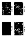
- Figures 6A through 6E provide photographic results of immunofluorescent staining assays on infected and uninfected cells employing antibodies of the invention.
- Example 1 is directed to stimulation of a murine host toward production of antibodies to a commercially available anti-T4 monoclonal antibody, "OKT4", the fusion of spleen cells with myeloma cells, the screening, cloning and growth of hybridoma cells, and the isolation of monoclonal antibodies therefrom.
- Example 2 relates to the characterization of monoclonal antibodies so produced by fluorescent immunoassay, by Western blot assay for reactivity with HIV protein, and by screening for capacity to effect in vitro neutralizaitn of HIV infectivity.
- Example 3 relates to a first procedure for development of hybridoma cell lines capable of providing in the medium of their growth monoclonal antibodies to the commercially available antibody "OKT4A”.
- Example 4 relates to characterization of monoclonal antibodies so produced by means of immunofluorescence assay, Western blot assay, in vitro neutralization assay, ELISA assay, and fluorescent cell staining assays.
- Example 5 relates to a second procedure for development of hybridoma cell lines capable of providing in the medium of their growth monoclonal antibodies to the commercially available antibody "OKT4A” and to characterization of monoclonal antibodies so produced by means of Western blot assay, and in vitro neutralization assay.
- hybrid tumor cell lines are produced using standard immunological techniques such as described in Oi and Herzenberg, Selected Methods Cell Immunology , 351-372 (1979) and Godding, "Antibody Production By Hybridomas", J. Immunol.Meth. , 39 , p. 285-308 (1980).
- Spleen cells from mice, hyperimmunized with monoclonal anti-T4 are fused with a mouse myeloma cell line in the presence of polyethylene glycol. The supernatant from growth of each "hybridoma" cell culture is tested for the presence of the desired antibody activity.
- Selected hybridoma cells are cloned to propagate cell lines which produce an antibody in their growth culture supernatant, which antibody has highly specific anti-anti-T4 activity.
- mice each were subject to splenic injection with monoclonal anti-human lymphocyte T4 antibody (OKT4, Ortho Diagnostics, Rahway, N.J.).
- OKT4 Ortho Diagnostics, Rahway, N.J.
- lyophilized OKT4 material was brought up in 1 ml of distilled water. Dialysis was employed to remove and replace the original phosphate buffer with 50mM MES buffer (Sigma Chemicals), pH 6.0. The material was then subjected to high pressure liquid chromatography separation on FPLC® apparatus (Pharmacia, Laboratory Separation Division, Piscataway, N.J. 08854). Separation was carried out using a Mono Q column under recommended procedures except that the salt gradient was changed to 0 to 0.5 M NaCl. Aliquots (20 ⁇ l) of each 0.5 ml fraction were assayed for activity using freshly collected human lymphocytes (105 cells per ml).
- mice Each of 4 mice was initially given a splenic injection totalling approximately 100 ⁇ l of inoculant (approximately 1.5 ⁇ g OKT4 per mouse). Fourteen and twenty-eight days later the mice were given booster injections of 100 ⁇ l inoculant.
- mice Four days after the final booster, the mice were sacrificed and spleens were removed aseptically and placed in petri dishes (on ice) containing Dulbecco's Modified Eagle's Medium (Gibco). The spleens were trimmed of fat and connective tissue, passed through 100 gauge stainless steel mesh. The resulting individual spleen cells were pelleted by centrifugation for 10 minutes at 1000 rpm. The cell pellet was washed twice with media (as above) and was resuspended in RPMI 1640 and the cell concentration was determined by counting in a hemocytometer in the presence of 0.2% trypan blue.
- Dulbecco's Modified Eagle's Medium Gibco
- Spleen and NS1/1 cells were combined in a ratio of 4:1 and centrifuged at 1000 rpm for 10 minutes.
- the supernatant fluid was aspirated away and cell fusion was conducted at 37°C using polyethylene glycol (PEG) 1500, molecular weight 500-600.
- PEG polyethylene glycol
- the procedure was carried out with constant gentle stirring by addition of the following, at the times indicated: 1.0 ml of 50% PEG in RPMI 1640 added over one minute, with one minute stirring, 1.0 ml RPMI 1640 containing 15% horse serum over one minute; 1.0 ml of RPMI 1640 containing 15% horse serum added over one minute; and 8 ⁇ l RPMI 1640 containing 15% horse serum added over 2 minutes.
- the resulting fused cells were centrifuged at 1000 rpm for 10 minutes, resuspended in RPMI 1640 containing 15% horse serum and also containing penicillin G and streptomycin at 100 units and 100 mg per ml, respectively.
- the cells were plated at 0.1 ml per well in 96-well plates previously equilibrated in 5% CO2. Plates were incubated overnight at 37°C in 5% CO2.
- HAT medium (13.6 ⁇ g/ml hypoxanthine 0.176 ⁇ g/ml aminopterin and 3.88 ⁇ g/ml thymidine) in RPMI 1640 containing 15% horse serum.
- This medium selectively allows spleen cell NS1/1 hybrids to survive, screening out unfused NS1/1 cells or those fused to other cells. Unfused primary spleen cells from adult mice will not survive in culture for more than a few days.
- FLISA fluorescent-linked immunosorbent assay
- FITC-labelled OKT4 100 ⁇ l was incubated in each well overnight and in the dark at 4°C. Wells were washed 10 times with PBS/Tween 20 solution as above. To each well was added 200 ⁇ l of 5 x 10 ⁇ 5 N NaOH and 5.6 x 10 ⁇ 4 N NH4OH solution. The plates were maintained at room temperature for 15 minutes and then shaken for 1 minute. After transfer to Titertek Microtitration 1 x 8 plates, the wells were fluorometrically (excitation 490 nm; emission 530 nm) using a Corona Electric model MTP 100F microplate reader.
- Formal cloning of hybridoma cells obtained from the positive wells was conducted by diluting the cells into additional wells at a ratio such that there was approximately 1 cell per 3 wells.
- formal cloning from an active well produced formal clones which appeared to be subclones of the same hybridoma cell but in several instances revealed different clone populations over three generations.
- Subclones from the same original well were named with the parent number and an additional number (e.g., clones obtained from well JT4C7 were labeled JT4C7-1, JT4C7-2, etc.).
- a fluorescent-linked assay was carried out according to the same general procedure employed for antibody screening in Example 1, except that various dilutions of rabbit anti-mouse IgG Fc were deposited in test wells. Fluorescence results are set out in Table 1 for antibodies derived from clones JT4Cl through JT4C-13 and JT4C16. Fluorescence results for varous subclones of JT4C7 are set out in Table 2.
- Table 2 reveals that of the subclones of JT4C7, clones JT4C7-12 and JT4C7-9 are respectively of the highest and lowest relative affinity in this test procedure.
- Antibodies derived from all nineteen positive clones identified in Example 1 were assayed by Western blot analysis for immunoreactivity with proteins of the HIV virion. More specifically, HTLV-IIIB particles were disrupted with SDS (0.1%) and dithiothriotol (0.003M) and the material was placed on a 7.5% polyacrylamide gel, the gel was electrophoresed using standard procedures and materials were transferred to nitrocellulose filter paper. Filters were initially incubated with 20% heat inactivated horse serum in PBS for 1 hour and then washed with PBS. Filters were then incubated with antibody supernatants of each clone for 3 hours at room temperature with gentle shaking and then overnight at 4°C with gentle shaking.
- JT4C12 and JT4C16 antibodies were of the IgG3 isotype.
- Hybridoma culture supernatants were tested for HIV neutralization capacity in the following manner. Three day growth supernatants were diluted 1:5 in complete medium [500 ml RPMI 1640; 6 ml 100 x Penicillin/ Streptomycin; 6 ml 100 x L-glutamine; 100 ml FCS; and, 1.2 ml Polybrene Stock (1 mg/ml)]. 200 ⁇ l of the medium-diluted sample was added to all but two wells of a 24 well microtiter plate and two wells received an equal quantity of medium alone. Additional medium was added to one of the "medium-only" wells and each remaining well received 200 ⁇ l of high titer HIV virus stock.
- H9 cells were incubated in complete medium for 30 minutes at 37°C at a density of 1 x 10⁶ cells/ml, then centrifuged and resuspended in fresh complete medium at a density of 5 x 10⁶ cell/ml. 200 ⁇ l of the cell suspension was added to each well (bringing the total volume to 600 ⁇ l) and the plates were incubated for 1 hour at 37°C, whereupon 150 ⁇ l was transferred to a duplicate plate containing 2.0 ml of fresh complete medium per well. Cultures were incubated at 37°C in a CO2 incubator.
- Example 1 The general hybridoma forming and screening procedures of Example 1 were repeated using the commercially available monoclonal anti-human lymphocyte T4 antibody designated OKT4A (Ortho Diagnostics, Rahway, N.J.) which was purified using a Mono S (rather than Mono Q) column.
- OKT4A Ortho Diagnostics, Rahway, N.J.
- the specific immunization procedure varied slightly from that of Example 1 in that the initial injection was intraperitoneal and involved approximately 15 ⁇ g of purified antibody.
- the second intraperitoneal inoculation was seven days later and consisted of approximately 10 ⁇ g of purified antibody. Thirteen days later, the final booster of about 5 ⁇ g of antibody was administrated by splenic injection.
- Example 3 In order to characterize the antibodies produced by the positive clones described in Example 3, tests were conducted essentially as described in Example 2. Briefly, supernatant antibodies were reactive on the Western blot assay with HIV protein having a molecular weight of from about 60,000 to about 80,000. The assays predominantly indicated reaction with an approximately 67,000 molecular weight protein, with some antibodies showing reactivity with a 78,000 molecular weight species. Significantly no reactivity was noted with 41 Kd or 120 Kd fractions usually characterized as the major immunologically significant HIV envelope glycoproteins.
- JT1-6E12 and JT2-8E9 were of an IgM isotype and the antibody of clone JT1-1F3 was of the IgG1 isotype.
- Table 3 are the results of the fluorescent-linked assay for employing the JT1-1F3 antibody.
- Example 2(C) Neutralization studies carried out concurrently with those of Example 2(C) also indicated initial negative neutralization results and, in the second trial, only slight evidence of neutralization capacity on the RT assay. Nonetheless, the IgG1-secreting JT1-1F3 clone was selected for ascites amplification and further antibody ELISA screening for reactivity with HIV protein.
- varying concentrations of HTLV-IIIB protein were coated onto microtiter plates, blocked (20% horse serum/PBS, 37°C, 2 hours), and dried overnight.
- Culture supernatant (20 ⁇ l with 80 ⁇ l PBS) or HAT medium control was added as a first antibody.
- peroxidase conjugated rabbit anti-mouse IgG was added as the second antibody.
- the ELISA procedure was repeated on plates initially coated with varying concentrations of uninfected H9 cell membrane preparations [106, 6 x 10⁵, 4 x 10⁵, 2 x 10⁵, 6 x 10⁴, 4 x 10⁴, 2 x 10⁴, 1 x 10⁴, 6 x 10³, 4 x 10³, 2 x 10³ cells/ml].
- the results of this test procedure are graphically represented in Figure 2 and indicate no reactivity with non-infected preparations.
- the HIV protein ELISA was repeated using the same varying concentrations of protein from the HTLV-IIIB HIV variant, the Haitian HIV variant designated AL1212C (obtained from Dr. David Hall, Massachusetts General Hospital, Boston, MA) and the African HIV variant designated 906 (obtained form Dr. Jerone Groopman, New England Deaconess Hospital, Boston, Massachusetts).
- the HT1-1F3 antibody recognized a common epitope of all three HIV variants.
- Ascites fluid derived JT1-1F3 antibody was then tested for in vitro neutralization activity as indicated by the RT assay described above and by syncitium induction.
- a comparison was made between varying levels of JT1-1F3 antibody (ascites fluid IgG fraction), a negative control (ascites fluid/pristane) and a positive control in the form of serum from an HTLV-IIIB-infected patient.
- HIV strains IIIB, AL1212C and 906 were incubated with Mab JR1-1F3, control ascites or neutralizing human serum (in a 1:5 dilution in phosphate buffered saline) for 90 minutes at 4°C.
- H9 cells (5 x 10⁶) were then added to each well and incubated for 1 hour at 37°C. Aliquots (150 ⁇ l) were removed from each well and added to 2.0 ml fresh medium. The cultures were split 1:1 on day 4. Reverse transcriptase activity and syncytium induction were monitored on day 4 and day 7. The day 7 results of this procedure are provided in Table 4, wherein the relative syncytium induction is indicated as follows: (-), 0/200 cells; (+/-), 1-5/ 200 cells; (+), >10% cells; (++), >25% of cells; and (+++), >50% of cells.
- the molecular weight of the JT1-1F3 antibody reactive species was determined by immunoblotting.
- Purified HTLV-IIIB (5 and 10 ⁇ g) was analyzed by SDS-PAGE and Coomasie blue/silver staining (Fig. 4, Lanes B and C). Similar aliquots were analyzed by SDS-PAGE, transferred to nitrocellulose paper and analyzed for reactivity with JT1-1F3 antibody. A single reactive species was detectable at approximately 67 kd (Fig. 4, Lanes E and F).
- JT1-1F3 antibody suggested that it might also be useful in detecting HIV virus-infected cells.
- JT1-1F3 antibody ascites fluid IgG fraction
- the extend of HT1-1F3 finding was determined by a fluoresceinated rabbit anti-mouse IgG.
- Figure 6A there was little if any detectable binding of antibody to uninfected H9 cells.
- focal and diffuse binding of the antibody was detectable with HTLV-IIIB- infected H9 cells (Fig. 6B).
- MHC major histocompatibility
- JT1-1F3 Subcloning of the JT1-1F3 resulted in the selection of subclones JT1-1F3-E5 and JT1-1D7 as respectively producing progressively higher levels of IgG1 antibody than JT1-1F3, JT1-1F3-E5 and JT1-1D7 is set out in Table 5 below. Reactivity within the ELISA assay format for the antibodies produced by these three hybridomas is set out in Table 6 below.
- the hybridoma forming and screening procedures of Examples 1 and 3 were again repeated using OKT4A, with the following variations in the immunization procedure.
- the initial immunization involved interperitoneal injection of 10 ⁇ g of Mono S purified OKT4A antibody; the second intraperitoneal injection (7 ⁇ g) was given seventeen days later; and the final, 7 ⁇ g intravenous dose was administered 18 days later.
- an IgG1-producing positive clone, designated JT2-N15 was selected for further study.
- JT1-1F3 and its subcones JT1-1F3-E5 and JT1-1D7 clone JT2-N15 produced a monoclonal antibody which was reactive on the Western blot assay with HIV protein having a molecular weight of about 65-67,000.
- Culture media from growth of JT2-N15 was positive in the ELISA assay and the results in a preliminary neutralization assay with respect to HIV-IIIB infectivity are set out in Table 7 below.
- a third collection from surviving animals generally provided 3-5 ml of fluid which, at times, was substantially less active than either of the materials from the first and second collection.
- Neutralization data shown for JT1-1F3 in Tables 4 and 5 was based on ascites pooled from three collections whereas data for JT1-1F3-E5, JT1-1D7 and JT2-N15 in Tables 5 and 7 was based on ascites materials pooled from first and second collections only.
- antibodies produced by JT1-1F3 and its subclones and JT2-N15 are of the IgG1 isotype, it is expected that antibodies of differing isotypes will be equally useful.
- Antibodies of the IgG2 isotype may be more useful in procedures involving complement-mediated cytolytic reactions.
- the anti-Leu3a antibody of Chanh et al. is not indicated as reactive with any HIV-derived protein other than gp120 as shown in Figure 4 at page 3894 of Chanh et al., supra .
- hybrid hybridomas e.g., mouse/human
- human/human hybridomas prepared, for example, in a manner consistent with Borrebaeck, TIBTECH , June, 1986, pp. 147-153; Abrams et al., Methods in Enzymology , 121 , pp. 107-119 91986); Kozbor et al., Methods in Enzymology , 121 , pp. 120-140 (1986); and Masuho et al., Biochem. & Biophys. Res. Comm. , 135(2) , pp.
- immunologically active polyeptides for use in diagnostic and therapeutic methods of the invention by means or expression of DNA sequences encoding therefor in suitably transformed or transfected procaryotic and eucaryotic host cells in culture.
- Anti-HIV therapeutic methods of the invention will be understood to comprise the administration of effective amounts of antibodies or antibody fragments of the invention to a patient infected with HIV or at risk of HIV infection in order to generate passive immunity involving neutralizing infectivity of HIV in vivo .
- combination of products of the invention with immunologically acceptable diluents, adjuvants and carriers is contemplated in order to form immunologically effective anti-HIV therapeutic compositions.
- Anti-HIV therapeutic methods of the invention within the contemplation of the invention also comprehend active immunization using biologically active HIV protein fractions reactive with the monoclonal anti-monoclonal anti-human lymphocytes.
- Such products may be obtained directly from virus preparations by well known affinity purification processes involving forming immunological reaction mixtures between HIV proteins and antibodies of the invention, followed by isolation of the desired protein.
- the 60,000 to 80,000 molecular weight HIV protein recognized by the JT1-1F3 in procedures of Example 4 is a prime candidate for vaccine use. The same is expected to be true for recombinant expression products based on HIV DNA which can be immunologically identified (and/or purified) by means of antibodies of the invention.
- Diagnostic methods of the invention wherein polypeptide products are employed to detect and quantify HIV particles in biological fluids such as blood are expected to form an essential part in preliminary screening for patients who would benefit from passive immunization according to the invention and in the monitoring therapeutic regimens of the invention.
- antibodies of the invention are selectively reactive with the surfaces of HIV-infected cells is indicative of a variety of diagnostic and therapeutic utilities including histological screen overwhelminging of tissue (e.g., lymphatic cells) and fluid (e.g., blood) samples for detection of HIV infection, possibly at early stages of infection not readily detectable by assays based on screening for antibodies.
- Recognition of infected cells by antibodies of the invention allows for segregation of such cells from cell populations comprising both infected and non-infected cells. It is thus contemplated that blood of AIDS patients may be subjected to extracorporeal treatment to remove or selectively kill infected lymphocytes through use of antibodies of the invention.
- T4 protein which is believed to provide the common receptor for HIV infection by all variants (e.g., AL1212C, 906, ARC, LAV, HTLV-IIIRF, HTLV-IIIB) -- rather than any specific HIV variant, it is expected that diagnostic and therapeutic methods of the invention will be applicable to detection and treatment of infection involving all HIV variants.
- Use of polypeptides of the invention as diagnostic and research tools is expected to provide additional information into the nature of the interaction between HIV and host cells.
- monoclonal antibodies of the invention will be useful in identifying the precise primary, secondary and tertiary structural conformation of region(s) of surface proteins of HIV and host cells which are specifically and necessarily interactive in the recognition and association processes involved in HIV infection of cells. This information, in turn, would allow for generation of immunologically active materials of the invention through use of synthetic and recombinant-produced peptide immunogens.
Landscapes
- Chemical & Material Sciences (AREA)
- Health & Medical Sciences (AREA)
- Organic Chemistry (AREA)
- Life Sciences & Earth Sciences (AREA)
- Proteomics, Peptides & Aminoacids (AREA)
- Immunology (AREA)
- General Health & Medical Sciences (AREA)
- Genetics & Genomics (AREA)
- Medicinal Chemistry (AREA)
- Molecular Biology (AREA)
- Biochemistry (AREA)
- Biophysics (AREA)
- Virology (AREA)
- Gastroenterology & Hepatology (AREA)
- Preparation Of Compounds By Using Micro-Organisms (AREA)
- Pharmaceuticals Containing Other Organic And Inorganic Compounds (AREA)
- Medicines Containing Material From Animals Or Micro-Organisms (AREA)
- Peptides Or Proteins (AREA)
- Micro-Organisms Or Cultivation Processes Thereof (AREA)
- Medicines Containing Antibodies Or Antigens For Use As Internal Diagnostic Agents (AREA)
Priority Applications (1)
| Application Number | Priority Date | Filing Date | Title |
|---|---|---|---|
| EP91202465A EP0468601B1 (fr) | 1987-02-19 | 1988-02-18 | Procédés et matériaux pour la détection et la thérapie d'HIV |
Applications Claiming Priority (6)
| Application Number | Priority Date | Filing Date | Title |
|---|---|---|---|
| US1628287A | 1987-02-19 | 1987-02-19 | |
| US16282 | 1987-02-19 | ||
| US6406687A | 1987-06-29 | 1987-06-29 | |
| US64066 | 1987-06-29 | ||
| US14637188A | 1988-02-03 | 1988-02-03 | |
| US146371 | 1988-02-03 |
Related Child Applications (1)
| Application Number | Title | Priority Date | Filing Date |
|---|---|---|---|
| EP91202465.0 Division-Into | 1991-09-23 |
Publications (3)
| Publication Number | Publication Date |
|---|---|
| EP0280468A2 true EP0280468A2 (fr) | 1988-08-31 |
| EP0280468A3 EP0280468A3 (en) | 1990-09-05 |
| EP0280468B1 EP0280468B1 (fr) | 1994-06-08 |
Family
ID=27360536
Family Applications (2)
| Application Number | Title | Priority Date | Filing Date |
|---|---|---|---|
| EP88301369A Expired - Lifetime EP0280468B1 (fr) | 1987-02-19 | 1988-02-18 | Méthodes et matériaux de détection et de thérapie du VIH |
| EP91202465A Expired - Lifetime EP0468601B1 (fr) | 1987-02-19 | 1988-02-18 | Procédés et matériaux pour la détection et la thérapie d'HIV |
Family Applications After (1)
| Application Number | Title | Priority Date | Filing Date |
|---|---|---|---|
| EP91202465A Expired - Lifetime EP0468601B1 (fr) | 1987-02-19 | 1988-02-18 | Procédés et matériaux pour la détection et la thérapie d'HIV |
Country Status (5)
| Country | Link |
|---|---|
| EP (2) | EP0280468B1 (fr) |
| JP (1) | JP2583555B2 (fr) |
| AT (2) | ATE106945T1 (fr) |
| AU (1) | AU609447B2 (fr) |
| DE (2) | DE3855761T2 (fr) |
Cited By (5)
| Publication number | Priority date | Publication date | Assignee | Title |
|---|---|---|---|---|
| EP0385909A3 (fr) * | 1989-03-03 | 1991-04-10 | MicroGeneSys, Inc. | Kit ou composition pour la prévention ou le traitement des infections par HIV-1 |
| WO1992022579A1 (fr) * | 1991-06-19 | 1992-12-23 | Immulogic Pharmaceutical Corporation | PEPTIDES MIMANT LA gp120 |
| EP1083226A4 (fr) * | 1998-05-25 | 2001-12-19 | Asahi Chemical Ind | Dispositif pour la separation cellulaire et procede de separation |
| EP0771326A4 (fr) * | 1994-07-13 | 2002-01-16 | Progenics Pharm Inc | Articles manufactures permettant d'eliminer le vih-1 d'un echantillon, et procedes faisant appel a ces articles |
| CN112362864A (zh) * | 2021-01-12 | 2021-02-12 | 广州科方生物技术股份有限公司 | 一种降低免疫诊断试剂背景发光值的处理剂及其制备方法 |
Families Citing this family (1)
| Publication number | Priority date | Publication date | Assignee | Title |
|---|---|---|---|---|
| AP237A (en) * | 1990-05-29 | 1993-04-29 | Cedars Sinai Medical Center | Immunoreagents reactive with a conserved epitope of human immunodeficiency virus type 1 (HIV-1) gp120 and methods of use. |
Family Cites Families (8)
| Publication number | Priority date | Publication date | Assignee | Title |
|---|---|---|---|---|
| GB8423659D0 (en) * | 1984-09-19 | 1984-10-24 | Pasteur Institut | Cloned dna sequences |
| US4652599A (en) * | 1984-04-23 | 1987-03-24 | The United States Of America As Represented By The Department Of Health And Human Services | Method of continuous production of retroviruses (HTLV-III) from patients with AIDS and pre-AIDS using permissive cells |
| FR2580177B2 (fr) * | 1985-04-15 | 1989-06-02 | Pasteur Institut | Antigenes apparentes a la glycoproteine d'enveloppe du virus du sida, notamment precurseurs de cette glycoproteine, procedes d'obtention de ces antigenes et moyens mis en oeuvre dans ces procedes, applications de ces antigenes a la preparation de compositions immunogenes ou pour le diagnostic du sida ou des affections qui lui sont apparentees |
| DK152517B (da) * | 1985-08-21 | 1988-03-07 | Nordisk Gentofte | Fremgangsmaade til paavisning af infektioes aids-virus |
| DK155886D0 (da) * | 1986-04-04 | 1986-04-04 | Bo Arne Hofmann | Vaccine og fremgangsmaade til fremstilling af denne |
| WO1988000472A1 (fr) * | 1986-07-23 | 1988-01-28 | Aphton Corporation | Immunogenes et procedes ameliores de production d'immunogenes |
| DE3751751T2 (de) * | 1986-08-21 | 1996-10-17 | Univ Columbia | Für das t-zell oberflächenprotein t4 kodierende dna und verwendung von t4-fragmenten bei der behandlung von aids |
| FI885441L (fi) * | 1987-03-23 | 1988-11-23 | Hiver Ltd | Nya vaccin. |
-
1988
- 1988-02-17 AU AU11903/88A patent/AU609447B2/en not_active Ceased
- 1988-02-18 AT AT88301369T patent/ATE106945T1/de not_active IP Right Cessation
- 1988-02-18 AT AT91202465T patent/ATE147759T1/de not_active IP Right Cessation
- 1988-02-18 DE DE3855761T patent/DE3855761T2/de not_active Expired - Fee Related
- 1988-02-18 EP EP88301369A patent/EP0280468B1/fr not_active Expired - Lifetime
- 1988-02-18 DE DE3889945T patent/DE3889945T2/de not_active Expired - Fee Related
- 1988-02-18 EP EP91202465A patent/EP0468601B1/fr not_active Expired - Lifetime
- 1988-02-19 JP JP63038258A patent/JP2583555B2/ja not_active Expired - Lifetime
Cited By (6)
| Publication number | Priority date | Publication date | Assignee | Title |
|---|---|---|---|---|
| EP0385909A3 (fr) * | 1989-03-03 | 1991-04-10 | MicroGeneSys, Inc. | Kit ou composition pour la prévention ou le traitement des infections par HIV-1 |
| WO1992022579A1 (fr) * | 1991-06-19 | 1992-12-23 | Immulogic Pharmaceutical Corporation | PEPTIDES MIMANT LA gp120 |
| EP0771326A4 (fr) * | 1994-07-13 | 2002-01-16 | Progenics Pharm Inc | Articles manufactures permettant d'eliminer le vih-1 d'un echantillon, et procedes faisant appel a ces articles |
| EP1083226A4 (fr) * | 1998-05-25 | 2001-12-19 | Asahi Chemical Ind | Dispositif pour la separation cellulaire et procede de separation |
| CN112362864A (zh) * | 2021-01-12 | 2021-02-12 | 广州科方生物技术股份有限公司 | 一种降低免疫诊断试剂背景发光值的处理剂及其制备方法 |
| CN112362864B (zh) * | 2021-01-12 | 2021-04-30 | 广州科方生物技术股份有限公司 | 一种降低免疫诊断试剂背景发光值的处理剂及其制备方法 |
Also Published As
| Publication number | Publication date |
|---|---|
| ATE106945T1 (de) | 1994-06-15 |
| DE3889945T2 (de) | 1995-01-19 |
| EP0280468A3 (en) | 1990-09-05 |
| ATE147759T1 (de) | 1997-02-15 |
| JPS63294795A (ja) | 1988-12-01 |
| AU1190388A (en) | 1988-09-01 |
| EP0468601B1 (fr) | 1997-01-15 |
| AU609447B2 (en) | 1991-05-02 |
| DE3889945D1 (de) | 1994-07-14 |
| JP2583555B2 (ja) | 1997-02-19 |
| DE3855761D1 (de) | 1997-02-27 |
| DE3855761T2 (de) | 1997-06-05 |
| EP0280468B1 (fr) | 1994-06-08 |
| EP0468601A1 (fr) | 1992-01-29 |
Similar Documents
| Publication | Publication Date | Title |
|---|---|---|
| CA1339857C (fr) | Anticorps monoclonaux qui peuvent se fixer a la proteine gp120 et qui neutralisent le vih-1 | |
| US5217895A (en) | Monoclonal anti-idiotypic antibodies specific for anti-T4 antibodies and cross-reactive with HIV | |
| US6057421A (en) | Variable heavy and light chain regions of murine monoclonal antibody 1F7 | |
| Ikuta et al. | Expression of human immunodeficiency virus type 1 (HIV-1) gag antigens on the surface of a cell line persistently infected with HIV-1 that highly expresses HIV-1 antigens | |
| US5854400A (en) | Monoclonal antibodies which neutralize HIV-1 infection | |
| EP0586505A1 (fr) | Anticorps d'heteroconjugues pour le traitement des infections a l'hiv | |
| US5180660A (en) | Methods and materials for HIV detection | |
| Zaghouani et al. | Induction of antibodies to the envelope protein of the human immunodeficiency virus by immunization with monoclonal anti-idiotypes. | |
| US5140105A (en) | Methods and materials for HIV detection | |
| US5731189A (en) | Human monoclonal antibodies to human immunodeficiency virus | |
| EP0492560A2 (fr) | Anticorps monoclonaux humains contre la glycoprotéine transmembranaire (gp41) de HIV-1, et peptides associés | |
| US5169752A (en) | Methods and materials for HIV detection | |
| CA2053921C (fr) | Peptides synthetiques pour vaccin anti-vih | |
| EP0280468B1 (fr) | Méthodes et matériaux de détection et de thérapie du VIH | |
| Kang et al. | Anti-idiotype monoclonal antibody elicits broadly neutralizing anti-gp120 antibodies in monkeys. | |
| Corre et al. | Anti‐idiotypic antibodies to human anti‐gp120 antibodies bind recombinant and cellular human CD4 | |
| JPH05506561A (ja) | HIV―1 MN gp120のヒトモノクローナル抗体 | |
| EP0535154A1 (fr) | Heterohybridomes produisant des anticorps monoclonaux humains contre le vih-1 | |
| Modrow et al. | Use of synthetic oligopeptides in identification and characterization of immunological functions in the amino acid sequence of the envelope protein of HIV-1 | |
| Marchalonis et al. | Autoantibodies against peptide-defined epitopes of T-cell receptors in retrovirally infected humans and mice | |
| Sutor et al. | Neutralization of HIV-1 by anti-idiotypes to monoclonal anti-CD4. Potential for idiotype immunization against HIV | |
| US6083504A (en) | Human monoclonal antibodies directed against the transmembrane glycoprotein (GP41) of human immunodeficiency virus-1 (HIV-1) | |
| Boudet et al. | Anti-idiotypic antibodies to the third variable domain of gp120 induce an anti-HIV-1 antibody response in mice | |
| Dickey et al. | Murine monoclonal antibodies biologically active against the amino region of HIV-1 gp120: isolation and characterization | |
| Wahren et al. | B and T cell reactivities after immunization of macaques with HIV subcomponents |
Legal Events
| Date | Code | Title | Description |
|---|---|---|---|
| PUAI | Public reference made under article 153(3) epc to a published international application that has entered the european phase |
Free format text: ORIGINAL CODE: 0009012 |
|
| AK | Designated contracting states |
Kind code of ref document: A2 Designated state(s): AT BE CH DE ES FR GB GR IT LI LU NL SE |
|
| PUAL | Search report despatched |
Free format text: ORIGINAL CODE: 0009013 |
|
| AK | Designated contracting states |
Kind code of ref document: A3 Designated state(s): AT BE CH DE ES FR GB GR IT LI LU NL SE |
|
| 17P | Request for examination filed |
Effective date: 19901023 |
|
| 17Q | First examination report despatched |
Effective date: 19910121 |
|
| RTI1 | Title (correction) | ||
| GRAA | (expected) grant |
Free format text: ORIGINAL CODE: 0009210 |
|
| AK | Designated contracting states |
Kind code of ref document: B1 Designated state(s): AT BE CH DE ES FR GB GR IT LI LU NL SE |
|
| PG25 | Lapsed in a contracting state [announced via postgrant information from national office to epo] |
Ref country code: NL Effective date: 19940608 Ref country code: GR Free format text: LAPSE BECAUSE OF FAILURE TO SUBMIT A TRANSLATION OF THE DESCRIPTION OR TO PAY THE FEE WITHIN THE PRESCRIBED TIME-LIMIT Effective date: 19940608 Ref country code: ES Free format text: THE PATENT HAS BEEN ANNULLED BY A DECISION OF A NATIONAL AUTHORITY Effective date: 19940608 Ref country code: BE Effective date: 19940608 Ref country code: AT Effective date: 19940608 |
|
| REF | Corresponds to: |
Ref document number: 106945 Country of ref document: AT Date of ref document: 19940615 Kind code of ref document: T |
|
| XX | Miscellaneous (additional remarks) |
Free format text: TEILANMELDUNG 91202465.0 EINGEREICHT AM 18/02/88. |
|
| REF | Corresponds to: |
Ref document number: 3889945 Country of ref document: DE Date of ref document: 19940714 |
|
| ITF | It: translation for a ep patent filed | ||
| PG25 | Lapsed in a contracting state [announced via postgrant information from national office to epo] |
Ref country code: SE Effective date: 19940908 |
|
| ET | Fr: translation filed | ||
| NLV1 | Nl: lapsed or annulled due to failure to fulfill the requirements of art. 29p and 29m of the patents act | ||
| PG25 | Lapsed in a contracting state [announced via postgrant information from national office to epo] |
Ref country code: LU Free format text: LAPSE BECAUSE OF NON-PAYMENT OF DUE FEES Effective date: 19950228 |
|
| PLBE | No opposition filed within time limit |
Free format text: ORIGINAL CODE: 0009261 |
|
| STAA | Information on the status of an ep patent application or granted ep patent |
Free format text: STATUS: NO OPPOSITION FILED WITHIN TIME LIMIT |
|
| 26N | No opposition filed | ||
| PGFP | Annual fee paid to national office [announced via postgrant information from national office to epo] |
Ref country code: FR Payment date: 19990209 Year of fee payment: 12 |
|
| PGFP | Annual fee paid to national office [announced via postgrant information from national office to epo] |
Ref country code: GB Payment date: 19990218 Year of fee payment: 12 |
|
| PGFP | Annual fee paid to national office [announced via postgrant information from national office to epo] |
Ref country code: DE Payment date: 19990301 Year of fee payment: 12 |
|
| PGFP | Annual fee paid to national office [announced via postgrant information from national office to epo] |
Ref country code: CH Payment date: 19990310 Year of fee payment: 12 |
|
| PG25 | Lapsed in a contracting state [announced via postgrant information from national office to epo] |
Ref country code: GB Free format text: LAPSE BECAUSE OF NON-PAYMENT OF DUE FEES Effective date: 20000218 |
|
| PG25 | Lapsed in a contracting state [announced via postgrant information from national office to epo] |
Ref country code: LI Free format text: LAPSE BECAUSE OF NON-PAYMENT OF DUE FEES Effective date: 20000229 Ref country code: CH Free format text: LAPSE BECAUSE OF NON-PAYMENT OF DUE FEES Effective date: 20000229 |
|
| GBPC | Gb: european patent ceased through non-payment of renewal fee |
Effective date: 20000218 |
|
| REG | Reference to a national code |
Ref country code: CH Ref legal event code: PL |
|
| PG25 | Lapsed in a contracting state [announced via postgrant information from national office to epo] |
Ref country code: FR Free format text: LAPSE BECAUSE OF NON-PAYMENT OF DUE FEES Effective date: 20001031 |
|
| PG25 | Lapsed in a contracting state [announced via postgrant information from national office to epo] |
Ref country code: DE Free format text: LAPSE BECAUSE OF NON-PAYMENT OF DUE FEES Effective date: 20001201 |
|
| REG | Reference to a national code |
Ref country code: FR Ref legal event code: ST |
|
| PG25 | Lapsed in a contracting state [announced via postgrant information from national office to epo] |
Ref country code: IT Free format text: LAPSE BECAUSE OF NON-PAYMENT OF DUE FEES;WARNING: LAPSES OF ITALIAN PATENTS WITH EFFECTIVE DATE BEFORE 2007 MAY HAVE OCCURRED AT ANY TIME BEFORE 2007. THE CORRECT EFFECTIVE DATE MAY BE DIFFERENT FROM THE ONE RECORDED. Effective date: 20050218 |