EP0282317A2 - Facteur de croissance purifié provenant de plaquettes sanguines et son procédé de purification - Google Patents
Facteur de croissance purifié provenant de plaquettes sanguines et son procédé de purification Download PDFInfo
- Publication number
- EP0282317A2 EP0282317A2 EP88302116A EP88302116A EP0282317A2 EP 0282317 A2 EP0282317 A2 EP 0282317A2 EP 88302116 A EP88302116 A EP 88302116A EP 88302116 A EP88302116 A EP 88302116A EP 0282317 A2 EP0282317 A2 EP 0282317A2
- Authority
- EP
- European Patent Office
- Prior art keywords
- rpdgf
- sis
- polypeptide
- cells
- pdgf
- Prior art date
- Legal status (The legal status is an assumption and is not a legal conclusion. Google has not performed a legal analysis and makes no representation as to the accuracy of the status listed.)
- Withdrawn
Links
Images


Classifications
-
- C—CHEMISTRY; METALLURGY
- C07—ORGANIC CHEMISTRY
- C07K—PEPTIDES
- C07K14/00—Peptides having more than 20 amino acids; Gastrins; Somatostatins; Melanotropins; Derivatives thereof
-
- C—CHEMISTRY; METALLURGY
- C07—ORGANIC CHEMISTRY
- C07K—PEPTIDES
- C07K16/00—Immunoglobulins [IG], e.g. monoclonal or polyclonal antibodies
- C07K16/18—Immunoglobulins [IG], e.g. monoclonal or polyclonal antibodies against material from animals or humans
- C07K16/22—Immunoglobulins [IG], e.g. monoclonal or polyclonal antibodies against material from animals or humans against growth factors ; against growth regulators
-
- A—HUMAN NECESSITIES
- A61—MEDICAL OR VETERINARY SCIENCE; HYGIENE
- A61P—SPECIFIC THERAPEUTIC ACTIVITY OF CHEMICAL COMPOUNDS OR MEDICINAL PREPARATIONS
- A61P17/00—Drugs for dermatological disorders
-
- A—HUMAN NECESSITIES
- A61—MEDICAL OR VETERINARY SCIENCE; HYGIENE
- A61P—SPECIFIC THERAPEUTIC ACTIVITY OF CHEMICAL COMPOUNDS OR MEDICINAL PREPARATIONS
- A61P43/00—Drugs for specific purposes, not provided for in groups A61P1/00-A61P41/00
-
- C—CHEMISTRY; METALLURGY
- C07—ORGANIC CHEMISTRY
- C07K—PEPTIDES
- C07K14/00—Peptides having more than 20 amino acids; Gastrins; Somatostatins; Melanotropins; Derivatives thereof
- C07K14/435—Peptides having more than 20 amino acids; Gastrins; Somatostatins; Melanotropins; Derivatives thereof from animals; from humans
- C07K14/475—Growth factors; Growth regulators
- C07K14/49—Platelet-derived growth factor [PDGF]
-
- A—HUMAN NECESSITIES
- A61—MEDICAL OR VETERINARY SCIENCE; HYGIENE
- A61K—PREPARATIONS FOR MEDICAL, DENTAL OR TOILETRY PURPOSES
- A61K38/00—Medicinal preparations containing peptides
-
- C—CHEMISTRY; METALLURGY
- C07—ORGANIC CHEMISTRY
- C07K—PEPTIDES
- C07K2319/00—Fusion polypeptide
-
- C—CHEMISTRY; METALLURGY
- C07—ORGANIC CHEMISTRY
- C07K—PEPTIDES
- C07K2319/00—Fusion polypeptide
- C07K2319/70—Fusion polypeptide containing domain for protein-protein interaction
- C07K2319/74—Fusion polypeptide containing domain for protein-protein interaction containing a fusion for binding to a cell surface receptor
- C07K2319/75—Fusion polypeptide containing domain for protein-protein interaction containing a fusion for binding to a cell surface receptor containing a fusion for activation of a cell surface receptor, e.g. thrombopoeitin, NPY and other peptide hormones
Definitions
- the present invention pertains in general to highly purified recombinant platelet-derived growth factor (rPDGF) and methods for obtaining such material.
- rPDGF platelet-derived growth factor
- the present invention relates to affinity chromatographic purification of rPDGF employing monoclonal antibodies and to such monoclonal antibodies, as well as to a method of production of crude PDGF in quantities sufficient to be useful.
- PDGF platelet derived growth factor
- Unreduced PDGF is a 27-35kd mw protein.
- the variation in the number of bands observed on some separating gels may be due to glycosylation differences, protease action during purification, or the presence of more than one molecular species.
- Reduction of PDGF yields 2 or more smaller bands on gels, in a molecular weight range of 10-18kd
- the model favored by most in the field is that the native 27-35kd mw species consists of 2 smaller, dissimilar subunits of approximately 18kd and 16kd molecular weights, called respectively the "A" and "B" subunits (or alternatively PDGF A chain and PDGF B chain).
- Simian sarcoma virus was isolated from the fibrosarcoma of a woolly monkey. This virus causes oncogenic transformation of cells, and causes sarcomas in some animals.
- the complete SSV genome has been cloned and sequenced, and the oncogene region ( v-sis ) was identified and found to be potentially capable of coding for a fusion protein of 28-33kd molecular weight.
- Antisera raised to peptides based on this sequence immunoprecipitated a protein of 28kd from SSV-infected cells. This protein was called p28sis (Robbins et al., Nature 305 :605-608 (1983)).
- v-sis chromosomal clones corresponding to c-sis were isolated from a human liver library by Gallo et al. ( Nature 292 :31 (1981); and Josephs et al. Science 219 :503-505 (1983)) and Aaronson et al. ( Cell 37 :123 (1983)).
- v-sis a number of human tumor cell lines were screened for expression of c-sis RNA by Gallo et al. ( Nature 295 :116-119 (1982)) and a high percentage of tumors of mesenchymal origin were found to contain a 4.2 kb c-sis RNA transcript.
- Gallo et al. ( Science 223 :487-490 (1984)) disclosed the sequence of all six of the exons of the human liver c-sis chromosomal gene that are homologous to v-sis .
- This disclosed DNA sequence predicted a protein product almost identical to the published amino terminal sequence of the PDGF B chain.
- this DNA sequence predicted the remainder of the PDGF B chain amino acid sequence which had not been derived by protein sequencing.
- Josephs et al., Science 225 : 636-639 (1984) disclosed a 2.7 kb cDNA clone from HUT102 tumor cells. While not a complete clone of the 4.2 kb RNA, it apparently contained all the sequence necessary for coding for an active PDGF B chain. When placed in a vector downstream from a SV40 early promoter, the vector was capable of transforming 3T3 cells.
- PDGF and analogs thereof have been expressed in prokaryotic and eukaryotic cells transformed with vectors including exogenous genes.
- Murray et al. European Patent Application No. 177,957, Hannick et al., Mol. Cell. Biol., 6 : 1304-1314 (1986); Hannick et al., Mol. Cell. Biol., 6 : 1343-1348 (1986); King et al., Proc. Int'l Acad. Sci. (USA), 82 : 5295-5299 (1985); Kelly et al., EMBO J.
- the present invention provides a purified and isolated polypeptide having a sufficient part of the structural conformation of rPDGF B, having an epitope for binding to a monoclonal antibody specific for an epitope of a B chain of PDGF, having one or more of the biological properties of naturally-occurring PDGF and being characterized by a purity of greater than 95% as determined by reducing SDS-PAGE
- rPDGF B shall mean biologically active homodimers of recombinant PDGF B chain unless otherwise specified.
- the polypeptide of the present invention is the product of prokaryotic or eukaryotic expression of an exogenous DNA sequence carried on an autonomously replicating DNA plasmid or viral vector.
- the polypeptide possesses the structural conformation of rPDGF B v-sis as set forth in FIG. 1 or any naturally occurring variant thereof or the polypeptide possesses the structural conformation of rPDGF B c-sis as set forth in FIG. 2 or any naturally occurring variant thereof.
- the present invention further relates to a class of monoclonal antibodies having an affinity to specifically bind to an epitope found in the B chain of PDGF.
- the present invention further provides for a method for purifying a polypeptide of the present invention comprising contacting a substrate-bound monoclonal antibody having an affinity to specifically bind to an epitope found in rPDGF B with a solution containing a polypeptide having at least one epitope found in rPDGF B and eluting the polypeptide from the substrate-bound monoclonal.
- the present invention provides also a method for expression of rPDGF B in significant quantities which comprises introduction of a PDGF B gene in a suitable vector into Chinese hamster ovary (CHO) cells, followed by induction of gene amplification, to obtain quantities (>0.4 ⁇ g/106 cells/24h; concentration of >1 ⁇ g/ml in 5 day medium) of crude rPDGF B which can be subjected to the purification procedure described herein.
- the invention further relates to cell lines useful in such expression systems.
- the present invention also provides a method for promoting wound healing comprising the step of administrating an effective amount of a polypeptide according to the present invention.
- an isolated and purified polypeptide having at least part of the structural conformation of active rPDGF B was obtained.
- the polypeptides of the present invention comprise only part or all of rPDGF B and are essentially free of other PDGF related molecules, i.e., PDGF A chain, PDGF A homodimer and PDGF A,B heterodimers.
- the purified polypeptides of the present invention are characterized as having an epitope for binding to a monoclonal antibody specific for an epitope found in rPDGF B and have a purity of greater than 95% as determined by SDS-PAGE.
- the method for purifying the polypeptides of the present invention comprises passing a solution con- taining the crude polypeptide over a chormatographic column to which is bound a monoclonal antibody specific for an epitope found in rPDGF B and then eluting the bound rPDGF B from the column.
- biologically active rPDGF B or “active rPDGF B” refer to rPDGF B active in an in vitro mitogenic assay.
- monoclonal antibodies useful in methods of the present invention include a monoclonal antibody [30] expressed by a hybridoma deposited as ATCC No. 9366 with the American Type Culture Collection, 12301 Parklawn Drive, Rockville, Maryland 20852 on March 18, 1987; a monoclonal antibody [133] expressed by a hybridoma deposited as ATCC No. HB 9357 with the American Type Culture Collection, 12301 Parklawn Drive, Rockville, Maryland 20852 on March 13, 1987; a monoclonal antibody [155] expressed by a hybridoma deposited as ATCC No.
- HB 9354 with the American Type Culture Collection, 12301 Parklawn Drive, Rockville, Maryland 20852 on March 13, 1987; a monoclonal antibody [232] expressed by a hybridoma deposited as ATCC No. 9372 with the American Type Culture Collection, 12301 Parklawn Drive, Rockville, Maryland 20852 on March 18, 1987; a monoclonal antibody [52] expressed by a hybridoma deposited as ATCC No. HB 9361 with the American Type Culture Collection, 12301 Parklawn Drive, Rockville, Maryland 20852 on March 13, 1987; a monoclonal antibody [191] expressed by a hybridoma deposited as ATCC No.
- a preferred method includes conducting the purification at a temperature of about 4°C.
- structural conformation refers to the structure of a mitogenically active polypeptide having all or part of the amino acid sequence of rPDGF B.
- the expression systems useful for the production of the polypeptides of the present invention comprise cell culture systems, preferably CHO cells.
- other systems are contemplated by the present invention and include for example one or more of the following: modification of the sites for protease cleavage, use of an alternate leader sequence to increase level of secretion from host cells of the polypeptides of the present invention.
- the present invention also provides cell lines producing rPDGF B including a CHO cell line producing a rPDGF B v-sis deposited as ATCC No. CRL 9359 with the American Type Culture Collection, 12301 Parklawn Drive, Rockville, Maryland 20852 on March 13, 1987 and a CHO cell line producing a rPDGF B c-sis deposited as ATCC No. CRL 9358 with the American Type Culture Collection, 12301 Parklawn Drive, Rockville, Maryland 20852 on March 13, 1987.
- the present invention also provides rPDGF B c-sis/v-sis and rPDGF B v-sis /chicken growth hormone (CGH) fusion polypeptides, DNA segments encoding them and transformation vectors containing such DNA segments.
- CGH chicken growth hormone
- the present invention provides a cv-sis cell line deposited as ATCC Nos. CRL 9360 and a CGH vector deposited in E. coli AM7 as ATCC No. 67352 with the American Type Culture Collection, 12301 Parklawn Drive, Rockville, Maryland 20852 on March 13, 1987.
- the biologically active rPDGF B produced by the prokaryotic or eukaryotic expression of the cloned rPDGF B genes of the present invention can be used for the in vivo treatment of mammalian species by physicians and/or veterinarians.
- the amount of active ingredient will, of course, depend upon the severity of the condition being treated, the route of administration chosen, and the specific activity of the rPDGF B, and will be determined by the attending physician or veterinarian.
- the amount of rPDGF B determined to produce a therapeutic response in a mammal is referred to as "PDGF B therapeutically effective" amount.
- Such therapeutically effective amounts are readily ascertained by one of ordinary skill in the art.
- the rPDGF B may be administered by any route appropriate to the condition being treated.
- the rPDGF B is applied topically to a wound.
- Compositions for topical application of the rPDGF B of the present are readily ascertained by one of ordinary skill in the art. It will be readily appreciated by those skilled in the art that the preferred route will vary with the condition being treated.
- rPDGF B While it is possible for the rPDGF B to be administered as the pure or substantially pure compound, it is preferable to present it as a pharmaceutical formulation or preparataion.
- the formulations of the present invention both for veterinary and for human use, comprise a therapeutically effective amount of rPDGF B as above described, together with one or more pharmaceutically acceptable carriers therefor and optionally other therapeutic ingredients.
- the carrier(s) must be "acceptable” in the sense of being compatible with the other ingredients of the formulation and not deleterious to the recipient thereof.
- the formulation should not include oxidizing or reducing agents and other substances with which peptides are known to be incompatible.
- the formulations may conveniently be presented in unit dosage form and may be prepared by any of the methods well known in the art.
- All methods include the step of bringing into association the active ingredient with the carrier which constitutes one or more accessory ingredients.
- the formulations are prepared by uniformly and intimately bringing into association the active ingredient with liquid carriers or finely divided solid carriers or both, and then, if necessary, shaping the product into the desired formulation.
- Formulations suitable for parenteral administration conveniently comprise sterile aqueous solutions of the active ingredient with solutions which are preferably isotonic with the blood of the recipient. Such formulations may be conveniently prepared by dissolving solid active ingredient in water to produce an aqueous solution, and rendering said solution sterile may be presented in unit or multi-dose containers, for example sealed ampoules or vials. Formulations suitable for topical applications comprise a therapeutically effective amount of rPDGF with pharmaceutically acceptable topical adjuvants.
- the v-sis gene utilized in the present invention was derived from the plasmid pC60, a clone of the simian sarcoma virus retroviral genome (Wong-Staal et al., Science 213 : 226-8 (1981)); obtained from R. Gallo (National Institutes of Health, Bethesda, Md.).
- the library was generated using the cosmid vector pTL5 (Lund et al., Proc. Natl. Acad. Sci. (USA), 29 : 520-524 (1982)) and the procedures described by Steinmetz et al. , Cell 28 : 489-498 (1982).
- the library was screened with a v-sis probe which had been subcloned from a clone of simian sarcoma virus obtained from Devare et al., J. Virol. 42 : 1108 (1982) according to the procedure set forth in 1(a).
- FIG. 3 A map of clone U2-OS56.1 is shown in FIG 3.
- the boxed areas in FIG. 3 represent exon sequences (Josephs et al., Science 223 : 487 (1984) and Gazit et al., Cell 39 : 89 (1984) ).
- Exons 2 through 7 are homologous to v-sis , and the junctions with non-homologous regions represent the borders of the exons.
- Exon 1 while apparently necessary for initiation of translation of the RNA transcribed from this gene (Gazit et al., Cell 39 : 89 (1984)), does not code for peptide sequences which appear in the final processed protein (Johnsson et al., Embo J. 3 : 921 (1984)).
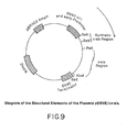
- pDSVE The vector chosen for expression of rPDGF B genes in Chinese hamster ovary DHFR ⁇ cells was called pDSVE.
- This vector was constructed using four basic DNA sequence elements. One of these elements provided DNA sequences necessary for selection and autonomous replication in bacterial cells. These characteristics are provided by the origin of replication and ampicillin-resistance gene DNA sequences in the region spanning nucleotides 2448 through 4362 (standard numbering system) of the plasmid pBR322.
- This 1918 base pair DNA fragment, derived from the pBR322 derivative pSV08 obtained from Dr. R. Tjian, University of California, Berkeley, was structurally modified by the addition of the following Hind III linker immediately adjacent to nucleotide 2448: 5 ⁇ -AAGCTTG-3 ⁇ .
- a second element provided DNA sequences constituting a viral promoter that is functional in mammalian and other vertebrate cells.
- a DNA fragment containing a simian virus 40 (SV40) early promoter was generated by first digesting SV40 DNA with restriction endonuclease enzyme Pvu II, producing three Pvu II/ Pvu II fragments of differing sizes.
- Pvu II restriction endonuclease enzyme
- One of these fragments contained the sequence ( Hind III at position number 5171 to Pvu II at position number 270) which codes for the counterclockwise "early gene" promoter and origin of replication of the SV40 virus.
- a third element provided a signal for terminating transcription of genes inserted into the vector.
- a DNA fragment containing the terminator sequence of the early SV40 genes was obtained by first digesting the complete SV40 genome with restriction endonuclease Hpa I, and then converting a blunt end into a Sal I recognition site by the attachment of a Sal I linker. This unligated Hpa I/ Sal I SV40 sequence was thereafter digested with Eco RI, and the 2030 bp Sal I/ Eco RI fragment was isolated by agarose gel electrophoresis.
- the fourth element contained in pDSVE provided a means for selecting mammalian cells containing the vector.
- the dihydrofolate reductase (DHFR) gene codes for an enzyme which allows cells to grow in media lacking thymidine and hypoxanthine, as well as allowing the cells to grow in media containing certain levels of the drug methotrexate.
- This gene comprised an approximately 2,500 bp mouse DHFR minigene, with Eco RI and Hind III restriction endonuclease sticky ends, isolated from plasmid pMG-1 as in Gasser et al., Proc. Natl. Acad. Sci. (USA) 79 : 6522-6526 (1982).
- the four DNA fragments containing these elements were ligated together with T4 DNA ligase, to produce the vector pDSVE.
- a diagram of this vector is shown in FIG. 5.
- Synthetic DNA linkers containing the recognition site for the restriction endonuclease Sal I were added to the blunt-ended c-sis fragment in a 21 ⁇ l reaction containing 1 ⁇ g of kinased linkers and T4 DNA ligase. After incubation overnight at 14° C, phenol/chloroform extraction and ethanol precipitation, the linkered DNA was restricted in a 100 ⁇ l reaction with 26 units of Sal I for 3 hours. The restricted DNA was ethanol precipitated, dissolved in a small volume of buffer and electrophoresed in a 1% low-melting temperature agarose gel. The 23 kb band was cut out of the gel and purified in accordance with the procedures described by Maniatis, ibid .
- the 23 kb band was ligated to pDSVLd DNA which had been linearized with Sal I, in a 20 ⁇ l reaction containing T4 DNA ligase.
- the ligated DNA was transformed into E . coli K12 strain DH1 (ATCC #33849). Ampicillin-resistant colonies were screened by hybridization with a 32P-labelled v-sis probe, according to procedures described by Maniatis ibid .
- a hybridizing clone was selected and plasmid DNA was prepared the clone.
- An insert from this clone, pDSVLd/ c-sis was excisable by restriction with Sal I. Restriction mapping confirmed that this clone contained the expected 23 kb c-sis DNA fragment.
- pDSVLd/ did not readily transform NIH-3T3 cells, and after introduction into COS-1 cells, or Chinese hamster ovary (CHO) cells pDSVLd/ c-sis did not lead to production of a product having detectable levels of PDGF activity.
- a related plasmid constructed by Gazit et al. ( ibid .) had been shown to be capable of transforming NIH-3T3 cells. Since pDSVLd/ c-sis contained a 6.7 kb region of DNA between the putative exon 1 region and exon 2 region which was not present in the Gazit et al.
- a new mammalian c-sis expression vector was therefore constructed, in which the 6.7 kb DNA fragment between the Hind III sites at 5.2 kb and 11.9 kb on the c-sis map was deleted.
- This new vector was constructed using the mammalian expression vector pDSVE described in section 2(a) above. It was found that pDSVE usually yields higher expression levels of the inserted gene in CHO cells than did pDSVLd.
- One microgram of pDSVE was restricted with Sal I, phenol/chloroform extracted amd ethanol precipitated, and used in the ligation described below.
- the effect of this procedure is to add a Sal I site near the Bam HI site at 3.9 kb on the map of U2-OS56.1 in FIG. 3.
- the 0.6 kb Sal I to Hind III fragment was isolated from the reaction mixture by electrophoresis through a 1.0% low-melting temperature agarose gel and extraction. This fragment is referred to below as the Exon 1 fragment.
- a DNA fragment containing the v-sis gene and 178 bp of SSV DNA upstream of it was inserted into pDSVE.
- the initial translation product resulting from transcription of this vector is a fusion of the SSV envelope glycoprotein and rPDGF B v-sis (Robbins et al., Nature 305 : 605 (1983)). Processing of the amino terminal portion of the fusion product may then result in production of a mature rPDGF B protein lacking SSV envelope protein sequences.
- the plasmids were mapped with the restriction enzymes Hind III, Xba I and Pst I to identify clones containing inserts in the correct orientation for transcription by the SV40 early promoter in pDSVE.
- One such clone was named pDSVE/ v-sis and was used in the examples described below. Its structure is diagrammed in FIG. 7.
- a mammalian expression vector in which the mature rPDGF B protein region is encoded by the v-sis gene, but the translation initiation signal and amino terminal region of the rPDGF B pre-protein is encoded by the c-sis gene the following procedure may be used.
- One microgram of pDSVE was linearized with Sal I, followed by phenol/chloroform extraction and ethanol precipitation.
- Three micrograms of the RF form of the M13mp18/ v-sis (Kpn-Xba) subclone described in Example 1 was restricted with Sst I and Sal I.
- a DNA fragment corresponding to the putative rPDGF B c-sis protein precursor amino terminal region was synthesized according to published procedures (McBride and Caruthers, Tetrahedron Lett. 24 : 245 (1983)). This fragment, the sequence of which is shown in FIG. 8, corresponds to the region spanning nucleotide 111 to nucleotide 203 of the rPDGF B protein cDNA clone described by Josephs et al., Science 225 : 636 (1984) .
- a Sal I site was added at the upstream end to permit ligation to the pDSVE vector. The Sst I site at the downstream end occurs naturally at this position in both c-sis and v-sis .
- the final expression vector was assembled in a 3-way ligation reaction containing 100 ng of Sal I-cut pDSVE, 2.6 ng of the synthetic Sal I/ Sst I c-sis fragment, and 25 ng of the Sst I/ Sal I v-sis fragment. After overnight incubation at 14° C in the presence of T4 DNA ligase, the reaction products were transformed into E . coli K12 strain DH1. Duplicate replica filters were made of ampicillin resistant colonies. One of the filters was hybridized to a radiolabeled v-sis probe as described above; the other was hybridized to radiolabeled synthetic Sal I/ Sst I c-sis fragment.
- Example 2 In order to achieve high level production of biologically active rPDGF B, the expression plasmids described in Example 2 were introduced into a mammalian cell line. The procedures used are essentially those described in PCT Patent Application No. US84/02021, 5863, which is incorporated by reference herein.
- CHO DHFR ⁇ cells (DuX-811) cells described by Urlaub et. al., Proc. Natl. Acad. Sci. (USA) 77 : 4461 (1980), lack the enzyme dihydrofolate reductase (DHFR) due to mutations in the structural genes and therefore require the presence of glycine, hypoxanthine, and thymidine in the culture media.
- Plasmids pDSVE/ c-sis , pDSVE/ v-sis or pDSVE/ cv-sis were transfected along with carrier DNA into CHO DHFR ⁇ cells growing in media containing hypoxanthine, thymidine, and glycine in 60 mm culture plates. The plasmid and carrier DNA mixture was transfected into CHO DHFR ⁇ cells.
- the cells were dispersed by typsinization into several 100 mm culture plates in media lacking hypoxanthine and thymidine. Only those cells which have stably transformed with the DHFR gene, and thereby the rPDGF B gene, survive in this media.
- transformant colonies After 7-21 days, colonies of surviving cells became apparent. These transformant colonies, after dispersion by typsinization, may be continuously propagated as a mixed population in media lacking hypoxanthine and thymidine, creating new cell strains (e.g., CHO-pDSVE/ v-sis , CHO-pDSVE/ cv-sis ). Alternatively, individual colonies may be trypsinized and transferred to the wells of microtiter dishes, creating new, clonal cell lines (e.g., CHO-pDSVE/ cv-sis ).
- Cell lines CHO-pDSVE/ v-sis , CHO-pDSVE/ c-sis and CHO-pDSVE/ c-sis were respectively deposited on March 13, 1987 with the American Type Culture Collection, 12301 Parklawn Drive, Rockville, Maryland 20852, as ATCC Nos. CRL 9359, CRL 9360 and CRL 9358.
- Culture fluids from the above cell strains including those derived by growth of CHO-pDSVE/ c-sis clones in microtiter wells, were tested in a bioassay and in a radioimmunoassay (RIA) as, described in Example 4, for the presence of rPDGF B.
- Representative 5 day culture fluids from CHO-pDSVE/ v-sis and CHO-pDSVE/ cv-sis cells contained approximately 100 to 200 ng/ml of rPDGF B as determined in both assays.
- Culture fluids from one of the CHO-pDSVE/ c-sis clones, which clone was used in subsequent work contained approximately 200 ng of rPDGF B per milliliter. Culture fluids from cells transformed only with pDSVE did not contain detectable amounts of rPDGF B.
- the quantity of recombinant rPDGF B produced by the cell strains described above may be increased by gene amplification, yielding new cell strains of greater productivity.
- the enzyme dihydrofolate reductase which is the product coded for by the DHFR gene, may be inhibited by the drug methotrexate (MTX). More speci- fically, cells propagated in media lacking hypoxanthine and thymidine are inhibited or killed by MTX. Under the appropriate conditions (e.g., minimal concentrations of MTX), cells resistant to and able to grow in MTX may be obtained. These cells are usually found to be resistant to MTX due to an amplification of the number of their DHFR genes, resulting in increased production of DHFR enzyme.
- MTX drug methotrexate
- the surviving cells may, in turn, be treated with increasing concentrations of MTX, resulting in cell strains containing even greater numbers of DHFR genes.
- Passenger genes e.g., rPDGF B
- carried on the expression vector along with the DHFR gene are frequently found also to be increased in their gene copy number.
- cell strains CHO-pDSVE/ c-sis , CHO-pDSVE/ v-sis or CHO-pDSVE/ cv-sis were subjected to increasing MTX concentrations (0 nM, 30 nM and 100 nM).
- Representative 5 day culture media samples from each amplification step of CHO-pDSVE/ c-sis were assayed by RIA and determined to contain 0.2, 2.0 and 2.0 ⁇ g/ml, respectively, of rPDGF B.
- Representative 5 day culture media samples from each amplification step of CHO-pDSVE/ v-sis and CHO-pDSVE/ cv-sis contained rPDGF B at 0.15, 1.0 and 1.0 ⁇ g/ml, respectively.
- 1 x 10⁶ cells were plated in 5 ml of media in 60 mm dishes. Twenty-four hours later the media were removed and replaced with 5 ml of serum-free media (high glucose DMEM supplemented with 0.1 mM non-essential amino acids and 2mM L-glutamine).
- rPDGF B was allowed to accumulate for 5 days in the serum-free media. The media was collected for RIA assay and the cells were trypsinized and counted.
- the number of cells per plate was 2.5 x 10⁶.
- the effective production rates for these culture conditions were thus 0.8 and 0.4 ⁇ g/106 cells/24 hours.
- the cells in the CHO-pDSVE/ v-sis and CHO-pDSVE/ cv-sis cultures described immediately above were a genetically heterogeneous population. Standard screening procedures were employed for these cells, as well as for pDSVE/ v-sis cells, in order to isolate genetically homogeneous clones with the highest production capacity. See Section 1, Part 2, of "Points to Consider in the Characterization of Cell Lines Used to Produce Biologics", June 1, 1984, Office of Biologics Research Review, Center for Drugs and Biologics, U.S. Food and Drug Administration.
- the productivity of the rPDGF B producing CHO cell lines described above may be improved by appropriate cell culture techniques.
- the propagation of mammalian cells in culture generally requires the presence of serum in the growth media.
- a method for production of rPDGF B from CHO cells in media that does not contain serum is expected to greatly facilitate the purification of rPDGF B from the culture medium when standard protein purification techniques are used.
- the method described below is capable of economically producing rPDGF B in serum-free media in large quantities sufficient for production.
- rPDGF B-producing CHO cells grown in standard cell culture conditions, are used to seed spinner cell culture flasks.
- the cells are propagated as a suspension cell line in the spinner cell culture flask in media consisting of 50-50 mixture of high glucose DMEM and Ham's F12 (Gibco) supplemented with 5% fetal calf serum, 2mM L-glutamine, 0.05 mM non-essential amino acids and the appropriate concentration of methotrexate.
- Suspension cell culture allows the rPDGF B-producing CHO cells to be expanded easily to large volumes.
- the CHO cells, grown in suspension, are used to seed roller bottles at an initial seeding density of 1.5 x 10⁷ viable cells per 850 cm2 roller bottle in 200 ml of media.
- the cells are allowed to grow to confluency as an adherent monolayer over a three-day period.
- the medium used for this phase of the growth is the same as used for growth in suspension.
- the serum-containing media is removed and replaced with 100 ml of serum-free media; 50-50 mixture of high glucose DMEM and Ham's F12 supplemented with 0.05 mM non-essential amino acids and 2mM L-glutamine.
- roller bottles are returned to the roller bottle incubator for a period of 1-3 hours and the media again is removed and replaced with 100 ml of fresh serum-free media.
- the 1-3 hour incubation of the serum-free media reduces the concentration of contaminating serum proteins.
- rPDGF B accumulates in the serum-free culture media.
- the conditioned media is removed and replaced with fresh serum-free medium for a second production cycle.
- a representative seven-day, serum-free medium sample contained rPDGF B c-sis at 1 ⁇ g/ml as judged by the RIA and mitogenesis assay.
- each 850 cm2 roller bottle contained from 0.75 to 1.5 x 10⁸ cells and thus the rate of production of rPDGF B c-sis in the 7-day, 100 ml culture was 0.10 to 0.19 ⁇ g/106 cells/24 hours.
- Cells are grown in spinner flasks and innoculated into roller bottles exactly as described above. After 3 days of growth to confluency, the medium is removed and replaced with 100 ml of the same medium containing 1% serum. The bottole are returned to the incubator and after 7 days the medium is removed. Another 100 ml may be added to the cells for another 7 day cycle of accumulation of rPDGF B in the medium. This cycle may be repeated several times before production levels fall off significantly. A representative 7-day sample contained 3 ⁇ g/ml of rPDGF B c-sis .
- rPDGF B active rPDGF B produced by CHO cells containing a foreign rPDGF B gene were unexpected, being 20-100 fold higher than the levels reportedly produced by a variety of other cell types into which a rPDGF B gene had previously been introduced (Huang et al., Cell 39 : 79 (1984); Johnsson et al., Proc. Natl. Acad. Sci. USA 82 : 1721 (1985); Kelly et al., EMBO J. 4 : 3399 (1985); or cells in which an endogenous PDGF B gene is expressed (Nilsson et al., Proc. Natl. Acad.
- the levels of active rPDGF B secreted into the culture media by CHO and other vertebrate cells may also be increased by altering the amino acid sequence of the precursor protein which is "upstream" of or amino terminal to the mature, secreted form of the protein.
- the portion of the rPDGF B chain gene encoding the region of rPDGF B c-sis upstream of amino acid number 82 in Figure 2 may be deleted and replaced with a DNA sequence coding for the amino terminal region of another protein, which protein is known to be secreted by CHO or other vertebrate cells in large quantity.
- the upstream DNA sequence may be replaced with a DNA sequence coding for the amino terminal region of the protein erythropoeitin, which protein can be secreted from CHO cells in large quantity.
- rPDGF B samples were tested for biological activity in a mitogenic assay.
- NIH 3T3 cells were plated at a density of 3 x 10⁴ cells per well in a 24-well plate (area approximately 2 cm2), and allowed to grow to confluency at 37° C in a cell growth medium consisting of Dulbecco's minimum essential medium ("DMEM"), supplemented with penicillin (100 units per ml) and streptomycin (100 ⁇ g per ml), and containing 10% heat-inactivated calf serum.
- DMEM Dulbecco's minimum essential medium
- penicillin 100 units per ml
- streptomycin 100 ⁇ g per ml
- PDGF standards and assay samples were added to the wells and incubation at 37° C continued for 18 hours. Media was removed, and the cells were labeled for exactly one hour at 37° C in one ml of fresh supplemented DMEM containing 5% heat inactivated calf serum and 20 ⁇ Ci per ml of Me-3H-thymidine (New England Nuclear, 20 Ci/m Mole). After one hour,the cells were chilled, medium was removed, and cell sheets were washed with cold phosphate-buffered saline (PBS), then with cold 5% Trichloracetic acid (TCA) which was allowed to stand for 5 minutes on ice.
- PBS cold phosphate-buffered saline
- TCA Trichloracetic acid
- TCA was removed, cells were washed once with fresh cold 5% TCA, the wash was aspirated off and cell sheets were dried.
- the contents of each well were dissolved in 1.0 ml of 0.25 M sodium hydroxide, transferred to a glass vial containing 10 ml of Aquasol II® scintillation cocktail (New England Nuclear Boston, MA), and counted in a Beckman liquid scintillation counter.
- a standard curve was constructed covering the concentration range of 2 to 120 ng per ml of PDGF, and PDGF activity of assay samples was calculated from the standard curve.
- the specific radioimmunoassay used to detect and quantitate rPDGF B employed PDGF purified from human platelets and rabbit polyclonal antisera raised against PDGF from the same source.
- Human platelet PDGF (0.5 ⁇ g) was iodinated using ,[ 125 I] Na and Iodo-GenTM (Pierce Chemical Co., Rockford, Ill.) by the procedure of Fraker and Speck, Biochem. Biophys. Res. Commun. 80 : 849 (1978).
- 125 I]PDGF (30,000 cpm) was incubated with an amount of antiserum which resulted in half-maximal precipitation of the labeled PDGF, in a 60 ⁇ l reaction containing RIA buffer (10 mM Tris-HCl, pH 8; 150 mM NaCl; 0.4% NonidetTM P-40 (Sigma, St. Louis, MO); and 0.1% BSA) for 16 hours at 4° C.
- RIA buffer (10 mM Tris-HCl, pH 8; 150 mM NaCl; 0.4% NonidetTM P-40 (Sigma, St. Louis, MO); and 0.1% BSA) for 16 hours at 4° C.
- a standard curve was generated by pre-incubation of the antiserum with varying amounts of unlabeled human platelet PDGF for 1 hour at room temperature, prior to addition of the 125 I-PDGF.
- rPDGF B proteins synthesized in and secreted by CHO cells containing sis vectors
- the cells were first incubated with 35 S-cysteine for 20 hours to label cysteine-containing proteins.
- the label incorporated into proteins is believed to approach a steady state distribution during this time period.
- Labeled PDGF-related proteins were specifically detected by immunoprecipitation with antiserum to human platelet PDGF and analyzed by SDS-PAGE and flourography.
- CHO-pDSVE/ c-sis , CHO-pDSVE/ v-sis , CHO-pDSVE/ cv-sis or CHO-pDSVE (as a control) cells were grown to approximately 80% confluency in 10 cm culture plates. The cells were washed twice with cysteine-deficient medium. Four milliliters of cysteine-deficient medium containing 0.625 mCi of 35S-cysteine was added to each plate. After incubation for 20 hours at 37° C, the media were collected and made 1 mM in PMSF and 0.025% in sodium azide to inhibit protease action and bacterial growth.
- the cells on each plate were lysed by addition of 1.4 ml of disruption buffer (0.025 M Tris-HCl, pH 8.0; 0.05 M NaCl; 0.5% sodium deoxycholate; 0.5% NonidetTM P-40; 1 mM PMSF).
- Insoluble material was removed by centrifugation for 1 min. in a Beckman Microfuge. Aliquots of media or cell extracts containing equal amounts of TCA-insoluble labeled material were pre-incubated with rabbit non-immune serum. Non-specifically-reacted proteins were removed by addition of Protein A-Sepharose (BioRad, Richmond, California) and centrifugation as above. Anti-PDGF serum was added to each supernatant.
- Protein A-SepharoseTM After incubation at 4° C for 16 hours, specifically-reacted proteins were removed by addition of Protein A-SepharoseTM for 1 hour at room temperature, followed by centrifugation as above. The Protein A-SepharoseTM/immune complexes were washed 6 times with RIA buffer. Bound immune complexes were disrupted and released from the protein A-SepharoseTM by boiling in 1% SDS for 5 minutes. Each supernatant was split into two equal portions, and gel loading buffer (1% SDS, 20% glycerol, 0.1% bromophenol blue) either with or without reducing agent (3% 2-mercaptoethanol) was added to each aliquot. The samples were boiled again and then electrophoresed through an 18% SDS-polyacrylamide gel. The gels were treated with En3HanceTM (New England Nuclear, Boston, MA), dried, and exposed to X-ray film for 2-10 days.
- En3HanceTM New England Nuclear, Boston, MA
- rPDGF B The major stable intracellular form of rPDGF B in cells transformed by sis -containing vectors ran as a homogeneous band of 23 kd. After reduction, this form was converted to three smaller species of approximately 8.5 kd, 7.5 kd and 7 kd.
- the major extracellular forms of rPDGF B secreted by these cells were heterogeneous.
- the two species with the greatest intensities on the autoradiograms had mobilities corresponding to 27.5 kd and 25 kd.
- a 30.5 kd species was somewhat less intense.
- a fourth species of much lower intensity was similar in size to the major intracellular form, i.e. 23 kd.
- the reduced forms of extracellular rPDGF B were also heterogeneous.
- the most intense band had a mobility corresponding to 13.5 kd. Somewhat less intense bands of 17 kd, 16 kd and 12 kd were also present.
- an expression plasmid was constructed for introduction into E . coli .
- the plasmid was designed to code for a fusion protein consisting of the amino terminal 120 amino acids of chicken growth hormone (CGH) (Souza et al., J. Exp. Zoology 232 : 465 (1984)) and the carboxy terminal 136 amino acids of the protein encoded by the v-sis gene.
- CGH chicken growth hormone
- the complete sequence of the resulting protein, called CGP1 are shown in FIG. 11.
- pCGP1 Ampicillin-resistant colonies containing v-sis were identified by hybridization to a v-sis probe. Plasmid DNA was isolated from several such colonies and analyzed by restriction mapping with Xba I, Bam HI and Eco RI. One clone, called pCGP1, had v-sis DNA inserted in the correct orientation for expression and was selected for further use.
- pCGPl DNA was transformed into the E. coli strain K12 for expression.
- Cells of E. Coli K12 strain AM7 transformed with pCGP1 were deposited on March 13, 1987 with the American Type Culture Collection, 12301 Parklawn Drive, Rockville, Maryland 20852 as Deposit No. 67352.
- a 20-milliliter culture was grown overnight at 28° C in Luria broth.
- One milliliter of this culture was used to innoculate one liter of Luria broth, and the larger culture was incubated at 28° C on a rotary platform. Absorbance of the culture was monitored and when the absorbance at 600 nm was 0.5, the culture was shifted to 37° for 13.5 hours to induce expression of the fusion protein.
- the cells were pelleted by centrifugation and resuspended in 12 ml of 10 mM Tris-HCl (pH 7.5) and 1 mM EDTA. The cells were broken in a French pressure chamber and insoluble material was pelleted by low speed centrifugation.
- Rabbits were injected with CGP1 and bled according to the following schedule: On day 0, a preimmune bleed was taken, and each rabbit was injected intradermally in multiple sites on the back with a total of 250 ⁇ g CGP1 in 1.0 ml of a mixture containing 50% emulsified BACTO-Freunds complete adjuvant H37 Ra (DIFCO 3113-60), as available from DIFCO, Detroit, Michigan in PBS. Fourteen days later, the rabbits were intradermally injected with 250 ⁇ g of CGP1 mixed with 50% Freunds incomplete adjuvant (DIFCO, BACTO 0639-60-6), as before. A second, identical booster injection was done on day 30, and on day 42 the rabbits were test bled. The resulting sera were tested for the production of anti-CGP1 antibodies.
- DIFCO 3113-60 50% emulsified BACTO-Freunds complete adjuvant H37 Ra
- Activity curves were constructed by plotting rabbit serum dilution (10 ⁇ 1 to 10 ⁇ 5) against counts per minute of bound 125I-protein-A, and the titer of each serum was determined to be the reciprocal of the dilu-tion at which 50% of the maximum count was bound.
- Two of three rabbits were strongly positive for antibodies to CGP1 protein (#304 and #305), producing titers ranging from 5.6 x 10³ and 1.5 x 10³ initially to 2.6 x 10⁴ and 4 x 10³, respectively.
- Immunoglobulin absorbed to the column was eluted with 0.58% acetic acid in 0.15 M sodium chloride and was located in resulting fractions by U.V. absorbance.
- the fractions containing immunoglobulin were neutralized with 1.0 M Tris buffer (pH 8.0).
- Tris buffer pH 8.0
- immunoglobulin concentration was determined by U.V. absorbance at 280 nM using an extinction coefficient of 1.46.
- the yield of rabbit immunoglobulin was 42.9 mg.
- cyanogen bromide-activated SepharoseTM 4B (Pharmacia Fine Chemicals, Uppsala, Sweden) was swelled in 0.001 M HCl and washed on a glass funnel with 600 ml of the same solution.
- the wet gel was transferred to a 50 ml centrifuge tube and washed with 20 ml of coupling buffer (0.1 M sodium bicarbonate and 0.5 M sodium chloride at pH 8.5). After a 4 minute centrifugation at 600 x g, the supernatant fluid was removed and the 42.9 mg of rabbit immunoglobulin in 9.7 ml of coupling buffer was added to the SepharoseTM 4B.
- coupling buffer 0.1 M sodium bicarbonate and 0.5 M sodium chloride at pH 8.5
- the mixture was tumbled end over end at 4° C overnight, centrifuged as above, and the supernatant tested for the presence of residual immunoglobulin. Less than 0.3% of original immunoglobulin remained in the supernatant. Thus the efficiency of coupling was better than 99.7%.
- the immunoglobulin-coupled SepharoseTM 4B was washed with 4 cycles of 0.1 M sodium acetate, (pH 4.0), and 0.5 M sodium chloride, followed by coupling buffer.
- the gel was equilibrated with buffer E (0.0026 M KCl, 0.1369 M NaCl, 0.0014 M KH2P04, and 0.008 M Na2HP04), poured into a 1.5 x 10 cm glass column, and washed at 4°C with 100 ml of buffer E.
- Culture fluids harvested after 4-7 days from roller bottle cultures of CHO-pDSVE/ cv-sis , CHO-pDSVE/ v-sis or CHO-pDSVE/ cv-sis cells were centrifuged at 10,000 X g for 20 minutes, and the supernatant fluid passed through a 0.45 ⁇ filter.
- One hundred to 400 ml of filtered medium was adsorbed to a 10 ml anti-CGP1-Sepharose affinity column at a flow rate of 30 to 40 ml/hr, at 4° C.
- the column was washed sequentially with 20 column volumes of buffer F (0.5M NaCl, 0.025M Tris-HCl, pH 7.5, and 0.1% NonidetTM P-40 and at least 5 column volumes of buffer G (0.15M NaCl, 0.01% NonidetTM P-40).
- buffer F 0.5M NaCl, 0.025M Tris-HCl, pH 7.5, and 0.1% NonidetTM P-40
- buffer G 0.15M NaCl, 0.01% NonidetTM P-40
- Bound rPDGF B was eluted with buffer H (0.2 M acetic acid, 0.15 M NaCl and 0.01% NP40) at a rate of 30 ml/hour.
- rPDGF B containing fractions were located by testing 50 ⁇ l aliquots in the Bio-Rad protein assay (Bio-Rad, Richmond, CA), and by immunological assay in a dot immunobinding assay employing rabbit anti-CGP1 antiserum as described below. Protein-containing fractions were pooled, dialyzed against 0.1 M acetic acid and 0.01% NonidetTM P-40 at 4° C, and then lyophilized.
- the nitrocellulose paper was "blocked” by shaking in a solution of 10% dried, non-fat milk powder (Carnation, Los Angeles, CA) in PBS (pH 7.4) (0.15M NaCl, 10 mM Na2HP04 (pH 7.4), incubated with a 1:50 dilution of rabbit anti-CGP1 serum in PBS-1% dried milk powder, and washed three times with PBS containing 0.025% TweenTM 20.
- the detection of bound antibody was carried out with a VectaStainTM Kit, according to the manufacturer's instructions (Vector Laboratories, Burlingame, CA). Approximate rPDGF B concentrations in column fractions were estimated by color intensity of column fraction "dots" by comparison with standard "dots”.
- Lyophilized rPDGF B fractions from the rabbit immunoglobulin affinity column were reconstituted in 10 mM acetic acid. Aliquots (0.2 to 2 ⁇ g) were loaded (either non-reduced or reduced with 5% 2-mercaptoethanol) into wells of either 15% or 18% polyacrylamide gels, 1.5 mm thick. Electophoresis was performed in Tris-glycine buffer (0.025 M Tris, 0.192 M glycine) containing 0.1% SDS, at 80-90 volts for 10-12 hours.
- Protein bands were visualized by development with Coomassie Blue stain or with silver stain (RAPID-Ag-STAIN, ICN Radiochemicals, Irvine CA) , or protein was transferred from the developed gel to nitrocellulose paper (BA83, Schleicher and Scheull) by an electrophoretic transfer technique (BioRad Trans-Blot Cell, catalogue no. 170-3910) employing electrical current to drive the resolved bands from the gel onto the surface of the paper. The transfer was carried out in a buffer containing 0.15M glycine, 0.02M Tris base and 20% methanol (vol/vol), for 6h at 60V at room temperature.
- Coomassie Blue and silver-stained gels showed two major protein bands occurring in the area of a 25,400 mw marker for unreduced samples, and two bands close to a 14,400 mw marker for reduced samples. From the silver-stained gels, the rPDGF B was estimated to be more than 95% pure. Nitrocellulose blots were blocked with 10% goat serum and 2% BSA in PBS for 1 hour, incubated with the primary antibody for 2 to 3 hours, washed with KP (Kirkegaard and Perry, Gaithersburg, Maryland) wash solution, incubated with secondary biotinylated goat anti-rabbit serum in PBS for 1.5 hours, and the antibody detection carried out using a VectaStainTM kit as above (see Example 6(c)). The primary antibody employed for the immunoblot was rabbit anti-CGP1 serum, (#305) diluted 1:50.
- rPDGF B v-sis Purified as described in Example 6
- Sera were titered as described in Example 5(c), using rPDGF B v-sis bound to the microtiter plate wells; generally, the titers were > 1 X 10⁴,
- antisera were capable of specifically detecting as little as 1 ng of rPDGF B or 5 ng of PDGF purified from human platelets in a dot immunobinding assay (as described in Example 6(c)), at a dilution of 1:100.
- the antisera also specifically reacted with human platelet PDGF radioiodinated as described in Example 4(b), or rPDGF B radioiodinated by the Bolton-Hunter technique (Bolton et al., Biochem. J. 133 : 529 (1973)), in a radioimmunoassay performed as described in Example 4(b).
- mice Five female mice, of strain Balb/c, (Jackson Laboratories, Ann Arbor, Maine) were each injected initially with 10 ⁇ g of rPDGF B v-sis purified as described in Example 6, in approximately 0.3 ml of a solution consisting of 0.15 ml of PBS [0.15 m NaCl and 0.01 M Na2HPO4 (pH 7.4)] emulsified by sonication with 0.15 ml Freunds complete adjuvant (Calbiochem-Behring modification, Calbiochem-Behring Corporation, La Jolla, California). The mice received multiple subcutaneous injections on the back.
- each mouse received a booster immunization intraperitoneally, which immunization consisted of 10 ⁇ g rPDGF B v-sis in 0.3 ml of PBS emulsified in MPL/TDM special emulsion R-700 (RIBI Immunochemical Research, Inc., Hamilton, Montana).
- the mice received 0.15 ml in multiple sites on the back, as before, and 0.15 ml intraperitoneally. Twenty days later, the mice were bled and sera were assayed for antibodies to rPDGF B v-sis .
- rPDGF B v-sis (700 ⁇ g/ml) in 0.035 M NaHC03 and 0.015M Na2C03 was bound to the wells of a serological 96-well plate by incubating 50 ⁇ l of antigen solution overnight at room temperature. Removal of non-bound antigen was followed by blocking unbound sites with 250 ⁇ l per well of PBS containing 5% bovine serum albumin for one hour at room temperature. Fifty microliters of mouse sera dilutions, from 10 ⁇ 1 to 10 ⁇ 5 in PBS, were added to the antigen-containing, blocked wells, incubated at room temperature for three hours and removed.
- mice were then washed three times with KP wash solution. Each well received 50 ⁇ l PBS containing 1 X 10⁵ cpm 125I-rabbit antimouse immunoglobulin G (IgG) (NEX 1610, New England Nuclear) with 0.1% bovine serum albumin. Wells were incubated for 90 minutes, washed five times with KP wash solution and counted in a Beckman 5500 gamma counter. Three mice were producing antibodies to rPDGF B v-sis with titers of greater than or equal to 103. These three were given two more booster immunizations, identical to the first booster one week after the bleed and ten days after the bleed.
- IgG 125I-rabbit antimouse immunoglobulin G
- mice were killed by cervical dislocation and rinsed with 70% ethanol.
- Spleens were removed asepticaly and placed in petri dishes (on ice) containing Dulbecco's Modified Eagle's Medium (Gibco) with pencillin G and streptomycin at 200 units and 200 ⁇ g per ml, respectively.
- the spleens were trimmed of fat and connective tissue, passed through two rinses in fresh media (as above) and placed in a sterile stomacher bag with 10 ml of the same medium.
- Disruption of the spleens in the stomacher apparatus (Stomacher Lab-Blender 80, Seward Laboratories, London, England) for 90 seconds was followed by filtration through four layers of sterile gauze, and the resulting spleen cells were pelleted by centrifugation for 10 minutes at 1000 rpm in an IEC-HN-SII centrifuge.
- the cell pellet was washed twice with media (as above) containing penicillin-streptomycin and once with media containing no antibiotics.
- the cell pellet was resuspended in fresh medium and the cell concentration was determined by counting in a hemocytometer in the presence of 0.2% trypan blue.
- Mouse myeloma cells SP 2/0 derived from Balb/c strain, were grown in HB1O1 medium (NC-200, New England Nuclear Research Products) containing 1% heat-inactivated fetal bovine serum (FBS). The cells were pelleted by centrifugation at 1000 rpm for 10 minutes, as for the spleen cells, and washed with Dulbecco's modified Eagle's Medium (DMEM) containing no antibiotic. The cell concentration was determined by counting in a hemocytometer in 0.2% trypan blue after resuspension in the same medium.
- DMEM Dulbecco's modified Eagle's Medium
- Spleen and SP 2/0 cells were combined in a ratio of 3:1 and centrifuged at 1000 rpm for 10 minutes as above.
- the supernatant fluid was aspirated away and cell fusion was conducted at 37° C using polyethylene glycol (PEG) 1500, molecular weight 500-600.
- PEG polyethylene glycol
- This procedure was carried out with constant gentle stirring by addition of the following, at the times indicated: 1.0 ml of 50% PEG in DMEM added over one minute, with one minute stirring; 1.0 ml DMEM containing 10% fetal bovine serum (FBS) over one minute; 1.0 ml of DMEM containing 10% FBS added over one minute; and 8 ml DMEM containing 10% FBS added over 2 minutes.
- FBS fetal bovine serum
- the resulting fused cells were centrifuged at 1000 rpm for 10 minutes as above, resuspended in approximately 45 ml of DMEM containing 10% FBS and also containing pencillin G and streptomycin at 100 units and 100 ⁇ g per ml, respectively.
- the cells were plated at 0.1 ml per well in 96-well plates previously equilibrated in 9% CO2. Plates were incubated at 37° C in 9% CO2.
- HAT medium (13.6 ⁇ g/ml hypoxanthine, 0.176 ⁇ g/ml aminopterin and 3.88 ⁇ g/ml thymidine) in DMEM containing 10% FBS.
- This medium selectively allows spleen cell-SP 2/0 hybrids to survive, screening out unfused SP 2/0 cells or those fused to other SP 2/0 cells. Unfused primary spleen cells from adult mice will not survive in culture for more than a few days.
- ELISA enzyme-linked immunosorbent assay
- Wells were washed five times with wash solution, flicked dry, and the color reaction developed with 100 ⁇ l of ABTS color reagent (Kirkegaard and Perry Laboratories, Gaithersburg, Maryland). The wells were scanned at 414 nm employing a Titertek Multiscan (Flow Laboratories, Springfield, Virginia).
- 173 were significantly positive for reaction with rPDGF B v-sis .
- cells from 22 wells were selected for further expansion and cloning.
- Cells from each of the selected wells were diluted into 3 X 96 wells each, giving rise to individual clones, or in some cases, a small number of clones per well.
- culture fluids from all wells containing one or more cell clones were tested by ELISA assay as described above.
- Anti-rPDGF B v-sis antibody was detected in some wells of plates representing 15 of the 22 original master plate wells, and corresponding cells were subjected to further cloning. Of these 15, the cells corresponding to wells with highest titer were selected to be recloned to give single-cell clones.
- Selected positive single cell hybridoma clones were expanded in culture, cells were harvested, centrifuged at 700 rpm in an IEC-HNSII centrifuge for 5 minutes, washed once with DMEM (no serum) and once with PBS. Washed hybridoma cells were suspended at 2 X 10⁷ cells/ml in PBS, and 5 X 10⁶ cells were injected intraperitoneally into female Balb/c mice (Jackson Laboratories, Bar Harbor, Maine) 6 to 8 weeks of age or into female CDFI hybrid mice from the same source.
- Ascites fluid developed in injected mice at 8 to 14 days.
- the ascites fluid was harvested, centrifuged at 2000 rpm (IEC-HN-SII) for 10 minutes, and the clear supernatant fluid was utilized further as a source of monoclonal antibody.
- Reactivity and titer of the monoclonal antibodies in the ascites fluids were assayed against rPDGF B v-sis by a plate binding assay essentially as described in Example 5(c) wherein rPDGF B v-sis was coated at 0.7 ⁇ g/ml, and serial ten-fold dilutions of ascites fluids were made in PBS containing 1% BSA.
- the murine monoclonal antibody was detected with 125I-Rabbit anti-mouse IgG antibody (NEX 161, New England Nuclear, Boston, MA) approximately 105 cpm (Beckman 5500 gamma counter) per well.
- Titers of individual ascites fluids were calculated as the reciprocal of the dilution at which 50% of maximum binding occurred. Titers of ascites from 12 different monoclonal antibody-producing clones ranged from 1.5 X 10⁴ to 7.5 x 10⁵. Results are given in Table 1.
- the blocking solution was removed and the blot was reacted with one of 9 primary antibodies (8 rPDGF B monoclonals and 1 rabbit anti-platelet PDGF serum) for 3 hours and washed with KP wash solution.
- Bound primary antibody was detected by reaction with a secondary biotinylated horse anti-mouse IgG antibody, an avidin-horseradish peroxidase complex, and substrate color reagent HRP (BioRad 1.70-6534) containing 4-chloro-1-napthol; 60 mg of the last reagent was dissolved in 20 ml methanol, diluted into 100 ml TBS (0.2 M NaCl, 0.05 M Tris, pH 7.4) containing 60 ⁇ l of 30% hydrogen peroxide.
- TBS 0.2 M NaCl, 0.05 M Tris, pH 7.4
- the twelve selected monoclonal antibody clones were screened for their immunoglobulin subtype utilizing a mouse immunoglobulin subtype identification kit (catalogue no. 100 036, Boehringer-Mannheim, Indianapolis, IN). For each antibody tested, 9 wells of a 96-well tray were coated with 50 ⁇ l of a solution of 0.7 ⁇ g/ml rPDGF B v-sis in carbonate-bicarbonate buffer (as in Example 5) overnight. Remaining protein binding sites were blocked by incubation with 200 ⁇ l per well of 2% BSA in PBS for 2 hours at room temperature.
- each well was added 50 ⁇ l of one of the monoclonal antibodies (approximately 0.5 ⁇ g of immunoglobulin), and incubation was continued for 3 hours at room temperature.
- the wells were washed with 200 ⁇ l washing buffer (10 mM Tris-HCl, pH 7.0; 0.05% Tween 20; 0.01% thimerisol) four times.
- Monoclonal antibodies raised against rPDGF B v-sis were tested for the abilitiy to neutralize the biological activity of rPDGF B v-sis in the mitogenesis assay for 3H-thymidine incorporation into murine NIH cells (as in Example 4).
- the immunoglobulin fraction from each of 6 ascites fluids, generated from 6 different hybridomas, was isolated by chromatography on a protein A-agarose column. The specific technique utilized was Bio-Rad's Affi-Gel Protein A MAPS II monoclonal antibody purification system.
- the immunoglobulin fractions of the monoclonal antibody ascites fluids were analyzed by SDS- PAGE on 12.5% gels. Approximately 2 ⁇ g of each immunoglobulin was run in comparison with the same quantity of immunoglobulin from unfractionated ascites. All samples were reduced by treatment at 100°C for 5 minutes in 5% 2-mercaptoethanol before applying to the gel. Electrophoresis and silver-staining of gels were performed as described in Section 6(d). The stained gels revealed a high degree of purity of all immunoglobulin fractions and the presence of amounts of heavy- and light-chain IgG expected from the amounts loaded. Each immunoglobulin sample assayed contained heavy- and light-chain components which migrated on the gel to the expected molecular weight positions of 50,000 and 25,000 respectively.
- Sensitive and very specific immunoassays may employ monoclonal and polyclonal antibodies to rPDGF B according to the present invention in a variety of combinations and variations, and may utilize a variety of detection schemes.
- the present invention comprehends all of the possible combinations and variations that utilize monoclonal antibodies directed against native, non-reduced, dimeric PDGF according to the present invention, and is not limited to the specific example described here.
- Such assays may be useful for the diagnosis and prediction of prognosis in certain pathological conditions, e.g., some forms of cancer.
- Purified monoclonal antibody 162 (50 ⁇ l) was adsorbed to the wells of Nunc 96-well microtiter plates at 5 ⁇ g/ml in 0.05 M sodium carbonate (pH 9.2) in an overnight incubation at room temperature. The solution was aspirated and the remaining protein adsorption sites were blocked with 150 ⁇ l of 2% gelatin for 45 minutes at room temperature. The solution was aspirated and the wells were rinsed once with PBS containing 0.025% TweenTM 80. Fifty microliters of antigen in PBS containing 0.025% TweenTM 80 and 0.2% gelatin were added to each well, before incubation for 1.5 to 2 hours at room temperature.
- the antigen solution was removed and the wells were washed once with PBS containing 0.025% TweenTM 80.
- a rabbit polyclonal antiserum raised against rPDGF B was diluted 1:1000 in PBS containing 0.025% TweenTM 80 and 0.2% gelatin, and 50 ⁇ l was added to each well. After incubation at room temperature for 1 to 1.5 hours, the solution was removed. The wells were washed once with PBS containing 0.025% TweenTM 80.
- Purified monoclonal antibody immunoglobulins 52 and 162 were coupled to CnBr-activated SepharoseTM4B for the development of affinity columns. The coupling was carried out as described in Example 6(d). The ratio of immunoglobulin to Sepharose 4B was 3.5 mg per ml of SepharoseTM for monoclonal 52, and for 162 it was 3.0 mg/ml.
- Affinity purification of rPDGF B from rPDGF B producing cell may be performed using monoclonal antibody 52 and 162 columns ("Seph 52" and "Seph 162").
- the operation of the two monoclonal affinity columns was essentially as described in Example 6 for the rabbit anti-CGP1 immunoglobulin affinity columns.
- Protein-containing (PDGF) fractions were located by the Bio-Rad protein assay, mitogenic assay for 3H-thymidine incorporation, or, in some cases, by dot immunobinding assay. rPDGF B containing fractions were pooled and stored for later analysis as described below.
- rPDGF B chromatographed on Seph 52 or Seph 162 affinity columns
- SDS-PAGE SDS-PAGE.
- Unlabeled rPDGF B was detected by silver staining, a technique which permits detection of small quantities of a protein, or by staining with Coomassie Brilliant Blue. Although silver staining is generally a more sensitive method, some proteins are better detected with the Coomassie stain.
- Affinity column-purified rPDGF B was iodinated by the method of Bolton and Hunter, ibid .
- the labeled rPDGF B (2 x 10⁵ cpm) was electrophoresed through a SDS-polyacrylamide gel, dried, and exposed to X-ray film for 16 hours.
- the only bands detected in the autoradiogram corresponded to rPDGF B polypeptides. Since even 50 cpm in a band would have been detectable under these conditions, this analysis shows that the purity of this particular batch of rPDGF B, purified by these methods, was greater than 99%.
- rPDGF B fractions recovered from the rabbit immunoglobulin affinity column, and pooled fraction samples were tested for biological activity in a mitogenic assay as described in Example 4a. All rPDGF B-containing affinity column fractions were active in the mitogenic assay, and the concentrations calculated from the bioassay standard curve correlated well with the concentrations determined by BioRad protein assay and by dot immunobinding analysis. The specific activity of rPDGF B was similar to several batches of PDGF purified from human platelets.
- rPDGF B purified as described in Example 10 was subjected to N-terminal sequence analysis and the first 14 amino acids of rPDGF B c-sis were identified as Ser-Leu-Gly-Ser-Leu-Thr-Ile-Ala-Glu-Pro-Ala-Met-Ile-Ala. These are the same 14 amino acids which occur at the N-terminus of the B chain of PDGF purified from human platelets (Johnsson et al, supra ).
- the small amounts of amino termini beginning at amino acids number 33 and 80 are likely to be the result of the action of specific proteolytic enzymes found inside CHO cells and platelets. For some purposes it may be desirable to obtain a rPDGF B product which does not contain these internal cleavages. This result may be achieved by altering the amino acid sequence at the cleavage sites in a way that prevents recognition by the specific proteolytic enzymes involved. Thus the particular amino acid residues number 32, 33, 79 and/or 80, as well as immediately adjacent residues, may be changed to alternate amino acids by changing the DNA sequence coding for these residues.
- Glycosylation of proteins may be either of two types.
- the most common type is N-linked, in which sugar resides are linked to asparagine at sites having the sequence asn-X-thr/ser.
- O-linked glycosylation sugar resides are linked to serines or threonines; no other consensus sequence for this type of glycosylation has been identified.
- PDGF purified from human platelets contains approximately 7% carbohydrate by weight (Deuel et al., J. Biol. Chem. 256 :8896 (1981)) .
- the distribution of sugar residues between A and B chains is unknown. However, it can be deduced that the B chain does not contain N-linked carbohydrate, since the sequence asn-X-thr/ser does not occur within it. It remains to be determined whether O-linked glycosylation occurs in the B chain of platelet PDGF.
- rPDGF B v-sis produced by CHO cells contains carbohydrate
- two types of analyses were performed. The first analysis monitored changes in the electrophoretic mobility of the protein during SDS-PAGE, following incubation with the glycosidases neuraminidase, endoglycosidase F or O-glycanase. Results of these experiments indicated that only a very minor component of the protein was altered by treatment with these enzymes.
- the second form of carbohydrate analysis involved release of sugar residues by acid methanolysis, and quantitation by gas chromatographic analysis, according to the procedure of Zanetta et al., J. Chromatogr. 69 :291 (1972).
- the results of this analysis were: fucose, 0.025% wt/wt; galactose, 0.125%; mannose, 0.075%; N-acetylglucosamine, 0.038%; sialic acid, 0.525%.
- rPDGF B v-sis produced by CHO cells contains less than 1% by weight of carbohydrate.
- PDGF purified from human platelets has been shown to be chemotactic for fibroblasts, smooth muscle cells, granulocytes and monocytes (Deuel et al., J. Clin. Invest . 69 : 1046 (1982); Seppa et al., J. Cell Biol. 92 : 584 (1982); Grotendorst et al., Proc. Natl. Acad. Sci. USA 78 : 3669 (1981). To determine if rPDGF B shares these properties, it was tested in chemotaxis assays as described by Deuel et al., supra . The rPDGF B was found to be chemotactic for all of the above cell types, to a degree equal to PDGF purified from human platelets.
- Wound chambers are constructed from 3.0 x 3.5 cm pieces of stainless steel wire cloth (40 mesh; 0.010 mesh wire). The wire cloth is rolled around a rod to produce a cylinder 3.5 cm in length and 0.95 cm in diameter. One end of the cylinder is compressed to form a blunt end while a silicone rubber septum is attached to the open end. The completed wound chamber is heat sterilized (120° C for 30 min.) prior to implantation.
- Wound chambers are implanted subcutaneously on the dorsum of male Sprague-Dawley rats (350-400gm) into pockets made by a 2 cm scalpel incision and blunt dissection to the level on the panniculus carnosus. Following implantation of the chambers, the wounds are closed with stainless steel clips. In this manner, each animal receives two chambers at shoulder level and two additional chambers at the level of the hind legs.
- 0.1 ml control vehicle or 0.1 ml test article is injected into the chamber by penetration of the silicone rubber plug with a hypodermic needle.
- the pattern of injection is systematically varied among animals to prevent anatomical location of chambers from influencing the results.
- the chambers are removed and the contents scraped out onto a tared weighing boat for determination of net weight.
- total protein is determined by the BioRad protein assay, total DNA by the method of Vytasek, Anal. Biochem. 120 : 243 (1982), and total hydroxyproline by the method of Berg, Methods Enzymol. 82 : 372 (1982).
- Table II shows the effect of daily administration of 5.0 ⁇ g rPDGF B c-sis on the accumulation of granulation tissue within the wound chambers.
- the data show that the total net weight of accumulated tissue was almost three-fold higher than that obtained from chambers injected with control vehicles. Similar increases were noted in the total amounts of protein, DNA and hydroxyproline deposited within the chambers.
- the maximum load tolerated by wounded skin was measured with the Tensometer 10. Measurements were performed on strips stretched at 10 mm per minute, with the maximum load recorded on a graph. Breaking strength measurements were not performed on wounds showing evidence of infection, excessive hemorrhage or poor coaptation (less than 5% of all wounds).
- Decreased wound healing is a characteristic feature of many disease states.
- a classical example is insulin-dependent diabetes wherein impaired circulation and decreased monocyte/macrophage functions are associated with a diminished rate of wound healing. Since these conditions could result in a decreased delivery of growth factors to the wound site, it was of interest to ascertain if direct delivery of recombinant human PDGF b-sis could bypass this defect.
- rats were weighed and then made diabetic by a single intravenous injection of streptozotocin (70 mg/Kg body wt.) at the time of implantation of subcutaneous wound chambers. The wound chambers are described in Example 10(F).
- wound chambers were injected daily with either 0.1 ml control vehicle or vehicle containing 1.0 ⁇ g recombinant PDGF b-sis .
- Treatment continued for nine (9) successive days at which time the rats were again weighed, blood samples were obtained for plasma glucose analysis, and wound chambers were removed for determination of granulation tissue formation. The results obtained were then compared with those from a concurrent group of non-diabetic rats whose wound chambers had been injected with control vehicle.
- rPDGF-B has shown efficacy in promoting the healing of rabbit ear punch wounds.
- 6 mm diameter plugs of tissue are removed down to the level of cartilage.
- Regrowth of tissue involves both granulation tissue and epithelium.
- three measurements were made on cross sections through the center of each wound at various times post-wounding. Two of the measurements, i.e. maximum depth of granulation tissue and distance between advancing edges of granulation tissue , were concerned with the growth of granulation tissue.
- the third measurement, of the reepithelialization gap was concerned with the regrowth of epithelium across the wound.
- monoclonal antibodies which recognize epitopes present on the A and/or B chains of PDGF may be useful for purification of PDGF.
- PDGF is the major mitogen for connective tissue cells
- the highly purified PDGF of the present invention will be useful for stimulating wound healing.
- PDGF is released from platelets only at the site of wounds and it is both mitotic and chemotactic for connective tissue cells.
- One study showed greater collagen synthesis in PDGF-treated wounds of rats than when one of several other hormones were applied.
Landscapes
- Health & Medical Sciences (AREA)
- Chemical & Material Sciences (AREA)
- Organic Chemistry (AREA)
- Life Sciences & Earth Sciences (AREA)
- General Health & Medical Sciences (AREA)
- Medicinal Chemistry (AREA)
- Proteomics, Peptides & Aminoacids (AREA)
- Molecular Biology (AREA)
- Genetics & Genomics (AREA)
- Biophysics (AREA)
- Biochemistry (AREA)
- Nuclear Medicine, Radiotherapy & Molecular Imaging (AREA)
- General Chemical & Material Sciences (AREA)
- Public Health (AREA)
- Veterinary Medicine (AREA)
- Bioinformatics & Cheminformatics (AREA)
- Animal Behavior & Ethology (AREA)
- Chemical Kinetics & Catalysis (AREA)
- Gastroenterology & Hepatology (AREA)
- Pharmacology & Pharmacy (AREA)
- Engineering & Computer Science (AREA)
- Toxicology (AREA)
- Zoology (AREA)
- Dermatology (AREA)
- Immunology (AREA)
- Preparation Of Compounds By Using Micro-Organisms (AREA)
- Peptides Or Proteins (AREA)
- Medicines Containing Antibodies Or Antigens For Use As Internal Diagnostic Agents (AREA)
- Micro-Organisms Or Cultivation Processes Thereof (AREA)
- Medicines That Contain Protein Lipid Enzymes And Other Medicines (AREA)
Priority Applications (1)
| Application Number | Priority Date | Filing Date | Title |
|---|---|---|---|
| EP93120821A EP0622456A1 (fr) | 1987-03-13 | 1988-03-10 | Vecteurs procaryotiques et eucaryotiques produisant des polypeptides contenant des épitopes de PDGF-B |
Applications Claiming Priority (4)
| Application Number | Priority Date | Filing Date | Title |
|---|---|---|---|
| US25344 | 1987-03-13 | ||
| US07/025,344 US5175255A (en) | 1987-03-23 | 1987-03-23 | Methods for purification of platelet-derived growth factor |
| US15204588A | 1988-02-19 | 1988-02-19 | |
| US152045 | 1988-02-19 |
Related Child Applications (3)
| Application Number | Title | Priority Date | Filing Date |
|---|---|---|---|
| EP93104885.4 Division-Into | 1988-03-10 | ||
| EP93120821.9 Division-Into | 1988-03-10 | ||
| EP93104885A Division EP0559234A1 (fr) | 1987-03-13 | 1988-03-10 | Anticorps monoclonaux spécifiques pour un épitope dans la chaîne B du facteur de croissance provenant de plaquettes et leur application en chromatographie par affinité |
Publications (2)
| Publication Number | Publication Date |
|---|---|
| EP0282317A2 true EP0282317A2 (fr) | 1988-09-14 |
| EP0282317A3 EP0282317A3 (fr) | 1990-05-30 |
Family
ID=26699628
Family Applications (3)
| Application Number | Title | Priority Date | Filing Date |
|---|---|---|---|
| EP93104885A Withdrawn EP0559234A1 (fr) | 1987-03-13 | 1988-03-10 | Anticorps monoclonaux spécifiques pour un épitope dans la chaîne B du facteur de croissance provenant de plaquettes et leur application en chromatographie par affinité |
| EP93120821A Ceased EP0622456A1 (fr) | 1987-03-13 | 1988-03-10 | Vecteurs procaryotiques et eucaryotiques produisant des polypeptides contenant des épitopes de PDGF-B |
| EP88302116A Withdrawn EP0282317A3 (fr) | 1987-03-13 | 1988-03-10 | Facteur de croissance purifié provenant de plaquettes sanguines et son procédé de purification |
Family Applications Before (2)
| Application Number | Title | Priority Date | Filing Date |
|---|---|---|---|
| EP93104885A Withdrawn EP0559234A1 (fr) | 1987-03-13 | 1988-03-10 | Anticorps monoclonaux spécifiques pour un épitope dans la chaîne B du facteur de croissance provenant de plaquettes et leur application en chromatographie par affinité |
| EP93120821A Ceased EP0622456A1 (fr) | 1987-03-13 | 1988-03-10 | Vecteurs procaryotiques et eucaryotiques produisant des polypeptides contenant des épitopes de PDGF-B |
Country Status (8)
| Country | Link |
|---|---|
| EP (3) | EP0559234A1 (fr) |
| JP (1) | JPH01502669A (fr) |
| KR (1) | KR890700609A (fr) |
| AU (1) | AU624789B2 (fr) |
| FI (2) | FI945466A0 (fr) |
| NO (1) | NO175312C (fr) |
| NZ (1) | NZ223867A (fr) |
| WO (1) | WO1988007057A1 (fr) |
Cited By (22)
| Publication number | Priority date | Publication date | Assignee | Title |
|---|---|---|---|---|
| US5128321A (en) * | 1986-08-13 | 1992-07-07 | Zymogenetics, Inc. | PDGF analogs and methods of use |
| GB2288118A (en) * | 1994-03-29 | 1995-10-11 | Univ Manchester | Wound healing composition |
| US5474982A (en) * | 1986-08-13 | 1995-12-12 | Zymogenetics, Inc. | PDGF analogs and methods of use |
| US5512545A (en) * | 1991-01-25 | 1996-04-30 | British Biotech Pharmaceuticals Limited | PDGF-B analogues |
| US5583103A (en) * | 1988-06-28 | 1996-12-10 | La Jolla Cancer Research Foundation | Inhibition of transforming growth factor beta activity |
| US5654270A (en) * | 1988-06-28 | 1997-08-05 | La Jolla Cancer Research Foundation | Use of fibromodulin to prevent or reduce dermal scarring |
| US5654267A (en) * | 1988-12-20 | 1997-08-05 | La Jolla Cancer Research Center | Cooperative combinations of ligands contained within a matrix |
| US5662904A (en) * | 1991-03-28 | 1997-09-02 | The Victoria University Of Manchester | Anti-scarring compositions comprising growth factor neutralizing antibodies |
| US5726149A (en) * | 1988-06-28 | 1998-03-10 | La Jolla Cancer Research Foundation | Treatment of glomerulonephritis with decorin |
| US5770228A (en) * | 1991-11-04 | 1998-06-23 | Zymogenetics, Inc. | Platelet derived growth factor gel formulation |
| US5895755A (en) * | 1990-07-23 | 1999-04-20 | Zymogenetics, Inc. | DNA molecules encoding PDGF molecules having protease resistant basic cleavage sites |
| US5972335A (en) * | 1994-03-29 | 1999-10-26 | The Victoria University Of Manchester | Wound healing |
| US6635743B1 (en) | 1996-03-22 | 2003-10-21 | Human Genome Sciences, Inc. | Apoptosis inducing molecule II and methods of use |
| US7214375B1 (en) | 1989-09-29 | 2007-05-08 | La Jolla Cancer Research Foundation | Methods of decreasing the accumulation of extracellular matrix associated with glomerulonephritis with anti-TGF-β-specific antibodies |
| US7429646B1 (en) | 1995-06-05 | 2008-09-30 | Human Genome Sciences, Inc. | Antibodies to human tumor necrosis factor receptor-like 2 |
| EP1997829A1 (fr) | 2001-12-21 | 2008-12-03 | Human Genome Sciences, Inc. | Protéines de fusion d'albumine |
| US7597886B2 (en) | 1994-11-07 | 2009-10-06 | Human Genome Sciences, Inc. | Tumor necrosis factor-gamma |
| EP2206720A1 (fr) | 2000-04-12 | 2010-07-14 | Human Genome Sciences, Inc. | Protéines de fusion d'albumine |
| US7820798B2 (en) | 1994-11-07 | 2010-10-26 | Human Genome Sciences, Inc. | Tumor necrosis factor-gamma |
| US7964190B2 (en) | 1996-03-22 | 2011-06-21 | Human Genome Sciences, Inc. | Methods and compositions for decreasing T-cell activity |
| EP2357192A1 (fr) | 1999-02-26 | 2011-08-17 | Human Genome Sciences, Inc. | Endokine alpha humain et methodes d'utilisation |
| CN108949870A (zh) * | 2018-08-06 | 2018-12-07 | 苏州锐妥欣生物技术有限公司 | 一种生产重血小板源生长因子的方法 |
Families Citing this family (2)
| Publication number | Priority date | Publication date | Assignee | Title |
|---|---|---|---|---|
| IL96682A0 (en) * | 1989-12-15 | 1991-09-16 | Amgen Inc | Production of biologically active platelet-derived growth factor from high expression host cell systems |
| AU2003297282A1 (en) * | 2002-11-14 | 2004-06-15 | Cornell Research Foundation, Inc. | Protection of cardiac myocardium |
Family Cites Families (6)
| Publication number | Priority date | Publication date | Assignee | Title |
|---|---|---|---|---|
| US4479896A (en) | 1981-12-11 | 1984-10-30 | Antoniades Harry N | Method for extraction localization and direct recovery of platelet derived growth factor |
| US4543439A (en) * | 1982-12-13 | 1985-09-24 | Massachusetts Institute Of Technology | Production and use of monoclonal antibodies to phosphotyrosine-containing proteins |
| CA1219232A (fr) * | 1983-08-17 | 1987-03-17 | Richard A. Lerner | Recepteurs monoclonaux induits par un polypeptide contre des ligands |
| US4801542A (en) | 1984-10-12 | 1989-01-31 | Zymogenetics, Inc. | Expression of biologically active PDGF analogs in eucaryotic cells |
| EP0259632B1 (fr) * | 1986-08-13 | 1995-12-13 | Zymogenetics, Inc. | Expression d'analogues de PDGF biologiquement actifs dans des cellules eucaryotiques |
| AU2451988A (en) * | 1988-10-28 | 1990-05-03 | Jewish Hospital Of St. Louis, The | Recombinant platelet-derived growth factor a-chain derived from endothelial cells |
-
1988
- 1988-03-07 AU AU17153/88A patent/AU624789B2/en not_active Ceased
- 1988-03-07 WO PCT/US1988/000701 patent/WO1988007057A1/fr not_active Ceased
- 1988-03-07 FI FI945466A patent/FI945466A0/fi not_active Application Discontinuation
- 1988-03-07 JP JP63503959A patent/JPH01502669A/ja active Pending
- 1988-03-07 FI FI885224A patent/FI885224A7/fi not_active IP Right Cessation
- 1988-03-10 EP EP93104885A patent/EP0559234A1/fr not_active Withdrawn
- 1988-03-10 EP EP93120821A patent/EP0622456A1/fr not_active Ceased
- 1988-03-10 EP EP88302116A patent/EP0282317A3/fr not_active Withdrawn
- 1988-03-11 NZ NZ223867A patent/NZ223867A/xx unknown
- 1988-11-11 NO NO885055A patent/NO175312C/no unknown
- 1988-11-14 KR KR1019880701460A patent/KR890700609A/ko not_active Ceased
Cited By (44)
| Publication number | Priority date | Publication date | Assignee | Title |
|---|---|---|---|---|
| US5128321A (en) * | 1986-08-13 | 1992-07-07 | Zymogenetics, Inc. | PDGF analogs and methods of use |
| US5474982A (en) * | 1986-08-13 | 1995-12-12 | Zymogenetics, Inc. | PDGF analogs and methods of use |
| US6436900B1 (en) | 1988-06-28 | 2002-08-20 | La Jolla Cancer Research Foundation | Methods of treating a pathology or a fibrotic condition by administering decorin |
| US5583103A (en) * | 1988-06-28 | 1996-12-10 | La Jolla Cancer Research Foundation | Inhibition of transforming growth factor beta activity |
| US5654270A (en) * | 1988-06-28 | 1997-08-05 | La Jolla Cancer Research Foundation | Use of fibromodulin to prevent or reduce dermal scarring |
| US6277812B1 (en) | 1988-06-28 | 2001-08-21 | The Burnham Institute | Methods for inhibiting TGF-β activity |
| US5726149A (en) * | 1988-06-28 | 1998-03-10 | La Jolla Cancer Research Foundation | Treatment of glomerulonephritis with decorin |
| US6046162A (en) * | 1988-06-28 | 2000-04-04 | La Jolla Cancer Research Foundation | Suppression of cell proliferation by decorin |
| US5830504A (en) * | 1988-12-20 | 1998-11-03 | La Jolla Cancer Research Foundation | Cooperative combinations of ligands contained within a matrix |
| US5654267A (en) * | 1988-12-20 | 1997-08-05 | La Jolla Cancer Research Center | Cooperative combinations of ligands contained within a matrix |
| US7214375B1 (en) | 1989-09-29 | 2007-05-08 | La Jolla Cancer Research Foundation | Methods of decreasing the accumulation of extracellular matrix associated with glomerulonephritis with anti-TGF-β-specific antibodies |
| US5895755A (en) * | 1990-07-23 | 1999-04-20 | Zymogenetics, Inc. | DNA molecules encoding PDGF molecules having protease resistant basic cleavage sites |
| US5905142A (en) * | 1990-07-23 | 1999-05-18 | Zymogenetics, Inc. | Protease resistant PDGF and methods of use |
| US5512545A (en) * | 1991-01-25 | 1996-04-30 | British Biotech Pharmaceuticals Limited | PDGF-B analogues |
| US5662904A (en) * | 1991-03-28 | 1997-09-02 | The Victoria University Of Manchester | Anti-scarring compositions comprising growth factor neutralizing antibodies |
| US5770228A (en) * | 1991-11-04 | 1998-06-23 | Zymogenetics, Inc. | Platelet derived growth factor gel formulation |
| US5972335A (en) * | 1994-03-29 | 1999-10-26 | The Victoria University Of Manchester | Wound healing |
| GB2288118A (en) * | 1994-03-29 | 1995-10-11 | Univ Manchester | Wound healing composition |
| US8093363B2 (en) | 1994-11-07 | 2012-01-10 | Human Genome Sciences, Inc. | Tumor necrosis factor-gamma |
| US7820798B2 (en) | 1994-11-07 | 2010-10-26 | Human Genome Sciences, Inc. | Tumor necrosis factor-gamma |
| US7597886B2 (en) | 1994-11-07 | 2009-10-06 | Human Genome Sciences, Inc. | Tumor necrosis factor-gamma |
| US7824675B2 (en) | 1995-04-27 | 2010-11-02 | Human Genome Sciences, Inc. | Use of an antibody that binds human tumor necrosis factor receptor-like 2 |
| US7429646B1 (en) | 1995-06-05 | 2008-09-30 | Human Genome Sciences, Inc. | Antibodies to human tumor necrosis factor receptor-like 2 |
| US6635743B1 (en) | 1996-03-22 | 2003-10-21 | Human Genome Sciences, Inc. | Apoptosis inducing molecule II and methods of use |
| US7964190B2 (en) | 1996-03-22 | 2011-06-21 | Human Genome Sciences, Inc. | Methods and compositions for decreasing T-cell activity |
| EP2357192A1 (fr) | 1999-02-26 | 2011-08-17 | Human Genome Sciences, Inc. | Endokine alpha humain et methodes d'utilisation |
| EP2206720A1 (fr) | 2000-04-12 | 2010-07-14 | Human Genome Sciences, Inc. | Protéines de fusion d'albumine |
| EP2213743A1 (fr) | 2000-04-12 | 2010-08-04 | Human Genome Sciences, Inc. | Protéines de fusion d'albumine |
| EP2236152A1 (fr) | 2000-04-12 | 2010-10-06 | Human Genome Sciences, Inc. | Protéines de fusion d'albumine |
| EP2267026A1 (fr) | 2000-04-12 | 2010-12-29 | Human Genome Sciences, Inc. | Protéine de fusion d'albumine |
| EP2275557A1 (fr) | 2000-04-12 | 2011-01-19 | Human Genome Sciences, Inc. | Protéines de fusion d'albumine |
| EP2357008A1 (fr) | 2000-04-12 | 2011-08-17 | Human Genome Sciences, Inc. | Protéines de fusion d'albumine |
| EP2216409A1 (fr) | 2000-04-12 | 2010-08-11 | Human Genome Sciences, Inc. | Protéines de fusion d'albumine |
| EP2311872A1 (fr) | 2000-04-12 | 2011-04-20 | Human Genome Sciences, Inc. | Protéines de fusion d'albumine |
| EP2295456A1 (fr) | 2000-04-12 | 2011-03-16 | Human Genome Sciences, Inc. | Protéines de fusion d'albumine |
| EP2298355A2 (fr) | 2000-04-12 | 2011-03-23 | Human Genome Sciences, Inc. | Protéines de fusion d'albumine |
| EP2277910A1 (fr) | 2001-12-21 | 2011-01-26 | Human Genome Sciences, Inc. | Protéines de fusion d'albumine |
| EP2277888A2 (fr) | 2001-12-21 | 2011-01-26 | Human Genome Sciences, Inc. | Protéine chimérique d'albumine et érythropoïetine |
| EP2277889A2 (fr) | 2001-12-21 | 2011-01-26 | Human Genome Sciences, Inc. | Protéines chimériques d'albumine et interféron beta |
| EP2261250A1 (fr) | 2001-12-21 | 2010-12-15 | Human Genome Sciences, Inc. | Protéines de fusion d'albumine |
| EP1997829A1 (fr) | 2001-12-21 | 2008-12-03 | Human Genome Sciences, Inc. | Protéines de fusion d'albumine |
| EP2990417A1 (fr) | 2001-12-21 | 2016-03-02 | Human Genome Sciences, Inc. | Protéines de fusion d'albumine et insuline |
| CN108949870A (zh) * | 2018-08-06 | 2018-12-07 | 苏州锐妥欣生物技术有限公司 | 一种生产重血小板源生长因子的方法 |
| CN108949870B (zh) * | 2018-08-06 | 2021-05-28 | 智享生物(苏州)有限公司 | 一种生产重组血小板源生长因子的方法 |
Also Published As
| Publication number | Publication date |
|---|---|
| FI945466A7 (fi) | 1994-11-21 |
| NO885055D0 (no) | 1988-11-11 |
| JPH01502669A (ja) | 1989-09-14 |
| KR890700609A (ko) | 1989-04-26 |
| FI945466L (fi) | 1994-11-21 |
| FI885224L (fi) | 1988-11-11 |
| NO175312C (no) | 1994-09-28 |
| AU1715388A (en) | 1988-10-10 |
| AU624789B2 (en) | 1992-06-25 |
| NO885055L (no) | 1989-01-12 |
| FI885224A0 (fi) | 1988-11-11 |
| EP0282317A3 (fr) | 1990-05-30 |
| FI885224A7 (fi) | 1988-11-11 |
| WO1988007057A1 (fr) | 1988-09-22 |
| EP0559234A1 (fr) | 1993-09-08 |
| NO175312B (no) | 1994-06-20 |
| FI945466A0 (fi) | 1994-11-21 |
| EP0622456A1 (fr) | 1994-11-02 |
| NZ223867A (en) | 1991-08-27 |
Similar Documents
| Publication | Publication Date | Title |
|---|---|---|
| EP0282317A2 (fr) | Facteur de croissance purifié provenant de plaquettes sanguines et son procédé de purification | |
| CA1341382C (fr) | Methodes de synthese d'un recepteur de l'hormone de croissance et de la proteine liante de l'hormone de croissance, ainsi que les produits resultants | |
| JP3121823B2 (ja) | 抗体におけるまたは抗体に関する改良 | |
| JPH1072366A (ja) | ヘマトクリット値上昇作用を有する医薬組成物 | |
| IE61146B1 (en) | Amphiregulin glycoprotein exhibiting bifunctional cell growth regulatory activity | |
| US5115096A (en) | Amphiregulin: a bifunctional growth modulating glycoprotein | |
| IE66495B1 (en) | Bone morphogenetic protein | |
| JP3732385B2 (ja) | 哺乳動物の血管系における内膜の過形成を阻害するための医薬組成物 | |
| JPH02503867A (ja) | Il‐2レセプター特異的キメラ抗体 | |
| WO1988003170A1 (fr) | Proteines associees a des agents tensioactifs hydrophobes pulmonaires | |
| DE3853842T2 (de) | Wachstumsregler zusammenhängend mit Blutplättchen. | |
| NO324206B1 (no) | Antistoffer mot interferon alfa-/beta-reseptorer og fremgangsmater for fremstilling og anvendelse derav, samt DNA-molekyl, ekspresjonsvektor, vertscelle, farmasoytisk sammensetning, hybridom og sett. | |
| US5175255A (en) | Methods for purification of platelet-derived growth factor | |
| US5618715A (en) | Oncostatin M and novel compositions having anti-neoplastic activity | |
| AU626791B2 (en) | Oncostatin m and novel compositions having anti-neoplastic activity | |
| JPH0625295A (ja) | 新規生理活性物質エピモルフィン、それをコードする 遺伝子及びエピモルフィンに対する抗体 | |
| WO1988004324A1 (fr) | Proteines associees a des tensioactifs hydrophobes pulmonaires | |
| EP0625990B1 (fr) | Complexes de glycoproteines et glycoproteines | |
| PT92613B (pt) | Processo para a obtencao de factor de crescimento de celulas endoteliais | |
| EP0303625B1 (fr) | Competiteur polypeptidique pour l'immunoglobuline e | |
| EP0132021B1 (fr) | Polypeptides TGF, oligopeptidiques antigéniques dérivés de ceux-ci, anticorps produits de ceux-ci | |
| US5451506A (en) | Oncostatin M and novel compositions having anti-neoplastic activity | |
| WO1991003251A1 (fr) | Compositions du ligand cr2 et procede de modulation des fonctions de cellules immunitaires | |
| JPH06293800A (ja) | 新規生理活性物質エピモルフィン、それをコードする 遺伝子及びエピモルフィンに対する抗体 | |
| KR960016305B1 (ko) | 온코스타틴 m 및 항신생물 활성을 갖는 신규 조성물 |
Legal Events
| Date | Code | Title | Description |
|---|---|---|---|
| PUAI | Public reference made under article 153(3) epc to a published international application that has entered the european phase |
Free format text: ORIGINAL CODE: 0009012 |
|
| AK | Designated contracting states |
Kind code of ref document: A2 Designated state(s): AT BE CH DE ES FR GB GR IT LI LU NL SE |
|
| PUAL | Search report despatched |
Free format text: ORIGINAL CODE: 0009013 |
|
| AK | Designated contracting states |
Kind code of ref document: A3 Designated state(s): AT BE CH DE ES FR GB GR IT LI LU NL SE |
|
| 17P | Request for examination filed |
Effective date: 19900914 |
|
| 17Q | First examination report despatched |
Effective date: 19920918 |
|
| STAA | Information on the status of an ep patent application or granted ep patent |
Free format text: STATUS: THE APPLICATION IS DEEMED TO BE WITHDRAWN |
|
| 18D | Application deemed to be withdrawn |
Effective date: 19960928 |