EP0299906A2 - In vivo Knochenbeschaffenheitsmessung - Google Patents
In vivo Knochenbeschaffenheitsmessung Download PDFInfo
- Publication number
- EP0299906A2 EP0299906A2 EP88630138A EP88630138A EP0299906A2 EP 0299906 A2 EP0299906 A2 EP 0299906A2 EP 88630138 A EP88630138 A EP 88630138A EP 88630138 A EP88630138 A EP 88630138A EP 0299906 A2 EP0299906 A2 EP 0299906A2
- Authority
- EP
- European Patent Office
- Prior art keywords
- bone
- cortex
- diameter
- velocity
- determining
- Prior art date
- Legal status (The legal status is an assumption and is not a legal conclusion. Google has not performed a legal analysis and makes no representation as to the accuracy of the status listed.)
- Withdrawn
Links
- 210000000988 bone and bone Anatomy 0.000 title claims abstract description 175
- 238000001727 in vivo Methods 0.000 title claims abstract description 11
- 238000005259 measurement Methods 0.000 title description 30
- 238000002604 ultrasonography Methods 0.000 claims abstract description 59
- 238000000034 method Methods 0.000 claims abstract description 39
- 230000001054 cortical effect Effects 0.000 claims abstract description 37
- 229910052500 inorganic mineral Inorganic materials 0.000 claims abstract description 27
- 239000011707 mineral Substances 0.000 claims abstract description 27
- 230000037182 bone density Effects 0.000 claims abstract description 10
- 230000005540 biological transmission Effects 0.000 claims description 10
- RBFQJDQYXXHULB-UHFFFAOYSA-N arsane Chemical compound [AsH3] RBFQJDQYXXHULB-UHFFFAOYSA-N 0.000 claims description 8
- 230000037361 pathway Effects 0.000 description 22
- 241000283073 Equus caballus Species 0.000 description 10
- IPGGELGANIXRSX-RBUKOAKNSA-N 3-methoxy-2-[(1r,6r)-3-methyl-6-prop-1-en-2-ylcyclohex-2-en-1-yl]-5-pentylphenol Chemical compound COC1=CC(CCCCC)=CC(O)=C1[C@H]1[C@H](C(C)=C)CCC(C)=C1 IPGGELGANIXRSX-RBUKOAKNSA-N 0.000 description 8
- 210000000236 metacarpal bone Anatomy 0.000 description 7
- 241000282414 Homo sapiens Species 0.000 description 6
- 241000283086 Equidae Species 0.000 description 5
- 229920005439 Perspex® Polymers 0.000 description 5
- 230000008859 change Effects 0.000 description 5
- 238000012937 correction Methods 0.000 description 5
- 239000004926 polymethyl methacrylate Substances 0.000 description 5
- 238000004364 calculation method Methods 0.000 description 4
- 230000000694 effects Effects 0.000 description 4
- 230000005484 gravity Effects 0.000 description 4
- 238000012360 testing method Methods 0.000 description 4
- 241001465754 Metazoa Species 0.000 description 3
- 208000001132 Osteoporosis Diseases 0.000 description 3
- 210000001185 bone marrow Anatomy 0.000 description 3
- 238000011835 investigation Methods 0.000 description 3
- XLYOFNOQVPJJNP-UHFFFAOYSA-N water Substances O XLYOFNOQVPJJNP-UHFFFAOYSA-N 0.000 description 3
- 102000008186 Collagen Human genes 0.000 description 2
- 108010035532 Collagen Proteins 0.000 description 2
- 241000282412 Homo Species 0.000 description 2
- 238000005452 bending Methods 0.000 description 2
- 229920001436 collagen Polymers 0.000 description 2
- 238000011161 development Methods 0.000 description 2
- 239000003814 drug Substances 0.000 description 2
- 210000003414 extremity Anatomy 0.000 description 2
- 239000000463 material Substances 0.000 description 2
- 238000007620 mathematical function Methods 0.000 description 2
- 238000000691 measurement method Methods 0.000 description 2
- 210000001037 metacarpus Anatomy 0.000 description 2
- 210000001519 tissue Anatomy 0.000 description 2
- 229910052695 Americium Inorganic materials 0.000 description 1
- 238000012935 Averaging Methods 0.000 description 1
- 241000283690 Bos taurus Species 0.000 description 1
- 241001517013 Calidris pugnax Species 0.000 description 1
- 208000030136 Marchiafava-Bignami Disease Diseases 0.000 description 1
- 208000029725 Metabolic bone disease Diseases 0.000 description 1
- 239000005862 Whey Substances 0.000 description 1
- 102000007544 Whey Proteins Human genes 0.000 description 1
- 108010046377 Whey Proteins Proteins 0.000 description 1
- 238000010521 absorption reaction Methods 0.000 description 1
- 230000004075 alteration Effects 0.000 description 1
- LXQXZNRPTYVCNG-UHFFFAOYSA-N americium atom Chemical compound [Am] LXQXZNRPTYVCNG-UHFFFAOYSA-N 0.000 description 1
- 230000003321 amplification Effects 0.000 description 1
- 230000008901 benefit Effects 0.000 description 1
- 230000037118 bone strength Effects 0.000 description 1
- 238000002591 computed tomography Methods 0.000 description 1
- 238000004590 computer program Methods 0.000 description 1
- 230000002596 correlated effect Effects 0.000 description 1
- 238000009795 derivation Methods 0.000 description 1
- 238000001514 detection method Methods 0.000 description 1
- 238000002059 diagnostic imaging Methods 0.000 description 1
- 238000010586 diagram Methods 0.000 description 1
- 230000037213 diet Effects 0.000 description 1
- 235000005911 diet Nutrition 0.000 description 1
- 238000011549 displacement method Methods 0.000 description 1
- 230000009977 dual effect Effects 0.000 description 1
- 238000011156 evaluation Methods 0.000 description 1
- 238000002474 experimental method Methods 0.000 description 1
- 238000003384 imaging method Methods 0.000 description 1
- 238000012804 iterative process Methods 0.000 description 1
- 238000002595 magnetic resonance imaging Methods 0.000 description 1
- 230000002503 metabolic effect Effects 0.000 description 1
- 238000003199 nucleic acid amplification method Methods 0.000 description 1
- 238000009546 plain radiography Methods 0.000 description 1
- 238000002600 positron emission tomography Methods 0.000 description 1
- 230000008569 process Effects 0.000 description 1
- 238000001303 quality assessment method Methods 0.000 description 1
- 238000012552 review Methods 0.000 description 1
- 210000004872 soft tissue Anatomy 0.000 description 1
- 238000012549 training Methods 0.000 description 1
- 230000007306 turnover Effects 0.000 description 1
- 210000001364 upper extremity Anatomy 0.000 description 1
- 210000000689 upper leg Anatomy 0.000 description 1
Images



Classifications
-
- A—HUMAN NECESSITIES
- A61—MEDICAL OR VETERINARY SCIENCE; HYGIENE
- A61B—DIAGNOSIS; SURGERY; IDENTIFICATION
- A61B8/00—Diagnosis using ultrasonic, sonic or infrasonic waves
- A61B8/08—Clinical applications
- A61B8/0858—Clinical applications involving measuring tissue layers, e.g. skin, interfaces
-
- A—HUMAN NECESSITIES
- A61—MEDICAL OR VETERINARY SCIENCE; HYGIENE
- A61B—DIAGNOSIS; SURGERY; IDENTIFICATION
- A61B8/00—Diagnosis using ultrasonic, sonic or infrasonic waves
- A61B8/08—Clinical applications
- A61B8/0875—Clinical applications for diagnosis of bone
Definitions
- This invention relates to methods and apparatus for the measurement of bone quality in-vivo. While a preferred embodiment of the invention will be described as applied to the measurement of bone quality in horses, the invention is equally applicable to the performance of similar measurements in humans and other animals.
- BMD kg m ⁇ 3 bone mineral density
- CBD kg m ⁇ 3 compact bone density
- J GN m ⁇ 2 a modulus of elasticity
- BMD can be derived from the bone mineral content (BMC kg m ⁇ 1) if the cross sectional area is known.
- BMC can be determined using single photon absorptiometry with the ability to detect changes of less than 3%.
- cortical and trabecular or cancellous There are essentially two types of bone that can be measured (i.e. cortical and trabecular or cancellous). Cortical bone is dense and compact contributing mainly to the strength of long bones, whereas trabecular bone has a faster turnover and more rapidly reflects generalised changes in the skeletal system (e.g. in osteoporosis). In man it is measurement of trabecular bone that is considered more important for the early detection of metabolic bone changes, however accurate cortical bone measurements are not ruled out.
- bone quality includes bone mass or density, measured as its mineral content, and stiffness as estimated by the modulus of elasticity. Both these parameters have been shown to be related to bone strength. Noninvasive measurements of bone mineral content have been considered of diagnostic importance for over 50 years. The current trend is not to use plain radiography, but to develop more sophisticated methods. A variety of different techniques for bone mineral content have been successfully used to quantify aspects of bone both clinically (e.g. in age-related osteoporosis) and experimentally (e.g. by immobilisation and spaceflight).
- the modulus of elasticity (E) is the property of a material which relates to its bending strength and stiffness and ultimately to its fracture threshold.
- the method involves passing a beam of ultrasound transversely from a transmitting to a receiving transducer.
- dB decibels
- an electrical image of the ultrasound beam leaving one transducer and arriving at the other is recorded on an oscilloscope.
- the time of flight through the bone is then measured and, by knowing the distance between the transducers, the velocity of ultrasound (m s ⁇ 1) can be simply determined.
- This value for ultrasound velocity is referred to as the "apparent transverse velocity" (Ca) because it assumes that the pathway of the ultrasound beam is directly between the two transducers. In fact, the fastest pathway of the ultrasound beam is confined to the cortex and passes around the medullary cavity. The ultrasound velocity through cortical bone (approx 3000 m s ⁇ 1) is double that through the medulla (approx 1500 m s ⁇ 1). Thus the actual distance travelled by the ultrasound beam is greater than the distance between the transducers. For this reason, an "apparent transverse velocity" measurement cannot be used to obtain a sufficiently accurate estimate of bone quality.
- the object of this invention is to provide a practical and reproducible method for estimating the transverse cortical bone ultrasound velocity and the cross sectional area which can then be used with absorptiometry measurements to make estimates of bone quality in vivo.
- the invention therefore provides a method of determining in-vivo an estimate of the modulus of elasticity of a bone, comprising the steps of passing a beam of ultrasound from a transmitting transducer through the bone to a receiving transducer, determining the distance between said transducers and correcting that distance to obtain a measure of the diameter of said bone, measuring the time of transmission of ultrasound beam along a first path through the cortex of said bone and along a second path through the cortex and medulla of said bone, calculating the velocity of said beam along said first path and along said second path and using said velocity values to calculate an estimate of the transverse cortical bone velocity and the ratio of the diameter of the medulla to the diameter of the cortex, calculating an estimate of the cross sectional area of the cortex, measuring the bone mineral content of the bone and using said estimated cross sectional area and said measured bone mineral content to calculate an estimate of the modulus of elasticity of the bone.
- a number of mathematical functions and constants must first be determined for the particular bone under investigation by experimental and mathematical means.
- the velocity of sound in the medulla has been experimentally determined to be about 1470 ms ⁇ 1 and this value is deemed to be constant in the calculations referred to above.
- the ovality of the external cortical bone cross-section and the medulla must be taken into account.
- the bone is approximately elliptical and empirical values for constants which take the ovality of the sections of the bone into account have been experimentally determined.
- a further factor is introduced to take into account the splint bones (MC2 and MC4) in the calculation of the cross-sectional area of the bone. The presently preferred method will be described in greater detail below.
- the invention provides an apparatus for determining in-vivo an estimate of the modulus of elasticity of a bone comprising a pair of transducers for transmitting an ultrasound beam through the bone along a first path through the cortex of said bone and along a second path through the cortex and medulla of said bone, means for determining the distance between said transducers and for correcting that distance to obtain a measure of the diameter of said bone, means for determining the time of transmission of said ultrasound beam along said first and second paths, means for calculating the velocity of said beam along said first and second paths and for calculating an estimate of the transverse cortical bone velocity and the ratio of the diameter of the medulla to the diameter of the cortex, and means for calculating an estimate of the cross-sectional area of the cortex and for calculating from this information and from separately derived information an estimate of the modulus of elasticity of the bone.
- the estimate of ultrasound velocity is gained from knowing the distance between two transducers and measuring the time of flight of the transmitted beam.
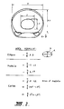
- the velocity of sound in the medulla is of the order 1500 ms ⁇ 1 and that in the cortical bone of the order of 3000 m s ⁇ 1 it is readily seen that the shortest time of flight must be for the sound to travel around the cortex ( Figure 1).
- C a is the first estimate of the transverse bone velocity and as the bone velocity must be greater than the apparent velocity, by iteration (appendix B), it is possible to calculate both C bE and R.
- K is an ovality factor for the external cortical bone cross section and L is an ovality factor for the medulla.
- M is a factor to include the area of the splint bones (MC2 and MC4) as these bones were included in the determination of the BMC.
- the compact bone density, CBD E is estimated using equation (1).
- CBD M mass of wet bone/volume of wet bone (9)
- the modulus of elasticity, J M is determined using equation (2), CBD M and C bM .
- the modulus of elasticity, J E is determined with the same equation but using CBD E and C bE .
- the ultrasound time of flight measurements were obtained using a pulse module (Panametrics Model 5055), a preamplifier (Panametrics Model 5660B), two 2.25 MHz, 13 mm diameter ultrasound transducers and and 2 ( Figure 1A) mounted on an electronic digital caliper 3 (Max-cal Model 950-101) connected to a computer (not shown) via an interface (Max-cal Model 960-101) and a digital storage oscilloscope with signal averaging capacity (Tektronix Model 468).
- An Apple II Plus microcomputer was used to apply the various iterative and calculative algorithms.
- the BMC values were determined using a Norland Digital bone Densitometer (Prototype Model 2781, Large Animal Scanning System, LASS) with an Americium source (241Am).
- the method embodying the invention comprises the steps of
- Ten metacarpal bones of approximately the same size were selected from the left forelimbs of thoroughbred horses aged from 2 to 5 years. After removal of all soft tissues, the bones were stored at -15°C until examined. The lateral bone diameter, the times of flight through both the cortex alone and the corticomedullary pathways (Fig. 3) and the BMC were determined at 10 sites along the shaft of the bone as shown in Figure 4. From these measurements C a , R, C bE and CSA E (using factors determined later) were estimated. The bones were then cut to provide 10 transverse cross sections ( Figure 4). The transverse sections 1, 3, 5, 7 and 9 were cut using a band saw to provide approximately 1cm thick sections and radiographed to record the cross sectional profile.
- dorsal segments of as large a size as practical, were cut to allow direct transverse cortical bone velocity measurements (C bM ) to be made.
- the bone marrow was removed as a plug from all bone sections and the ultrasound velocity of the bone marrow was determined in the dorsopalmar, lateral and longitudinal directions using the same ultrasound equipment.
- the modulus of elasticity, J was determined using equation (2) from two sets of measurements.
- the first estimate, J M is from the directly measured C bM and the directly measured CBD M .
- the other estimate, J E was made from C bE and the CBD E from equation (1). Both determinations are made in this report.
- C bM and C bE are summarised in Table 2. From the radiographs of the individual bone segments an estimate of ovality of the medulla and the ovality of the external cortical boundary was made.
- the average ovality factor for the cortex (K) ranged from 0.75 to 0.90 with a mean of 0.81.
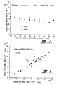
- the areas of the MC2, 3 and 4 were estimated by counting squares and average correction factors were determined for the sections along the bones. Using these correction factors the cross sectional areas were estimated from the ultrasound measurements and Figure 7 shows CSA E versus CSA M for positions along the shaft.
- Figure 8 shows the result of a comparison for the 50 bone sites for sections 1, 3, 5, 7 and 9.
- Tables 3 and 4 summarise for each bone the average BMC (5 readings at each site), BMC M , BMD E , CBD M , CBD E , J M and J e .
- the values for bone mineral content in the horse differed from those in man because of the size of the bone being measured.
- An 241Am source was used instead of 125I.
- the readings for bone mineral density (1060 ⁇ 20 kg m ⁇ 3) were compared with Greenfield et al (1120 ⁇ 150 kg m ⁇ 3) they were similar.
- the measurement of bone mineral density to this precision helps to overcome the volumetric problems identified by Ruff and Hayes (1984) found in using just bone mineral content.
- the calculated estimates for transverse cortical bone velocity, bone mineral density, compact bone density and modulus of elasticity compared favourably with the measured values for the same parameters.
- Schryver (1978) measured the bending properties of the metacarpals of ponies and found the elastic modulus to be 18.4 ⁇ 0.14 GN m ⁇ 2 for the cranial cortex of the mid-shaft. He obtained values of 16.2 to 20.2 GN M ⁇ 2 for different parts of the cortex of the radius and femur and was able to show that diet, exercise and sex of the pony affected the elastic modulus, ultimate strength and energy absorption of the bone.
- transverse cortical bone velocity has been determined rather than the apparent transverse velocity because it is more uniform along the shaft, is less subject to geometrical effects, allow an estimation of a modulus of elasticity and should provide better correlation between individuals.
- T m time through cortex + time through medulla
- Equation (5) then gives a revised estimate of C b . This process is continued until the change in C b per cycle is less than some chosen value e.g. 1 m s ⁇ 1. It takes only five cycles on average to achieve a suitable result.
Landscapes
- Health & Medical Sciences (AREA)
- Life Sciences & Earth Sciences (AREA)
- Biomedical Technology (AREA)
- Heart & Thoracic Surgery (AREA)
- Veterinary Medicine (AREA)
- Biophysics (AREA)
- Nuclear Medicine, Radiotherapy & Molecular Imaging (AREA)
- Pathology (AREA)
- Radiology & Medical Imaging (AREA)
- Engineering & Computer Science (AREA)
- Public Health (AREA)
- Physics & Mathematics (AREA)
- Medical Informatics (AREA)
- Molecular Biology (AREA)
- Surgery (AREA)
- Animal Behavior & Ethology (AREA)
- General Health & Medical Sciences (AREA)
- Orthopedic Medicine & Surgery (AREA)
- Rheumatology (AREA)
- Ultra Sonic Daignosis Equipment (AREA)
- Investigating Or Analyzing Materials By The Use Of Ultrasonic Waves (AREA)
Applications Claiming Priority (2)
| Application Number | Priority Date | Filing Date | Title |
|---|---|---|---|
| AUPI321687 | 1987-07-16 | ||
| AU3216/87 | 1987-07-16 |
Publications (2)
| Publication Number | Publication Date |
|---|---|
| EP0299906A2 true EP0299906A2 (de) | 1989-01-18 |
| EP0299906A3 EP0299906A3 (de) | 1990-06-13 |
Family
ID=3772341
Family Applications (1)
| Application Number | Title | Priority Date | Filing Date |
|---|---|---|---|
| EP88630138A Withdrawn EP0299906A3 (de) | 1987-07-16 | 1988-07-14 | In vivo Knochenbeschaffenheitsmessung |
Country Status (2)
| Country | Link |
|---|---|
| US (1) | US4976267A (de) |
| EP (1) | EP0299906A3 (de) |
Cited By (14)
| Publication number | Priority date | Publication date | Assignee | Title |
|---|---|---|---|---|
| WO1991006245A1 (en) * | 1989-10-24 | 1991-05-16 | The Adelaide Bone And Joint Research Foundation Inc | Vibrational analysis of bones |
| EP0516353A1 (de) * | 1991-05-22 | 1992-12-02 | Walker Magnetics Group, Inc. | Ultraschallwandleranordnung |
| GB2257253A (en) * | 1991-06-17 | 1993-01-06 | Christian Mcdonald Langton | Ultrasound bone analyser |
| WO1994018889A1 (en) * | 1993-02-17 | 1994-09-01 | John Koblanski | Ultrasonic scanning apparatus |
| WO1994022375A1 (en) * | 1993-04-07 | 1994-10-13 | Osteo Sciences Corporation | System and method for external acoustic bone velocity measurement |
| EP0663182A1 (de) * | 1994-01-14 | 1995-07-19 | IGEA S.r.L. | Ultraschall-Messgerät zur Bestimmung der Knochendichte und der Knochenstruktur |
| US5452722A (en) * | 1992-06-22 | 1995-09-26 | Mccue Ultrasonics, Ltd. | Ultrasound bone analyser |
| US5720290A (en) * | 1993-04-07 | 1998-02-24 | Metra Biosystems, Inc. | Apparatus and method for acoustic analysis of bone using optimized functions of spectral and temporal signal components |
| US5755228A (en) * | 1995-06-07 | 1998-05-26 | Hologic, Inc. | Equipment and method for calibration and quality assurance of an ultrasonic bone anaylsis apparatus |
| US5785041A (en) * | 1996-03-26 | 1998-07-28 | Hologic Inc. | System for assessing bone characteristics |
| US6004272A (en) * | 1995-06-07 | 1999-12-21 | Hologic, Inc. | Ultrasonic bone testing apparatus with repeatable positioning and repeatable coupling |
| EP0570936B1 (de) * | 1992-05-20 | 2000-08-09 | Aloka Co. Ltd. | Vorrichtung zur Bestimmung der Eigenschaften von Knochen |
| US6352512B1 (en) | 1995-06-07 | 2002-03-05 | Hologic, Inc. | Bone analysis apparatus and method for calibration and quality assurance of an ultrasonic bone analysis apparatus |
| US7727152B2 (en) * | 2001-02-28 | 2010-06-01 | The Research Foundation Of State University Of New York | Method and apparatus for scanning confocal acoustic diagnostic for bone quality |
Families Citing this family (29)
| Publication number | Priority date | Publication date | Assignee | Title |
|---|---|---|---|---|
| US6277076B1 (en) | 1988-05-11 | 2001-08-21 | Lunar Corporation | Ultrasonic densitometer with pre-inflated fluid coupling membranes |
| US5293870A (en) * | 1989-11-17 | 1994-03-15 | Board Of Regents The University Of Texas System | Method and apparatus for elastographic measurement and imaging |
| US5107837A (en) * | 1989-11-17 | 1992-04-28 | Board Of Regents, University Of Texas | Method and apparatus for measurement and imaging of tissue compressibility or compliance |
| IL94616A (en) * | 1990-06-04 | 1994-06-24 | Medicano Systems Ltd | Apparatus and method for calculating the mechanical properties of a solid |
| US5426979A (en) * | 1990-06-04 | 1995-06-27 | Medicano Systems Ltd. | Frequency spectrum apparatus for determining mechanical properties |
| US5247934A (en) * | 1991-08-09 | 1993-09-28 | Trustees Of The University Of Pennsylvania | Method and apparatus for diagnosing osteoporosis with MR imaging |
| US5140988A (en) * | 1991-08-22 | 1992-08-25 | Animal Ultrasound Services, Inc. | Detection of abnormal bone structure in animals and carcasses with ultrasound |
| US5259384A (en) * | 1992-07-30 | 1993-11-09 | Kaufman Jonathan J | Ultrasonic bone-assessment apparatus and method |
| US5836876A (en) * | 1993-03-03 | 1998-11-17 | Washington University | Method and apparatus for determining bone density and diagnosing osteoporosis |
| US5402781A (en) * | 1993-03-03 | 1995-04-04 | Washington University | Method and apparatus for determining bone density and diagnosing osteoporosis |
| US5429143A (en) * | 1993-11-22 | 1995-07-04 | Marzluff; Joseph | Device and method for determining hole integrity in surgical applications |
| JP3207318B2 (ja) | 1994-05-11 | 2001-09-10 | アロカ株式会社 | 超音波骨評価装置 |
| US5535750A (en) * | 1994-09-30 | 1996-07-16 | Kabushiki Kaisha Ishikawa Seisakusho, Ltd. | Method and apparatus for evaluating the progress of osteoporosis by ultrasonic signals |
| JP2883290B2 (ja) * | 1995-04-10 | 1999-04-19 | アロカ株式会社 | 超音波骨評価装置 |
| US5879301A (en) * | 1996-02-16 | 1999-03-09 | Orthologic Corp. | Ultrasonic bone assessment method and apparatus |
| US5651363A (en) * | 1996-02-16 | 1997-07-29 | Orthologic Corporation | Ultrasonic bone assessment method and apparatus |
| IT1292288B1 (it) * | 1997-04-24 | 1999-01-29 | Igea Srl | Dispositivo di misura ad ultrasuoni per la rilevazione della densita' e struttura ossea. |
| US5931795A (en) * | 1997-11-07 | 1999-08-03 | Manly; Philip | Method for evaluating human bone strength |
| US8202219B2 (en) * | 2004-02-23 | 2012-06-19 | Cyberlogic, Inc. | Ultrasonic bone assessment apparatus and method |
| US7033321B1 (en) * | 2004-10-25 | 2006-04-25 | Artann Laboratories, Inc. | Ultrasonic water content monitor and methods for monitoring tissue hydration |
| EP1921976A2 (de) * | 2005-08-12 | 2008-05-21 | University of Washington | Verfahren und gerät zur vorbereitung von organen und geweben für eine laparoskopische operation |
| WO2007120890A2 (en) * | 2006-04-13 | 2007-10-25 | The Research Foundation Of State University Of New York | Phased array ultrasound with electronically controlled focal point for assessing bone quality via acoustic topology and wave transmit functions |
| US7901356B2 (en) * | 2006-12-13 | 2011-03-08 | Cyberlogic, Inc. | Ultrasonic bone assessment apparatus and method |
| US9615814B2 (en) * | 2006-12-13 | 2017-04-11 | Cyberlogic, Inc. | Ultrasonic bone assessment apparatus and method |
| US7862510B2 (en) * | 2007-02-09 | 2011-01-04 | Cyberlogic, Inc. | Ultrasonic bone assessment apparatus and method |
| US8187185B2 (en) * | 2007-08-08 | 2012-05-29 | Hitachi Aloka Medical, Ltd. | Ultrasound diagnostic apparatus |
| US9008394B2 (en) * | 2008-11-26 | 2015-04-14 | General Electric Company | Methods and apparatus for determining brain cortical thickness |
| US8421479B2 (en) * | 2009-06-30 | 2013-04-16 | Navisense | Pulsed echo propagation device and method for measuring a parameter |
| JP6389116B2 (ja) * | 2014-12-22 | 2018-09-12 | ジーイー・メディカル・システムズ・グローバル・テクノロジー・カンパニー・エルエルシー | 超音波画像表示装置及びその制御プログラム |
Family Cites Families (7)
| Publication number | Priority date | Publication date | Assignee | Title |
|---|---|---|---|---|
| FR2318420A1 (fr) * | 1975-07-17 | 1977-02-11 | Guiset Jacques | Procede et dispositif pour la mesure d'une pression ou d'une distance par ultra-sons |
| US4361154A (en) * | 1978-07-28 | 1982-11-30 | Massachusetts Institute Of Technology | Method for establishing, in vivo, bone strength |
| IL69471A (en) * | 1983-08-12 | 1987-07-31 | Benjamin Gavish | Method and device for non-invasively monitoring the instantaneous fluctuations in the viscoelastic properties of a soft tissue |
| DE3329134C2 (de) * | 1983-08-12 | 1985-06-20 | Deutsche Forschungs- und Versuchsanstalt für Luft- und Raumfahrt e.V., 5000 Köln | Vorrichtung zur Messung von Querschnitten an Objekten, insbesondere an Körperteilen |
| JPS60116345A (ja) * | 1983-11-30 | 1985-06-22 | 富士通株式会社 | 超音波診断装置 |
| JPS60163643A (ja) * | 1984-02-07 | 1985-08-26 | テルモ株式会社 | 超音波測定装置 |
| US4913157A (en) * | 1986-06-03 | 1990-04-03 | Analog Devices, Inc. | Ultrasound method and apparatus for evaluating, in vivo, bone conditions |
-
1988
- 1988-07-14 EP EP88630138A patent/EP0299906A3/de not_active Withdrawn
- 1988-07-18 US US07/220,083 patent/US4976267A/en not_active Expired - Fee Related
Cited By (20)
| Publication number | Priority date | Publication date | Assignee | Title |
|---|---|---|---|---|
| US5368044A (en) * | 1989-10-24 | 1994-11-29 | The Adelaide Bone And Joint Research Foundation, Inc. | Vibrational analysis of bones |
| WO1991006245A1 (en) * | 1989-10-24 | 1991-05-16 | The Adelaide Bone And Joint Research Foundation Inc | Vibrational analysis of bones |
| EP0516353A1 (de) * | 1991-05-22 | 1992-12-02 | Walker Magnetics Group, Inc. | Ultraschallwandleranordnung |
| GB2257253A (en) * | 1991-06-17 | 1993-01-06 | Christian Mcdonald Langton | Ultrasound bone analyser |
| GB2257253B (en) * | 1991-06-17 | 1995-01-11 | Christian Mcdonald Langton | Ultrasound bone analyser |
| EP0570936B1 (de) * | 1992-05-20 | 2000-08-09 | Aloka Co. Ltd. | Vorrichtung zur Bestimmung der Eigenschaften von Knochen |
| US5452722A (en) * | 1992-06-22 | 1995-09-26 | Mccue Ultrasonics, Ltd. | Ultrasound bone analyser |
| WO1994018889A1 (en) * | 1993-02-17 | 1994-09-01 | John Koblanski | Ultrasonic scanning apparatus |
| US5720290A (en) * | 1993-04-07 | 1998-02-24 | Metra Biosystems, Inc. | Apparatus and method for acoustic analysis of bone using optimized functions of spectral and temporal signal components |
| WO1994022375A1 (en) * | 1993-04-07 | 1994-10-13 | Osteo Sciences Corporation | System and method for external acoustic bone velocity measurement |
| US6371916B1 (en) | 1993-04-07 | 2002-04-16 | Metra Biosystems, Inc. | Acoustic analysis of bone using point-source-like transducers |
| EP0663182A1 (de) * | 1994-01-14 | 1995-07-19 | IGEA S.r.L. | Ultraschall-Messgerät zur Bestimmung der Knochendichte und der Knochenstruktur |
| US5755228A (en) * | 1995-06-07 | 1998-05-26 | Hologic, Inc. | Equipment and method for calibration and quality assurance of an ultrasonic bone anaylsis apparatus |
| US6004272A (en) * | 1995-06-07 | 1999-12-21 | Hologic, Inc. | Ultrasonic bone testing apparatus with repeatable positioning and repeatable coupling |
| US6135964A (en) * | 1995-06-07 | 2000-10-24 | Hologic, Inc. | Ultrasonic bone testing apparatus with repeatable positioning and repeatable coupling |
| US6352512B1 (en) | 1995-06-07 | 2002-03-05 | Hologic, Inc. | Bone analysis apparatus and method for calibration and quality assurance of an ultrasonic bone analysis apparatus |
| US5935073A (en) * | 1995-09-26 | 1999-08-10 | Hologic, Inc. | Equipment and method for calibration and quality assurance of an ultrasonic bone analysis apparatus |
| US5785041A (en) * | 1996-03-26 | 1998-07-28 | Hologic Inc. | System for assessing bone characteristics |
| US6029078A (en) * | 1996-03-26 | 2000-02-22 | Hologic, Inc. | System for assessing bone characteristics |
| US7727152B2 (en) * | 2001-02-28 | 2010-06-01 | The Research Foundation Of State University Of New York | Method and apparatus for scanning confocal acoustic diagnostic for bone quality |
Also Published As
| Publication number | Publication date |
|---|---|
| US4976267A (en) | 1990-12-11 |
| EP0299906A3 (de) | 1990-06-13 |
Similar Documents
| Publication | Publication Date | Title |
|---|---|---|
| US4976267A (en) | Method and apparatus for the measurement of bone quality in vivo | |
| Freeman et al. | Evaluation of the use of muscle condition score and ultrasonographic measurements for assessment of muscle mass in dogs | |
| Laugier | Instrumentation for in vivo ultrasonic characterization of bone strength | |
| MARKEL et al. | Noninvasive monitoring techniques for quantitative description of callus mineral content and mechanical properties. | |
| US4913157A (en) | Ultrasound method and apparatus for evaluating, in vivo, bone conditions | |
| Li et al. | Could clinical ultrasound improve the fitting of spinal orthosis for the patients with AIS? | |
| Schneider et al. | In vivo measurements of cortical thickness and porosity at the proximal third of the tibia using guided waves: Comparison with site-matched peripheral quantitative computed tomography and distal high-resolution peripheral quantitative computed tomography | |
| KR20140035932A (ko) | 환자의 뼈 조직의 품질을 평가하기 위한 초음파 장치 | |
| Rodriguez et al. | Skeletal muscle estimation: a review of techniques and their applications | |
| Braun | Ultrasonographic examination of the left kidney, the urinary bladder, and the urethra in cows | |
| Cunningham et al. | Ultrasound velocity and attenuation at different skeletal sites compared with bone mineral density measured using dual energy X-ray absorptiometry | |
| JP3954981B2 (ja) | 超音波診断装置 | |
| KR101750108B1 (ko) | 외부 팬텀 없는 영상 신호에서 골밀도 값 예측 | |
| McCartney et al. | Combined 2.25 MHz ultrasound velocity and bone mineral density measurements in the equine metacarpus and their in vivo applications | |
| CN103237501B (zh) | 用于估算骨的无机物密度的方法和装置 | |
| Jeffcott et al. | Non‐invasive measurement of bone: A review of clinical and research applications in the horse | |
| Weiss et al. | Ultrasonic protocols for separately measuring subcutaneous fat and skeletal muscle thickness in the calf area | |
| Njeh et al. | Factors influencing the speed of sound through the proximal phalanges | |
| Pattijn et al. | Structural and radiological parameters for the nondestructive characterization of trabecular bone | |
| Serpe et al. | Broadband ultrasound attenuation value dependence on bone width in vitro | |
| Chen et al. | Sophisticated imaging technology in the assessment of osteoporosis risk | |
| Singh | Ultrasonic non-destructive measurements of cortical bone thickness in human cadaver femur | |
| Arpinar et al. | Correlation between mechanical vibration analysis and dual energy X-ray absorptiometry (DXA) in the measurement of in vivo human tibial bone strength | |
| McCarthy et al. | Ultrasonic transmission velocity and single photon absorptiometric measurement of metacarpal bone strength: an in vitro study in the horse | |
| Tóth et al. | Assessment of the mineral density and mineral content of the equine third metacarpal and first phalanx bone by dual energy x-ray absorptiometry |
Legal Events
| Date | Code | Title | Description |
|---|---|---|---|
| PUAI | Public reference made under article 153(3) epc to a published international application that has entered the european phase |
Free format text: ORIGINAL CODE: 0009012 |
|
| AK | Designated contracting states |
Kind code of ref document: A2 Designated state(s): CH DE ES FR GB GR IT LI NL SE |
|
| PUAL | Search report despatched |
Free format text: ORIGINAL CODE: 0009013 |
|
| AK | Designated contracting states |
Kind code of ref document: A3 Designated state(s): CH DE ES FR GB GR IT LI NL SE |
|
| 17P | Request for examination filed |
Effective date: 19900803 |
|
| 17Q | First examination report despatched |
Effective date: 19920930 |
|
| STAA | Information on the status of an ep patent application or granted ep patent |
Free format text: STATUS: THE APPLICATION IS DEEMED TO BE WITHDRAWN |
|
| 18D | Application deemed to be withdrawn |
Effective date: 19940625 |