EP0307792A2 - Dispositif pour prélever un échantillon de tissus - Google Patents
Dispositif pour prélever un échantillon de tissus Download PDFInfo
- Publication number
- EP0307792A2 EP0307792A2 EP88114664A EP88114664A EP0307792A2 EP 0307792 A2 EP0307792 A2 EP 0307792A2 EP 88114664 A EP88114664 A EP 88114664A EP 88114664 A EP88114664 A EP 88114664A EP 0307792 A2 EP0307792 A2 EP 0307792A2
- Authority
- EP
- European Patent Office
- Prior art keywords
- smear
- stem
- swab
- cell
- disc
- Prior art date
- Legal status (The legal status is an assumption and is not a legal conclusion. Google has not performed a legal analysis and makes no representation as to the accuracy of the status listed.)
- Withdrawn
Links
- 238000001574 biopsy Methods 0.000 title 1
- 239000000463 material Substances 0.000 claims abstract description 43
- 239000011148 porous material Substances 0.000 claims abstract description 33
- 238000003745 diagnosis Methods 0.000 claims abstract description 6
- 229920005830 Polyurethane Foam Polymers 0.000 claims description 17
- 239000011496 polyurethane foam Substances 0.000 claims description 17
- 238000003780 insertion Methods 0.000 claims description 8
- 230000037431 insertion Effects 0.000 claims description 8
- 239000006260 foam Substances 0.000 claims description 6
- 239000000853 adhesive Substances 0.000 claims description 5
- 230000001070 adhesive effect Effects 0.000 claims description 5
- NIXOWILDQLNWCW-UHFFFAOYSA-M Acrylate Chemical compound [O-]C(=O)C=C NIXOWILDQLNWCW-UHFFFAOYSA-M 0.000 claims description 3
- 208000027418 Wounds and injury Diseases 0.000 abstract description 6
- 230000006378 damage Effects 0.000 abstract description 4
- 208000014674 injury Diseases 0.000 abstract description 4
- 210000004027 cell Anatomy 0.000 description 47
- 210000000056 organ Anatomy 0.000 description 19
- 238000000034 method Methods 0.000 description 12
- 229920000742 Cotton Polymers 0.000 description 6
- 238000011084 recovery Methods 0.000 description 6
- 238000000605 extraction Methods 0.000 description 5
- 210000004400 mucous membrane Anatomy 0.000 description 5
- 238000002360 preparation method Methods 0.000 description 5
- 206010028980 Neoplasm Diseases 0.000 description 4
- 201000011510 cancer Diseases 0.000 description 4
- 210000004696 endometrium Anatomy 0.000 description 4
- 238000002474 experimental method Methods 0.000 description 4
- 239000000835 fiber Substances 0.000 description 4
- 238000004519 manufacturing process Methods 0.000 description 4
- 238000012546 transfer Methods 0.000 description 4
- 210000004291 uterus Anatomy 0.000 description 4
- LFQSCWFLJHTTHZ-UHFFFAOYSA-N Ethanol Chemical compound CCO LFQSCWFLJHTTHZ-UHFFFAOYSA-N 0.000 description 3
- 239000004793 Polystyrene Substances 0.000 description 3
- 239000003522 acrylic cement Substances 0.000 description 3
- 238000013461 design Methods 0.000 description 3
- 238000001514 detection method Methods 0.000 description 3
- 238000011161 development Methods 0.000 description 3
- 230000018109 developmental process Effects 0.000 description 3
- 238000012360 testing method Methods 0.000 description 3
- 239000001856 Ethyl cellulose Substances 0.000 description 2
- ZZSNKZQZMQGXPY-UHFFFAOYSA-N Ethyl cellulose Chemical compound CCOCC1OC(OC)C(OCC)C(OCC)C1OC1C(O)C(O)C(OC)C(CO)O1 ZZSNKZQZMQGXPY-UHFFFAOYSA-N 0.000 description 2
- 210000003484 anatomy Anatomy 0.000 description 2
- 244000052616 bacterial pathogen Species 0.000 description 2
- 230000015572 biosynthetic process Effects 0.000 description 2
- 235000019325 ethyl cellulose Nutrition 0.000 description 2
- 229920001249 ethyl cellulose Polymers 0.000 description 2
- 239000011521 glass Substances 0.000 description 2
- 235000015097 nutrients Nutrition 0.000 description 2
- 229920003023 plastic Polymers 0.000 description 2
- 239000004033 plastic Substances 0.000 description 2
- 239000002984 plastic foam Substances 0.000 description 2
- 210000001519 tissue Anatomy 0.000 description 2
- 210000001215 vagina Anatomy 0.000 description 2
- FHVDTGUDJYJELY-UHFFFAOYSA-N 6-{[2-carboxy-4,5-dihydroxy-6-(phosphanyloxy)oxan-3-yl]oxy}-4,5-dihydroxy-3-phosphanyloxane-2-carboxylic acid Chemical compound O1C(C(O)=O)C(P)C(O)C(O)C1OC1C(C(O)=O)OC(OP)C(O)C1O FHVDTGUDJYJELY-UHFFFAOYSA-N 0.000 description 1
- 241001264766 Callistemon Species 0.000 description 1
- 206010014733 Endometrial cancer Diseases 0.000 description 1
- 206010014759 Endometrial neoplasm Diseases 0.000 description 1
- 238000005299 abrasion Methods 0.000 description 1
- 238000010521 absorption reaction Methods 0.000 description 1
- 230000006978 adaptation Effects 0.000 description 1
- 230000001476 alcoholic effect Effects 0.000 description 1
- 229940072056 alginate Drugs 0.000 description 1
- 235000010443 alginic acid Nutrition 0.000 description 1
- 229920000615 alginic acid Polymers 0.000 description 1
- 230000001580 bacterial effect Effects 0.000 description 1
- 230000000740 bleeding effect Effects 0.000 description 1
- 210000000601 blood cell Anatomy 0.000 description 1
- 235000019994 cava Nutrition 0.000 description 1
- 229920002678 cellulose Polymers 0.000 description 1
- 239000001913 cellulose Substances 0.000 description 1
- 210000003679 cervix uteri Anatomy 0.000 description 1
- 239000011248 coating agent Substances 0.000 description 1
- 238000000576 coating method Methods 0.000 description 1
- 230000000052 comparative effect Effects 0.000 description 1
- 210000000805 cytoplasm Anatomy 0.000 description 1
- 238000009826 distribution Methods 0.000 description 1
- 238000013399 early diagnosis Methods 0.000 description 1
- 230000000694 effects Effects 0.000 description 1
- 238000005516 engineering process Methods 0.000 description 1
- 238000004299 exfoliation Methods 0.000 description 1
- 239000004744 fabric Substances 0.000 description 1
- 239000002657 fibrous material Substances 0.000 description 1
- 210000000936 intestine Anatomy 0.000 description 1
- 230000003902 lesion Effects 0.000 description 1
- 239000007788 liquid Substances 0.000 description 1
- 230000003211 malignant effect Effects 0.000 description 1
- 239000002184 metal Substances 0.000 description 1
- 230000000877 morphologic effect Effects 0.000 description 1
- 238000009595 pap smear Methods 0.000 description 1
- 244000052769 pathogen Species 0.000 description 1
- 230000001717 pathogenic effect Effects 0.000 description 1
- 229920002223 polystyrene Polymers 0.000 description 1
- 229920002635 polyurethane Polymers 0.000 description 1
- 239000004814 polyurethane Substances 0.000 description 1
- 238000005096 rolling process Methods 0.000 description 1
- 229920006395 saturated elastomer Polymers 0.000 description 1
- 230000028327 secretion Effects 0.000 description 1
- 210000003491 skin Anatomy 0.000 description 1
- 238000010186 staining Methods 0.000 description 1
- 210000002784 stomach Anatomy 0.000 description 1
- 230000000472 traumatic effect Effects 0.000 description 1
- 210000003708 urethra Anatomy 0.000 description 1
- 238000004804 winding Methods 0.000 description 1
- 239000002023 wood Substances 0.000 description 1
Images


Classifications
-
- A—HUMAN NECESSITIES
- A61—MEDICAL OR VETERINARY SCIENCE; HYGIENE
- A61B—DIAGNOSIS; SURGERY; IDENTIFICATION
- A61B10/00—Instruments for taking body samples for diagnostic purposes; Other methods or instruments for diagnosis, e.g. for vaccination diagnosis, sex determination or ovulation-period determination; Throat striking implements
- A61B10/02—Instruments for taking cell samples or for biopsy
- A61B10/0291—Instruments for taking cell samples or for biopsy for uterus
Definitions
- swab a swab, a smear body made of fiber materials such as. B. from cotton, from cellulose two-one / two-acetate, from ethyl cellulose or from alginate.
- these swab devices intended for human organs, for. B. in the gynecological area of the portio surface or from the cervical canal, cell material (or germs) or scraped off and the be further examined. This means that the material scraped off with the above-mentioned swab device, which adheres to its swab body, must be recovered by this device.
- the recovery takes place directly by rolling out the smear body onto a slide; (these are usually "glass plates”). Special, known preparation steps then follow the transfer to the slide.
- a gynecological smear is usually fixed in alcohol and then a staining procedure, e.g. B. after Papanicolaou to make the cells visible for microscopic assessment, it being particularly important to show the fine morphological structures in nuclei and cytoplasm.
- Cell and germ extraction techniques place high demands on the extraction technology and the preparation, particularly with regard to the diagnostic significance of the preparations (early cancer detection or germ and pathogen diagnosis).
- a removal instrument for the extraction of swab material must have a highly efficient mode of operation, without being traumatic or damaging to the cell. This means that the instrument must be adapted or adaptable to the anatomy of the organ to be removed, e.g. B. to optimally promote material from crypts of different positions and depths.
- the known swab device with a swab body (swab) made of cotton wool has serious disadvantages with regard to cell recovery from the organ and cell recovery on the slide (see G. Schlueter et al., "More exact cell fix cytology with new swab and preparation technique" in "Diagnostik 17" (1984) No. 5, pp. 27-29 - MMVtechnik für publishing house, D-8000 Kunststoff 80).
- the swab body is made by winding cotton wool as a swab onto the end of a stem in the usual way.
- a cell brush has been recommended for performing cervical smears, which is made like a bottle brush and is intended to brush the cell material off the organ wall when it is used.
- the individual tufts of bristles are held by two coiled metal wires, which necessarily protrude slightly above the foremost tufts of bristles. Even if you round this protruding double wire end, put it is still a dangerous tip for the procedure, protruding beyond the bristle area, which can easily lead to injuries. This is especially true when the cell brush has to be inserted at an angle or handled clumsily. Furthermore, a hard-edged spatula has already been created as a smear device.
- Rinsing and aspiration methods are already known for obtaining samples from the uterus. However, these have not proven themselves because the degree of exfoliation of the endometrium (mucous membrane of the uterine wall) is controlled hormonally. These procedures therefore only promote cells that can be detached from the mucous membrane in loose bandages.
- the object of the present invention is therefore, in particular, to provide a device for obtaining smear material in which the disadvantages of the known smear devices are at least largely avoided; in particular, a device is to be created with which, with simple handling, a well-preserved, representative cell material which is sufficient in terms of quantity for diagnosis can be obtained and transferred well to the slide.
- the smear body as preferred porous disk attached at least approximately centrally to the insertion of the stem.
- Such a flexible pore body can adapt well to the special outline shape of even narrower body cavities and it is well suited to absorbing and transporting a comparatively large amount of cell material.
- the upper side of the pane facing the insertion of the device can rest well against the wall of the body channel section, while when the device is withdrawn the back of the pane can additionally lie against this wall. A large number of pores are therefore available, into which cell material can be taken up and removed.
- a rigid abutment disk can be provided on the handle of the device on the back of such a porous disk.
- the device is particularly well suited for the removal of cell material or the like at places which are to be dabbed off somewhat under pressure.
- the smear body is designed as a flag-like strip and is attached to the insertion of the stem of the device.
- a particularly advantageous embodiment of the device according to the invention is that the smear body consists of a polyurethane foam layer or the like. Body-friendly, process-neutral sponge-like foam layer. It is particularly expedient if this polyurethane foam layer has about 600 to 2,500 pores / cm2, preferably about 1,500 pores / cm2.
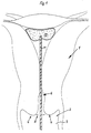
- Fig. 1 shows a highly schematic longitudinal section through a uterus designated as a whole and the inner end 2 of the vagina 3. With 4 the vaginal vault, with 5 the portio vaginalis and with 6 the cervical canal.
- the circular disc 15 folds through from above and / or from Side pressure to the outer end of the stem around. Parts of the circular disk 15 can be supported on one side or circularly against the stem 11 and the circular disk 15 adapts to the lumen of the body cavity 18 with the formation of folds. If the swab device 10 is first pushed through such a body cavity 18 in the direction of the arrow 19 (FIG. 4), the narrowed circular disk 15 can unfold again approximately into the original disk shape (FIG. 2). If the smear device 10 is then withdrawn through the aforementioned body cavity 18, the circular disk 15 folds away from its attachment point on the stem 11 in a cup-like manner, as can be seen clearly from FIG. 5. Then, in the starting position (FIG.
- the rear side of the disk 15 facing the outer end 10 of the stem 11 can adapt to the inner wall of the cavity 19 and the corresponding starting point Take coating material with you.
- the above-described mode of operation is described below by way of example for use on the cervical canal 6. Since the cervical canal (endocervix) can be narrow, the smear device 10 is carried out with a circular disc 15 with a diameter D of 10 mm. The swab is inserted into the cervical canal 6 while rotating.
- the circular disk 15 folded like an umbrella can narrow and widen depending on the design of the channel.
- the stem 11 is rotated, the pores 16 of the polyurethane foam disc 15 then rubbing against the body wall and absorbing cell material.
- the swab 10 is then withdrawn.
- the circular disk 15 turns upside down, as shown in FIG. 5, so that when the pull-out is also carried out, the underside of the circular disk 15 is occupied with cell material from the entire length of the cervical canal 6 contacted.
- the cells are then transferred from the circular disk 15 to the slides provided for the examination, that is to say generally to corresponding glass plates.
- Another application example is the removal of smear material on organ surfaces with depressions, e.g. B. in the portio vaginalis 5.
- a smear device 10, e.g. B. with a circular disc diameter D 20 mm, but without abutment disc 21, is placed under slight pressure on the portio surface, the stem 11 is directed towards the cervix. Since this is recessed, the porous, slightly flexible circular disk 15 made of polyurethane foam takes on a slight conical shape. It can adapt to the different, anatomically present recesses of the portio vaginalis 5. Whose surface is wiped while rotating the circular disc 15. The cell transfer from the circular disc 15 to the slide then takes place in the manner already mentioned.
- the smear device 10 is similar to that in conjunction with Fig. 4 u. 5 described mode of operation housed folded in a tube (endoscope). Only after this application tube has reached the organ cavity to be examined is the swab body 13 of the swab device 10 according to the invention pushed out of the endoscope. Then the cells can be obtained either blindly (in small caves such as the uterus) or under an endoscopic view. After the smear process, the porous circular disk 15 withdrawn into the application tube and removed together with this from the organ cavity. The cell is obtained from the smear body 13 again in the manner already described.
- the advantageous mode of operation of the swab device 10 according to the invention was tested in a comparative experiment.
- a germ recovery was carried out from an infected wound area of the skin with the aid of a smear device 10, which has a circular disk 15 and an abutment disk 21 to stabilize it.
- This was compared to a germ recovery certificate using a cotton swab.
- the swab device 10 according to the invention showed a very good absorption capacity for the secretion and also proved to be particularly suitable with regard to the abrasion ability.
- the germ transfer took place in a nutrient medium in which the polyurethane disc 15 was rinsed out.
- the swab body 13 can also have a cylindrical or olive-shaped outline shape according to the invention. It has turned out to be particularly advantageous in special applications if the smear body 13 a is designed as a flag-like strip 25.
- the flag-like strip can e.g. B. have dimensions of 8 mm x 40 mm. As can be seen in FIG.
- swab bodies 13 made of polyurethane foam can adapt well to the different shapes of the body part to be examined on the one hand, without causing injuries, on the other hand, the pores 16 can absorb the cells to be wiped off particularly well with this material and later release them to a correspondingly large extent.
- An acrylate adhesive has proven to be particularly advantageous for fastening the swab body 13 made of polyurethane foam.
- Acrylate adhesive is tissue-friendly and process-inherent.
- Other fabrics also come as adhesives friendly and process-internal materials in question, e.g. polystyrene adhesive.
- acrylic adhesive has proven to be particularly advantageous.
- a somewhat modified embodiment of the smear device can consist in the fact that the smear body 13 does not consist of a pore body, but of a structured, e.g. B. woven nonwoven. In such a nonwoven, the nonwoven structure replaces the pores 16 in the pore body 14. The fineness of the mesh and the diameter of the warp and weft threads then influence the surface structure of the smeared body.
- a particularly advantageous application of a smear body made of fleece is that the flag-like strip (25) consists of fleece. He can e.g. B. be particularly tearproof.
- the following advantages result: a relatively large-area connection between the stem 11 and the pore body 14 is possible and, moreover, when the smear is carried out, which is usually done by performing a rotating and advancing movement, a particularly good guidance of the pore body 14 (see Fig.8a).
- the stem 11 is particularly simple in terms of production technology.
Landscapes
- Health & Medical Sciences (AREA)
- Life Sciences & Earth Sciences (AREA)
- Medical Informatics (AREA)
- Molecular Biology (AREA)
- Reproductive Health (AREA)
- Engineering & Computer Science (AREA)
- Biomedical Technology (AREA)
- Heart & Thoracic Surgery (AREA)
- Gynecology & Obstetrics (AREA)
- Pathology (AREA)
- Surgery (AREA)
- Animal Behavior & Ethology (AREA)
- General Health & Medical Sciences (AREA)
- Public Health (AREA)
- Veterinary Medicine (AREA)
- Investigating Or Analysing Biological Materials (AREA)
- Absorbent Articles And Supports Therefor (AREA)
Applications Claiming Priority (2)
| Application Number | Priority Date | Filing Date | Title |
|---|---|---|---|
| DE19873731407 DE3731407A1 (de) | 1987-09-18 | 1987-09-18 | Vorrichtung zum gewinnen von abstrichmaterial |
| DE3731407 | 1987-09-18 |
Publications (2)
| Publication Number | Publication Date |
|---|---|
| EP0307792A2 true EP0307792A2 (fr) | 1989-03-22 |
| EP0307792A3 EP0307792A3 (fr) | 1989-11-15 |
Family
ID=6336315
Family Applications (1)
| Application Number | Title | Priority Date | Filing Date |
|---|---|---|---|
| EP88114664A Withdrawn EP0307792A3 (fr) | 1987-09-18 | 1988-09-08 | Dispositif pour prélever un échantillon de tissus |
Country Status (2)
| Country | Link |
|---|---|
| EP (1) | EP0307792A3 (fr) |
| DE (1) | DE3731407A1 (fr) |
Families Citing this family (2)
| Publication number | Priority date | Publication date | Assignee | Title |
|---|---|---|---|---|
| DE4005266A1 (de) * | 1990-02-20 | 1991-08-22 | Geka Brush Georg Karl Gmbh | Vorrichtung zur durchfuehrung von abstrichen fuer cytologische untersuchungen |
| DE102012022525A1 (de) | 2012-11-16 | 2014-05-22 | Urotech Medizinische Technologie Gmbh | Vorrichtung zum Aufnehmen einer Probenaufnahmeeinrichtung sowie diese Vorrichtung und die Probenaufnahmeeinrichtung enthaltendes Probenaufnahmeset und Verfahren zur Probenaufnahme bzw. -entnahme |
Family Cites Families (3)
| Publication number | Priority date | Publication date | Assignee | Title |
|---|---|---|---|---|
| GB1378393A (en) * | 1971-12-03 | 1974-12-27 | Armour Pharma | Diagnostic device for obtaining cytologic samples |
| US3776219A (en) * | 1972-09-15 | 1973-12-04 | Metpath Inc | Cervical scraper |
| DE3101109C2 (de) * | 1981-01-15 | 1982-10-14 | Dieter von Dipl.-Ing. 8000 München Zeppelin | Gynäkologisches Abstrich-Instrument |
-
1987
- 1987-09-18 DE DE19873731407 patent/DE3731407A1/de not_active Withdrawn
-
1988
- 1988-09-08 EP EP88114664A patent/EP0307792A3/fr not_active Withdrawn
Also Published As
| Publication number | Publication date |
|---|---|
| DE3731407A1 (de) | 1989-04-06 |
| EP0307792A3 (fr) | 1989-11-15 |
Similar Documents
| Publication | Publication Date | Title |
|---|---|---|
| DE3877022T2 (de) | Vorrichtung fuer cervicale cytologie. | |
| DE69633966T2 (de) | Uterus-endometrialische Musterbürste | |
| DE2812709C2 (de) | Vorrichtung zur Gewinnung von Zellmaterialien aus Körperhöhlen | |
| DE69624338T2 (de) | Vorrichtung zur entnahme von mukosa aus der gebärmutter | |
| DE69510461T2 (de) | Einweg-vorrichtung zum nachweis oder zur analyse einer körperflüssigkeit | |
| DE602004010240T3 (de) | Tupfer zur aufnahme von biologischen proben | |
| DE69633750T2 (de) | Vorrichtung zur automatischen Biopsie- und Weichgewebeentnahme | |
| DE3924291C2 (de) | Biopsiekanäle zur Entnahme von Gewebeproben | |
| DE2033665A1 (de) | Medizinisches Instrument lnsbe sondere zur Fetal Blutentnahme | |
| DE69230829T2 (de) | Biopsievorrichtung | |
| DE4305226A1 (en) | Additional device for needles for transcutaneous biopsy - involves structure insertable in proximal end of needle, pushed in between inner walling of needle and tissue cylinder to be investigated | |
| DE2348438A1 (de) | Vorrichtung zur entnahme von proben aus der gebaermutterschleimhaut | |
| DE60031178T2 (de) | Vorrichtung zur gewinnung von biologischen proben | |
| DE3816477A1 (de) | Vorrichtung zur entnahme von biologischem material | |
| DE10054621A1 (de) | Biopsienadel | |
| DE2235337B2 (de) | Sauggerät für die Entnahme von Zellenproben | |
| DE1566122A1 (de) | Medizinisches Geraet zum Entnehmen zellularer oder bakterieller Proben,insbesondere aus dem Koerperinnern | |
| EP0526721A1 (fr) | Endoscope pour introduire dans une cavité d'organe d'un être vivant | |
| DE69433888T2 (de) | Proben-entnahme | |
| DE69433382T2 (de) | Vorrichtung zur wiederholten probenentnahme mittels beweglicher probenröhrchen | |
| EP0307792A2 (fr) | Dispositif pour prélever un échantillon de tissus | |
| DE2829118A1 (de) | Diagnosegeraet | |
| DE69507312T2 (de) | Vorrichtung für einmaligen gebrauch zur analyse einer körperlichen flüssigkeit | |
| AT392411B (de) | Vorrichtung zur aufnahme und abgabe von zellmaterial, insbesondere fuer gynaekologischen krebsabstrich | |
| EP0943292A1 (fr) | Sonde en spirale |
Legal Events
| Date | Code | Title | Description |
|---|---|---|---|
| PUAI | Public reference made under article 153(3) epc to a published international application that has entered the european phase |
Free format text: ORIGINAL CODE: 0009012 |
|
| AK | Designated contracting states |
Kind code of ref document: A2 Designated state(s): AT CH DE GB LI |
|
| PUAL | Search report despatched |
Free format text: ORIGINAL CODE: 0009013 |
|
| AK | Designated contracting states |
Kind code of ref document: A3 Designated state(s): AT CH DE GB LI |
|
| 17P | Request for examination filed |
Effective date: 19891107 |
|
| 17Q | First examination report despatched |
Effective date: 19920312 |
|
| STAA | Information on the status of an ep patent application or granted ep patent |
Free format text: STATUS: THE APPLICATION HAS BEEN WITHDRAWN |
|
| 18W | Application withdrawn |
Withdrawal date: 19920609 |