EP0316764A2 - Nachweis von Tumorassozierten Anzeigern in biologischen Flüssigkeiten - Google Patents
Nachweis von Tumorassozierten Anzeigern in biologischen Flüssigkeiten Download PDFInfo
- Publication number
- EP0316764A2 EP0316764A2 EP88118727A EP88118727A EP0316764A2 EP 0316764 A2 EP0316764 A2 EP 0316764A2 EP 88118727 A EP88118727 A EP 88118727A EP 88118727 A EP88118727 A EP 88118727A EP 0316764 A2 EP0316764 A2 EP 0316764A2
- Authority
- EP
- European Patent Office
- Prior art keywords
- tumor
- sample
- antibody
- solid phase
- antigen
- Prior art date
- Legal status (The legal status is an assumption and is not a legal conclusion. Google has not performed a legal analysis and makes no representation as to the accuracy of the status listed.)
- Granted
Links
Images



Classifications
-
- G—PHYSICS
- G01—MEASURING; TESTING
- G01N—INVESTIGATING OR ANALYSING MATERIALS BY DETERMINING THEIR CHEMICAL OR PHYSICAL PROPERTIES
- G01N33/00—Investigating or analysing materials by specific methods not covered by groups G01N1/00 - G01N31/00
- G01N33/48—Biological material, e.g. blood, urine; Haemocytometers
- G01N33/50—Chemical analysis of biological material, e.g. blood, urine; Testing involving biospecific ligand binding methods; Immunological testing
- G01N33/53—Immunoassay; Biospecific binding assay; Materials therefor
- G01N33/575—Immunoassay; Biospecific binding assay; Materials therefor for cancer
- G01N33/57515—Immunoassay; Biospecific binding assay; Materials therefor for cancer of the breast
-
- G—PHYSICS
- G01—MEASURING; TESTING
- G01N—INVESTIGATING OR ANALYSING MATERIALS BY DETERMINING THEIR CHEMICAL OR PHYSICAL PROPERTIES
- G01N33/00—Investigating or analysing materials by specific methods not covered by groups G01N1/00 - G01N31/00
- G01N33/48—Biological material, e.g. blood, urine; Haemocytometers
- G01N33/50—Chemical analysis of biological material, e.g. blood, urine; Testing involving biospecific ligand binding methods; Immunological testing
- G01N33/53—Immunoassay; Biospecific binding assay; Materials therefor
- G01N33/575—Immunoassay; Biospecific binding assay; Materials therefor for cancer
- G01N33/5756—Immunoassay; Biospecific binding assay; Materials therefor for cancer involving tumour-associated glycolinkage [TAG]
Definitions
- the invention relates to an improved immunoassay method for detection of cryptic tumor-associated (CTA) antigens in biological fluids.
- CTA tumor-associated
- the invention is an immunoassay method for detection of CTA antigens in body fluids which incorporates sample digestion with neuraminidase to unmask tumor-associated epitopes prior to testing for the CTA antigens.
- immunoassays for tumor-associated antigens are well known in the art.
- immunoassays are lcommercially available for detection of various tumor-associated antigens such as carcinoembryonic antigen (CEA). alpha-fetoprotein (AFP). CA125, CA19-9 and CA15-3.
- CEA carcinoembryonic antigen
- AFP alpha-fetoprotein
- CA125, CA19-9 and CA15-3 are tumor-associated antigens.
- One problem associated with the presently available assays is that they do not detect tumor-associated antigens at very early stages of disease. Another problem is that many of the immunoassays are not sensitive or specific enough to detect a particular antigen in all samplesTherefore, many of the available assays are used only for monitoring levels of particular antigens in known cancer patients as a patient management tool or for research use. It is therefore highly desirable to develop new assay procedures which will detect tumor-associated antigens at an earlier stage of disease and to increase specificity and sensitivity of these immunoassays.
- the present invention is an improved method of detection of cryptic tumor-associated (CTA) antigens in body fluids such as serum, plasma, pleural effusion, sputum, urine, nipple discharge or feces.
- the method incorporates neuraminidase treatment of the sample without any necessity for prior preparation of cells.
- the neuraminidase treatment removes sialic acid to unmask CTA epitopes and frees the CTA antigens for reaction with antibody reagents resulting in a more sensitive and specific immunoassay.
- CTA antigens body fluid samples such as serum, plasma, whole blood, urine or pleural effusions are treated with neuraminidase in order to expose CTA antigens.
- "cryptic tumor-associated antigens" or “CTA antigens” shall mean any tumor-associated antigen which is hindered or prevented from reacting fully with antibodies specific for the antigens due to interference with sialic acid which may be attached to the CTA antigens or located in close spatial proximity to specific CTA epitopes so as to interfere with binding of the antigens by certain antibodies. Once the sialic acid is removed by neuraminidase treatment, the CTA antigens are free to react with antibodies in an immunoassay procedure.
- the assay can be a homologous or a heterologous format, that is, the same or different antibodies may be used on the solid phase and labelled phase to detect the tumor-associated antigen. Further, the assay can be configured as direct or competitive formats and the assay can be a one-step assay or may preferably be accomplished in two or more steps.
- an antibody (polyclonal or monoclonal) specific for a tumor-associated antigen is adsorbed or covalently bound onto a solid phase such as polystyrene beads, microparticles, wells, strips, plates or other suitable plastic or paper solid supports which are well known to those skilled in the art.
- the solid phase antibody is referred to herein as the anchor antibody.
- the anchor antibody is used to bind a specific tumor-associated antigen from a body fluid sample.
- the sample can be treated with neuraminidase simultaneously with the first incubation.
- the sample is added to the anchor antibody and treated with neuraminidase in a first step.
- the sample is pretreated with neuraminidase prior to incubation with the anchor antibody.
- any antigen in the sample which is not bound by the anchor antibody and any residual serum components and neuraminidase are removed by washing.
- the tumor-associated antigen in the sample is reacted in a second incubation with a labelled probe antibody which may be the same or different from the anchor antibody.
- Labels which are useful in the assay include enzymes, radioisotopes and fluorescent labels and any other chemical or biological entity which will produce a detectable signal and are well known to those skilled in the art.
- the labelled probe is an antibody-enzyme conjugate such as antibody-horseradish peroxidase.
- Unbound labelled probe antibody is removed by washing, and then the label is detected in the solid phase as a measure of the presence or amount of the tumor-associated antigen.
- an enzyme is used as the label, a substrate is added which forms a colored product in the presence of the enzyme label. For example, if horseradish peroxidase is used, o-phenylenediamine is added as a substrate to form a colored product which can be measured visually or by spectrophotometric determination at an absorbance of 492 nm.
- a labelled antibody specific for a tumor-associated antigen is mixed with a body fluid sample suspected of containing a tumor-associated antigen.
- the sample is pretreated with neuraminidase or treated simultaneously with neuraminidase and the labelled antibody in the first incubation.
- a solid phase such as microparticles coated with a tumor-associated antigen is added to the neuraminidase-treated mixture in order to digest the antigen on the solid phase so that any labelled antibody not bound by the sample can be bound to the solid phase antigen.
- the neuraminidase treatment of the solid phase antigen is critical to the assay because digestion of the solid phase antigen increases immunoreactivity of the antigen with the labelled antibody.
- the solid phase is then separated from the other components of the assay as by centrifugation, washing, filtration or a combination of these in order to remove any sample antigen-labelled antibody complexes, leaving only solid phase-bound labelled antibody .
- a substrate for the label is added to the solid phase; to produce a detectable signal such as fluorescence or color. The more sample antigen-antibody complex formed, the lower the amount of labelled antibody bound to the solid phase and the lower the color or fluorescence formed in the reaction.
- the neuraminidase used to treat the sample may be from any source well known in the art.
- a preferred source of neuraminidase is obtained from Clostridium perfringens such as C. perfringens Type X from Sigma Chemical, St. Louis, Missouri.
- This example describes a cryptic tumor-associated antigen assay utilizing neuraminidase treatment of the sample.
- a mucin glycoprotein immunogen was isolated and purified by S400 polyacrylamide gel permeation chromatography and high performance liquid chromatography (HPLC) from the ascites fluid of a pool of several cancer patients manifesting a variety of cancer types.
- the purified mucin glycoprotein was used to inject BALB/c mice three times using a RIBI adjuvant, RIBI Immunochem Research, Inc. , Hamilton, Montana, with a final intravenous boost with neuraminidase-treated glycoprotein.
- mice were sacrificed and spleens removed for fusion with myeloma cells such as SP2/0.
- the hybridoma cells were cultured in hypoxanthine aminoptern thymidene (HAT) medium, and hybrids were screened against the purified glycoprotein and neuraminidase-treated glycoprotein, red blood cell ghosts, white blood cells and a meconium glycosphingolipid preparation.
- This antibody was reactive with Paragloboside (PG) (Gal ⁇ 1 4-GlcNAc ⁇ 1, 3-Gal ⁇ 1, 4-Glc ⁇ 1,1 Ceramide) and with mucinous glycoproteins prepared from ascitic fluids.
- PG Paragloboside
- sialosyl ⁇ (2,3) lacto-N-norhexaosylceramide and sialosyl ⁇ (2,3) lacto-N-isooctaosylceramide suggested specificity for the Gal ⁇ 1, 4-GlcNAc ⁇ 1, R sequence similar to the I or i antigen determinants.
- a monoclonal antibody designated F36/22 was conjugated to horseradish peroxidase by the periodate method of Nakane and Pierce, J. Histochem. Cytochem. , 22:1084-1091 (1974).
- the F36/22 antibody was obtained from Health Research. Inc., Roswell Park Division, Buffalo, New York, and is described in detail in Croghan et al., Cancer Research , 43 :4980-4988 (1983); Cancer Research , 43 :1741-1747 (1983); and Canadian patent No. 1,215,331 issued on December 16, 1986, incorporated by reference herein.
- the antibody produced by the F36/22 hybridoma cell line is available through the American Type Culture Collection, Rockville, Maryland under ATCC Number HB 8215.
- BSA bovine serum albumin
- PBS phosphate buffered saline
- neuraminidase C. perfringens , Type X, Sigma Chemical Co.
- One unit of neuraminidase is defined as that amount liberating 1.0 micromole N-acetyl neuraminic acid per minute at pH 5.0 at 37°C using the manufacturer-indicated substrate.
- the coated wells are incubated with the serum, buffer and neuraminidase for 1 hour at 37°C. Unbound serum components are removed from the wells by aspiration and washing with distilled water.
- This example illustrates a competitive assay approach for cryptic tumor-associated antigens utilizing treatment of the sample with neuraminidase to free the cryptic tumor-associated antigens for reaction.
- 2 ml of 50 mM NaIO4 is prepared by adding 21.4 mg NaIO4 plus 2 ml of 0.01 M Na2Co3, pH 9.5. Equal volumes of periodate solution and alkaline phosphatase (10 mg/ml), i.e., 300 ul alkaline phosphatase and 300 ul periodate, are added together and incubated at room temperature for 2 hours. A 9 ml G 25 Column, Pharmacia Biotechnology Products, Piscataway, New Jersey, is equilibrated with 0.01 M Na2CO3, pH 9.5. The activated alkaline phosphatase is then passed over the column and approximately 1 ml fractions are collected and read at 280 nm for collection of the first fraction at this reading.
- the concentration of the activated alkaline phosphatase enzyme is adjusted to 1 mg/ml using 0.01 M Na2CO4, assuming an extinction coefficient of one.
- 1 M MgCl2 and 0.1 M ZnCl2 are added to a final concentration of 1 mM and 0.1 mM, respectively.
- the activated alkaline phosphatase enzyme is conjugated to the 85/34 antibody described in Example 1 using a molar ratio of 1:1.
- the antibody and enzyme are incubated together for 4 to 6 hours at room temperature in the dark.
- 10 mg NaBH4 and 2 ml 0.01M NaCO3 (pH 9.5) are added.
- 35 ul of NaBH4 solution is then added for each ml of reaction solution.This is incubated overnight at 4°C.
- a volume of acetone equal to 1/10 of the NaBH4 volume is added.
- the conjugate is diluted with a diluent comprising 0.1 M Tris, 0.5 M NaCl, 20% calf serum and 3% Triton to bring the antibody concentration to 100 ug/ml.
- Another diluent which also works well is 12 mg/ml BSA in PBS, pH 7.4.
- the antibody-alkaline phosphatase conjugate is stored at -20°C
- a 2.5% solution of latex microparticles (0.21 microns, commercially available from Seradyn, Indianapolis, Indiana) was made by diluting 41.66 ml microparticles in 458.34 ml distilled water.
- the microparticles were prewashed in an ion exchange media using a monosized mixed bed resin by mixing the diluted microparticles and the resin at a 2:1 ratio (500 ml particles:250 g resin).
- the particle/resin mixture is rotated at room temperature for 1 hour and then filtered through a coarse scintered glass funnel.
- the resin is then washed with distilled water 2-3 times but with no more than 400 ml water.
- the particles are then centrifuged at 10,000 rpm for 20 minutes, the supernatant poured off and the particles resuspended in distilled water to a final volume of 500 ml.
- 1 ml of the prewashed 2.5% microparticles is mixed at room temperature with 1 ml of 0.2 mg/ml carbodiimide (EDAC) (0.1 g EDAC, Sigma Chemical Co., St. Louis, Missouri, in 50 ml MES buffer described below) and 2 ml 5 mM MES buffer.
- EDAC carbodiimide
- the MES buffer is made by mixing 0.8 g of 5 mM MES, Sigma Chemical, with 800 ml distilled water and adjusting the pH to 4.75 with 6N NaOH.
- the microparticle mixture is vortexed for 10 seconds, and then a 1:5 dilution of a DU145 antigen preparation (S300 gel permeation chromatography-purified fraction obtained from culture media) in PBS is added.
- the DU145 cell line is available from the American Type Culture Collection, Rockville, Maryland under ATCC# HTB-81E and is a human prostate carcinoma, metastasis to brain, described in detail in Internatl. J. Cancer , 21 :274-281 (1978). This cell line is grown in 90% Eagle's minimum essential medium with 10% fetal bovine serum.
- microparticles plus DU145 antigen are vortexed for 5 seconds at low speed and then rotated for 2 hours at room temperature followed by centrifugation at 10,000 rpm for 30 minutes.
- the supernatant is carefully removed, taking care not to disturb the antigen-coated microparticle pellet, and the pellet is washed with a PBS/Tween solution by adding 1 ml PBS/Tween and breaking up the pellet into a uniform suspension using a vortex and a pipette tip. This wash step is repeated five times and then the washed microparticles are pulled through a 25 g needle.
- the microparticles are then resuspended to 10 ml in PBS/Tween and centrifuged at 10,000 rpm for 20 minutes.
- the pellet is again washed in PBS/Tween as described above and centrifuged at 10,000 rpm for 15 minutes. Next, the supernatant is removed with care not to disturb the pellet. Then the pellet is washed by adding 1 ml of an overcoat buffer consisting of 50 mM Tris, 100 mM NaCl, 0.1% BSA, pH 8.0 and breaking up the pellet in a uniform suspension using a vortex and a pipette tip. The overcoat buffer is added in increments of 1 ml up to 5 mls while continuing to mix the microparticles with the pipette and vortexing. The mixture is brought up to 10 ml with PBS/Tween and stored overnight at 45°C.
- an overcoat buffer consisting of 50 mM Tris, 100 mM NaCl, 0.1% BSA, pH 8.0 and breaking up the pellet in a uniform suspension using a vortex and a pipette tip.
- the overcoat buffer is added in increments of 1 m
- microparticle mixture is centrifuged again at 10,000 rpm for 30 minutes, followed by another series of washing steps as outlined above. Finally, the washed microparticles are resuspended by adding 1 ml portions of PBS/Tween buffer while breaking up the pellet into a uniform suspension with a pipette and vortex and bringing the final volume to 10 ml.
- the coated microparticles are stored at 2-8°C.
- a diluent buffer containing Tris, 12.1 g/L; EDTA, 0.336 g/L; NaCl, 29.22 g/L; BSA, 0.5 g/L and distilled water containing 250 mU/ml neuraminidase ( C. perfringens , Type X, Sigma Chemical Co.) is added to 50 ul of a patient's serum sample (diluted 1:10 in diluent buffer) and incubated for 5 minutes. Next, 100 ul of the 85/34 antibody-alkaline phosphatase conjugate is added and incubated for 25 minutes, followed by addition of 80 ul of DU145 coated microparticles.
- reaction solution is incubated for 20 minutes at 34°C and then a methylylumbelliferone fluorophore substrate (MUP) which fluoresces in the presence of alkaline phosphatase is added. Fluorescence is measured at 448 nm.
- MUP methylylumbelliferone fluorophore substrate
- the amount of fluorescence is inversely proportional to the amount of cryptic tumor antigen in the sample, i.e., a low amount of fluorescence is indicative of a high amount of antigen in the sample and vice versa.
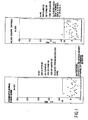
- Figures 1-6 illustrate the results of testing using the CTA assays described in Examples 1 and 2.
- Figures 1, 2 and 3 show results of testing using the Example 1 assay.
- the cutoff for all assays ( Figures 1, 2 and 3) was established as the mean of the optical density (O.D.) at 490 nm plus two standard deviations from the mean, of the Blood Donors (Normal) population, i.e., 0.890 O.D. That cutoff was selected for optimal specificity of the assay.
- Figure 1 demonstrates that none of a group of documented normal human sera was positive by the CTA assay of Example 1, and that only 1 out of 45 normal blood donor sera tested positive. This positive sample was also borderline positive when tested by another commercially available research use assay (Centacor CA 15-3, Centacor, Malvern, Pennsylvania).
- Figure 2 demonstrates testing of benign breast disease, early breast cancer and Stage IV breast cancer in human sera compared to the normal blood donor population shown in Figure 1. As can be seen in this figure, 3 of 29 benign breast disease sera, 21 of 68 early breast cancer sera and 72 of 93 Stage IV breast cancer sera tested positive by the CTA assay.
- Figure 3 shows a head-to-head comparison of the CTA assay to the CA 15-3 assay.
- the 67 sera are from a documented specimen panel obtained from the University of Toronto Medical Center, Toronto, Canada and the Wisconsin Clinical Cancer Center, Madison, Wisconsin. Normal women were documented by breast examination, family histories and normal menstrual cycles.
- the horizontal dashed line represents the cut-off (with a background of 0.2 subtracted) separating persons with normal levels of the CTA marker from elevated levels associated with cancer, and is based on the mean plus two standard deviations of the blood donor population. The cutoff level chosen is greater than the mean plus three standard deviations of the documented FIG. 3 Normal Women population to enhance specificity.
- CA 15-3 negative specimens are indicated by circles, CA 15-3 positive specimens are indicated by triangles.
- the CA 15-3 assay showed 11 of 67 blood donor samples to be positive, while the CTA assay showed only 1 of 67 of these samples to be positive.
- the CA 15-3 assay resulted in a positive result for 1 of 32 benign breast disease samples, while the CTA assay found none of these samples to be positive.
- the CA 15-3 assay found 8 of 66 Stage I and II breast cancer samples to be positive and 49 of 92 Stage IV breast cancer samples positive, while the CTA assay determined 11 of 66 of the Stage I and II samples to be positive and 66 of 92 of the Stage IV samples to be positive.
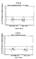
- Figures 4-6 show test results using the CTA assay of Example 2.
- Figure 4 shows a comparison of the CTA assay of Example 2 with the CA 15-3 assay.
- the CTA assay found only one sample to be positive, while the CA 15-3 assay found two positives.
- Testing on an additional normal population Figure 5 showed that the CTA assay and the CA 15-3 assay each detected 1 of 25 samples as positive.
- the CTA assay detected 17 of 28 samples as positive, while the CA 15- 3 assay detected 16 of 28.
- the neuraminidase treatment of a sample in the assay for cryptic tumor antigens offers many advantages.
- Third, the cryptic tumor antigen assay of the invention is more sensitive than previous assays as evidenced by greater detection of tumor-associated antigens in cancer patients than prior art assays.
- Fourth, the assay has a lower false positive rate than prior art assays.
- the assay is convenient and simple to perform.
Landscapes
- Health & Medical Sciences (AREA)
- Life Sciences & Earth Sciences (AREA)
- Immunology (AREA)
- Engineering & Computer Science (AREA)
- Molecular Biology (AREA)
- Biomedical Technology (AREA)
- Chemical & Material Sciences (AREA)
- Hematology (AREA)
- Urology & Nephrology (AREA)
- Food Science & Technology (AREA)
- Biochemistry (AREA)
- Cell Biology (AREA)
- Biotechnology (AREA)
- Medicinal Chemistry (AREA)
- Physics & Mathematics (AREA)
- Analytical Chemistry (AREA)
- Microbiology (AREA)
- General Health & Medical Sciences (AREA)
- General Physics & Mathematics (AREA)
- Pathology (AREA)
- Measuring Or Testing Involving Enzymes Or Micro-Organisms (AREA)
- Micro-Organisms Or Cultivation Processes Thereof (AREA)
- Measurement Of The Respiration, Hearing Ability, Form, And Blood Characteristics Of Living Organisms (AREA)
- Peptides Or Proteins (AREA)
- Preparation Of Compounds By Using Micro-Organisms (AREA)
Applications Claiming Priority (2)
| Application Number | Priority Date | Filing Date | Title |
|---|---|---|---|
| US12343987A | 1987-11-20 | 1987-11-20 | |
| US123439 | 1987-11-20 |
Publications (3)
| Publication Number | Publication Date |
|---|---|
| EP0316764A2 true EP0316764A2 (de) | 1989-05-24 |
| EP0316764A3 EP0316764A3 (en) | 1990-07-25 |
| EP0316764B1 EP0316764B1 (de) | 1994-06-08 |
Family
ID=22408693
Family Applications (1)
| Application Number | Title | Priority Date | Filing Date |
|---|---|---|---|
| EP88118727A Expired - Lifetime EP0316764B1 (de) | 1987-11-20 | 1988-11-10 | Nachweis von Tumorassozierten Anzeigern in biologischen Flüssigkeiten |
Country Status (6)
| Country | Link |
|---|---|
| EP (1) | EP0316764B1 (de) |
| JP (1) | JPH01165964A (de) |
| KR (1) | KR970001318B1 (de) |
| AT (1) | ATE107033T1 (de) |
| AU (1) | AU616919B2 (de) |
| DE (1) | DE3850046D1 (de) |
Cited By (3)
| Publication number | Priority date | Publication date | Assignee | Title |
|---|---|---|---|---|
| EP0313244A3 (en) * | 1987-10-09 | 1990-08-01 | Oncogen Limited Partnership | Method for increasing the sensitivity of assays for a ligand |
| US5459043A (en) * | 1989-09-15 | 1995-10-17 | Genetic Systems Corporation | Hybridoma CT43 producing a monoclonal antibody to a novel mucin epitope which correlates with the presence of colorectal cancer |
| WO2004037272A1 (ja) * | 2002-10-22 | 2004-05-06 | Japan Science And Technology Agency | デング熱ウィルス感染阻害剤 |
Families Citing this family (1)
| Publication number | Priority date | Publication date | Assignee | Title |
|---|---|---|---|---|
| EP1750265B1 (de) | 2003-06-23 | 2009-07-29 | Sony Corporation | Aufzeichnungsmediumgehäuse |
Family Cites Families (3)
| Publication number | Priority date | Publication date | Assignee | Title |
|---|---|---|---|---|
| US4743543A (en) * | 1985-09-09 | 1988-05-10 | Coulter Corporation | Method for enhancing and/or accelerating immunoassay detection of human carcinoma tumor associated antigen in a pathology sample |
| JPS62201364A (ja) * | 1985-10-22 | 1987-09-05 | セントコ−・インコ−ポレ−テツド | 高分子量 癌胎児性抗原の免疫検定法 |
| CA1340927C (en) * | 1987-10-09 | 2000-03-14 | Peter S. Linsley | Method for increasing the sensitivity of assays for target ligand |
-
1988
- 1988-11-10 DE DE3850046T patent/DE3850046D1/de not_active Expired - Lifetime
- 1988-11-10 AT AT88118727T patent/ATE107033T1/de not_active IP Right Cessation
- 1988-11-10 EP EP88118727A patent/EP0316764B1/de not_active Expired - Lifetime
- 1988-11-16 AU AU25620/88A patent/AU616919B2/en not_active Ceased
- 1988-11-18 KR KR1019880015200A patent/KR970001318B1/ko not_active Expired - Lifetime
- 1988-11-19 JP JP63293442A patent/JPH01165964A/ja active Pending
Cited By (4)
| Publication number | Priority date | Publication date | Assignee | Title |
|---|---|---|---|---|
| EP0313244A3 (en) * | 1987-10-09 | 1990-08-01 | Oncogen Limited Partnership | Method for increasing the sensitivity of assays for a ligand |
| US5646002A (en) * | 1987-10-09 | 1997-07-08 | Oncogen Limited Partnership | Method for increasing the sensitivity of assays for target ligand |
| US5459043A (en) * | 1989-09-15 | 1995-10-17 | Genetic Systems Corporation | Hybridoma CT43 producing a monoclonal antibody to a novel mucin epitope which correlates with the presence of colorectal cancer |
| WO2004037272A1 (ja) * | 2002-10-22 | 2004-05-06 | Japan Science And Technology Agency | デング熱ウィルス感染阻害剤 |
Also Published As
| Publication number | Publication date |
|---|---|
| KR890008328A (ko) | 1989-07-10 |
| KR970001318B1 (ko) | 1997-02-05 |
| EP0316764A3 (en) | 1990-07-25 |
| ATE107033T1 (de) | 1994-06-15 |
| AU2562088A (en) | 1989-05-25 |
| DE3850046D1 (de) | 1994-07-14 |
| EP0316764B1 (de) | 1994-06-08 |
| JPH01165964A (ja) | 1989-06-29 |
| AU616919B2 (en) | 1991-11-14 |
Similar Documents
| Publication | Publication Date | Title |
|---|---|---|
| EP0615129B1 (de) | Verfahren zum selektiven Nachweis von perinuklearen anti-neutrophilen cytoplasmischen Antikörpern bei ulzerativen Kolitis oder primärer sclerotischer Cholangitis | |
| JP3486413B2 (ja) | 前立腺特異抗原の免疫検定法 | |
| EP0440044A1 (de) | Verfahren zur Vermeidung humaner Anti-Maus-Antikörper-Störungen in den in vitro diagnostischen Bestimmungsverfahren | |
| CN1362970A (zh) | 对雌激素受激亮氨酸氨基太酶的单克隆抗体 | |
| KR880006358A (ko) | 새로운 뮤친 에피토프에 대한 모노클로날항체 생산용 잡종체 | |
| JPS61500315A (ja) | 血清中の免疫複合体を検出する方法 | |
| US5288614A (en) | Method for the detection of malignant diseases | |
| CA2008304C (en) | Assay for bone alkaline phosphatase | |
| US5591830A (en) | Detection of complexes which include basement membrane components as diagnostic of cancer and other diseases | |
| US5646002A (en) | Method for increasing the sensitivity of assays for target ligand | |
| JPS62231172A (ja) | 免疫複合体の検定法 | |
| EP0316764B1 (de) | Nachweis von Tumorassozierten Anzeigern in biologischen Flüssigkeiten | |
| US6015680A (en) | Human lung adenocarcinoma-related monoclonal antibody and immunoassay method which uses the same | |
| EP0651826A1 (de) | Test auf den infektiösen peritonitisvirus der feliden mittels monoklonaler idiotypischer und anti-idiotypischer antikörper | |
| EP0221561A2 (de) | Menschlichen Brustkrebs anzeigendes Antigen und darauf basierende Teste | |
| US5358850A (en) | Sandwich immunoassay of β-n-acetylglucosaminidase and monoclonal antibody used therein | |
| CA2281262C (en) | Anti-human medullasin monoclonal antibody, process for producing the same and immunoassay using the same | |
| EP0447490A1 (de) | Nachweis der basismembrankomponenten, diagnose von krebs und sonstigen krankheiten. | |
| CA2044154C (en) | Method of assaying d-vanillylmandelic acid, and reagent and kit therefor | |
| EP0738890B1 (de) | Methode zur Bestimmung von Cholinesterase und Methode zur Unterscheidung zwischen Leberzirrhose und Hepatitis | |
| Sato et al. | Treponema pallidum specific IgM haemagglutination test for serodiagnosis of syphilis. | |
| JPH10150999A (ja) | ヒト前立腺特異抗原−α1−アンチキモトリプシン複合体に対するモノクローナル抗体及びそれを用いたヒト前立腺特異抗原−α1−アンチキモトリプシン複合体の免疫学的検出法 | |
| JPH0272198A (ja) | 抗ヒト肺腺癌モノクローナル抗体 | |
| JPH03504272A (ja) | 免疫抑制の予知および診断のための胎盤イソフェリチン |
Legal Events
| Date | Code | Title | Description |
|---|---|---|---|
| PUAI | Public reference made under article 153(3) epc to a published international application that has entered the european phase |
Free format text: ORIGINAL CODE: 0009012 |
|
| AK | Designated contracting states |
Kind code of ref document: A2 Designated state(s): AT BE CH DE ES FR GB IT LI NL |
|
| PUAL | Search report despatched |
Free format text: ORIGINAL CODE: 0009013 |
|
| AK | Designated contracting states |
Kind code of ref document: A3 Designated state(s): AT BE CH DE ES FR GB IT LI NL |
|
| 17P | Request for examination filed |
Effective date: 19910117 |
|
| 17Q | First examination report despatched |
Effective date: 19921023 |
|
| GRAA | (expected) grant |
Free format text: ORIGINAL CODE: 0009210 |
|
| AK | Designated contracting states |
Kind code of ref document: B1 Designated state(s): AT BE CH DE ES FR GB IT LI NL |
|
| PG25 | Lapsed in a contracting state [announced via postgrant information from national office to epo] |
Ref country code: IT Free format text: LAPSE BECAUSE OF FAILURE TO SUBMIT A TRANSLATION OF THE DESCRIPTION OR TO PAY THE FEE WITHIN THE PRESCRIBED TIME-LIMIT;WARNING: LAPSES OF ITALIAN PATENTS WITH EFFECTIVE DATE BEFORE 2007 MAY HAVE OCCURRED AT ANY TIME BEFORE 2007. THE CORRECT EFFECTIVE DATE MAY BE DIFFERENT FROM THE ONE RECORDED. Effective date: 19940608 Ref country code: BE Effective date: 19940608 Ref country code: ES Free format text: THE PATENT HAS BEEN ANNULLED BY A DECISION OF A NATIONAL AUTHORITY Effective date: 19940608 Ref country code: NL Effective date: 19940608 Ref country code: AT Effective date: 19940608 |
|
| REF | Corresponds to: |
Ref document number: 107033 Country of ref document: AT Date of ref document: 19940615 Kind code of ref document: T |
|
| ET | Fr: translation filed | ||
| REF | Corresponds to: |
Ref document number: 3850046 Country of ref document: DE Date of ref document: 19940714 |
|
| PG25 | Lapsed in a contracting state [announced via postgrant information from national office to epo] |
Ref country code: DE Effective date: 19940909 |
|
| PG25 | Lapsed in a contracting state [announced via postgrant information from national office to epo] |
Ref country code: GB Effective date: 19941110 |
|
| NLV1 | Nl: lapsed or annulled due to failure to fulfill the requirements of art. 29p and 29m of the patents act | ||
| PG25 | Lapsed in a contracting state [announced via postgrant information from national office to epo] |
Ref country code: CH Effective date: 19941130 Ref country code: LI Effective date: 19941130 |
|
| PLBE | No opposition filed within time limit |
Free format text: ORIGINAL CODE: 0009261 |
|
| STAA | Information on the status of an ep patent application or granted ep patent |
Free format text: STATUS: NO OPPOSITION FILED WITHIN TIME LIMIT |
|
| 26N | No opposition filed | ||
| GBPC | Gb: european patent ceased through non-payment of renewal fee |
Effective date: 19941110 |
|
| PG25 | Lapsed in a contracting state [announced via postgrant information from national office to epo] |
Ref country code: FR Effective date: 19950731 |
|
| REG | Reference to a national code |
Ref country code: CH Ref legal event code: PL |
|
| REG | Reference to a national code |
Ref country code: FR Ref legal event code: ST |