EP0325461A2 - Gerät zur Bilderzeugung mittels magnetischer Resonanz - Google Patents
Gerät zur Bilderzeugung mittels magnetischer Resonanz Download PDFInfo
- Publication number
- EP0325461A2 EP0325461A2 EP89300513A EP89300513A EP0325461A2 EP 0325461 A2 EP0325461 A2 EP 0325461A2 EP 89300513 A EP89300513 A EP 89300513A EP 89300513 A EP89300513 A EP 89300513A EP 0325461 A2 EP0325461 A2 EP 0325461A2
- Authority
- EP
- European Patent Office
- Prior art keywords
- gradient field
- magnetic resonance
- slice
- image
- field
- Prior art date
- Legal status (The legal status is an assumption and is not a legal conclusion. Google has not performed a legal analysis and makes no representation as to the accuracy of the status listed.)
- Withdrawn
Links
Images





Classifications
-
- G—PHYSICS
- G01—MEASURING; TESTING
- G01R—MEASURING ELECTRIC VARIABLES; MEASURING MAGNETIC VARIABLES
- G01R33/00—Arrangements or instruments for measuring magnetic variables
- G01R33/20—Arrangements or instruments for measuring magnetic variables involving magnetic resonance
- G01R33/44—Arrangements or instruments for measuring magnetic variables involving magnetic resonance using nuclear magnetic resonance [NMR]
- G01R33/48—NMR imaging systems
- G01R33/4828—Resolving the MR signals of different chemical species, e.g. water-fat imaging
-
- G—PHYSICS
- G01—MEASURING; TESTING
- G01R—MEASURING ELECTRIC VARIABLES; MEASURING MAGNETIC VARIABLES
- G01R33/00—Arrangements or instruments for measuring magnetic variables
- G01R33/20—Arrangements or instruments for measuring magnetic variables involving magnetic resonance
- G01R33/44—Arrangements or instruments for measuring magnetic variables involving magnetic resonance using nuclear magnetic resonance [NMR]
- G01R33/48—NMR imaging systems
- G01R33/483—NMR imaging systems with selection of signals or spectra from particular regions of the volume, e.g. in vivo spectroscopy
- G01R33/485—NMR imaging systems with selection of signals or spectra from particular regions of the volume, e.g. in vivo spectroscopy based on chemical shift information [CSI] or spectroscopic imaging, e.g. to acquire the spatial distributions of metabolites
-
- G—PHYSICS
- G01—MEASURING; TESTING
- G01R—MEASURING ELECTRIC VARIABLES; MEASURING MAGNETIC VARIABLES
- G01R33/00—Arrangements or instruments for measuring magnetic variables
- G01R33/20—Arrangements or instruments for measuring magnetic variables involving magnetic resonance
- G01R33/44—Arrangements or instruments for measuring magnetic variables involving magnetic resonance using nuclear magnetic resonance [NMR]
- G01R33/48—NMR imaging systems
- G01R33/54—Signal processing systems, e.g. using pulse sequences ; Generation or control of pulse sequences; Operator console
- G01R33/56—Image enhancement or correction, e.g. subtraction or averaging techniques, e.g. improvement of signal-to-noise ratio and resolution
-
- G—PHYSICS
- G01—MEASURING; TESTING
- G01R—MEASURING ELECTRIC VARIABLES; MEASURING MAGNETIC VARIABLES
- G01R33/00—Arrangements or instruments for measuring magnetic variables
- G01R33/20—Arrangements or instruments for measuring magnetic variables involving magnetic resonance
- G01R33/44—Arrangements or instruments for measuring magnetic variables involving magnetic resonance using nuclear magnetic resonance [NMR]
- G01R33/48—NMR imaging systems
- G01R33/54—Signal processing systems, e.g. using pulse sequences ; Generation or control of pulse sequences; Operator console
- G01R33/56—Image enhancement or correction, e.g. subtraction or averaging techniques, e.g. improvement of signal-to-noise ratio and resolution
- G01R33/561—Image enhancement or correction, e.g. subtraction or averaging techniques, e.g. improvement of signal-to-noise ratio and resolution by reduction of the scanning time, i.e. fast acquiring systems, e.g. using echo-planar pulse sequences
- G01R33/5615—Echo train techniques involving acquiring plural, differently encoded, echo signals after one RF excitation, e.g. using gradient refocusing in echo planar imaging [EPI], RF refocusing in rapid acquisition with relaxation enhancement [RARE] or using both RF and gradient refocusing in gradient and spin echo imaging [GRASE]
-
- G—PHYSICS
- G01—MEASURING; TESTING
- G01R—MEASURING ELECTRIC VARIABLES; MEASURING MAGNETIC VARIABLES
- G01R33/00—Arrangements or instruments for measuring magnetic variables
- G01R33/20—Arrangements or instruments for measuring magnetic variables involving magnetic resonance
- G01R33/44—Arrangements or instruments for measuring magnetic variables involving magnetic resonance using nuclear magnetic resonance [NMR]
- G01R33/48—NMR imaging systems
- G01R33/54—Signal processing systems, e.g. using pulse sequences ; Generation or control of pulse sequences; Operator console
- G01R33/56—Image enhancement or correction, e.g. subtraction or averaging techniques, e.g. improvement of signal-to-noise ratio and resolution
- G01R33/561—Image enhancement or correction, e.g. subtraction or averaging techniques, e.g. improvement of signal-to-noise ratio and resolution by reduction of the scanning time, i.e. fast acquiring systems, e.g. using echo-planar pulse sequences
- G01R33/5615—Echo train techniques involving acquiring plural, differently encoded, echo signals after one RF excitation, e.g. using gradient refocusing in echo planar imaging [EPI], RF refocusing in rapid acquisition with relaxation enhancement [RARE] or using both RF and gradient refocusing in gradient and spin echo imaging [GRASE]
- G01R33/5616—Echo train techniques involving acquiring plural, differently encoded, echo signals after one RF excitation, e.g. using gradient refocusing in echo planar imaging [EPI], RF refocusing in rapid acquisition with relaxation enhancement [RARE] or using both RF and gradient refocusing in gradient and spin echo imaging [GRASE] using gradient refocusing, e.g. EPI
Definitions
- the present invention relates to a magnetic resonance imaging (MRI) system and, in particular, to a magnetic imaging system which can obtain a high-speed image by separating two substances having magnetic resonance frequencies somewhat different in their chemical shift.
- MRI magnetic resonance imaging
- a projection reconstruction method by Lauterbur, Fourier transform method by Kumar, Welti and Ernst, spin warp method (modified Fourier transform method, and so on).
- the magnetic resonance imaging system for obtaining an image by virtue of a magnetic resonance imaging takes a longer time in the collection of data than the other medical image diagnosing apparatus, e.g., an ultrasonic diagnosing apparatus or X-ray CT (Computer tomography) apparatus. Therefore, an artifact is produced by the movement of the object, e.g., a patient, such as the respiration movement and it is thus difficult to obtain an image of a high-speed moving section or site, such as the heart and circulatory system. Furthermore, the patient experiences great pain during the imaging period due to longer imaging time involved.
- the other medical image diagnosing apparatus e.g., an ultrasonic diagnosing apparatus or X-ray CT (Computer tomography) apparatus. Therefore, an artifact is produced by the movement of the object, e.g., a patient, such as the respiration movement and it is thus difficult to obtain an image of a high-speed moving section or site, such as the heart and circulatory system. Furthermore, the patient experiences great pain during the imaging period due
- ultra high-speed Fourier transform methods As a method for obtaining an image at high speed by a magnetic resonance imaging technique, ultra high-speed Fourier transform methods have been proposed, such as an echo planar method by Mansfield and ultra high-speed Fourier transform method by Hutchison.
- the magnetic resonance data is collected in accordance with a pulse sequence as shown in Fig. 1.
- the ultra high-speed Fourier method also referred to as a multiple echo Fourier method is different from teh echo planar method of Fig. 1 in that, as shown in Fig. 2, the phase encode gradient field Ge is applied in a pulse-like fashion for each inversion of the read-out gradient field. In other respects, both are similar to each other.
- the excited magnetization in the slice of the object produces a phase dispersion resulting from the spatial inhomogeneities of the static field and hence an apparent transverse relaxation occurs at a time T2 * which is shorter than that transverse relaxation time T2.
- T2 * which is shorter than that transverse relaxation time T2.
- this method takes double a time from the excitation of magnetization until the application of a 180° high frequence pulse.
- the magnetic resonance data collection time and the time required from the 180° pulse application are added to the time (i.e. imaging time) from the excitation of the magnetization until the collection of the magnetic resonance data is completed. That total time offers a bar to the implementation of high-speed imaging.
- phase encode gradient field is very small, thus being liable to produce a phase encode error resulting from the spatial or time inhomogeneities of a static field.
- phase encode error causes a deformation or blurring of an image reconstructed.
- a magnetic resonance signal even if coming from the nuclei of specific atoms in the sample, produces a frequency difference called a "chemical shift" due to a difference in the chemical circumference.
- the Dixon method is known as the method for obtaining a water and fat separated image through the utilization of the chemical shift.
- two imaging operations are required in obtaining a water/fat-separated image.
- the Dixon method uses the conventional imaging technique and takes a longer time for the respective imaging than the aforementioned ultra high-speed imaging method. Hence it takes longer time to obtain a water/fat-separated image.
- Figs. 3A and 3B show a pulse sequence for obtaining a water/fat-separated image by the Dixon method.
- a gradient field Gs for the slice and 90° selective excitation pulse RF are applied to the object and, upon applying a 180° pulse RF 2 following a time TE/2, the nuclear spin dispersed is brought into an in-phase relation after TE/2, producing an echo; At this time, magnetic resonance signals of the nuclei of hydrogen in water and fat have the same phase. Under these situations, the read-out gradient field Gr is applied, allowing the collection of data for one echo. By repeating these operations a plurality of times while varying little by little the phase encode gradient field Ge, it is possible to collect whole magnetic resonance data necessary for the reconstruction of an image.
- the image is reconstructed by the magnetic resonance data thus obtained, then it is possible to get an image corresponding to a sum of the water and fat images, noting that this image contains a chemical shift, that is, a positional displacement corresponding to a difference between the frequence of the water and that of the fat.
- the application timing of the 180° pulse RF 2 is changed a time TE'/2 from a 90° pulse RF 1 and magnetic resonance data is collected a time AT after the occurrence of an echo following that time ⁇ T.
- the time AT is so set that a phase difference between the magnetic resonance signals of the nuclei of hydrogen in the water and fat is just 180°.
- the image reconstructed based on the magnetic resonance data thus obtained corresponds to an image which is a difference of the image of the water and that of the fat. It is thus possible to separate the image of the water and that of the fat by computation from those two reconstructed images.
- the Dixon method two imaging steps are needed to obtain a water/fat-separated image. If, however, the time AT in the sequence of Fig. 3B is set to a predetermined value, that is, the time at which a phase difference between both the water and the fat signal is n/2 or -j f /2, then the water and fat image information can be obtained by one imaging operation in a manner separated into an imaginary and a real part of the image data, respectively.
- the method for obtaining the water/fat-separated image through the utilization of the above is referred to as a modified Dixon method. Even the modified Dixon method employs conventional imaging method which is longer in the imaging process than the ultra high-speed imaging method and hence it takes longer time to obtain a water/fat-separated image.
- the magnetic resonance imaging system applies a high frequency magnetic field and gradient field to an object in a homogeneous static magnetic field in a predetermined pulse sequence and detects a magnetic resonance signal from the sample to obtain an image.
- the magnetic resonance imaging system of the present invention includes a data pick-up section and image processing unit.
- the data pick-up section uses a predetermined pulse sequence so timed that a nuclear magnetization phase difference between two predetermined substances somewhat differing in their magnetic resonance frequency due to a chemical shift is set to ⁇ /2 or -n/2 at a predetermined time and acquires all magnetic resonance data necessary to reconstruct an image of a slice excited by the aforementioned high frequency magnetic field into magnetic resonance, within a period of time in which the aforementioned predetermined nuclear magnetization is relaxed by the relaxation of the transverse magnetization.
- the image processing section obtains a magnetic resonance image of at least one of the two substances as obtained in a form separated into image information's real and imaginary parts by subjecting magnetic resonance data acquired by the data pick-up unit to a two-dimensional complex Fourier transform.
- a magnetic resonance image of at least one of the aforementioned two substances for a very small period of time.
- a magnetic resonance imaging system includes a correction section adapted to correct an influence from the inhomogeneities of the magnetic field in the acquiring process of all magnetic resonance data by the data pick-up section which is necessary to reconstruct a slice image, whereby it is possible to achieve the ultra high-speed imaging of the two substances as their separate images, while correcting any influence from the inhomogeneities of the static field.
- an image such as a water/fat-separated image, corresponding to the nuclear magnetization of two predetermined substances somewhat differing in their magnetic resonance frequency due to the chemical shift.
- the system equipped with the correction section can eliminate any influence from a phase dispersion of the magnetization resulting from the inhomogeneities of the static field. It is also possible to correct a phase encode error which is caused by the use of a weaker gradient magnetization (for phase encode) in the ultra high-speed imaging process. This it is possible to gain a correct separated image free from any chemical shift artifact and deformation and hence to achieve the imaging of the aforementioned two substances as a separate image on an active site, such as the heart and circulatory system.
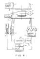
- static field magnet 1 having a static field coil system is driven by excitation power supply 2 and gradient field coil 3 is driven by gradient field drive circuits 4, 5 and 6 which in turn are controlled by gradient field controller 7.
- Static field magnet 1 applies a homogeneous static field to an object P (for example, a human subject or a patient) usually lying on a couch.
- Gradient field coil 3 generates a gradient field having a magnetic field distribution of a gradually varying field strength along two right-angled intersecting directions x and y in a slice, that is, an object cross-sectional portion of interest, and along a direction z perpendicular to the slice plane.
- the z-, x- and y-direction gradient fields are assumed as being a slicing gradient field Gs, read-out gradient field Gr and phase encode gradient field Ge, respectively.
- a high frequency field which is generated from probe 9 by a high frequency signal of transmitting section 8 is applied to the object P.
- Transmitting section 8 is operated under control of pulse sequencer 10.
- probe 9 which is formed of a high frequency coil is commonly employed for a transmitting coil for the generation of a high frequency field and for a receiving coil for receiving a magnetic resonance signal relating to the nuclei of various atoms in the object.
- duplexer 12 is provided which is connected between probe 9 and transmitting section 8 and receiving processor section 11.
- duplexer 12 is not necessary and the transmit- and receive-only coils are connected to transmitting section 8 and receiving processor section 11, respectively.
- a magnetic resonance signal that is, a magnetic resonance echo signal, which is received by probe 9 is amplified, detected, and picked up, as magnetic resonance echo data, by means of receiving processor section 11.
- the picked-up magnetic resonance echo data is, after being analog/digital (A/D) converted, supplied to computer 13.
- Computer 13 receives the magnetic resonance echo data from receiving processor section 11 and subjects the echo data to a Fourier transform processing to achieve an image reconstruction as image data.
- Computer 13 controls pulse sequencer 10 via interface 14.
- Pulse sequencer 10 controls gradient field controller 7 and transmitting section 8. If necessary, power supply 2 and receiving processor section 11 are controlled by means of pulse sequencer 10.
- the image data which is obtained from computer 11 is supplied to display 14 where an image is displayed.
- a nuclear spin which is excited by a 90° selection excitation pulse into magnetic resonance causes a phase dispersion in accordance with a time constant T2 *.
- the phase-dispersed nuclear spin is refocused by a 180° pulse which is applied a time TE/2 after the application of the 90° pulse and an echo is produced after TE/2.
- the nuclear spin of 1 H in the water and fat has the same phase on the rotation coordinate system.
- the read-out gradient field Gr is repeatedly inversion-switched to produce a multi-echo, and during this period of time, the encode gradient field Ge of a predetermined amplitude is applied to the object P. This is what is called an ordinary echo planar method.
- ⁇ represents a difference of the resonance frequencies of the nuclei of 1 H in the water and fat, noting that the fat is about 3 to 4 ppm lower than the water.
- the following is a pulse sequence for obtaining the image data of the water/fat-separated image of the object's slice in accordance with the present invention.
- the pulse sequence is controlled by pulse sequencer 10.
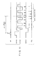
- the pulse sequence as shown in Fig. 5 is basically the same as that of the conventional echo planar method shown in Fig. 1.
- the slicing gradient field Gs is applied in the direction z and, in this state, a 90° selective excitation pulse (a high frequence pulse for 90°-flipping only the magnetization of the object's slice of interest) is applied, causing the predetermined nuclear magnetization of a specific slice of the object to be selectively excited to generate magnetic resonance.
- the gradient field Gs for slice is inverted, after the application of the 90° selective excitation pulse, so as to refocus a magnetization.
- a 180° pulse is applied as a high frequency field RF after the application of the 90° selective excitation pulse and gradient field for slice, that is, a time TE'/2 after the application of the 90° selective excitation pulse.
- a magnetic resonance echo signal is generated a time TE'/2 from the application of a 180° pulse, and a readout gradient field Gr of the direction x and encode gradient field Ge of the direction y are applied after the time AT to sequentially produce a plurality of magnetic resonance echoes.
- the read-out gradient field Gr is alternately switched, in the positive and negative alternate fashion, in one direction (for example, the direction x) on an x,y plane and, at the same time, the phase encode gradient field Ge is statically applied in the other direction, that is, the direction y, on the x,y plane during the time period in which the read-out gradient field Gr is repetitively applied in the positive and negative alternate fashion.
- the phase encode gradient field Ge is statically applied in the other direction, that is, the direction y, on the x,y plane during the time period in which the read-out gradient field Gr is repetitively applied in the positive and negative alternate fashion.
- the whole magnetic resonance data of the slice is acquired during the time T2 * in which the magnetization in the slice plane excited by the 90° high frequency pulse and refocused by the 180° pulse to be relaxed due to the relaxation of the transverse magnetization containing an inhomogeneities of the static field.
- the magnetic resonance echo signal is picked up with the use of a pulse sequence, including only the high frequence field RF and gradient field Gs and not including the read-out gradient field Gr and encode gradient field Ge, and then subjected to a one-dimensional Fourier transformation to obtain a water/fat spectrum.
- a pulse sequence including only the high frequence field RF and gradient field Gs and not including the read-out gradient field Gr and encode gradient field Ge, and then subjected to a one-dimensional Fourier transformation to obtain a water/fat spectrum.
- AT 0
- the water/fat spectrum is obtained as a "real-part" spectrum of the same sign, noting that the water spectrum SP W and fat spectrum SP f are indicated by the solid and broken lines, respectively. It is only necessary that, with the proper adjustment of AT from that state, the fat spectrum SP f emerge as an imaginary part as indicated by the solid line in Fig. 6.
- the water/fat-separated image thus obtained may be displayed individually independently or as a water/fat-combined image.
- the position of the water/fat image is displaced due to a difference in their resonance frequency and that image is reconstructed after being adjusted properly.
- the images of the water and fat may be displayed in a different color.
- the value AT for imparting a phase difference to the magnetic resonance signals of the nuclei of H in the water and fat need not necessarily be adjusted by the 180° pulse and may be obtained simply adjusting the data pick-up timing, that is, the read-out gradient field, encode gradient field and A/D conversion timing or selective excitation pulse.
- the sequence for picking up the magnetic resonance echo may not be obtained from the echo planar method and may be any sequence so long as it is a sequence for ultra high-speed imaging.
- the 180° pules is not necessarily required and it may be possible to pick up the data of a free induction decay (FID) signal subsequent to the application of the 90° selection excitation pulse.
- FID free induction decay
- the adjustment of the AT is conducted during a period of time from the application of the 90° selection excitation pulse to the picking-up of associated data.
- the first embodiment of the present invention is tantamount to, so to speak, the application of the ultra high-speed imaging method to the modified Dixon method.
- the conventional ultra high-speed imaging method poses a problem, such as the degradation of a high-speed characteristic because of the requirement of the refocusing process of a phase-dispersed magnetization resulting from the inhomogeneities of the static field or the occurrence of a phase encode error due to a static field and consequent degradation of a reconstructed image.
- an image of two substances whose resonance frequencies somewhat differ due to a chemical shift involved, an image quality is greatly affected by an error, such as a spatial phase error resulting from the inhomogeneities of the static field.
- a magnetic resonance imaging system is directed to solving the aforementioned problem.
- a separated substance/substance image that is, an image of two kinds of substances whose resonance frequencies somewhat differ due to a chemical shift, can be obtained in an ultra high-speed imaging process in comparison with the conventional system and in a high quality free from any adverse effect from the inhomogeneous static field.
- Fig. 7 is a view showing an arrangement of a magnetic resonance imaging system according to the second embodiment of the present invention.
- static field magnet 101 and gradient field coil 103 are driven by excitation power supply 102 and driver circuit 104, respectively, which are in turn controlled by systems controller 110.
- Static field magnet 101 applies a homogeneous static field to an object P on couch 106.
- Gradient field coil 103 applies a gradient field having a magnetic intensity distribution of a gradually varying intensity along two directions x and y intersecting at right angles in a cross-section of interest of the object P, that is, a slice plane, and direction z perpendicular to the slice plane.
- the z-direction gradient field, x-direction gradient field and y-direction gradient field as a slicing gradient field Gs, read-out gradient field Gr and phase encode gradient field Ge, respectively.
- a high frequency magnetic field which is generated from probe 107 by a high frequency signal from transmitter 108, is applied to the object P.
- Transmitter 108 is operated under control of system controller 110.
- probe 107 is commonly employed for a transmitting coil for the generation of a high frequency-magnetic field and receiving coil for the reception of a magnetic resonance signal relating to various types of the nuclei of atoms in the object P
- transmit- and receive-only coils may separately be provided as in the same way as in the first embodiment of the present invention.
- a magnetic resonance signal that is, a magnetic resonance echo signal, which is received by probe 107, is transferred to data acquisition section 111 after being amplified and detected by receiver 109.
- the transfer of the data is effected under control of system controller 110.
- Data acquisition section 111 which is controlled by system controller 110, acquires a magnetic resonance echo signal from receiver 109 and subjects it to an A/D conversion, and send it to computer 112.
- Computer 112 is controlled by an operation via console 113 and it subjects the magnetic resonance echo data which is input from data acquisition section 111 to a Fourier transformation and hence an image reconstruction processing to obtain image data.
- Computer 112 also controls system controller 110 and the image data of computer 112 is supplied to image display 114 where the image is displayed.
- Fig. 8 shows one form of a pulse sequence for obtaining a water/fat-separated image data relating to a slice of an object P in accordance with the present invention.
- the pulse sequence is controlled by system controller 110.
- the pulse sequence of Fig. 8 is fundamentally based on that of the conventional ultra high-speed Fourier method of Fig. 2, but is different from the latter in that the gradient field Ge for phase encode is applied before the application of a 180° high frequency pulse following the application of a 90° selective excitation pulse, that a selective excitation pulse is used as a 180° high frequency pulse, and that the 180° selective excitation high frequency pulse is employed with the slicing gradient field Gs applied.
- a slicing gradient field Gs is applied in the direction z and, in this state, a 90° selective excitation pulse (a high frequency pulse for 90° flipping the magnetization of a predetermined slice of interest) is applied as a high frequency field RF, causing a selective excitation of a predetermined nuclear magnetization of the predetermined slice plane of interest of the object P.
- the gradient field Gs is inverted to refocus a magnetization.
- gradient field Ge phase encode is applied in the direction y.
- a time TE/2 after the application of the 90° selective excitation pulse a 180° selective excitation pulse is applied as a high frequency magnetic field RF, while, at the same time, the slicing gradient field Gs is being applied.
- the magnetic resonance echo signal is produced a time TE/2 after the application of the 180° pulse, while in the pulse sequence of Fig.
- an x-direction read-out gradient field Gr and y-direction gradient field Ge are applied without a wait for a lapse of a time TE/2 following the application of the 180° pulse to sequentially produce -n-th to n-th magnetic resonance echoes. That is, in one direction of the x and y plane intersecting at right angles to the z direction, for example, the x direction, the gradient field Gr are repetitively applied in a positive and negative alternate switching fashion and, in the other direction of the x and y plane, for example, the y-direction, the gradient field Ge for phase encode is applied in a pulse-like fashion for each inversion of the read-out gradient field.
- the echo signal train is Fourier-transformed and image-reconstructed to generate the image data of the slice.
- Figs. 9 and 10 show a flow of processing for obtaining a water/fat-separated image in accordance with the present invention with an effect of an inhomogeneous static field corrected.
- Fig. 12 shows a pulse sequence for measuring the inhomogeneities of the static image field.
- the slice's nuclear spin is selectively excited by a selective excitation pulse and z direction gradient field Gz (that is, Gs).
- Gz that is, Gs
- the x and y direction gradient fields Gx and Gy are applied so as to incorporate the x and y direction positional information in the excited slice plane into the phase of the nuclear spin.
- the subsequent application of a 180° pulse results in the generation of a spin echo signal.
- the imaging processing is carried out, at step 34, for obtaining a water/fat-separated image, the processing including the processing for correcting an effect resulting from the inhomogeneities of the static field.
- the water/fat-separated imaging step 34 comprises, as shown in Fig. 10, a step 34a for subjecting the inhomogeneous field distribution to a coordinate conversion and interpolation, step 34b for correcting a phase error resulting from the inhomogeneous magnetic field, coordinate conversion step 34c and step 34d for correcting an image shift resulting from the chemical shift frequency of the fat image.
- the present invention is not restricted to the aforementioned embodiments.
- the 180° pulse has been explained as being applied subsequent to the application of the 90° selective excitation pulse, subsequent to exciting the magnetization in the slice plane by an a° (90° for instance) selective excitation pulse and gradient field Gs the switching of the read-out gradient field Gr and pulsatory application of the gradient field Ge may be performed (without the application of the 180° pulse as shown in Fig. 8), as shown in Fig. 11, to achieve high-speed imaging.
- a pulse sequence as shown in Fig. 11 eliminates the need for simultaneously applying a 180° pulse and gradient field Gs as in the pulse sequence shown in Fig. 8. That is, in order to selectively excite the magnetization in a slice plane of interest of the object P, the application of the gradient field Gs in the z-direction and that of a 90° selective excitation pulse (an angle a° other than 90° may be used) are effected to selectively excite a specific slice in the object P in which case the gradient field Gs is, subsequent to the application of the 90° selective excitation pulse, inverted so that a magnetization may be refocused.
- a read-out gradient field Gr is repetitively applied in a positive and negative alternate switching fashion in one direction of an x,y plane intersecting at right angles to the z direction, for example, in the x direction, without applying the 180° pulse and gradient field Gs as shown in Fig. 8, and, at the same time, a gradient field Ge is applied in a pulse-like fashion in the other direction of the x,y plane, for example, in the y direction each time the read-out gradient field Gr is inverted.
- a magnetic resonance signal an echo signal
- the present invention is not restricted to the aforementioned embodiments.
- the present invention has been explained as using the ultra high-speed Fourier transform method, it can use the echo planar method shown in Fig. 1.
- the gradient field Ge for phase encode is statically applied during all the time in which the read-out gradient field Gr is applied while being inverted.
Landscapes
- Physics & Mathematics (AREA)
- High Energy & Nuclear Physics (AREA)
- Condensed Matter Physics & Semiconductors (AREA)
- General Physics & Mathematics (AREA)
- Health & Medical Sciences (AREA)
- Spectroscopy & Molecular Physics (AREA)
- General Health & Medical Sciences (AREA)
- Nuclear Medicine, Radiotherapy & Molecular Imaging (AREA)
- Radiology & Medical Imaging (AREA)
- Engineering & Computer Science (AREA)
- Signal Processing (AREA)
- Optics & Photonics (AREA)
- Magnetic Resonance Imaging Apparatus (AREA)
Applications Claiming Priority (4)
| Application Number | Priority Date | Filing Date | Title |
|---|---|---|---|
| JP63010823A JP2656275B2 (ja) | 1988-01-22 | 1988-01-22 | 磁気共鳴映像装置 |
| JP10823/88 | 1988-01-22 | ||
| JP299330/88 | 1988-11-26 | ||
| JP63299330A JP2731195B2 (ja) | 1988-11-26 | 1988-11-26 | 磁気共鳴映像装置 |
Publications (2)
| Publication Number | Publication Date |
|---|---|
| EP0325461A2 true EP0325461A2 (de) | 1989-07-26 |
| EP0325461A3 EP0325461A3 (de) | 1990-08-16 |
Family
ID=26346166
Family Applications (1)
| Application Number | Title | Priority Date | Filing Date |
|---|---|---|---|
| EP89300513A Withdrawn EP0325461A3 (de) | 1988-01-22 | 1989-01-19 | Gerät zur Bilderzeugung mittels magnetischer Resonanz |
Country Status (2)
| Country | Link |
|---|---|
| US (1) | US4949042A (de) |
| EP (1) | EP0325461A3 (de) |
Cited By (2)
| Publication number | Priority date | Publication date | Assignee | Title |
|---|---|---|---|---|
| EP0372814A3 (de) * | 1988-11-30 | 1991-01-23 | Kabushiki Kaisha Toshiba | Vorrichtung zur Bilderzeugung mittels magnetischer Resonanz |
| CN104914389A (zh) * | 2014-12-18 | 2015-09-16 | 北京大学 | 基于自旋锁定技术探测震荡磁场的磁共振成像方法及应用 |
Families Citing this family (21)
| Publication number | Priority date | Publication date | Assignee | Title |
|---|---|---|---|---|
| JPH02215440A (ja) * | 1989-02-17 | 1990-08-28 | Toshiba Corp | 磁気共鳴映像装置 |
| JP2853232B2 (ja) * | 1990-02-02 | 1999-02-03 | 株式会社日立製作所 | 核磁気共鳴を用いた流体イメージング装置 |
| US5229717A (en) * | 1992-05-22 | 1993-07-20 | General Electric Company | Simultaneous two-contrast fast spin echo NMR imaging |
| US5594336A (en) * | 1995-06-02 | 1997-01-14 | Picker International, Inc. | Three point technique using spin and gradient echoes for water/fat separation |
| US5627469A (en) * | 1995-07-31 | 1997-05-06 | Advanced Mammography Systems, Inc. | Separation of fat and water magnetic resonance images |
| DE19718129A1 (de) * | 1997-04-29 | 1998-11-12 | Siemens Ag | Pulssequenz für ein Kernspintomographiegerät und Kernspintomographiegerät |
| JP3976845B2 (ja) * | 1997-07-22 | 2007-09-19 | 株式会社日立メディコ | 磁気共鳴イメージング装置 |
| US6263228B1 (en) | 1998-08-27 | 2001-07-17 | Toshiba America, Mri, Inc. | Method and apparatus for providing separate water-dominant and fat-dominant images from single scan single point dixon MRI sequences |
| JP3534669B2 (ja) * | 2000-01-27 | 2004-06-07 | ジーイー・メディカル・システムズ・グローバル・テクノロジー・カンパニー・エルエルシー | 磁気共鳴撮像装置 |
| US6466014B1 (en) * | 2000-08-29 | 2002-10-15 | Ge Medical Systems Global Technology Company, Llc | Suppression of fat signals in MR water images produced in Dixon imaging |
| DE10119784B4 (de) * | 2001-04-23 | 2004-04-15 | Siemens Ag | Stufenlose Überblendung zwischen Darstellungen mehrerer Spinkollektive an der Benutzer-Schnittstelle mit einem Eingabegerät bei der Kernspintomographie |
| JP2003339667A (ja) * | 2002-05-31 | 2003-12-02 | National Institute Of Advanced Industrial & Technology | 超高速磁気共鳴イメージング装置 |
| US7525310B2 (en) * | 2006-03-04 | 2009-04-28 | Raju Viswanathan | Signal acquisition and processing method and apparatus for magnetic resonance imaging |
| CA3065182C (en) | 2006-10-03 | 2022-05-17 | Duke University | Systems and methods for assessing pulmonary gas transfer using hyperpolarized 129xe mri |
| US8649846B2 (en) | 2007-07-11 | 2014-02-11 | Kabushiki Kaisha Toshiba | Magnetic resonance imaging apparatus and magnetic resonance imaging method |
| WO2011098941A1 (en) * | 2010-02-09 | 2011-08-18 | Koninklijke Philips Electronics N.V. | Coronary magnetic resonance angiography with signal separation for water and fat |
| JP5908878B2 (ja) | 2013-09-18 | 2016-04-26 | ジーイー・メディカル・システムズ・グローバル・テクノロジー・カンパニー・エルエルシー | 磁気共鳴装置およびプログラム |
| DE102014219320B4 (de) * | 2014-09-24 | 2016-06-16 | Siemens Healthcare Gmbh | Rekonstruktion eines MR-Bildes unter Berücksichtigung der chemischen Verschiebung |
| CN105785298B (zh) * | 2016-03-10 | 2018-08-17 | 大连锐谱科技有限责任公司 | 一种高精度三维化学位移成像方法 |
| DE102017206853A1 (de) * | 2017-04-24 | 2018-10-25 | Siemens Healthcare Gmbh | Automatisches Füllen von Messwarteschlangen mit variierenden Bildgebungsparametern |
| JP7202954B2 (ja) * | 2019-03-29 | 2023-01-12 | キヤノンメディカルシステムズ株式会社 | 磁気共鳴イメージング装置、画像処理装置及び画像処理方法 |
Family Cites Families (7)
| Publication number | Priority date | Publication date | Assignee | Title |
|---|---|---|---|---|
| GB1596160A (en) * | 1976-12-15 | 1981-08-19 | Nat Res Dev | Nuclear magnetic resonance apparatus and methods |
| CA1147806A (en) * | 1980-03-14 | 1983-06-07 | William A. Edelstein | Methods of producing image information from objects |
| JPH0714386B2 (ja) * | 1985-04-10 | 1995-02-22 | 株式会社日立製作所 | 核磁気共鳴を用いた検査装置 |
| JPS62148658A (ja) * | 1985-12-23 | 1987-07-02 | 株式会社日立製作所 | 核磁気共鳴を用いた検査方法 |
| US4720679A (en) * | 1985-12-31 | 1988-01-19 | Picker International, Inc. | Magnetic resonance imaging with phase encoded chemical shift correction |
| DE3604280A1 (de) * | 1986-02-12 | 1987-08-13 | Philips Patentverwaltung | Verfahren zur bestimmung der raeumlichen und der spektralen verteilung der kernmagnetisierung in einem untersuchungsbereich und anordnung zur durchfuehrung des verfahrens |
| US4714885A (en) * | 1986-08-04 | 1987-12-22 | Elscint Ltd. | Magnetic resonance imaging |
-
1989
- 1989-01-19 EP EP89300513A patent/EP0325461A3/de not_active Withdrawn
- 1989-01-19 US US07/298,947 patent/US4949042A/en not_active Expired - Lifetime
Cited By (4)
| Publication number | Priority date | Publication date | Assignee | Title |
|---|---|---|---|---|
| EP0372814A3 (de) * | 1988-11-30 | 1991-01-23 | Kabushiki Kaisha Toshiba | Vorrichtung zur Bilderzeugung mittels magnetischer Resonanz |
| US5043665A (en) * | 1988-11-30 | 1991-08-27 | Kabushiki Kaisha Toshiba | Magnetic resonance imaging system |
| CN104914389A (zh) * | 2014-12-18 | 2015-09-16 | 北京大学 | 基于自旋锁定技术探测震荡磁场的磁共振成像方法及应用 |
| CN104914389B (zh) * | 2014-12-18 | 2017-10-31 | 北京大学 | 基于自旋锁定技术探测震荡磁场的磁共振成像方法及应用 |
Also Published As
| Publication number | Publication date |
|---|---|
| EP0325461A3 (de) | 1990-08-16 |
| US4949042A (en) | 1990-08-14 |
Similar Documents
| Publication | Publication Date | Title |
|---|---|---|
| US4949042A (en) | Magnetic resonance imaging system | |
| US5711300A (en) | Real time in vivo measurement of temperature changes with NMR imaging | |
| US6559642B2 (en) | Calibration method for use with sensitivity encoding MRI acquisition | |
| US8072215B2 (en) | Magnetic resonance device and method | |
| US6242916B1 (en) | Partial fourier acquistion of MR data over a limited field of view and image reconstruction | |
| US5759152A (en) | Phase-aligned NMR surface coil image reconstruction | |
| EP0634664A1 (de) | Vorabtastung mit einer schnellen Spinecho-Sequenz für ein Abbildgerät mit magnetischer Resonanz | |
| US4740748A (en) | Method of high-speed magnetic resonance imaging | |
| JP4427152B2 (ja) | Mriシステムによる画像形成方法及びmriシステム | |
| CN102597795B (zh) | 使用导航器的mr成像 | |
| JP5196408B2 (ja) | 多重ピークを備えた種の磁気共鳴スペクトロスコピー | |
| JPH0654828A (ja) | Nmrシステム | |
| JP3850495B2 (ja) | Nmrデータから画像を生成する方法及び装置 | |
| CN104685368A (zh) | 耐金属mr成像参考扫描 | |
| US6483308B1 (en) | Method and apparatus for processing MRI data acquired with a plurality of coils using dixon techniques | |
| US7197353B2 (en) | Sensitivity encoding MRI acquisition method | |
| US5043665A (en) | Magnetic resonance imaging system | |
| US6469505B1 (en) | Method and apparatus to reduce perturbation field effects in MR images by restricting the region of interest | |
| US20220057467A1 (en) | Epi mr imaging with distortion correction | |
| CN112470021A (zh) | 利用磁场调制进行磁共振层析成像的方法和装置 | |
| US4818942A (en) | Method of high-speed magnetic resonance imaging employing continuous wave readout gradient | |
| JP2000157507A (ja) | 核磁気共鳴撮影装置 | |
| US6466014B1 (en) | Suppression of fat signals in MR water images produced in Dixon imaging | |
| US6239597B1 (en) | Method and apparatus for rapid T2 weighted MR image acquisition | |
| US5541513A (en) | MRI center point artifact elimination using realtime receiver phase control |
Legal Events
| Date | Code | Title | Description |
|---|---|---|---|
| PUAI | Public reference made under article 153(3) epc to a published international application that has entered the european phase |
Free format text: ORIGINAL CODE: 0009012 |
|
| 17P | Request for examination filed |
Effective date: 19890206 |
|
| AK | Designated contracting states |
Kind code of ref document: A2 Designated state(s): DE GB NL |
|
| PUAL | Search report despatched |
Free format text: ORIGINAL CODE: 0009013 |
|
| AK | Designated contracting states |
Kind code of ref document: A3 Designated state(s): DE GB NL |
|
| 17Q | First examination report despatched |
Effective date: 19920924 |
|
| STAA | Information on the status of an ep patent application or granted ep patent |
Free format text: STATUS: THE APPLICATION IS DEEMED TO BE WITHDRAWN |
|
| 18D | Application deemed to be withdrawn |
Effective date: 19941221 |