EP0328106A2 - Procédé d'essai d'une substance et élément d'analyse immunologique - Google Patents
Procédé d'essai d'une substance et élément d'analyse immunologique Download PDFInfo
- Publication number
- EP0328106A2 EP0328106A2 EP89102245A EP89102245A EP0328106A2 EP 0328106 A2 EP0328106 A2 EP 0328106A2 EP 89102245 A EP89102245 A EP 89102245A EP 89102245 A EP89102245 A EP 89102245A EP 0328106 A2 EP0328106 A2 EP 0328106A2
- Authority
- EP
- European Patent Office
- Prior art keywords
- substance
- galactosidase
- galactoside
- derivative
- indolyl
- Prior art date
- Legal status (The legal status is an assumption and is not a legal conclusion. Google has not performed a legal analysis and makes no representation as to the accuracy of the status listed.)
- Withdrawn
Links
- 0 C1C2*=C=CCC12 Chemical compound C1C2*=C=CCC12 0.000 description 6
Images








Classifications
-
- G—PHYSICS
- G01—MEASURING; TESTING
- G01N—INVESTIGATING OR ANALYSING MATERIALS BY DETERMINING THEIR CHEMICAL OR PHYSICAL PROPERTIES
- G01N33/00—Investigating or analysing materials by specific methods not covered by groups G01N1/00 - G01N31/00
- G01N33/48—Biological material, e.g. blood, urine; Haemocytometers
- G01N33/50—Chemical analysis of biological material, e.g. blood, urine; Testing involving biospecific ligand binding methods; Immunological testing
- G01N33/53—Immunoassay; Biospecific binding assay; Materials therefor
- G01N33/536—Immunoassay; Biospecific binding assay; Materials therefor with immune complex formed in liquid phase
- G01N33/542—Immunoassay; Biospecific binding assay; Materials therefor with immune complex formed in liquid phase with steric inhibition or signal modification, e.g. fluorescent quenching
-
- G—PHYSICS
- G01—MEASURING; TESTING
- G01N—INVESTIGATING OR ANALYSING MATERIALS BY DETERMINING THEIR CHEMICAL OR PHYSICAL PROPERTIES
- G01N33/00—Investigating or analysing materials by specific methods not covered by groups G01N1/00 - G01N31/00
- G01N33/48—Biological material, e.g. blood, urine; Haemocytometers
- G01N33/50—Chemical analysis of biological material, e.g. blood, urine; Testing involving biospecific ligand binding methods; Immunological testing
- G01N33/53—Immunoassay; Biospecific binding assay; Materials therefor
- G01N33/543—Immunoassay; Biospecific binding assay; Materials therefor with an insoluble carrier for immobilising immunochemicals
- G01N33/54306—Solid-phase reaction mechanisms
-
- G—PHYSICS
- G01—MEASURING; TESTING
- G01N—INVESTIGATING OR ANALYSING MATERIALS BY DETERMINING THEIR CHEMICAL OR PHYSICAL PROPERTIES
- G01N33/00—Investigating or analysing materials by specific methods not covered by groups G01N1/00 - G01N31/00
- G01N33/48—Biological material, e.g. blood, urine; Haemocytometers
- G01N33/50—Chemical analysis of biological material, e.g. blood, urine; Testing involving biospecific ligand binding methods; Immunological testing
- G01N33/53—Immunoassay; Biospecific binding assay; Materials therefor
- G01N33/543—Immunoassay; Biospecific binding assay; Materials therefor with an insoluble carrier for immobilising immunochemicals
- G01N33/54366—Apparatus specially adapted for solid-phase testing
- G01N33/54386—Analytical elements
-
- G—PHYSICS
- G01—MEASURING; TESTING
- G01N—INVESTIGATING OR ANALYSING MATERIALS BY DETERMINING THEIR CHEMICAL OR PHYSICAL PROPERTIES
- G01N33/00—Investigating or analysing materials by specific methods not covered by groups G01N1/00 - G01N31/00
- G01N33/48—Biological material, e.g. blood, urine; Haemocytometers
- G01N33/50—Chemical analysis of biological material, e.g. blood, urine; Testing involving biospecific ligand binding methods; Immunological testing
- G01N33/58—Chemical analysis of biological material, e.g. blood, urine; Testing involving biospecific ligand binding methods; Immunological testing involving labelled substances
- G01N33/581—Chemical analysis of biological material, e.g. blood, urine; Testing involving biospecific ligand binding methods; Immunological testing involving labelled substances with enzyme label (including co-enzymes, co-factors, enzyme inhibitors or substrates)
Definitions
- This invention relates to a method of assaying a target substance of which existance is to be detected or of which amount is to be determined.
- This invention also relates to an immunoassay element for conducting the method of the present invention.
- a number of assay for detecting a substance contained in a biological fluid in an very small amount have been developed. Among these, the most sensitive assay is the immunoassay.
- Immunoassay was first invented by Berson and Yallow in 1958. They succeeded in quantifying insulin in blood using bovine insulin labelled with radioactive iodine and anti-insulin antibody. Thereafter, radioimmunoassay has been widely used.
- the markers other than the radioactive markers include enzymes, substrates of enzymes, coenzymes, enzyme inhibitors, bacteriophages, circulation reactant, complexes of metals or organic metals, organic prosthetic groups, chemiluminescent reactant and fluorescent molecules.
- B/F separation by which a bound substance and free substance are separated.
- Japanese Patent Disclosure (Kokai) No. 34155/84 discloses a method in which a sheet containing non-bound component is held.
- this method if the measurement is conducted while intimately contacting the sheet for reaction and the non-bound component-containing sheet, the above-described problems are brought about. Further, to separate the sheets at the time of measurement is troublesome and makes it difficult to automate the operation.
- the object of the present invention is to provide a method of assaying a target substance in a sample fluid, in which high degree of B/F separation is conducted, which has low background and noise, and so which has high sensitivity, precision and reproducibility.
- the present invention provides a method of assaying a target substance in a fluid sample utilizing a specific binding reaction between the target substance and a substance which specifically binds to the target substance, comprising the steps of reacting a) the target substance, b) the substance which specifically binds to the target substance, to which a biological active substance which does not bind to the target substance is attached, or to which a substance which specifically binds to a biological active substance which does not bind to the target substance is attached, c) a labelled substance which is the target substance or an analogue thereof labelled with ⁇ -D-galactosidase, or which is a substance which specifically binds to the target substance, labelled with ⁇ -D-galactosidase, d) a substance which specifically binds to the biological active substance, or the biological active substance, which is fixed to a carrier, which carrier exists in a porous reaction layer of an assay element, and e) a substance which specifically binds to ⁇ -
- the present invention also provides an immunoassay element comprising a biological active substance which does not binds to a target substance in a fluid sample, or a substance which specifically binds to a biological active substance which does not binds to a target substance in a fluid sample, which is fixed to a carrier contained in a porous reaction layer of the immunoassay element; and a substance which specifically binds to ⁇ -D-galactosidase and which changes a signal originated from ⁇ -D-galactosidase, which is fixed to said carrier or another carrier existing in the porous reaction layer of the immunoassay element.
- the present invention further provides a method of assaying a target substance in a fluid sample utilizing a specific binding reaction between the target substance and a substance which specifically binds to the target substance, comprising the steps of reacting a) the target substance, b) a labelled substance which is the target substance or an analogue thereof labelled with ⁇ -D-galactosidase, or a labelled substance which is a substance which specifically binds to the target substance, labelled with ⁇ -D-galactosidase, c) the substance which specifically binds to the target substance, which is fixed to a carrier contained in a porous reaction layer of an immunoassay element, and d) a substance which specifically binds ⁇ -D-galactosidase and which changes a signal originated from ⁇ -D-galactosidase, which is fixed to said carrier or another carrier which exists in a porous reaction layer of an assay element; and mesuring the change of the signal from ⁇ -D-
- the present invention still further provides an immunoassay element comprising a substance which specifically binds to a target substance in a fluid sample, which is fixed to a carrier existing in a porous reaction layer of the immunoassay element; and a substance which specifically binds to ⁇ -D-galactosidase and which changes a signal originated from ⁇ -D-galactosidase, which is fixed to said carrier or another carrier existing in a porous reaction layer of the immunoassay element.
- the fluid sample which may be subjected to the assaying method of the present invention may be any solution or colloidal solution.
- the fluid sample may be a biological fluid such as blood, plasma, serum, cerebrospinal fluid, saliva, amniotic fluid, milk, urine, sweat, broth and the like.
- the target substance contained in the fluid sample is the substance which is to be assayed, i.e., the substance of which existence is to be detected or of which amount is to be determined.
- the target substance may include polypeptides, proteins, conjugated proteins, carbohydrates, lipids, complex lipids, nucleic acids, hormones or hormone-like substances, vitamins, drugs and metabolites thereof, antibiotics and agricultural chemicals. It is necessary that it is possible to provide a substance such as an antibody which specifically reacts or binds the target substance. Non-limiting specific examples of the target substance will now be enumerated.
- Prealbumin albumin, ⁇ 1-acidic glycoprotein, ⁇ 1-antitrypsin, ⁇ 1-antichymotrypsin, ⁇ 1-lipoprotein, thyroxine-bound globulin, ceruloplasmin, Zn- ⁇ 2-glycoprotein, Gc-globulin, inter- ⁇ -trypsin inhibitor, ⁇ 1-macroglobulin, ⁇ 2-HS-glycoprotein, ⁇ 2-macroglobulin, haptoglobin, ⁇ 2-lipoprotein, hemopexin, transferin, ⁇ -lipoprotein, ⁇ 2-glycoprotein, ⁇ 2-macroglobulin, C-reactive protein, myoglobin, erythropoietin, immunoglobulin (IgG, IgM, IgA, IgD, IgE), complement component (C1q, C1r, C1s, C2, C3, C4, C5, C6, C7, C8, C9 and the like), fibrin
- Follicle stimulating hormone FSH
- luteotropic hormone LH
- growth hormone GH
- thyroid stimulating hormone TSH
- melanin stimulating hormone MHS
- vasopressin oxytocin
- insulin glucagon
- angiotensin I, II prolactin
- secretin dopamine
- serotonin somatostatin
- thyroxine T4
- triiodothyronine T3
- gastrin cortisol, aldosterone, catecholamine, estrogen, progesteron, testosterone, placental gonadotropin, placental lactogen, pituitary hormone releasing factors (TRH, FSH-RH, CRH, LH-RH and the like).
- ⁇ -fetoprotein carcinoembryonic antigen, ferritin, polyamine, pancreas carcinoembryonic antigen, basic fetoprotein, M-protein, prostate gland acid phosphatase, carbohydrate antigen (CA19-9, CA125, etc.), ganglioside.
- Benzoyl ecgonine cocaine, codeine, dextromethorphan, heroin, lysergic acid, morphine, quinidine, quinine, amycacin, gentamycin, kanamycin, neomycin, tobramycin, actinomycetin, karomycin, chloramphenicol, chloromycetin, chlorotetracycline, erythromycin, oxytetracycline, penicillin, cephalosporin, polymyxin B, terramycin, tetracycline, streptomycin, diphenylhydantoin, ethosuximide, phenobarbital, primidone, secobarbital, acetaminophenone, amitriptyline, carbamazepin, digoxin, disopyramide, lidocaine, methotrexate, N-acetylprocainamide, phenytoin, procainamide, propranolol, theophyll
- the monoclonal antibody specific to the target substance may be prepared by the well-known hybridoma technology. Using a monoclonal antibody is preferred in view of the high specificity. Monoclonal antibody in any class such as IgG, IgM, IgA, IgD and IgE, as well as a polyclonal antibody or antiserum may be used. Futher, Fab, Fab′ and F(ab′)2 fragments with antibody activity may also be used. These antibodies or fragments thereof may be used individually or in combination. In cases where the target substance is an antibody, the corresponding antigen may conveniently used as the target-binding substance.
- the preferred combination is antibody-antigen and biotin-avidin.
- the most preferred is biotin-avidin.
- the biological active substance or the biological substance-binding substance is chosen so as not to react or interact with the target substance or labelled substance hereinbelow described which is used in the assay method.
- ⁇ -D-galactosidases from animals, plants, or microorganisms which are described in, for example, Bunji Maruo, Nobuo Tamiya eds. "Enzyme Handbook", vol. 7 "Enzymes”, pp.617-663, (published by Asakura Shoten, 1982) are preferred. Among these, the most preferred is the ⁇ -D-galactosidase from E. coli.
- a substance which specifically binds to ⁇ -D-galactosidase and which changes a signal originated from ⁇ -D-galactosidase (hereinafter referred to as "signal-changing substance"), which is fixed to a carrier is employed.
- the carrier may be the same carrier on which the substance which specifically binds to the biological active substance described above, or the biological active substance is fixed, ot may be another carrier which exists in the porous reaction layer. In view of the better B/F separation, the latter mode is preferred.
- the substance which specifically binds to ⁇ -D-galactosidase and which changes a signal originated from ⁇ -D-galactosidase is one which gives influence on the enzyme activity of ⁇ -D-galactosidase.
- Preferred signal-changing substances are inhibitors of ⁇ -D-galactosidase, examples thereof may include organic mercury derivatives described in Japanese Patent Disclosure (Kokai) No. 249063/87, more preferably aryl mercury compounds. Also preferred are organic silver compounds, galactostatins, p-aminophenyl- ⁇ -D-thiogalactopyranoside and o-mercuryphenyl- ⁇ -D-galactoside chloride.
- the aryl mercury derivatives which may be employed as the signal-changing substance in the present invention are represented by the general formula of R(HgX) n .
- R represents benzene or naphthalene ring which may be substituted.
- substituents may include halogen atoms (F, Cl, Br, I) -CN, -NO2, -SH, alkyl groups which may be substituted and phenyl group which may be substituted.
- the "R1 and “R2 in the above formulae represent alkyl groups, phenyl group which may be substituted or hydrogen.
- the number of the substituents on the benzene ring and the naphthalene ring is not more than 7, and preferably not more than 3.
- the "X” in the above formula is a monovalent acid group.
- n in the above formula is an integer of 1 or 2. Specific examples of the aryl mercury derivatives are summarized in the Table 1 below.
- aryl mercury derivatives are described in detail in Beilsteins Handbuch der Organischen Chemie, 4th edition, vol. XIV, second supplement, pp. 1347-1424.
- the aryl mercury derivatives may be prepared by known methods, which are described in, for example, the reference just mentioned above.
- porous reaction layer means that the porous layer in which the reaction required in the present invention, which is hereinbelow described in detail, can be carried out.
- the porous reaction layer should have a porous structure having communication holes preferably having pore diameter of 1 - 300 ⁇ m in part of, or in the entire layer.
- the material for constituting the porous reaction layer is not limited.
- the porous reaction layer is made of particles (i.e., a material in powder form) with a particle size of 1 - 350 ⁇ m, and/or a fabric with a mesh size of 40 - 400 mesh.
- Preferred examples of the material in powder form may include diatomaceous earth, titanium dioxide, barium sulfate, zinc oxide, lead oxide, microcrystalline cellulose, siliceous sand, glass, silica gel, crosslinked dextrane, crosslinked polyacrylamide, agarose, crosslinked agarose and various synthetic polymer such as polystyrene, as well as the self-binding particles consisting essentially of the compounds having the reactive groups such as oxiranyl group, glycidyl group, aziridinyl group, hydroxy group, amino group, carboxy group, thiol group, isocyanate group and vinyl sulfonyl group, which are described in detail in Japanese Patent Disclosure (Kokai) Nos. 101760/82 and 101761/82. These materials may be employed individually or in combination.
- the size of the particles constituting the powder may preferably be 1 - 100 ⁇ m.
- the porous reaction layer is made of powder material such as microcrystalline cellulose or non-self adhering particles
- an appropriate adhesive or binder is used to mutually adhering the powder material or the particles.
- the adhesive which may be employed is described in Japanese Patent Disclosure (Kokai) Nos. 53888/74, 90859/80 and 67860/82.
- the reaction layer may be prepared by a known method described in, for example, Japanese Patent Disclosure (Kokai) Nos. 101760/82, 101761/82 and 70163/83.
- the content of the hydrophilic binder may be selected from the wide range, and may preferably be 0.1 - 25% by weight, more preferably 1.0 - 20% by weight with respect to the weight of the fibers or the filbers-particles mixture.
- various method may be employed individually or in combination.
- a useful method is to add a surface active agent to the medium of the dispersion.
- Preferred surface active agents are non- ionic and ionic surface active agents such as octylphenoxypolyethoxyethanol (commercially available from Rohm and Haas under the trade name of Triton X-100), nonylphenoxypolyglycidol (commercially available from Oline Co., Ltd.
- the concentration of the surface active agent may be selected from a wide range, and may preferably be 20 - 0.005% by weight, more preferably 15 - 0.1% by weight with respect to the weight of the fibers or fibers-particles mixture. Further, ultrasonic treatment, physical mixing and/or agitation treatment may also be employed for the preparation of the dispersion. These methods are more useful if they are combined with the above-mentioned treatment with the surface active agent.
- the fibers and/or particles constituting the porous reaction layer may be used as the carrier to which the substance which specifically binds to the biological active substance, biological active substance, or the signal-changing substance is fixed. Fixing of these substances may be attained by various well-known methods. That is, these substances may be fixed to the surface of the porous reaction layer by physical adsorption or by chemical bond directly or indirectly. In fixing these substances, the activity thereof should not be lost. Fixing these substances to a carrier without losing the activities may be conducted, by known methods such as, for example, disclosed in Eiji Ishikawa, Tadashi Kawai, Kiyoshi Miyai eds.
- the fixing operation may preliminarily be performed on the particles of fibers and thereafter the porous reaction layer may be formed.
- the porous reaction layer may be formed first and then the fixing operation can be performed on the formed reaction layer.
- fibers or particles to which the above-described proteins which do not participate the specific reaction may be added.
- the above-mentioned two kinds of carriers may exist in a single layer in the porous reaction layer.
- the porous reaction layer may comprise more than one sublayers and the two kinds of carriers exist in the separate sublayers.
- the mode of the assay element of the present invention is not limited as long as the above-mentioned reaction can be carried out. However, in view of the ease of manufacturing and in view of the ease of operation, those in the form of a film or sheet are preferred.
- the change of the signal originated from ⁇ -D-galactosidase is measured.
- the signal originated from ⁇ -D-galactosidase can be detected by measuring absorbance (colorimetry), fluorescent light or by measuring the generated color by, for example, measuring the reflection density.
- absorbance colorimetry
- fluorescent light or by measuring the generated color by, for example, measuring the reflection density.
- the most preferred method is the colorimetry, because it can be conducted using a relatively simple apparatus and judgement by gross examination can also be conducted.
- ultraviolet light, visible light and near infrared light may be employed.
- green, red or near infrared light may be preferably be employed.
- the coloring reaction may be conducted employing chlorophenolred- ⁇ -D-galactopyranoside derivatives (CPRG) of the following formula as the substrate of ⁇ -D-galactosidase, and the change of the absorbance in the range of 574 nm about 30 nm is preferably measured.
- CPRG chlorophenolred- ⁇ -D-galactopyranoside derivatives
- indolyl- ⁇ -D-galactopyranoside includes 3-indolyl- ⁇ -D-galactopyranoside, 5-bromo-3-indolyl- ⁇ -D-galactopyranoside and 5-bromo-4-chloro-3-indolyl- ⁇ -D-galactopyranoside, and the like.
- tetrazolium compound As a coloring agent, it is preferred to employ tetrazolium compound as a coloring agent.
- the tetrazolium compounds which may be employed in the present invention are described in, for example, M. M. Nachlas et al., Journal of Biological Chemistry, vol. 235, p.2739 (1960); A. G. E. Pearse et al., "Experientia” vol. 17, p. 136 (1961), Takuzo Oda et al, Okayama Medical Magazine, vol. 70, p. 128 (1958), M. M. Nachlas et al., Journal of American Chemical Society, vol. 78, p.6139 (1956); A.
- tetrazolium compounds include 3-(p-iodophenyl)-2-(p-nitrophenyl)-5-phenyl-2-H tetrazolium chloride, 3-(4,5-dimethyl-2-thiazolyl)-2,5-diphenyl-2-H tetrazolium bromide, 3,3′-(4,4′-biphenylene)-bis(2,5-diphenyl-2-H tetrazolium chloride), 3,3′-(3,3′-dimethoxy-4,4′-biphenylylene)-bis[2-(p-nitrophenyl)-5-phenyl-2-H tetrazolium chloride, 3,3′-(3,3′-dimethoxy-4,
- a catalyst of the coloring reaction the substances described in, for example, S. Nakamura et al., "Clinica Chimica Acta, vol. 101, p.321 (1980); and in J. F. Burd etal., Clinica Chimica Acta” vol. 46, p.223 (1973) may be employed.
- Specific examples of the catalyst include 5-methylphenazinium methyl sulfate, (1-methoxy)-5-methylphenazinium methyl sulfate, 9-dimethylaminobenzo- ⁇ -phenazoxonium chloride and dihydrolipoamide reductase (NAD+).
- reference numeral 1 denotes the target substances
- reference numeral 2 denotes ⁇ -D-galactosidasee
- reference numeral 3 denotes a conjugate of the target substance and ⁇ -D-galactosidase (i.e., the labelled substance)
- reference numeral 4 denotes the substance which specifically binds to the target substance
- reference numeral 5 denotes the biological active substance (or biological substance-binding substance)
- reference numeral 6 denotes the conjugate of the target substance which specifically binds to the target substance and the biological active substance
- reference numeral 7 denotes the biological substance-binding substance (or biological active substance)
- reference numeral 8 denotes the signal-changing substance
- reference numeral 9 denotes the reaction product (conjugate of the target substance 1 and the conjugate 6
- reference numeral 10 denotes the reaction product (conjugate) of the labelled substance 3 and the conjugate 6
- A denotes the carrier
- the binding reactions (1 and 6 or 3 and 6) are liquid phase reactions. Therefore, these reactions occur sufficiently more quickly than the binding reaction between the biological substance-binding substance (or biological active substance) 7 fixed to the carrier A, and the binding reaction between the labelled substance 3 and the signal-changing substance 8 fixed to the carrier B. Thus, after the reactions in the liquid phase are sufficiently conducted, the solid-liquid reactions occur. As a result of the liquid phase reactions, the non-reacted labelled substance 3 binds to the fixed signal-changing substance 8 on B (the binding site is 2 - 8), and the conjugate 9 binds to the substance 7 fixed on the carrier A (the binding site is 5 - 7).
- the binding reaction product 10 may bind to the fixed substance 7 or to the fixed signal-changing substance 8, by choosing the binding affinity between 5 and 7 is greater than the binding affinity between 2 and 8, it is possible to make the conjugate 10 preferentially binds to the fixed substance 7.
- the reacted labelled substance 3 is bound to the carrier A and the non-reacted labelled substance 3 is bound to the carrier B, so that the separation of the reacted and non-reacted labelled substance (B/F separation) can be conducted in the same porous reaction layer.
- the target substance 1 and the labelled substance 3 competitively react with the conjugate 6, the amount of the non-reacted labelled substance 3 varies depending on the amount of the target substance 1 in the fluid sample. Since the signal originated from the ⁇ -D-galactosidase 2 is changed by the signal-changing substance 8, the intensity of the signal and the concentration of the target substance 1 has a functional relationship. Therefore, by preparing a calibration curve using several standard samples containing known concentration of the target substance 1, the concentration of the target substance 1 in an un-known sample can be determined using the calibration curve.
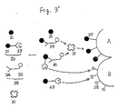
- biotin or a derivative thereof may be employed as the marker and in the measurement operation, avidin or a derivative thereof may be applied to the element together with the sample fluid or the labelled substance. This mode will now be described referring to Fig. 3′.
- reference numeral 21 denotes the target substance
- reference numeral 22 denotes the labelled substance
- reference numeral 23 denotes the conjugate of the target substance and the marker (labelled substance)
- reference numeral 24 denotes the target-binding substance
- reference numeral 25 denotes biotin or a derivative thereof
- reference numeral 26 denotes a conjugate of the target-binding substance and biotin or a derivative thereof
- reference numeral 27 denotes biotin or a derivative thereof
- reference numeral 28 denotes the signal-changing substance
- reference numeral 29 denotes the binding reaction product of the target substance and the conjugate
- reference numeral 30 denotes the binding reaction product of the conjugate 23 and the conjugate 26
- reference character A denotes a carrier to which the biotin 27 is fixed
- reference character B denotes a carrier to which the signal-changing substance 28 is fixed
- reference numeral 31 denotes avidin or a derivative thereof.
- the binding reaction of these reactants is a liquid phase reaction, so that these reaction occur suffiently quicker than the reaction between the immobilized biotin 27 and the avidin 31, or the reaction between the immobilized signal-changing substance 28 and the labelled substance 23.
- the liquid phase reactions are sufficiently conducted.
- the non-reacted labelled substance 23 in the liquid phase reacts with the signal-changing substance 28 fixed on the carrier B (the binding site is 2 - 8), the reaction product 29 reacts with the biotin 27 fixed on the carrier B through the avidin 31 (the binding site is 5 - 11 - 7).
- the binding reaction product 30 may bind to the biotin 27 or signal-changing substance 28, by choosing the binding affinity of 25 - 27 - 11 is greater than the binding affinity between 22 - 28, it is possible to preferentially make the labelled substance 23 or the reaction product 30 react with the biotin 27.
- reaction products 29 and 30 are bound to the carrier A through avidin 31, and the non-reacted labelled substance 23 is bound to the carrier B.
- the B/F separation can be attained in the reaction layer. Since the signal originated from the marker 22 is changed by the signal-changing substance 28, the concentration of the target substance in the sample fluid and the amount of the signal has a functional relationship. Therefore, by preliminarily preparing a calibration curve using standard samples containing known concentration of the target substance, the concentration of the target substance in an un-known sample may be determined.
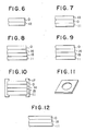
- Fig. 4 shows a schematic view of still another preferred embodiment of the assay element of the present invention.
- the porous reaction layer is made of two sublayers and the biological active substance or the biological substance-binding substance and the signal-changing substance are fixed to the separate sublayers. These sublayers may be formed in the same manner as forming the porous reaction layer described above.
- the assay element 5 has a light-transmitting support 11 on which the porous reaction layer 0 is formed.
- the material for constituting the support film include polymers such as cellulose acetate, polyethylene terephthalate, polycarbonate and polyvinyl compounds such as polystyrene, as well as inorganic materials such as glass.
- the reaction layer may directly be applied and/or formed into a film on the support. Alternatively, the support layer and the porous reaction layer are separately prepared and thereafter they may be laminated. In the embodiment shown in Fig. 5, the fluid sample is required to be dropped from the side of the reaction layer, but the measurement of the signal may be conducted from the either side of the element.
- the porous reaction layer 0 is formed on a light-reflecting support 12.
- the dropping of the fluid sample and the measurement of the signal are conducted from the side of the reaction layer, and the support 12 aids the measurement of the density of the reflection light.
- the support 12 may be made of a paper to which a water-proof treatment is performed, or may be made of the materials for forming the support 11 in Fig. 5. If necessary, to these materials, white pigment such as TiO2, BaSO4 and mica may be incorporated or applied.
- the embodiment shown in Fig. 7 also aims the similar purpose and the porous reaction layer 0 and a light-reflecting layer 13 are laminated on a light-transmitting support 11.
- the fluid sample is dropped from the side of the light-reflecting layer 13 and the measurement of the signal is conducted from the side of the light-transmitting support 11.
- the light-reflecting layers which are employed in the conventional elements may be employed.
- the particles and/or fibers for forming the porous reaction layer, in which the above-mentioned white pigments are incorporated are applied, formed into a film, or laminated.
- a coloring layer 15 is formed on the light-transmitting support 11.
- the coloring layer 15 is made of at least one layer of hydrophilic colloid containing the coloring agent and the substrate which are required for the measurement of the enzyme activity.
- the coloring agent and the substrate may be soluted or dispersed in a binder made of hydrophilic colloid to prepare a coating solution for forming the coloring layer 15.
- the conventional techniques widely used in the field of photography such as the oil-protecting dispersion technique, or direct dispersion technique may be employed.
- gelatin or derivatives thereof such as phthalic gelatin
- synthetic polymers such as polyvinyl alcohol, polyvinylpyrrolidone, polyvinyl imidazole, polyacrylamide and sodium polyacrylate
- cellulose derivatives such as hydroxyethyl cellulose and sodium carboxymethyl cellulose may be employed.
- the preferred are gelatin and phthalic gelatin.
- part of the macromolecular substance may be replaced with water-dispersible polymer such as macromolecular latex.
- macromolecular latex include those described in Japanese Patent Disclosure (Kokai) Nos. 116258/82 and 99752/83.
- 70% by weight of the hydrophilic colloid binder may be replaced with the macromolecular latex, it is preferred that the percentage of the hydrophilic colloid replaced with the macromolecular latex be not more than about 50% by weight.
- buffering agents which may be employed may include salts of citric acid, boric acid, phosphoric acid, tris(hydroxymethyl)aminomethane, barbital, glycine and Good's buffering agent.
- the content of the buffering agent may be appropriately selected.
- These buffering agent may also be contained in the layers other than the coloring layer.
- the preservative and the hardening agent which may be incorporated in the coloring layer the substances described in "Biochemistry Experiment 1, Chemistry of Proteins I", 1976, edited by Japan Biochemistry Association and published by Tokyo Kagaku Dojin), pp.66-67, "Experiment and Application, Affinity Chromatography” published by Kodansha Scientific, 1976 pp.
- Japanese Patent Disclosure (Kokai) No. 149927/85 may be employed.
- Specific examples of these substances may include gelatin, decomposition products of gelatin, albumin, cyclodextrin, non-reduced saccharides (such as sucrose, trehalose), polyethyleneglycol, amino acids such as glycine, glycylglycine, various ions such as ammonium sulfate and sodium azide.
- the content of these substances in the coloring layer may be appropriately selected.
- the coloring layer made of a hydrophilic colloid such as gelatin and gelatin derivative may also contain a hardening agent.
- the hardening agent widely used in the field of photograph may be employed.
- the hardening agents which may be employed are described in T. H. James ed., "The Theory of the Photographic Prorcess", 4th ed., pp. 77-87. That is, aldehydes, active olefins and active esters may be employed as the hardening agent.
- the content of the hardening agent in the coloring layer is usually 10 mg/m2 to 300 mg/m2.
- the surface active agent which may be incorporated in the coloring layer or in any other layers of the element include non-ionic and ionic surface active agents such as octylphenoxypolyethoxyethanol (commercially available from Rohm and Haas under the trade name of Triton X-100), nonylphenoxypolyglycidol (commercially available from Oline Co., Ltd. under the tradename of Surfactant 10G).
- the mordant which may be incorporated in the coloring layer serves to intensively collect the detection product required for the measurement of the enzyme activity and, in the case where the detection product is a dye to promote the absorbance or to shift the wavelength.
- Preferred mordant may include polymers such as described in Japanese Patent Disclosure (Kokai) No. 87461/83 and latex of these polymers.
- the conjugate of the target-binding substance and the biological active substance of the biological substance-binding substance (hereinafter referred to as "the conjugate” for short) and the labelled substance are not contained in the element.
- the conjugate and the labelled substance are not contained in the element have great freedom free from the restraint by the conjugate and the labelled substance.
- all of the conventional immunoassay elements are for assaying a certain substance. That is, the immunoassay element prepared for the measurement of a target substance M1 cannot be used for assaying a target substance M2. Therefore, to assay a plurality of target substances in a single sample, the same number of the elements as the target substances must be prepared and an element for a certain target substance must be selected in operation. In general, to regularly keep and administer many assay elements which have term for validity is troublesome and not economical. Further, selecting an element in each assay reduces the efficiency of the operation and also jeopardize the correctness of the measurement due to the mis-selection.
- the element of the present invention is not restricted to such a multipurpose type.
- the conjugate and the labelled substance may be contained in the assay element.
- this type of element will be hereinafter referred to as "incorporation type" for short
- the operation to drop the conjugate and the labelled substance may be omitted.
- both of the multipurpose type and incorporation type may be selected, which is an important feature of the present invention.
- a coloring layer 15 is formed on the light-transmitting support, and a conjugate-containing layer 14 is formed thereon.
- the conjugate is incorporated in a porous medium such that the area concentration of the conjugate is uniform. Therefore, when a certain amount of the fluid sample and a solution containing the labelled substance are dropped, a certain amount of the conjugate is eluted and diffused to the porous reaction layer together with the fluid sample.
- the material constituting the conjugate-containing layer the above-mentioned materials for constituting the porous reaction layer may be employed, and the materials may be applied, formed into a film or laminated.
- pulp, cellulose powder and natural, synthetic and semisynthetic fibers of various plants, animals, minerals and artificial products such as cotton, hemp, silk, wool, chitin, chitosan, cellulose ester, viscose rayon, copper-ammonia rayon, polyamides (e.g., 6-Nylon, 6,6- Nylon and the like), polyesters (e.g., polyethylene terephthalate and the like), polyolefins (polypropyrene, vinylon and the like), glass and asbestos may be employed. These materials may be employed individually or in combination. Further, water-absorptive European paper, Japanese paper, filter paper and brush polymer, as well as the woven fabrics, non-woven fabrics and synthetic paper made from one or more of the above-described fibers may also be employed.
- the position of the conjugate-containing layer is not restricted to the uppermost position.
- the materials constituting the conjugate-containing layer may be, in addition to those described above, hydrophilic macromolecules such as gelatin, gelatin derivatives, polysaccharides (e.g., agarose), carboxymethyl cellulose and hydroxyethyl cellulose, as well as homopolymers or copolymers of vinyl pyrrolidone derivatives, methacryl acid derivatives, vinyl alcohol and sulfonyl styrene. These may be used as a uniform binder and may be applied.
- the conjugate is incorporated in the porous reaction layer and the labelled substance is incorporated in the conjugate-containing layer in Fig. 9.
- the labelled substance-containing layer may be formed as in forming the conjugate-containing layer, and the conjugate may be incorporated in the porous reaction layer in the same manner as in incorporating the labelled substance therein.
- the conjugate and the labelled substance may also be incorporated in the porous reaction layer.
- Fig. 10 and 11 are a cross-sectional view and a perspective view, respectively, of a preferred embodiment of the assay element of the present invention.
- the entire element is placed in a plastic mount 17, and a fluid sample injection hole and a signal measuring hole are formed in the upper and lower portion of the mount 17, respectively.
- the element of the present invention may further comprise a spreading layer which aids the spreading of the sample fluid when it is dropped on the element.
- a spreading layer which aids the spreading of the sample fluid when it is dropped on the element.
- the conventional supplemental layers such as blood-separating layer which is necessary when the fluid sample is blood (whole blood), adhesive layer, protection layer and timing layer may be formed.
- supplemental layers and the above-described coloring layer, conjugate- or labelled substance-containing layer, and the timing layer may be formed individually or a layer having two or more functions in combination may be formed. The position of these layers may easily be determined by those skilled in the art depending on the function thereof.
- one of the preferred signal-changing substance is aryl mercury derivatives.
- aryl mercury derivatives are reacted under the presence of a buffer widely used in this field such as phosphate buffer, tris(hydroxymethyl)aminomethane, glycine and citric acid, the enzyme inhibitory activity of the aryl mercury derivatives is sharply decreased, so that the reliability of the assay is reduced.
- the present inventors have found that the aryl mercury derivatives are used in the presence of the compound of the formula [I] or [II] or a derivative thereof.
- the buffer containing the compound of the formula [I] or II] preferably has a pH of 6.5 - 8.5, more preferably 7.0 - 7.5.
- the pH may be adjusted by using, for example, a strong base such as sodium hydroxide and potassium hydroxide, or a strong acid such as hydrochloric acid to partly neutralize the compound.
- the concentration of the compound of the formula [I] or [II] in the buffer at the time of measurement varies depending on the sample and the assay system, usually, the concentration is preferably 0.001 - 10 M, more preferably 0.01 - 1 M.
- the buffer containing the compound of the formula [I] or [II] may preferably contain Mg2+ ion in the amount of 0.1 - 100 mM at the time of measurement.
- each layer constituting the assay element is determined not only by the function of the layer, but also the material for constituting the layer and also the existence of the other layers.
- the thickness of the porous reaction layer may preferably be 3 - 700 ⁇ m, more preferably be 10 -300 ⁇ m. Further, in cases where the porous reaction layer has a plurality of sublayers mainly composed of particles, or in cases where the element comprises other functional layers such as spreading layer, which are mainly composed of particles, the total of the thickness of the layers mainly composed of particles may preferably be 10 - 700 ⁇ m, more preferably 20 - 300 ⁇ m.
- the thickness of the porous reaction layer may preferably be 10 - 700 ⁇ m, more preferably 20 - 300 ⁇ m.
- the element comprises other functional layers such as spreading layer, which are mainly composed of fibers
- the total thickness of the layers mainly composed of the fibers may preferaby 40 - 1200 ⁇ m, more preferably 100 - 400 ⁇ m.
- the total thickness of the porous layers may preferably be 60 - 1500 ⁇ m, more preferably 100 - 900 ⁇ m.
- the thickness of such a layer may preferably be 5 - 200 ⁇ m, more preferably 10 - 100 ⁇ m. Further, the total thickness of these layers may also preferably be 5 - 200 ⁇ m, more preferably 10 - 100 ⁇ m.
- microcrystalline cellulose commercially available from Asahi Chemicals Inc., Osaka, Japan under the tradename of Avicel
- 800 ml of 2.5 M phosphate buffer pH 12.0
- 80.0 g of cyanogen bromide cyanide soluted in 800 ml of distilled water was added and the resulting mixture was allowed to react for 20 minutes while cooling in iced water.
- the thus treated Avicel was recovered by filtration and was sufficiently washed with water.
- the resultant was purified with Superose 6 Prep grade column (manufactured by Pharmacia) equilibrated with 0.15 M sodium chloride-containing 0.1 M phosphate buffer (pH 7.4) to obtain human IgG labelled with ⁇ -D-galactosidase.
- composition of Coating Solution (1) Deionized Gelatin 6.0 g Triton X-100 (manufactured by Room and Haas) 0.15 g 1,2-bis(vinylsulfonyl)ethane 0.01 g Distilled Water 54.0 g
- a coating solution (3) with the following composition in which the avidin-bound Avicel obtained in 1-(2) was dispersed was applied on the above-described layer containing p-aminophenylmercuric acetate-bound Avicel, and dried.
- Composition of Coating Solution (3) Avidin-bound Avicel containing bovine serum albumin and sucrose 22.5 g Triton X-100 2.50 g Polyvinylpyrrolidone 3.50 g n-butanol 52.0 g
- a coating solution (5) with the following composition was applied and dried to form a gelatin layer.
- Composition of Coating Solution (5) Deionized Gelatin 4.5 g Triton X-100 (manufactured by Room and Haas) 0.10 g 1,2-bis(vinylsulfonyl)ethane 0.007 g NeoTB 0.23 g Distilled Water 54.0 g
- Coating Composition (6) Avidin-bound Cellulose Powder D containing bovine serum albumin and sucrose 30.0 g Triton X-100 3.0 g Polyvinylpyrrolidone 1.5 g n-butanol 55.0 g
- the coating solution (5) described in 2-(2) was applied and dried to form a gelatin layer. Then a dispersion with the same composition as the coating solution (2) described in 1-(5) except not containing NeoTB was applied and dried to form p-aminophenylmercuric acetate-bound Avicel layer.
- a coating solution (7) with the following composition containing the human IgG labelled with ⁇ -D-galactosidase prepared in 1-(3) and the biotin-bound goat anti-human IgG antibody prepared in 1-(4) was applied and dried. Further, on this layer, the coating solution (6) described in 2-(2) was applied and dried. The sample was then cut into pieces sizing 1.5 cm x 1.5 cm to obtain an immunoassay element 3 of the present invention.
- composition of Coating Solution (7) Avidin-bound Avicel containing bovine serum albumin and sucrose 22.5 g Triton X-100 2.50 g Polyvinylpyrrolidone 3.30 g Human IgG labelled with ⁇ -D-galactosidase 2.0 mg Biotin-bound goat anti-human IgG antibody 2.0 mg Bovine serum albumin 38.0 mg n-butanol 55.0 mg
- rabbit anti-human IgG antibody labelled with ⁇ -D-galactosidase was obtained.
- B/F separation can be performed in the element and human IgG can be measured with high sensitivity.
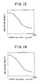
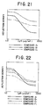
- Human IgG (0 - 640 ⁇ g/ml) dissolved in various 0.3 M solutions (pH 7.2) summerized in Table 2 below containing 1 mM of magnesium chloride and 6 wt% of bovine serum albumin was mixed with the human IgG labelled with ⁇ -D-galactosidase (10 ⁇ g/ml solution in the same buffer) prepared in 1-(3) and biotin-bound anti-human IgG antibody prepared in 1-(4) (20 ⁇ g/ml solution in the same buffer). Ten microliters of this mixture was dropped on the immunoassay element 1 prepared in 1-(5) and the element was incubated in sealed condition at 37°C for 10 minutes. Thereafter, the reflection density at 546 nm was measured from the side of the support layer. The results are shown in Figs. 17 and 18.
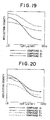
- Example 5 The same procedure as in Example 5 was repeated except that the tested element was element 2 obtained in Example 2. The results are shown in Figs. 19 and 20.
- composition of Coating Solution (8) Avidin-bound Avicel containing bovine serum albumin and sucrose 22.5 g Triton X-100 2.50 g Polyvinylpyrrolidone 3.30 g Human IgG labelled with ⁇ -D-galactosidase 0.16 mg Biotin-bound goat anti-human IgG antibody 0.60 mg Bovine serum albumin 38.0 mg n-butanol 55.0 g
- Human IgG (0 - 640 ⁇ g/ml) dissolved in various 0.3 M solutions (pH 7.2) summerized in Table 2 below containing 1 mM of magnesium chloride and 6 wt% of bovine serum albumin was mixed with the human IgG labelled with ⁇ -D-galactosidase (10 ⁇ g/ml solution in the same buffer) prepared in 1-(3) and biotin-bound anti-human IgG antibody prepared in 1-(4) (20 ⁇ g/ml solution in the same buffer). Ten microliters of this mixture was dropped on the immunoassay element 1 prepared in 1-(5) and the element was incubated in sealed condition at 37°C for 10 minutes. Thereafter, the reflection density at 546 nm was measured from the side of the support layer. The results are shown in Figs. 23 and 24.
- the thus treated Avicel was removed by filtration and was alternately washed with pure water, 0.1 M aqueous sodium hydrogen carbonate solution containing 0.15 M sodium chloride and 0.1 M sodium acetate buffer (pH 4.1) containing 0.15 M sodium chloride, and was suspended in 1200 ml of 1 M Tris-HCl buffer (pH 8.5). The suspension was stirred at room temperature for 20 hours to block the non-reacted reactive groups.
- 80 g of the antibody-bound Avicel was suspended in 240 ml of pure water containing 15.4 g of bovine serum albumin and 9.91 g of sucrose. The resulting suspension was freeze-dried to obtain goat anti-human IgG antibody-bound Avicel containing bovine serum albumin and sucrose.
- Human IgG solutions (0 - 640 ⁇ g/ml) in various 0.3 M buffers (pH 7.2) shown in Table 2 below containing 1 mM magnesium chloride and 6 wt% of bovine serum albumin were mixed with human IgG labelled with ⁇ -D-galactosidase (10 ⁇ g/ml solution in the same buffer) obtained in 1-(3).
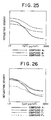
- Ten microliters of this mixture was dropped on the immunoassay element 6 obtained in 9-(2), and the element was incubated in sealed condition at 37°C for 10 minutes. Thereafter, the reflection density was measured at 546 nm from the side of the support layer. The results are shown in Figs. 25 and 26.
- Compound A1 is an especially preferred buffering compound
- Compounds B1 and C1 are conventional buffering compounds widely used in this field
- Compounds D1, E1 and F1 are analogues of Compound A1. All of the Compounds A1, D1, E1 and F1 were commercially available from Dojindo Laboratories.
- Example 5 The same procedure as in Example 5 was repeated except that the buffers employed were those summarized in Table 3 below. The results are shown in Figs. 29 and 30.
- Example 6 The same procedure as in Example 6 was repeated except that the buffers employed were those summerized in Table 3 below. The results are shown in Figs. 31 and 32.
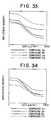
- Example 7 The same procedure as in Example 7 was repeated except that the buffers employed were those summerized in Table 3 below. The results are shown in Figs. 33 and 34.
- Example 8 The same procedure as in Example 8 was repeated except that the buffers employed were those summerized in Table 3 below. The results are shown in Figs. 35 and 36.
- Example 9 The same procedure as in Example 9 was repeated except that the buffers employed were those summerized in Table 3 below. The results are shown in Figs. 37 and 38.
- Example 10 The same procedure as in Example 10 was repeated except that the buffers employed were those summerized in Table 3 below. The results are shown in Figs. 39 and 40.
- Compound A2 and B2 are especially preferred buffering compounds
- Compounds C2 and D2 are conventional buffering compounds widely used in this field
- Compounds E2 and F2 are analogues of Compound A2 or B2. All of the Compounds A2, B2, E2 and F2 were commercially available from Dojindo Laboratories.
- the thus treated Avicel was removed and alternately washed with distilled water, 0.1 M aqueous sodium hydrogen carbonate solution containing 0.15 M sodium chloride and 0.1 M sodium acetate buffer (pH 4.1) containing 0.15 M sodium chloride, and was suspended in 1200 ml of 1 M tris-HCl buffer (pH 8.5). The suspension was stirred for 20 hours at room temperature to block the non-reacted reactive groups.
- the thus treated Avicel was removed by filtration, washed with water and lyophilized to obtain avidin-bound Avicel or goat anti-human IgG antibody-bound Avicel.
- the coating solutions (9) - (17) having the following compositions were prepared.
- Composition of coating solution (9) p-aminophenylmercuric acetate-bound Avicel 14.0 g Triton X-100 1.4 g Polyvinylpyrrolidone (Wako Pure Chemicals) 1.2 g n-butanol 34.0 g
- Composition of coating solution (10) Phenyl- ⁇ -D-thiogalactosidepyranoside-bound Avicel 14.0 g Triton X-100 1.4 g Polyvinylpyrrolidone (Wako Pure Chemicals) 1.2 g n-butanol 34.0 g
- Composition of coating solution (11) 5-aminoisophthalic acid-bound Avicel 14.0 g Triton X-100 1.4 g Polyvinylpyrrolidone (Wako Pure Chemicals) 1.2 g n-butanol 34.0 g
- Composition of coating solution (12) Avidin-bound Avicel 1
- Each sample was cut into pieces sizing 1.5 cm x 1.5 cm to obtain the assay elements.
- bovine serum albumin 5 wt% collagen peptide A (bovine bone) (Funakoshi Pharmaceuticals) 0.3 M bis(2-hydroxyethyl)iminotris(hydroxymethyl)methane buffer (pH 7.2) 1 mM magnesium chloride 15 ⁇ g/ml rabbit anti-human IgG antibody labelled with ⁇ -D-galactosidase 100 ⁇ g/ml biotin-bound goat anti-human IgG antibody
- bovine serum albumin 5 wt% of collagen peptide A (bovine bone) (Funakoshi Pharmaceuticals)
- 0.3 M sodium phosphate buffer (pH 7.2) 1 mM magnesium chloride 15 ⁇ g/ml rabbit anti-human IgG antibody labelled with ⁇ -D-galactosidase 100 ug/ml biotin-bound goat anti-human IgG antibody
- bovine serum albumin 5 wt% collagen peptide A (bovine bone) (Funakoshi Pharmaceuticals)
- 0.3 M N-2-hydroxyethylpiperazine-N′-2-hydroxypropane-3-sulfonic acid buffer pH 8.5
- 1 mM magnesium chloride 15 ⁇ g/ml rabbit anti-human IgG antibody labelled with glutamic acid dehydrogenase 100 ⁇ g/ml biotin-bound goat anti-human IgG antibody
- the reagent solution and the sample were mixed in the ratio of 4:1 (v/v) according to the combinations shown in Table 5. Immediately after mixing, the mixture was dropped on the assay element and the reflection density after incubation at 37°C for 20 minutes was measured. Table 5 No.
- the thus treated Avicel was removed by filtration and was alternately washed with distilled water, 0.1 M aqueous sodium hydrogen carbonate solution containing 0.15 M sodium chloride, and 0.1 M sodium acetate buffer (pH 4.1) containing 0.15 M sodium chloride, and was suspended in 1200 ml of 1 M Tris-HCl buffer (pH 8.5). The resulting suspension was stirred at room temperature for 20 hours to block the non-reacted reactive groups. The Avicel was removed by filtration and sufficiently washed with water to obtain Avicel to which biotin was bound.
- biotin-bound Avicel was suspended in 240 ml of pure water containing 10.0 g of bovine serum albumin and the resulting suspension was lyophilized to obtain biotin-bound Avicel containing bovine serum albumin.
- ⁇ -galactosidase-labelled human IgG was prepared in the same manner as in Example 1-(3).
- Biotin-bound goat anti-human IgG antibody was prepared in the same manner as in Example 1-(4).
- Example 1-(5) The same operation as in Example 1-(5) was repeated except that the biotin-bound Avicel did not contain sucrose, to obtain an immunoassay element.
- the human IgG in samples were measured in the same manner as in 1-(6). The results are shown in Fig. 42.
- the coating solution (5) described in 2-(2) was applied and dried to form a gelatin layer. Then a dispersion with the same composition as the coating solution (2) described in 1-(5) except not containing NeoTB was applied and dried to form p-aminophenylmercuric acetate-bound Avicel layer.
Landscapes
- Health & Medical Sciences (AREA)
- Life Sciences & Earth Sciences (AREA)
- Immunology (AREA)
- Engineering & Computer Science (AREA)
- Chemical & Material Sciences (AREA)
- Molecular Biology (AREA)
- Biomedical Technology (AREA)
- Hematology (AREA)
- Urology & Nephrology (AREA)
- Biotechnology (AREA)
- Microbiology (AREA)
- Cell Biology (AREA)
- Food Science & Technology (AREA)
- Medicinal Chemistry (AREA)
- Physics & Mathematics (AREA)
- Analytical Chemistry (AREA)
- Biochemistry (AREA)
- General Health & Medical Sciences (AREA)
- General Physics & Mathematics (AREA)
- Pathology (AREA)
- Chemical Kinetics & Catalysis (AREA)
- Measuring Or Testing Involving Enzymes Or Micro-Organisms (AREA)
Applications Claiming Priority (4)
| Application Number | Priority Date | Filing Date | Title |
|---|---|---|---|
| JP2963288 | 1988-02-09 | ||
| JP2963388 | 1988-02-09 | ||
| JP29633/88 | 1988-02-09 | ||
| JP29632/88 | 1988-02-09 |
Publications (2)
| Publication Number | Publication Date |
|---|---|
| EP0328106A2 true EP0328106A2 (fr) | 1989-08-16 |
| EP0328106A3 EP0328106A3 (fr) | 1990-12-19 |
Family
ID=26367850
Family Applications (1)
| Application Number | Title | Priority Date | Filing Date |
|---|---|---|---|
| EP19890102245 Withdrawn EP0328106A3 (fr) | 1988-02-09 | 1989-02-09 | Procédé d'essai d'une substance et élément d'analyse immunologique |
Country Status (1)
| Country | Link |
|---|---|
| EP (1) | EP0328106A3 (fr) |
Cited By (5)
| Publication number | Priority date | Publication date | Assignee | Title |
|---|---|---|---|---|
| EP0413561A3 (en) * | 1989-08-16 | 1992-02-26 | Microgenics Corporation | Substrates for beta-galactosidase |
| FR2667943A1 (fr) * | 1990-10-11 | 1992-04-17 | Toledano Jacques | Dispositif et procede pour la determination qualitative et quantitative rapide d'un ligand dans un fluide. |
| WO1996000393A1 (fr) * | 1994-06-24 | 1996-01-04 | Behringwerke Aktiengesellschaft | Procede de mise en oeuvre d'un dosage immunologique dans un systeme multiphase |
| US5527667A (en) * | 1993-05-25 | 1996-06-18 | Virginia Polytechnic Institute And State University | Water sample viral contamination detection system |
| US6703196B1 (en) * | 1997-12-24 | 2004-03-09 | Roche Diagnostics Corporation | General purpose structure of an analytical element and its application for analyte determination |
Families Citing this family (2)
| Publication number | Priority date | Publication date | Assignee | Title |
|---|---|---|---|---|
| JP2004525109A (ja) | 2001-02-15 | 2004-08-19 | キング・ファーマシューティカルズ・インコーポレイティッド | 安定化された甲状腺ホルモン医薬組成物、及びその製法 |
| US7101569B2 (en) | 2001-08-14 | 2006-09-05 | Franz G Andrew | Methods of administering levothyroxine pharmaceutical compositions |
Family Cites Families (2)
| Publication number | Priority date | Publication date | Assignee | Title |
|---|---|---|---|---|
| JPS58209994A (ja) * | 1982-05-10 | 1983-12-07 | Fujirebio Inc | 活性蛋白等固定化物を用いた抗原の測定方法 |
| JPS63159759A (ja) * | 1986-12-24 | 1988-07-02 | Fujirebio Inc | 酵素免疫測定法 |
-
1989
- 1989-02-09 EP EP19890102245 patent/EP0328106A3/fr not_active Withdrawn
Cited By (10)
| Publication number | Priority date | Publication date | Assignee | Title |
|---|---|---|---|---|
| EP0413561A3 (en) * | 1989-08-16 | 1992-02-26 | Microgenics Corporation | Substrates for beta-galactosidase |
| US5444161A (en) * | 1989-08-16 | 1995-08-22 | Microgenics Corporation | Substrates for β-galactosidase |
| FR2667943A1 (fr) * | 1990-10-11 | 1992-04-17 | Toledano Jacques | Dispositif et procede pour la determination qualitative et quantitative rapide d'un ligand dans un fluide. |
| WO1993010453A1 (fr) * | 1990-10-11 | 1993-05-27 | Jacques Toledano | Dispositif et procede de detection immunologique |
| US5527667A (en) * | 1993-05-25 | 1996-06-18 | Virginia Polytechnic Institute And State University | Water sample viral contamination detection system |
| WO1996000393A1 (fr) * | 1994-06-24 | 1996-01-04 | Behringwerke Aktiengesellschaft | Procede de mise en oeuvre d'un dosage immunologique dans un systeme multiphase |
| US6013457A (en) * | 1994-06-24 | 2000-01-11 | Dade Behring Marburg Gmbh | Method for carrying out an immunoassay in a multiphase system |
| US6258551B1 (en) * | 1994-06-24 | 2001-07-10 | Dade Behring Marburg Gmbh | Method for carrying out an immunoassay in a multiphase system |
| US6703196B1 (en) * | 1997-12-24 | 2004-03-09 | Roche Diagnostics Corporation | General purpose structure of an analytical element and its application for analyte determination |
| US7118875B2 (en) * | 1997-12-24 | 2006-10-10 | Roche Diagnostics Gmbh | Analyte determination using a general purpose structure of an analytical element |
Also Published As
| Publication number | Publication date |
|---|---|
| EP0328106A3 (fr) | 1990-12-19 |
Similar Documents
| Publication | Publication Date | Title |
|---|---|---|
| US4289747A (en) | Immunological determination using lectin | |
| US4868106A (en) | Analytical element and method for determining a component in a test sample | |
| EP0066648B2 (fr) | Element d'analyse à plusieurs couches utilisant une réaction de liaison spécifique | |
| US4228237A (en) | Methods for the detection and determination of ligands | |
| US20020160525A1 (en) | Immunoassay devices and materials | |
| EP0124352A2 (fr) | Essai protégé de liaison | |
| JP2786336B2 (ja) | 免疫分析要素および免疫分析方法 | |
| US5470713A (en) | Method and element for measuring analytes in biological fluids using immobilized binder-analyte labeled complex | |
| US5266460A (en) | Method of preparing immunological analytical element | |
| EP0327918A1 (fr) | Dispositif d'essai immunologique | |
| JP3715348B2 (ja) | 修飾アポペルオキシダーゼを用いる分析要素、組成物および方法 | |
| US4820633A (en) | Process for production of an immune reactive, porous carrier material for heterogeneous immunological analysis | |
| US7820402B2 (en) | Immunoassay element | |
| US4971916A (en) | Liposome based homogeneous immunoassay for diagnostic tests | |
| EP0184701B1 (fr) | Procédé pour déterminer un ligand | |
| JPH06148193A (ja) | 抗リン脂質抗体結合用担体及び該担体を用いた免疫学的測定法 | |
| JP2613107B2 (ja) | 抗体を測定する方法および試薬 | |
| JPS62242855A (ja) | 分析素子 | |
| JPH01203973A (ja) | 免疫学的分析素子 | |
| JPS63131063A (ja) | 免疫学的分析素子 | |
| JPH01308966A (ja) | 免疫学的分析方法及びその分析素子 | |
| JPH052021A (ja) | 免疫反応装置及び免疫学的測定装置並びに免疫学的測定法 | |
| JPS6290538A (ja) | 分析素子 | |
| JPH0269199A (ja) | ガラクトシドの酵素活性分析法 | |
| JPS63131062A (ja) | 免疫学的分析素子 |
Legal Events
| Date | Code | Title | Description |
|---|---|---|---|
| PUAI | Public reference made under article 153(3) epc to a published international application that has entered the european phase |
Free format text: ORIGINAL CODE: 0009012 |
|
| AK | Designated contracting states |
Kind code of ref document: A2 Designated state(s): DE GB |
|
| PUAL | Search report despatched |
Free format text: ORIGINAL CODE: 0009013 |
|
| AK | Designated contracting states |
Kind code of ref document: A3 Designated state(s): DE GB |
|
| 17P | Request for examination filed |
Effective date: 19910531 |
|
| STAA | Information on the status of an ep patent application or granted ep patent |
Free format text: STATUS: THE APPLICATION HAS BEEN WITHDRAWN |
|
| 18W | Application withdrawn |
Withdrawal date: 19910626 |
|
| R18W | Application withdrawn (corrected) |
Effective date: 19910626 |