EP0330080A2 - Procédé et moyen de détermination de dommages causés aux reins - Google Patents
Procédé et moyen de détermination de dommages causés aux reins Download PDFInfo
- Publication number
- EP0330080A2 EP0330080A2 EP89102714A EP89102714A EP0330080A2 EP 0330080 A2 EP0330080 A2 EP 0330080A2 EP 89102714 A EP89102714 A EP 89102714A EP 89102714 A EP89102714 A EP 89102714A EP 0330080 A2 EP0330080 A2 EP 0330080A2
- Authority
- EP
- European Patent Office
- Prior art keywords
- antibodies
- antibody
- villin
- bound
- fimbrin
- Prior art date
- Legal status (The legal status is an assumption and is not a legal conclusion. Google has not performed a legal analysis and makes no representation as to the accuracy of the status listed.)
- Withdrawn
Links
Images

Classifications
-
- A—HUMAN NECESSITIES
- A61—MEDICAL OR VETERINARY SCIENCE; HYGIENE
- A61B—DIAGNOSIS; SURGERY; IDENTIFICATION
- A61B5/00—Measuring for diagnostic purposes; Identification of persons
- A61B5/20—Measuring for diagnostic purposes; Identification of persons for measuring urological functions restricted to the evaluation of the urinary system
- A61B5/201—Assessing renal or kidney functions
-
- G—PHYSICS
- G01—MEASURING; TESTING
- G01N—INVESTIGATING OR ANALYSING MATERIALS BY DETERMINING THEIR CHEMICAL OR PHYSICAL PROPERTIES
- G01N33/00—Investigating or analysing materials by specific methods not covered by groups G01N1/00 - G01N31/00
- G01N33/48—Biological material, e.g. blood, urine; Haemocytometers
- G01N33/50—Chemical analysis of biological material, e.g. blood, urine; Testing involving biospecific ligand binding methods; Immunological testing
- G01N33/53—Immunoassay; Biospecific binding assay; Materials therefor
- G01N33/575—Immunoassay; Biospecific binding assay; Materials therefor for cancer
- G01N33/57525—Immunoassay; Biospecific binding assay; Materials therefor for cancer of the liver or pancreas
-
- G—PHYSICS
- G01—MEASURING; TESTING
- G01N—INVESTIGATING OR ANALYSING MATERIALS BY DETERMINING THEIR CHEMICAL OR PHYSICAL PROPERTIES
- G01N33/00—Investigating or analysing materials by specific methods not covered by groups G01N1/00 - G01N31/00
- G01N33/48—Biological material, e.g. blood, urine; Haemocytometers
- G01N33/50—Chemical analysis of biological material, e.g. blood, urine; Testing involving biospecific ligand binding methods; Immunological testing
- G01N33/53—Immunoassay; Biospecific binding assay; Materials therefor
-
- G—PHYSICS
- G01—MEASURING; TESTING
- G01N—INVESTIGATING OR ANALYSING MATERIALS BY DETERMINING THEIR CHEMICAL OR PHYSICAL PROPERTIES
- G01N33/00—Investigating or analysing materials by specific methods not covered by groups G01N1/00 - G01N31/00
- G01N33/48—Biological material, e.g. blood, urine; Haemocytometers
- G01N33/50—Chemical analysis of biological material, e.g. blood, urine; Testing involving biospecific ligand binding methods; Immunological testing
- G01N33/53—Immunoassay; Biospecific binding assay; Materials therefor
- G01N33/575—Immunoassay; Biospecific binding assay; Materials therefor for cancer
- G01N33/5753—Immunoassay; Biospecific binding assay; Materials therefor for cancer of the stomach or small intestine
-
- G—PHYSICS
- G01—MEASURING; TESTING
- G01N—INVESTIGATING OR ANALYSING MATERIALS BY DETERMINING THEIR CHEMICAL OR PHYSICAL PROPERTIES
- G01N33/00—Investigating or analysing materials by specific methods not covered by groups G01N1/00 - G01N31/00
- G01N33/48—Biological material, e.g. blood, urine; Haemocytometers
- G01N33/50—Chemical analysis of biological material, e.g. blood, urine; Testing involving biospecific ligand binding methods; Immunological testing
- G01N33/68—Chemical analysis of biological material, e.g. blood, urine; Testing involving biospecific ligand binding methods; Immunological testing involving proteins, peptides or amino acids
- G01N33/6893—Chemical analysis of biological material, e.g. blood, urine; Testing involving biospecific ligand binding methods; Immunological testing involving proteins, peptides or amino acids related to diseases not provided for elsewhere
-
- G—PHYSICS
- G01—MEASURING; TESTING
- G01N—INVESTIGATING OR ANALYSING MATERIALS BY DETERMINING THEIR CHEMICAL OR PHYSICAL PROPERTIES
- G01N2800/00—Detection or diagnosis of diseases
- G01N2800/34—Genitourinary disorders
- G01N2800/347—Renal failures; Glomerular diseases; Tubulointerstitial diseases, e.g. nephritic syndrome, glomerulonephritis; Renovascular diseases, e.g. renal artery occlusion, nephropathy
-
- Y—GENERAL TAGGING OF NEW TECHNOLOGICAL DEVELOPMENTS; GENERAL TAGGING OF CROSS-SECTIONAL TECHNOLOGIES SPANNING OVER SEVERAL SECTIONS OF THE IPC; TECHNICAL SUBJECTS COVERED BY FORMER USPC CROSS-REFERENCE ART COLLECTIONS [XRACs] AND DIGESTS
- Y10—TECHNICAL SUBJECTS COVERED BY FORMER USPC
- Y10S—TECHNICAL SUBJECTS COVERED BY FORMER USPC CROSS-REFERENCE ART COLLECTIONS [XRACs] AND DIGESTS
- Y10S436/00—Chemistry: analytical and immunological testing
- Y10S436/811—Test for named disease, body condition or organ function
Definitions
- the invention relates to a method for determining kidney damage, in which poly- or monoclonal antibodies are brought into contact with proteins occurring in the kidney and the reaction product obtained is then subjected to an immunological detection method, and to an agent with which this method can be carried out.
- Kidney damage is usually first noticeable through damage to the parenchyma, which occurs, for example, when taking kidney-damaging medications, such as phenacetin, in a healthy kidney or when an immunosuppressant is not taken in a transplanted kidney.
- kidney-damaging medications such as phenacetin
- an immunosuppressant is not taken in a transplanted kidney.
- the early detection of parenchymal damage to the kidney is an important medical problem.
- the invention is therefore based on the object of improving the method of the type mentioned at the outset in such a way that even minor or swelling kidney damage can be practically quantified.
- Another object of the invention is to provide a means for carrying out the method.
- the problem is solved by using antibodies which are specific for the proteins fimbrin or villin as antibodies.
- proteins villin and fimbrin which are two molecularly defined proteins of the proximal renal tubular epithelium, can be used in a practically quantitative manner as a yardstick for kidney damage. These proteins can be determined quantitatively in the urine. Both proteins are namely increased in Damage to the kidney that affects or involves the proximal tubule.
- a non-invasive, diagnostic method is available with which damage to the kidney, for example caused by circulatory disorders, impending acute kidney failure or impending transplant rejection, can be quantified at an early stage.
- the proteins villin and fimbrin can easily be detected in the urine with which they are excreted. Usually only about 0.1 ml of urine is excreted by the patient. In this respect, the detection method has no harmful or toxic effects on the patient, since the examination is carried out in the excreted urine in vitro.
- the proteins Villin and Fimbrin can be represented in pure form. In this respect, an exact determination of the measured values is possible, ie the proteins Fimbrin and Villin can be determined exactly quantitatively in the urine, so that this is a parameter for the severity of the Damage to the renal tubules is available.
- a calibration of the reagent used is advantageously carried out in order to quantitatively determine the content of the proteins villin and / or fimbrin in the urine.
- a typical, simple method with which the proteins villin and fimbrin can be quantitatively detected in urine consists of the following features:
- the proteins villin and fimbrin are isolated in a manner known per se from the intestinal epithelium of the chicken or the pig. This method is from A. Bretscher and K. Weber in Proc. Natl. Acad. Sci. USA 76 (1979), pp. 2321-2325 or J. Cell. Biol. 86 (1980) pages 335-340 or D. Drenckhahn et al. in Cell. Tiss. Res. 228 (1983). Reference is expressly made to the insulation processes explained therein.
- these proteins can be isolated from the intestinal epithelium of surgically removed pieces of human intestine.
- Antibodies against fimbrin and / or villin can be produced according to the literature references given above or else according to generally known methods. Such methods are described, for example, in the monograph by R.J. Mayer and J.H. Walker, entitled “Immunochemical methods in the biological sciences: Enzymes and proteins", appeared in Academic Press, London (1980).
- the isolated proteins in amounts of 50-100 ⁇ g with and without Freund's complete or incomplete adjuvant become rabbits or guinea pigs injected under the dorsal skin or at the level of the abdomen. The same amount of proteins is re-injected at three-week intervals. After the third or later injection, good titres of polyclonal antibodies are obtained, which are affinity-cleaned and tested for their specificity according to standard methods. Such a method has been described in the above publications by Drenckhahn et al., 1983, or Bretscher and Weber, 1979, 1980 or by Talian et al. in J. Cell. Biol. 97 (1983), pp. 1277-1282, to which reference is made.
- Villin monoclonal antibodies against Villin are already commercially available from Dianova, Hamburg, for the histological diagnosis of tumors of the intestine, in which Villin is also released.
- the poly- or monoclonal antibodies against the proteins villin and / or fimbrin are bound to the surface of reaction carriers.
- Binding is understood to mean either a physical adsorption of the antibodies to a substrate or a chemical covalent binding of these antibodies to the substrate.
- Filter materials materials from reaction vessels such as microtiter plates, column chromatography materials such as Sepharose and the like.
- the antibodies are brought into contact with the substrate material in an aqueous solution, advantageously in an aqueous buffer solution, until an adsorption equilibrium has formed.
- This equilibrium setting is temperature-dependent and takes about 3 hours at about 21 ° C and about 12 hours at 4 ° C.
- a plastic material in particular polystyrene, is preferred as the substrate material, which is usually used for the production of microtiter plates.
- the adsorbed antibodies are treated with protein solutions, for example albumin solutions, gelatin solutions or skimmed milk to saturate free protein binding sites, optionally with the addition of sodium chloride and / or detergent.
- protein solutions for example albumin solutions, gelatin solutions or skimmed milk to saturate free protein binding sites, optionally with the addition of sodium chloride and / or detergent.
- the antibody coatings treated in this way are then washed several times, after which treatment with a urine sample can take place.
- the incubation with urine is carried out until a reaction equilibrium has been established between the antibodies and the villin and / or fimbrin proteins.
- the incubation process advantageously takes about 30-90 minutes.
- the urine is then removed and, if microtiter plates are used, the holes are filled again with the washing solution specified above, which is changed several times at certain intervals.
- the antibody-protein binding complex thus produced is then subjected to an immunological quantification reaction in a second process step.
- the antibody-protein binding complex adhering to the substrate is again treated with antibodies against villin or fimbrin.
- a polyclonal antibody was used in the first process step, either the same polyclonal antibody or a monoclonal antibody against villin or fimbrin is used in the second process step.
- a monoclonal antibody is used in the first process step, then either a polyclonal antibody or a second monoclonal antibody different from the first antibody, which is directed against another binding site of the protein molecule, can be used as the antibody.
- the antibody used in this second method step is referred to below as the "second antibody”.
- the holes in the microtiter plates are also filled with a solution containing this second antibody for about 30-90 minutes and incubated.
- the second antibody solution is then removed, whereupon the holes are rinsed with a washing solution.
- a complex remains on the substrate, consisting of the first antibody, the bound proteins villin or fimbrin and the second antibody, hereinafter referred to as "quantification complex".
- This quantification complex can be subjected to a quantification reaction in several ways:
- the second antibody is subjected to covalent coupling with enzymes, for example peroxidase, alkaline phosphatase, glucose oxidase or beta-galactosidase.
- enzymes for example peroxidase, alkaline phosphatase, glucose oxidase or beta-galactosidase.
- the amount of the enzyme-labeled second antibodies bound in the quantification complex can then be determined quantitatively by an enzyme color reaction.
- Such quantification reactions are known per se, with usable enzymes and typical enzyme color reactions from L.A. Sternberger in "Immunocytochemistry", third edition, published by John Wiley & Sons, New York (1986) or from W.D. Kuhlmann in the monograph "Immunoenzyme techniques in cytochemistry", Verlag Chemie, Weinheim (1984) have been described. This is referred to.
- the second antibodies can be radioactively labeled using methods known per se, for example with iodine 125.
- radioactive labeling kits and substances are used which are supplied, for example, by the company NEN, Dreieich.
- the amount of bound second antibodies can be directly determined quantitatively by a particle counter or radiation counter.
- the complex can be treated with a third antibody which is specific for the bound second antibody.
- Antibodies against mouse immunoglobulins or Rabbit immunoglobulins are used. Such antibodies are commercially available in labeled form with enzymes or radioisotopes and are, for example, from the companies Dianova, Hamburg; Janssen, Belgium or NEN, Dreieich. This is followed by the quantification step already mentioned, either evaluating the color reaction quantitatively or determining the radioactive radiation.
- third antibodies can also serve in unlabelled form as bridge antibodies, which form enzyme-anti-enzyme immune complexes, which may be introduced in a fourth incubation step.
- enzyme-anti-enzyme immune complexes are also commercially available and are sold, for example, by Dianova, Hamburg or Janssen, Belgium.
- the quantification of villin or fimbrin in the urine is carried out by covalently coupling the second antibodies to biotin.
- Biotin is coupled to the second antibody by methods known per se, which have been described, for example, by NH Heggeness et al. in J. Cell. Biol. 73 (1977), pp. 783-788, which is incorporated by reference.
- biotin-N-hydroxysuccinimide ester is used, which is manufactured by Calbiochem. Hoechst is distributed.
- biotinylated second antibodies bound in the quantification complex are quantified by the biotin binding protein streptavidin.
- Streptavidin is covalently bound to the second antibody with the enzyme peroxidase or the other enzymes mentioned above.
- Enzyme streptavidin solutions of this type are commercially available and are marketed, for example, by Calbiochem-Hoechst.
- the streptavidin-antibody-complex obtained is then, if the enzyme is a peroxidase, mixed with 1,2-phenylenediamine and H2O2 solution, the colorless phenylenediamine being converted into a yellow dye by the reaction of peroxidase with hydrogen peroxide.
- the resulting dye can be determined by its absorbance at 492 nm compared to its reference wavelength of 620 nm in a UV photometer. The absorbance correlates with the amount of bound villin and / or fimbrin molecules, as can be shown below.
- villin and fimbrin can also be detected in the urine by a displacement assay.
- reaction carriers are coated with purified villin and / or fimbrin.
- a mixture of a urine sample to be examined and antibodies specific to villin or fimbrin is then added.
- the amount of bound antibodies can then be determined by direct labeling of the antibodies with enzymes or radioisotopes or by the other described Ver driving be quantified.
- the agent consists of mono- or polyclonal antibodies against fimbrin and / or villin.
- Antibodies which are already bound to a reaction carrier are preferably brought onto the market, the introduction of antibodies into microtiter plates being preferred.
- a trading unit can thus consist of a microtiter plate with a multiplicity of holes, each hole being provided with a specific amount of antibody against villin and / or fimbrin.
- the agent according to the invention advantageously has the above-mentioned second antibody for producing the quantitation complex.
- Biotin groups are advantageously covalently bound to this second antibody.
- Such a commercial unit can also - if not available in the laboratory - contain concentrates of the above-mentioned washing solutions and the quantification reagents streptavidin peroxidase, H2O2, phenylenediamine, which are used to quantify the quantification complex.
- the isolated purified proteins fimbrin and / or villin are advantageously supplied with each substrate unit for calibration of the measured values, advantageously in an amount of approximately 4 ⁇ g. These are distributed in lyophilized form in a container which is filled up with a certain amount of water or control urine by the consumer.
- the agent in addition to the antibodies against fimbrin and / or villin mentioned above under variant A, consists of the isolated proteins villin or fimbrin.
- the proteins are brought onto the market bound to reaction carriers.
- a commercial unit can consist of a microtiter plate, each hole being provided with a certain amount (for example 0.5-1 ⁇ g) of the isolated proteins villin and / or fimbrin.
- the second agent has the abovementioned mono- or polyclonal antibodies against villin or fimbrin, to which biotin groups are advantageously covalently bound.
- each unit of this embodiment advantageously also contains the isolated proteins villin and / or fimbrin for calibration purposes, as also described for variant A.
- this detection kit can also contain the above-mentioned washing solutions and the quantification reagents.
- Polyclonal antibodies to Fimbrin and / or Villin are made according to the method of Bretscher and Weber 1979, 1980 and Drenckhahn et al., 1983. Monoclonal antibodies are produced according to the method of Peter et al., 1985 or a monoclonal antibody against Villin from Dianova, Hamburg, is used.
- the isolated antibodies are dissolved in an immunoglobulin concentration of 5 ⁇ g / ml in 0.2 M carbonate buffer with a pH of 10.6.
- Immune serum which has not been purified is expediently diluted in a ratio of 1: 100 in the carbonate buffer.
- antibody is used as a general term for isolated immunoglobulins and immune serum.
- the diluted antibody solution For sufficient binding of the antibodies to the wall holes in the microtiter plates, 100 ⁇ l of the diluted antibody solution are left in the holes of the microtiter plates for about three hours at 21 ° C. or twelve hours at 4 ° C. The antibody solution is then removed from the holes, and the holes are then filled with about 400 ⁇ l of a 1% strength by weight gelatin solution in the carbonate buffer indicated above in order to saturate free binding sites.
- the gelatin solution is removed again and the holes are filled with approximately 400 ⁇ l washing buffer.
- This washing buffer consists of a saline solution buffered with phosphate (25 mM phosphate buffer, 100mM NaCl with a pH of 7.4), which is called PPK below. 0.05% by weight of the detergent Tween 20, which can be obtained from Serva, Heidelberg, is added to this buffer solution.
- the wash buffer is changed 3 times in 5-minute intervals.
- microtiter plate prepared in this way can either be exposed directly to the reaction with a urine sample to be examined or else dried and then packaged for use.
- the holes in the microtiter plate thus produced are each filled with 100 ⁇ l of undiluted urine to be examined, the urine then being left in the holes for about 1.5 hours. Then the urine is removed and the holes are again filled with 400 ⁇ l of the above-mentioned washing buffer, which is changed 3 times at 5-minute intervals.
- the holes are again incubated with 100 ⁇ l of the polyclonal antibodies against villin and / or fimbrin given for the coating of the plates.
- Biotin is first covalently coupled to these second antibodies using the method of Heggeness, 1977 (see above).
- the biotinylated antibodies are dissolved in an immunoglobulin concentration of 10 ⁇ g / ml in a buffer which consists of 150 mM NaCl, 20 mM Tris-HCl, 0.5 mM urea and 1% gelatin and has a pH of 7.4.
- This antibody solution is left in the holes for about 90 minutes. After removing the second antibody solution from the holes, the holes are each filled with 400 ml of PPK with 0.05% by weight of Tween 20, which is changed three times at 5-minute intervals.
- the quantification reaction is then carried out in the following manner.
- the holes are filled with 100 ⁇ l of the above. phosphate-buffered saline (PPK, pH 7.4) filled.
- PPK phosphate-buffered saline
- This solution contains the biotin-binding protein streptavidin, which is covalently coupled to the enzyme peroxidase.
- the peroxidase streptavidin is obtained as a stock solution from Calbiochem-Hoechst and used in a dilution of 1: 5,000 in PPK, pH 7.4. This streptavidin solution is left in the holes at 21 ° C. for about 1 hour and then removed.
- the holes are then filled with about 400 ⁇ l of the above-mentioned washing buffer (PPk, pH 7.4, 0.05% by weight Tween 20). This buffer solution is changed three times in 5-minute intervals.
- PPk pH 7.4, 0.05% by weight Tween 20
- the holes are each filled with 100 ul of a phosphate citrate buffer (0.2 M disodium hydrogen phosphate, 0.1 M citric acid monohydrate with a pH of 5), each containing 1 ml of buffer 1.5 mg 1,2-phenylenediamine (based on the Merck) and 1 ⁇ l of a 30% H2O2 solution (sold under the name "Perhydrol” by Merck).
- a phosphate citrate buffer 0.2 M disodium hydrogen phosphate, 0.1 M citric acid monohydrate with a pH of 5
- the phenylenediamine and H2O2 are added to the phosphate citrate buffer immediately before use. The holes are then incubated with this solution for 30 minutes.
- the reaction is terminated by adding 50 ⁇ l of 0.5 N sulfuric acid / hole.
- a yellow dye is formed by oxidation of the colorless phenylenediamine, the absorbance of which is measured at 492 nm compared to the reference wavelength of 620 nm in a UV photometer.
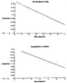
- This calibration curve was created in that defined amounts of villin (a) or fimbrin (b) were given to the urine of a person were added to the normal person and quantified using the above-mentioned immunological method.
- the microtiter well contains 1: 10 ⁇ g / ml, 2: 5 ⁇ g / ml, 3: 2.5 ⁇ g / ml, 4: 1.25 ⁇ g / ml, 5: 0.625 ⁇ g / ml and 6: 0.312 ⁇ g / ml protein .
- FIG. 2 graphically depicts a case study on a patient with a transplanted kidney over 30 days (abscissa), the villine content in the patient's urine samples being determined for each day.
- Fig. 2 From Fig. 2 it can be seen that the patient has a villin concentration with a baseline absorbance of 0.05.
- the absolute concentrations of villin in the patient's urine can be read from the calibration curve according to FIG. 1.
- the arrows drawn in FIG. 2 indicate rejection crises that have been clinically verified by increased creatinine values and clinical symptoms. Therapy for these rejection crises consisted of increased doses of immunosuppressive drugs, which resulted in a decrease in urine villi excretion, i.e. reflected in the decline in the risk of damage to the kidney.
Landscapes
- Health & Medical Sciences (AREA)
- Life Sciences & Earth Sciences (AREA)
- Engineering & Computer Science (AREA)
- Immunology (AREA)
- Biomedical Technology (AREA)
- Molecular Biology (AREA)
- Urology & Nephrology (AREA)
- Hematology (AREA)
- Chemical & Material Sciences (AREA)
- Physics & Mathematics (AREA)
- Pathology (AREA)
- General Health & Medical Sciences (AREA)
- Biochemistry (AREA)
- Medicinal Chemistry (AREA)
- Food Science & Technology (AREA)
- Analytical Chemistry (AREA)
- Microbiology (AREA)
- Cell Biology (AREA)
- General Physics & Mathematics (AREA)
- Biotechnology (AREA)
- Proteomics, Peptides & Aminoacids (AREA)
- Heart & Thoracic Surgery (AREA)
- Biophysics (AREA)
- Physiology (AREA)
- Medical Informatics (AREA)
- Surgery (AREA)
- Animal Behavior & Ethology (AREA)
- Public Health (AREA)
- Veterinary Medicine (AREA)
- Investigating Or Analysing Biological Materials (AREA)
- Preparation Of Compounds By Using Micro-Organisms (AREA)
- Medicines Containing Antibodies Or Antigens For Use As Internal Diagnostic Agents (AREA)
- Peptides Or Proteins (AREA)
- Measuring Or Testing Involving Enzymes Or Micro-Organisms (AREA)
Applications Claiming Priority (2)
| Application Number | Priority Date | Filing Date | Title |
|---|---|---|---|
| DE3805447A DE3805447A1 (de) | 1988-02-22 | 1988-02-22 | Verfahren und mittel zur bestimmung von nierenschaeden |
| DE3805447 | 1988-02-22 |
Publications (2)
| Publication Number | Publication Date |
|---|---|
| EP0330080A2 true EP0330080A2 (fr) | 1989-08-30 |
| EP0330080A3 EP0330080A3 (fr) | 1989-12-27 |
Family
ID=6347881
Family Applications (1)
| Application Number | Title | Priority Date | Filing Date |
|---|---|---|---|
| EP89102714A Withdrawn EP0330080A3 (fr) | 1988-02-22 | 1989-02-17 | Procédé et moyen de détermination de dommages causés aux reins |
Country Status (7)
| Country | Link |
|---|---|
| US (1) | US5141876A (fr) |
| EP (1) | EP0330080A3 (fr) |
| JP (1) | JPH01308236A (fr) |
| KR (1) | KR890012615A (fr) |
| AU (1) | AU624113B2 (fr) |
| BR (1) | BR8900796A (fr) |
| DE (1) | DE3805447A1 (fr) |
Cited By (2)
| Publication number | Priority date | Publication date | Assignee | Title |
|---|---|---|---|---|
| WO1993003381A1 (fr) * | 1991-07-26 | 1993-02-18 | Board Of Regents, The University Of Texas System | Analyse de constituant secretoire libre et procede de controle d'un rejet d'organe |
| WO2002010755A3 (fr) * | 2000-07-28 | 2003-10-09 | Marlene Lydia Rose | Rejet de greffe et pathologies associees |
Families Citing this family (1)
| Publication number | Priority date | Publication date | Assignee | Title |
|---|---|---|---|---|
| RU2161310C2 (ru) * | 1999-03-29 | 2000-12-27 | Ивановская государственная медицинская академия | Способ прогнозирования ранних послеоперационных осложнений у детей с аномалиями развития почек и верхних мочевых путей |
Family Cites Families (2)
| Publication number | Priority date | Publication date | Assignee | Title |
|---|---|---|---|---|
| US4465776A (en) * | 1982-09-27 | 1984-08-14 | Research Corporation | Monoclonal antibodies to vitamin B6 and immunoassay method |
| US4731326A (en) * | 1984-06-04 | 1988-03-15 | Ortho Diagnostic Systems Inc. | Disease diagnosis by detection of shed normal tissue antigens |
-
1988
- 1988-02-22 DE DE3805447A patent/DE3805447A1/de not_active Withdrawn
-
1989
- 1989-02-17 EP EP89102714A patent/EP0330080A3/fr not_active Withdrawn
- 1989-02-21 US US07/312,488 patent/US5141876A/en not_active Expired - Fee Related
- 1989-02-21 AU AU30170/89A patent/AU624113B2/en not_active Ceased
- 1989-02-22 KR KR1019890002079A patent/KR890012615A/ko not_active Withdrawn
- 1989-02-22 JP JP1042901A patent/JPH01308236A/ja active Pending
- 1989-02-22 BR BR898900796A patent/BR8900796A/pt not_active Application Discontinuation
Cited By (4)
| Publication number | Priority date | Publication date | Assignee | Title |
|---|---|---|---|---|
| WO1993003381A1 (fr) * | 1991-07-26 | 1993-02-18 | Board Of Regents, The University Of Texas System | Analyse de constituant secretoire libre et procede de controle d'un rejet d'organe |
| US5340721A (en) * | 1991-07-26 | 1994-08-23 | Board Of Regents, The University Of Texas System | Assay for free secretory component and methods for monitoring organ rejection |
| WO2002010755A3 (fr) * | 2000-07-28 | 2003-10-09 | Marlene Lydia Rose | Rejet de greffe et pathologies associees |
| US7192716B2 (en) | 2000-07-28 | 2007-03-20 | Marlene Lydia Rose | Organ transplant rejection and associated conditions |
Also Published As
| Publication number | Publication date |
|---|---|
| JPH01308236A (ja) | 1989-12-12 |
| AU3017089A (en) | 1989-08-24 |
| US5141876A (en) | 1992-08-25 |
| AU624113B2 (en) | 1992-06-04 |
| KR890012615A (ko) | 1989-09-18 |
| EP0330080A3 (fr) | 1989-12-27 |
| DE3805447A1 (de) | 1989-08-31 |
| BR8900796A (pt) | 1989-10-17 |
Similar Documents
| Publication | Publication Date | Title |
|---|---|---|
| DE69633780T2 (de) | Verfahren zum assay von troponin i und t und komplexen von troponin i und t und auswahl von antikörpern zur verwendung in immunoassays | |
| DE69733488T2 (de) | Verfahren zur besserung der wiederfindung troponin i und t | |
| DE69837015T2 (de) | Verfahren zur Stabilisierung und Messung von Troponinkomplexen | |
| DE2656155C2 (fr) | ||
| DE69113273T2 (de) | Zellnekrosnachweis durch prüfung von spektrin und seine abbauprodukte. | |
| DE2548963A1 (de) | Verfahren und mittel zur bestimmung der aktivitaet von creatinkinase-mb | |
| DE69730479T2 (de) | Marker und immunologisches Reagens für Dialyse verbundenen Amyloidosis, Diabetes Mellitus und Diabetes Mellitus-Komplikationen | |
| EP0032204B1 (fr) | Procédé et réactif pour la détermination immunologique d'enzymes | |
| DE2258822A1 (de) | Verfahren und mittel zur analyse von isoenzymmustern | |
| DE69001972T2 (de) | Mittel zum nachweis von gehirnkrankheiten in zusammenhang mit nervenscheidenmarkzerstoerung, insbesondere sklerosis multiplex. | |
| DE3205301C2 (fr) | ||
| DE19920704C1 (de) | Verwendung eines gegen Harn-Trypsin-Inhibitors gerichteten Antikörpers für die Diagnose des Ausbruchs von AIDS | |
| DE68912477T3 (de) | Verfahren zum messen der biologischen wirkung von antithrombin iii und reagenzkit zu dessen messung | |
| DE60108316T2 (de) | Medizinische Kit und Methode zur Bestimmung eines Arzneimittels | |
| DE69628713T2 (de) | Marker und Reagenz für Diabetes mellitus und Diabetes-mellitus-Komplikationen | |
| DE3883145T2 (de) | Verfahren zur Messung von humanen Insulin. | |
| DE69320661T2 (de) | Verfahren zur messung von heparin | |
| DE69711937T2 (de) | Entdecken von nekrosen des herzmuskels durch einen immunoassay und entsprechende antikorper | |
| DE3805447A1 (de) | Verfahren und mittel zur bestimmung von nierenschaeden | |
| DE68911574T2 (de) | Reagenzsystem zur Bestimmung des Komplexes des menschlichen Plasminogenaktivator-Inhibitors und des menschlichen Gewebeplasminogenaktivators und Testsatz dafür. | |
| EP0874994B1 (fr) | Determination des groupes terminaux acide sialique de la molecule transferrine humaine | |
| DE69523492T2 (de) | Immunoassay-Reagenzien, Testsätze und Verfahren | |
| DE69021631T2 (de) | Diagnostische Zusammenstellung zum Nachweis von rheumatischer Arthritis. | |
| DE69110067T2 (de) | Monoklonale Antikörper gegen nicht-A 1C glykosyliertes Hämoglobin. | |
| DE60133224T2 (de) | Organtransplantatenabstossung und verbundene bedingungen |
Legal Events
| Date | Code | Title | Description |
|---|---|---|---|
| PUAI | Public reference made under article 153(3) epc to a published international application that has entered the european phase |
Free format text: ORIGINAL CODE: 0009012 |
|
| AK | Designated contracting states |
Kind code of ref document: A2 Designated state(s): AT BE CH DE ES FR GB GR IT LI LU NL SE |
|
| PUAL | Search report despatched |
Free format text: ORIGINAL CODE: 0009013 |
|
| AK | Designated contracting states |
Kind code of ref document: A3 Designated state(s): AT BE CH DE ES FR GB GR IT LI LU NL SE |
|
| 17P | Request for examination filed |
Effective date: 19900627 |
|
| 17Q | First examination report despatched |
Effective date: 19920605 |
|
| STAA | Information on the status of an ep patent application or granted ep patent |
Free format text: STATUS: THE APPLICATION IS DEEMED TO BE WITHDRAWN |
|
| 18D | Application deemed to be withdrawn |
Effective date: 19960117 |