EP0340806A2 - Synthese von menschlichen Virusantigenen durch Hefe - Google Patents
Synthese von menschlichen Virusantigenen durch Hefe Download PDFInfo
- Publication number
- EP0340806A2 EP0340806A2 EP89110270A EP89110270A EP0340806A2 EP 0340806 A2 EP0340806 A2 EP 0340806A2 EP 89110270 A EP89110270 A EP 89110270A EP 89110270 A EP89110270 A EP 89110270A EP 0340806 A2 EP0340806 A2 EP 0340806A2
- Authority
- EP
- European Patent Office
- Prior art keywords
- yeast
- hbsag
- hepatitis
- protein
- surface antigen
- Prior art date
- Legal status (The legal status is an assumption and is not a legal conclusion. Google has not performed a legal analysis and makes no representation as to the accuracy of the status listed.)
- Granted
Links
Images


Classifications
-
- C—CHEMISTRY; METALLURGY
- C07—ORGANIC CHEMISTRY
- C07K—PEPTIDES
- C07K14/00—Peptides having more than 20 amino acids; Gastrins; Somatostatins; Melanotropins; Derivatives thereof
- C07K14/005—Peptides having more than 20 amino acids; Gastrins; Somatostatins; Melanotropins; Derivatives thereof from viruses
-
- A—HUMAN NECESSITIES
- A61—MEDICAL OR VETERINARY SCIENCE; HYGIENE
- A61K—PREPARATIONS FOR MEDICAL, DENTAL OR TOILETRY PURPOSES
- A61K39/00—Medicinal preparations containing antigens or antibodies
-
- C—CHEMISTRY; METALLURGY
- C12—BIOCHEMISTRY; BEER; SPIRITS; WINE; VINEGAR; MICROBIOLOGY; ENZYMOLOGY; MUTATION OR GENETIC ENGINEERING
- C12N—MICROORGANISMS OR ENZYMES; COMPOSITIONS THEREOF; PROPAGATING, PRESERVING, OR MAINTAINING MICROORGANISMS; MUTATION OR GENETIC ENGINEERING; CULTURE MEDIA
- C12N2730/00—Reverse transcribing DNA viruses
- C12N2730/00011—Details
- C12N2730/10011—Hepadnaviridae
- C12N2730/10111—Orthohepadnavirus, e.g. hepatitis B virus
- C12N2730/10122—New viral proteins or individual genes, new structural or functional aspects of known viral proteins or genes
Definitions
- the present invention relates to the biosynthesis of an antigen of human Hepatitis B virus (HBV) by yeast, brought about by an application of recombinant DNA techniques.
- Hepatitis B virus is recognized as a major, world-wide public health problem.
- Hepatitis B virus has been implicated in the etiology of hepatocellular carcinoma.
- HBsAg HBV surface antigen obtained from preparations of intact virus (Dane particles) or purified from the serum of hepatitis carriers. Skelly, J. et al., Nature 290 , 51 (1981) have reported the purification of water-soluble protein micelles of purified HBsAg.
- a significant limitation of this approach is that the amount of material which can be prepared depends upon the availability of donors. No technique is known for growing the virus in culture; therefore, in addition to limitations in the amount of source material, there is a risk of contamination of the vaccine with active virus or other components of donor serum, and a possible heterogeneity in the products obtained form various donors.
- a second approach has been the attempt to synthesize peptides eliciting antibodies against HBsAg based upon the amino acid sequence of the protein comprising the surface antigen (S-protein) and model studies predicting the most likely antigenic determinants. See, e.g. R. A. Lerner et al., Proc. Nat. Acad. Sci. USA 78 ,3403 (1981). Such work is in a highly preliminary stage, and it may be difficult to assess whether the approach can produce antigens having a practical degree of immunogenicity in a cost-effective manner.
- a third approach employing recombinant DNA techniques, is the synthesis of S-protein, HBsAg or an immunologically reactive equivalent by a microorganism, by endowing a microorganism with genetic capability to produce S-protein, HBsAg or an immunologically reactive equivalent in large amounts, in the absence of other viral gene products.
- This approach eliminates the possibility of contamination by virus or other viral components and permits large-scale production with economies of scale.
- the entire genome of HBV has been cloned in E .
- HBsAg The structure of HBsAg is believed to consist of two S-protein chains joined by intermolecular disulfide bonds and held in a prescribed confirmation by additional intramolecular disulfide bonds. One of the two chains appears to be glycosylated.
- HBsAg frequently appears in the form of spherical particles with a mean diameter of 22 nm, which are thought to aggregates of the S-protein dimers just described, and possibly contain lipids.
- HBsAg In the viral envelope, HBsAg is associated with the lipid-containing viral envelope, which is believed to be derived from membrane components of the host cell.
- the antigenicity and immunogenicity of HBsAg depend upon several factors, not all of which are well understood. It has been observed that reduction of the disulfide bonds reduces antigenicity and immunogenicity markedly (Mishiro, S. et al., J. Immunol . 124 , 1589 (1980)). Therefore, the tertiary configuration contributed by the intramolecular and intermolecular disulfide bonds is thought to contribute to antigenicity and immunogenicity. The contribution of other factors, such as the extent and nature of glycosylation and association with lipid is unclear, although all are thought to contribute to some degree. Aggregation into particles such as the above-mentioned 22 nm particles is thought to contribute significantly to enhancing immunogenicity.
- the S-protein has been synthesized in E. coli in the form of a fusion protein (Edman, J. C. et al., Nature 291, 503 (1981)).
- the product included 183 amino acids of pre-beta lactamase, 5-10 glycine residues, and 204 amino acids of S-protein lacking 22 amino acids of the amino terminal end.
- the fusion protein was immunoprecipitable with anti-HBsAg IgG.
- yeast is eucaryotic, so it was hoped that some of the post-translational processing steps which are carried out in a normal host cell might he carried out in yeast.
- S-protein The unprocessed translation product of the structural gene for surface antigen is termed S-protein.
- HBsAg The antigen isolated from plasma of infected donors, from Dane particles or from human hepatoma cell cultures, is termed HBsAg.
- Y-HBsAg The expression product of the surface antigen gene in yeast.
- the term, immunologically reactive equivalent of HBsAg is a general term for any immunologically cross-reactive composition comprising S-protein or a portion thereof, of which Y-HBsAg is an example.
- Yeast has never previously been used for expression of the genes of a virus which normally multiplies in a different organism.
- Prior art attempts to express heterologous proteins in yeast have yielded mixed results.
- a gene coding for a Drosophila gene has been reported capable of complementing a yeast ade 8 mutant, under conditions of selective pressure for genetic complementation. Isolation of a fuctional protein from the yeast strain was not reported.
- the gene for human leukocyte interferon has been expressed in yeast, under control of the yeast ADHl (alcohol dehydrogenase) promoter. In that instance, successful production of an active protein did not require post-translational processing or assembly of components.
- yeast vectors suitable for transfer and replication in yeast have been developed (Broach, J. R. et al., Gene 8 , 121 (1979) ; Hartley, J. L. et al., Nature 286 , 860 (1980).
- Most yeast vectors in current use are derived from E. coli vectors, such as pBR322, into which have been inserted a yeast origin of replication.
- yeast origin of replication Two types of yeast replication origins are available. The first, derived from a ubiquitous naturally-occurring yeast plasmid, commonly referred to as the 2 micron circle, confers the ability to replicate independently of yeast chromosomal DNA.
- arsl autonomous replication sequence
- yeast chromosomal replication origin derived from the yeast chromosomal replication origin, which also provides autonomous replication capability. Because both bacterial and yeast replication origins are present in the same vector, they can be used in either organism. Selection may be provided for in bacterial systems by the inclusion of antibiotic resistance genes, such as the ampicillin and tetracycline resistance genes of pBR322. Selection in yeast systems typically may be provided for by including a yeast gene complementing a mutation in a suitable auxotrophic host strain. The studies reported herein conveniently utilize yeast vectors containing a promoter isolated from the yeast gene coding for alcohol dehydrogenase (ADHl). (Bennetzen, J. L. et al., J. Biol. Chem. Vol. 257, p. 3018 (1981).
- ADHl alcohol dehydrogenase
- the ADHl promoter region was isolated from the 5′-flanking region of the yeast ADHl gene. A fragment containing approximately 1600 base pairs of the ADHl sequence extending from position -1550 to +17 within the coding region was fused to the yeast CYCl coding sequence. Studies on transcription of the attached CYCl coding sequence deomonstrated that transcript starting specificity could be transferred from one yeast gene to another. Smaller fragments, lacking all of the ADH coding region, have subsequently been constructed, and shown to be functional in the expression of human interferon. One such fragment, designated 921, is terminated after position -9, and was employed in the present studies.
- S-protein The translation product of the HBV surface antigen gene is termed the S-protein.
- S-protein has 226 amino acids whose sequence has been inferred from the nucleotide sequence of its gene and by partial sequence analysis.
- HBsAg as used herein includes the major surface antigenic component of HBV found in infected patients' serum and in Alexander cells, a hepatocellular carcinoma cell line which synthesizes and excretes 22 nm HBsAg particles (Alexander, J. J. et al., S. Afr. Med. J. 50 , 1124 (1976).
- HBsAg is used herein to include any modified form of the S-protein which contributes to its antigenic and immunogenic properties, including, but not limited to, dimerization, glycosylation, and particle assembly.
- the present invention relates to synthesis of HBsAg in yeast.
- Yeast expression vectors comprising a yeast promoter, ADHl, have been constructed.
- the product is antigenic (reactive with anti-HBsAg) , and a substantial portion is found associated with particles identical in electron microscopic appearance to those found in the serum of HBV-infected patients and in Alexander cells but having a smaller particle size diameter.
- the HBsAg synthesized by yeast has identical sedimentation behavior to purified, naturally-occurring HBsAg particles purified from Alexander cells as measured by sucrose gradient sedimentation.
- the present invention demonstrates synthesis and assembly of a higher ordered multi-component structure resulting from expression of a heterologous DNA coding segment in a microorganism.
- the present invention is believed to be the first instance of biosynthesis and particle assembly of a virus protein in a heterologous host, where the heterologous host (in this instance yeast) is far removed on an evolutionary scale from the normal host (man).
- the invention was made possible by the development of autonomously replicating DNA transfer vectors for yeast and also by the cloning and characterization of the HBV genome in bacteria.
- the promoter for the yeast ADHl gene was used to provide a high level of transcription of the inserted S-protein coding region.
- any yeast promoter could be employed instead, preferably an active promoter which provides a high level of transcription.
- yeast suitable active promoters of yeast include those for glyceraldehyde 3-phosphate dehydrogenase, aldolase, pyruvate kinase and phosphoglycerate kinase. It may be that heterologous promoters, such as the HBV S-protein promoter, may also be employed. However, at present, the use of yeast promoters is preferred.
- proteins of HBV should also be synthesizable by employing the principles and techniques of the present invention. Beyond that, the invention is applicable and will be particularly advantageous in any system where posttranslational processes are desired for making a biologically functional end product, including glycosylation, particle assembly, and possibly specific protein cleavage reactions.
- the S-protein gene has three potential points for initiation of translation.
- the first two are AUG codons located approximately 70 and 90 base pairs from the putative HBV S-protein promoter.
- the third which begins the known coding sequence of mature S-protein, is located 522 and 489 base pairs from the first and second, respectively.
- the third potential start point is therefore much farther away from the HBV S-protein promoter.
- it is not known which of the AUG codons is the actual starting point for translation. If translation is initiated at either the first or second potential start codons, the transcript either comprises the coding sequence for an unusually long leader of 163 amino acids which must be removed by post-translational processing, or it constitutes an unusual intron which is removed by post-transcriptional processing. If the third AUG is the actual initiation point, then there is an unusually large spacing between the promoter and the start codon.
- Y-HBsAg is a particulate, immunologically cross-reactive equivalent of HBsAg which differs from the latter in several ways, although its morphological appearance in the electron microscope is similar to HBsAg. Furthermore, the data demonstrates that Y-HBsAg may be at least as antigenic, per unit weight, as HBsAg, and Y-HBsAg may be at least as effective as HBsAg in eliciting antibody reactive to HBsAg in rodents and in primates.
- the rate of expression of the S-protein coding segment may be enhanced by a variety of means. These include modifying the expression vectors to optimize the spacing between the promoter and the start codon of the coding segment, in order to optimize the rate of translation initiation.
- a terminator sequence which directs termination of transcription at a point in the 3′ untranslated region following the stop condon of the coding segment, enhances expression, presumably by stabilizing the mRNA transcripts. In the absence of a termination signal, it has been observed that optimizing the length of the 3′ untranslated region itself enhanced expression.
- vectors adapted for cloning in yeast include genetic markers to insure growth of transformed yeast cells under selection pressure, for example, by including a TRPl gene to permit the growth of a trpl ⁇ host in medium lacking tryptophan.
- Host cell cultures containing such vectors may contain large numbers of untransformed segregants when grown under nonselective conditions, especially when grown to high cell densities. Therefore, it is advantageous to employ expression vectors which do not require growth under selection conditions, in order to permit growth to high densities and to minimize the proportion of untransformed segregants.
- Vectors which contain a substantial portion of the naturally occuring two circle plasmid are able to replicate stably with minimal segregation of untransformed cells, even at high cell densities, when transformed into host strains previously lacking two micron circles.
- host strains are termed circle zero ( cir 0) strains.
- the rate of cell growth at low cell densities may be enhanced by incorporating regulatory control over the promoter such that the expression of the S- protein coding region is minimized in dilute cultures such as early to middle log phase, then turned on for maximum expression at high cell densities.
- Such a control strategy increases the efficiency of cell growth in the fermentation process and further reduces the frequency of segregation of untransformed cells.
- coli transformation may be found in Methods in Enzymology , Vol. 68, Ray Wu, Ed., Academic Press (1979). Transformation of yeast protoplasts was carried out essentially as described by Beggs, (Nature 275, 104-109 (1978).
- E . coli strains useful for transformation include Xl776; K12 strain 294 (ATCC No. 31446) ; RRl and HB101.
- Yeast were grown on the following media: YEPD contained 1% (w/v) yeast extract, 2% (w/v) peptone, and 2% (w/v) glucose; and, in the case of plating media, 3% (w/v) agar. YNB plus CAA cotained 6.7 grams of yeast nitrogen base (Difco Laboratories, Minneapolis, Minnesota), 10 mg of adenine, 10 m of uracil, 5 g casamino acids (CAA) (Difco), 20 g glucose; and, in the case of plating media, 30 g agar per liter. Selection for tryptophan prototrophy was made on plates containing 6.7 g yeast nitrogen base (lacking amino acids), and supplemented for all growth requirements of the strain to be transformed except tryptophan.
- yeast nitrogen base lacking amino acids
- Two yeast vectors have been constructed, one having an ars l replication origin, the other comprising a 2 ⁇ circle replication origin.
- the plasmid pFRP-921 has been previously described by Hitzeman et al., Nature 293 , 717 (1981).
- the vector contains the ampicillin and tetracycline resistance genes and replication origin of bacterial plasmid pBR322, the yeast ars l replicaton origin and trp l gene together with the ADHl promoter fragment, designated 921, terminated after position -9 in the nucleotide sequence.
- a map of pFRP-921 is shown in Figure 1.
- Plasmid pMA56 contained the sequence of bacterial plasmid pBR322, a yeast trp l gene for selection in yeast, the yeast 2 ⁇ circle replication origin, and an ADHl promoter fragment designated 906, terminated after nucleotide -15 at the 3′-end. Steps and construction of pMA56 are outlined as follows and diagrammed in Figure 2.
- Plasmid YRp7′ (Stinchcomb, D. T. et al., Nature 282 , 39 (1979)) containing the yeast trp l and ars l sequences inserted at the Eco RI site of pBR322 was used as starting material.
- the plasmid contained two Eco RI sites (figure 2). One of these was deleted by partial digestion with Eco RI endonuclease, to digest, on the average, only one of the two sites per molecule.
- the ars l replication origin was bounded by a Pst I site and an Eco RI site.
- the plasmid YEp13 (Broach et al., supra ) contained the 2 ⁇ circle replicaton origin similarly bounded by a Pst I site and an Eco RI site. Therefore, cleavage of pFRT and YEp13 by Pst I and Eco RI endonucleases yielded, respectively, a large linear fragment lacking a yeast replication origin from pFRT and a small DNA fragment comprising the 2 ⁇ circle replication origin from YEp13.
- Plasmid pFRT was digested with Pst I endonuclease under partial digestion conditions to reduce the frequency of cleavage at the Pst I site within the ampicillin-resistance gene.
- the desired fragments were purified by preparative gel electrophoresis, mixed together and covalently joined in a DNA ligase-catalyzed reaction.
- the resulting plasmid, designated pMW5 was selected for ability to confer ampicillin resistance.
- the ADHl fragment 906 was inserted into pBR322 between the Bam HI and Eco RI sites. The promoter fragment was released by digestion with Bam HI and Eco RI endonucleases. A fragment of 1.5 kilobases (kb) , the ADHl-906 fragment, was isolated by preparative gel electrophoresis. Plasmid MW5 was simiarly digested with Eco RI and Bam HI endonucleases. The large fragment, having an Eco RI-specific end and a Bam HI-specific end, was isolated by preparative gel electrophoresis, mixed with the ADHl-906 fragment, and covalently joined by a DNA ligase-catalyzed reaction. The resulting plasmid, designated pMA56 and diagrammed in Figure 2, was selected by ampicillin resistance in E . coli .
- Plasmids pFRP-921 and pMA56 are structurally similar, differing primarily in having an ars l replication origin (pFRP-921) or a 2 ⁇ circle replication origin (pMA56), respectively.
- the ADHl promoter fragments differ slightly, as described. Both are similar in having bacterial replication origins and selection markers for growth in E . coli . Both contain a yeast TRPl gene to permit selection in yeast trp l host strains.
- the region is contained within the Tac I- Hpa I fragment of 835 base pairs length. This fragment includes 26 base pairs preceding the AUG codon for the N-terminal methionine of the S-protein. (Most of the region coding for the putative presequence described supra , as well as the first two AUG codons, are missing from the Tac I- Hpa I fragment). The fragment also contains the entire S-protein coding region (678 bp) , a TAA stop codon, and 128 bp following the stop codon.
- the 835 bp fragment was treated to provide EcoRI specific ends by addition of EcoRI linker oligonucleotides (obtained commercially from Collaborative Research, Waltham, Massachusetts).
- the linker oligonucleotides were joined to approximately 3-5 ⁇ g of the fragment by blunt-end ligation catalyzed by T4 DNA ligase.
- the fragment was then digested with EcoRI endonuclease to cleave unreacted and self-ligated linkers and to produce EcoRI-specific unpaired ("sticky”) ends.
- Plasmid pFRP-921 was digested with Eco RI endonuclease and treated with alkaline phosphatase to prevent self-ligation (Shine, J., U.S. Patent No. 4,264,731) .
- a mixture of the Tac I- Hpa I fragment with Eco RI ends and Eco RI-digested pFRP-921 was incubated with DNA ligase to form covalently closed circular DNA having the S-protein coding fragment inserted in the yeast vector.
- the resulting plasmids were used to transform E. coli, selecting for ampicillin resistance.
- Protoplasts of the yeast-recipient strain XV610-8C or GM3 C-2 were separately incubated with DNA from each of the four plasmids, pHBS-11, pHBS-12, pHBS-16 and pHBS-20, under the transformation conditions described, and plated on agar plates in medium lacking tryptophan. Surviving colonies, transformed to tryptophan prototrophy, were isolated. To test for HBsAg synthesis, yeast strains tranformed with each of the four plasmids were separately grown in liquid cultures, in medium lacking tryptophan, and harvested in mid-log phase.
- the cells were collected by centrifugation, and cell extracts were prepared by grinding the cells with glass beads in a buffer of 0.01M sodium phosphate (pH 7.4) containing 0.01M beta-mercaptoethanol, and 0.1% (v/v) NP-40 detergent [polyoxyethylene(9)octaphenol] .
- the presence of HBV surface antigen was assayed using a radioimmunoassay kit commercially available from Abbot Laboratories, North Chicago, Illinois.
- the plasmids containing the surface antigen coding fragment in correct orientation, pHBS-11 and pHBS-16 produced readily detectible amounts of surface antigen, whereas no detectible surface antigen was found in extracts of cells transformed with pHBS-12 or pHBS-20.
- a 200 ml culture of XV-610-8C containing pHBS-16 produced 1-2 ⁇ g of surface antigen protein.
- Cells containing pHBS-11 produced 1/2 to 1/3 as much surface antigen, possibly attributable to a lower copy number per cell of plasmids having the ars l replication origin.
- HBsAg synthesized by yeast was purified by a combination of equilibrium centrifugation in cesium chloride and sedimentation in a sucrose gradient. 100 Ml of cells grown to an O.D. of 2.0 at 650 nm were harvested by centrifugation to yield 0.150 ml packed cells. The cell extracts were prepared by grinding the cells with glass beads (as described above) such that, after centrifugation at 6,000 rpm for 15 minutes to remove cell debris, a total volume of 0.5 ml of extract resulted. The extract contained about 30 mg/ml protein and a total of about 1 ⁇ g HBsAg.
- the extract was layered on a discontinuous cesium chloride gradient from 1.1 g/cm3 to 1.4 g/cm3, and centrifuged in a swinging bucket rotor (SW41, Beckman Instruments, Fullerton, California) at 30,000 rpm for 24 hours. After centrifugation, fractions were collected and assayed as before.
- a control tube containing Alexander cell HBsAg was identically treated, as a marker.
- Yeast HBsAg comigrated with the Alexander cell HBsAg peak, with a bouyant density of 1.19 g/cm3.
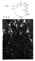
- HBsAg was synthesized in yeast in the form of particles or aggregates. The nature of these particles was further characterized by electron microscopy. HBsAg particles synthesized from yeast and purified as described were adsorbed onto carbon film grids and stained with uranyl acetate stain (2% (w/v) for 7 minutes). Under the electron microscope, particles of HBsAg synthesized in yeast were observed, had an identical appearance but smaller diameter compared with HBsAg from Alexander cells. In these studies Alexander cell HBsAg particles had a diameter of about 20 nm, whereas Y-HBsAg particle diameter was from about 16 to about 17 nm (see figure 3). These results are believed to be the first demonstration of assembly into a higher order structure of a heterologous protein in a microorganism host.
- the strain is a petite strain whose increased dependence upon carbohydrate metabolism may result in a higher activity for the ADH promoter.
- the high expression level observed in GM-3C-2 may also be the result of using a modified vector, pHBS-25, containing the S-protein coding fragment flanked by an ADHl promoter fragment and an ADH termination fragment.
- the Tac l- Hpa I HBV coding segment was fitted with Hin dIII oligonucleotide linkers and joined at the 5′-end to the ADHl promoter fragment ADHl-906 terminated in a Hin dIII linker sequence at its 3′ end.
- the 3′-end of the HBV segment was joined to a 450 bp Hin dIII- Bam HI fragment of the ADHl gene containing the coding region for the 43 C-terminal amino acids of ADH, the stop codon TAA and part of the 3′ untranslated region (see Bennetzen et al., J. Biol. Chem., Vol. 257, p. 3018 (1982) , such that both fragments were oriented in the same direction of transcription.
- the resulting composite segment, ADH-H-5-S-protein-ADH-terminator was flanked by Bam HI sites, permitting insertion at the Bam HI site of pMA56.
- the resulting vector was designated pHBS-25.
- pHBS25-3 and pHBS25-5 differ from pHBS25 in that the composite gene is inserted at the Sph l site of pMA56 rather than the nearby Bam HI site of pMA56.
- the transfer vector pHBS-16 and a yeast strain comprising the strain XV610-8C transformed by plasmid pHBS-16 have been placed on deposit in the American Type Culture Collection, 12301 Parklawn Drive, Rockville, Maryland.
- This example demonstrates removal of a 5′-untranslated segment of HBV-DNA.
- the DNA segment comprising the S-protein region isolated as described in Example 2 included an untranslated 26 base pair segment at the 5′-end of the coding region, lying between the promoter and the ATG start condon. The following procedure was developed to remove all, or all but one, of the bases of the 5′-untranslated region of the HBV-DNA preceding the S-protein coding region.
- the plasmid pHBS-5 was digested with Eco Rl endonuclease generating a fragment of approximately 850 base pairs including the S-protein coding region and flanking 3′-and 5′-untranslated regions, terminated by Eco Rl linker oligonucleotide segments.
- the HBV-DNA segment was reisolated by preparative gel electrophoresis, electroeluted and divided into samples which were digested with the exonuclease Bal -31 for varying times from 0.5 to 30 minutes at 37°C.
- the extent of exonuclease digestion was characterized qualitatively by digesting a portion of each sample with Xba I endonuclease.
- the S-protein coding region contains an Xba I site beginning 92 base pairs from the first base of the start condon. Therefore, samples in which Bal -31 digestion had proceeded beyond the Xba l site would yield only one fragment upon gel electrophoresis after Xba I endonuclease incubation while samples with fewer bases removed would yield two classes of fragment: a homogeneous large fragment and a heterogeneously sized small fragment. Samples yielding only one Xba I fragment were discarded. Samples yielding two size classes of fragments were blunt-ended by incubation with DNA polymerase I (Klenow fragment, see Klenow, H., et al., Proc. Nat. Acad. Sci.
- Linker oligonucleotides containing the Eco Rl recognition site were added by blunt-end ligation using T4 DNA ligase.
- Eco Rl specific cohesive ends were generated by digestion with Eco Rl endonuclease.
- the modified DNA was isolated by gel electrophoresis, electroeluted and joined to Eco Rl-digested, alkaline phosphatase treated pBR322, in a DNA ligase catalyzed reaction. The recombinant plasmids were then used to transform E . coli HB-101.
- a second screening strategy was used to detect clones in which the 5-untransalted region had been completely removed or terminated one base short of the start codon.
- the method exploited the observation that the first four bases of the S-protein coding sequence, ATGG, when joined to an Eco Rl linker oligonucleotide (GGAATTCC) generated a recognition site for the restriction endoculease Nco I: CCATGG.
- GGAATTCC Eco Rl linker oligonucleotide
- any S-protein coding segment in which the Bal -31 digestion was terminated precisely at the ATG start codon would be characterized by the generation of a new Nco I site when joined to an Eco Rl linker oligonucleotide.
- Bal -31 digests that retain only the last C of the 5′-untranslated region will also generate an Nco I recognition site when jointed to an Eco Rl linker oligonucleotide.
- Expression vector construction analogous to pHBS16, described in Example 2 was carried out by insertion of the modified HBV-DNA segment of pHBS5-3 in place of the corresponding segment in pHBS16.
- a "16-type" vector was prepared by Eco Rl endonuclease digestion and religation, followed by selection for a vector in which the HBV-DNA was deleted.
- Expression vectors constructed by the insertion of modified HBV-DNA segments at the Eco Rl site of the 16-type vector were characterized by the designation pHBS16-X, where X is a number characterizing the modification of HBV-DNA inserted at the Eco Rl site.
- the HBV-DNA segment transferred from pHBS5-3 to the 16 vector generated an expression plasmid designated phHBS16-3.
- the host strain for the 16 series expression vectors was Saccharomyces cerevisiae AB 35-D3-D a , leu2-3, leu2-112 , ura3-52 , trpl-289 , his4-580 , ade2 or Saccharomyces cerevisiae AB 35-14-D.
- Sample cultures of the host strain transformed with either pHBS16 or pHBS16-3 were grown under equivalent conditions and Y-HBsAg was qualitatively assayed by radioimmune assay as described in Example 3. Cells transformed with pHBS16-3 produced approximately 2.2 times as much Y-HBsAg per cell as those transformed by pHBS16.
- a further modification was made of the vector construction described in Example 4, in which the 3′-untranslated region removed during the Bal -31 digestion was restored.
- the strategy of this construction was to combine, at the Xba I site internal to the S-protein coding region, a fragment of the coding region derived from pHBS5-3, modified as described in Example 4, together with an unmodified fragment from pHBS5 having an intact 3′-untranslated region.
- the HBV-DNA segment of pHBS5 containing the 5′-untranslated region and promoter proximal part of the S-protein coding region was removed by the sequential action of Cla I endonuclease and Xba I endonuclease.
- the plasmid was first cleaved with Cla I endonuclease. The resulting unpaired ends were filled in using DNA polymerase I Klenow fragment in the presence of the four deoxynucleotide triphosphates to provide a linear vector with blunt ends.
- the DNA was then digested with Xba I endonuclease and alkaline phosphatase. The latter treatment was intended to insure that the ends generated by the foregoing series of steps could not rejoin to one another in the presence of DNA ligase, (Shine, J., supra).
- the modified HBV-DNA of pHBS5-3 containing the promoter proximal portion of the coding region for S-protein was also prepared by sequetial endonuclease digestion. Plasmid pHBS5-3 was first cleaved with Eco Rl endonuclease and blunt ended with DNA polymerase I Klenow fragment in the presence of the four deoxynucleotide triphosphates. The DNA was then cleaved with Xba endonuclease. The small fragment resulting from Xba I cleavage, approximately 100 base pairs having a blunt Eco Rl end and an Xba I end was isolated by gel electrophoresis and electroelution.
- the purpose of sequential endonuclease treatment in both instances was to insure that the 100 base pair fragment would be joined in correct orientation with the cleaved vector DNA.
- the 100 base pair fragment derived from pHBS5-3 was mixed with modified vector DNA derived from pHBS5, in the presence of DNA ligase under conditions permitting blunt end ligation as well as the joining of paired ends derived from the Xba I cuts.
- Transformants were selected and identifed by the existance of an Nco l site, derived from the small fragment from pHBS5-3 (See Example 4).
- the HBV-DNA segment modified as in pHBS6 was transfered to an expression vector of the 16 series as follows: The HBV-DNA region of pHBS6 was isolated by combined action of Nco I and Eco Rl endonucleases and blunt ended by incubation with DNA polymerase I Klenow fragment in the presence of the four deoxynucleotide triphosphates. The resulting 820 base pair fragment was isolated by gel electrophoresis and electroelution.
- the expression vector pHBS16 or the "16-vector" described in Example 4, was cleaved by Eco Rl endonuclease action, blunt ended using DNA polymerase 1 Klenow fragment in the presence of the four deoxynucleotide triphosphates, and treated with alkaline phosphatase.
- the HBV-DNA fragment was then joined to the treated 16 vector by blunt end ligation using T4 DNA ligase. Correct orientation of the fragments regenerated an Eco Rl site between the ADH promoter and the start codon of the S-protein coding region.
- DNA nucleotide sequence analysis was carried out confirming the structure of the resulting construction, designated pHBS16-5, diagrammed in Figure 5.
- the relative rate of expression of Y-HBsAg for yeast cells transformed by pHBS16-5 was measured under the same conditions as for pHBS16-3, described in Example 4. Expression of Y-HBsAg by Saccharomyces cerevisiae AB-35-D3-D transformed by pHBS16-5 was approximately 2.8 times greater per cell as measured by radioimmumo assay, than expression by cells transformed with pHBS16.
- a related construction using the HBV-DNA segment of pHBS6 was carried out using an identical procedure with the exception that the HBV-DNA fragment was excised by an Eco Rl endonuclease preparation having some Eco Rl* activity.
- the expression vector was found to have lost the Eco Rl site as well as the Nco I site adjacent to the ATG start codon of the S-protein coding region.
- the resulting expression plasmid was designated pHBS16-4.
- the nucleotide sequence adjacent to the S-protein start codon was 5′...ACTATCTGGCATGG...3′.
- the rate of expression in yeast cells transformed with pHBS16-4 was comparable to that of pHBS16-5 transformed cells, within experimental error.
- the structure of pHBS16-4 is diagrammed in Figure 5.
- the nucleotide sequence adjacent to the S-protein start codon of PHBS-16-3 was 5′...ACTATCTGGAATTCCCATGG...3′.
- the sequence for pHBS-16-5 was 5′...ACTATCTGGAATTCATGG...3′.
- the sequence difference between 16-3 and 16-5 was a consequence of blunt-ending the DNA after Eco Rl digestion of pHBS-6.
- This example describes details of the construction of a series of vectors for expression characterized by having the entire two micron circle plasmid DNA sequence within their sequence together with DNA segments comprising the promoter and transcription terminator sequences of the yeast ADH gene, with the S-protein coding region sandwiched between the ADH promoter and the ADH terminator regions.
- These vectors were designated "56" series vectors and their nomenclature is consistent with the nomenclature of the 16 series of expression vectors.
- the expression vector pHBS16-3 contains the S-protein gene modified as described for pHBS16-3
- pHBS56-5 contains HBV-DNA modified as described for pHBS16-5, as described in Examples 4 and 5 respectively.
- the full length two micron circle DNA provides for stable replication in a circle zero host strain in the absence of metabolic selection pressure.
- the ADH terminator was provided to enhance the stability of S-protein mRNA transcripts.
- the parent plasmid for the construction of 56 type vectors was pCl/l which was a hybrid plasmid between pBR-322 and a two micron circle plasmid joined at their Eco Rl sites.
- the two micron circle portion was previously modified to contain an inserted LEU2 gene of yeast and obtained from the plasmid pJBD219 described by Beggs, J. et al., Nature 275 , 104 (1978).
- the restriction map of pCl/l is shown in Figure 6.
- the precursor source for the ADH promoter and terminator segments was plasmid PAAH5 containing a 1500 base pair ADHl promoter fragment terminated at position ⁇ 9 in the nucleotide sequence (Hitzeman, R. A. et al., supra, and an approximately 450 base pair terminator unit from nucleotides 913 to 1368 in the ADH bene nucleotide sequence, joined by a Hin dIII site between the fragments and cloned into the BamHl site of the vector YEp13, (Broach, J.
- the HBV-DNA segment of pHBS5 was excised by Eco Rl digestion. The protruding ends were filled in using DNA polymerase I Klenow fragment and joined at both ends with Hin dIII linker oligonucleotides having the sequence CAAGCTTG. After Hin dIII endonuclease digestion to expose unpaired, Hin dIII specific ends on the HBV-DNA segment, the segment was joined to Hin dIII cut plasmid PAAH5, thereby placing the HBV S-protein coding sequence between the ADH promoter and terminator fragments.
- the ADH promoter and terminator sequences were each found to contain an Sph I site (recognition sequence GCATGC) making it possible to excise the entire composite gene comprising about 400 base pairs of ADHl promoter, HBV S-protein region and about 330 base pairs of ADHl terminator by digestion with Sph l endonuclease. Digestion of pHBS22 with Sph I endonuclease yielded the intact composite gene in a fragment of approximately 1500 base pairs. The fragment was joined with Sph I-cut vector pCl/l. E . coli .
- HB101 transformants were screened for ampicillin resistance and sensitivity to tetracycline, since the segment excised by Sph I endonuclease digestion of pCl/l deleted a portion of the tetracycline resistance gene of the pBR322 segment.
- the structure of the resulting vector, designated pHBS56 was further confirmed by restriction analysis.
- E . coli . HB101 transformants obtained from the products of the ligase reaction were cloned on plates containing ampicillin. Plasmid DNA from single colony isolates grown in culture was screened by restriction endonuclease analysis for the insertion and correct orientation of the S-protein coding region.
- the plasmid selected, pHBS-56 containing the HBV S-protein coding region in correct orientation with respect to the ADH promoter and terminator segments, is shown in Figure 6.
- Two additional 56-type vectors were constructed using a promoter fragment and promoter proximal region of the S-protein coding segment obtained from pHBS16-3 and from pHBS16-5. These constructions were designated pHBS56-3 and pHBS56-5, respectively.
- an Sph I - Xba I fragment was joined in a DNA ligase catalyzed reaction with the larger of the two Sph I - Xba I fragments obtained by digestion of pHBS56.
- the larger fragment approximately 1080 base pairs, extends from the Xba I site within the S-protein coding region to the Sph I site of the ADH terminator region.
- This fragment was isolated by gel electrophoresis and electroelution prior to joining to the Sph I - Xba I fragment of either pHBS16-3 or pHBS16-5, in a DNA ligase catalyzed reaction.
- the two composite genes thus constructed were identical except for the sequence of the 5′-untranslated region between the ADH promoter and the S-protein start codon, these differences arising from differences in the respective source vectors, pHBS16-3 and pHBS16-5 respectively.
- Both composite genes were sub-cloned, in separate reactions, at the Sph I site of PBR322, situated between the Bam HI site and the Sal I site of PBR322, for the purpose of obtaining amplified amounts of composite gene DNA.
- the sub-cloning vectors were selected by their ability to confer ampicillin resistance and tetracycline sensitivity phenotype to E . coli . HB101 transformants.
- the large fragment produced by Sph I cleavage of pHBS56 was treated with alkaline phosphatase, and isolated by gel electrophoresis and electroelution.
- the composite genes were excised from their sub-cloning vectors by Sph I cleavage and isolated by gel electrophoresis and electroelution, but without phosphatase treatment. These were combined, in separate reactions, with the large Sph I fragment of pHBS56 and joined in DNA ligase catalyzed reactions yielding pHBS56-3 and pHBS56-5, respectively.
- E . coli . HB101 transformants were selected by ampicillin resistance and tetracycline sensitivity and further characterized by restriction analysis.
- FIG. 7 A diagram of the construction steps and maps of the relevant vectors are shown in Figure 7. Although the nomenclature of the 56 type vectors is parallel with that of the 16 type vectors, it will be understood that the distinctions in the case of the 56 series vectors refer only to the 5′ untranslated region in each instance, and that the 3′ untranslated region is the same in each member of the series. Specifically, pHBS56-3 lacks the 40 base pair deletion in the 3′ untranslated region that occurs in pHBS-16-3.
- vectors of the 56 series were used to transofrm a circle zero yeast strain designated 2150-2-3.
- the strain was derived from a genetic cross between strain Y379-5-D cyh2 nib l ( rho ⁇ ) Livingston, D. Genetics 86 , 73 (1977) and DC 04 a Ade I Ade X leu2-04 (cir 0) (Broach, J., Cell , 21 , 501 (1980).
- the diploid strain resulting from the cross was permitted to sporulate and tetrads were disected by a standard procedure.
- One of the haploid spores gave rise to strain 2150-2-3 a Ade I Leu2-04 (cir 0 ).
- Yeast transformants were selected for Leu+ phenotype conferred by the presence of plasmids of the pHBS-56 series.
- the relative rates of Y-HBsAg expression in various plasmid-host combinations were compared in the following manner: A one liter volume of cell culture was grown to its limiting cell density, the cells harvested in crude lysates analyzed for total soluble protein and for Y-HBsAg, using radioimmunoassay, as described, supra. The results were expressed both as micrograms Y-HBsAg per liter of culture and Y-HBsAg as percent by weight of total yeast soluble protein. The latter provides a measure of the amount of cell metabolism devoted to Y-HBsAg production while the former provides a measure of the overall yield of Y-HBsAg obtainable upon growth of cultures to limiting density.
- Y-HBsAg Yields Vector pHBS- Host Culture Density O.D.660 % by weight of total yeast soluble protein ⁇ g Y-HBsAg per liter culture 16-3 AB-35-14D 2.0 0.1% 20 16-4 AB-35-14D 2.0 0.1% 20 16-5 AB-35-14D 2.0 0.1% 20 25 GM-3C-2 4.0 0.5% 200 56 2150-2-3 12.0 0.3% 300
- Y-HBsAg particles are purified from cell extracts by the method of Example 3, or by suitable methods known in the art. for example, as described in U.S patents 4,088,748 or 4,181,713.
- Purified HBsAg particles are dialyzed against physiological saline or phosphate-buffer saline and adjusted to 100 ⁇ g protein/ml final contentration.
- Guinea pigs are subjected subcutaneously at 9, 14 and 56 day intervals with 1 ml of the HBsAg preparation.
- the serum of the test animals is sampled at 0, 28, 56 and 84 days and assayed for antibody titre agaist Dane particles or HBsAg purified from Alexander cells.
- HBsAg synthesized by yeast is immunogenic and is capable of eliciting antibodies cross-reactive with naturally-occurring HBsAg.
- HBsAg synthesized by yeast has the advantage of being available in significantly larger quantities than that obtained from Dane particles or carrier serum. A more uniform product is obtainable at an advantageous cost per unit, which may be expected to decrease with increasing production volume. Furthermore, thre is no danger of accidental infection, since there is no intact HBV, and can be no intact HBV, in the surface antigen prepared from yeast. By contrast, viral proteins purified from serum or other natural sources always pose the danger of viral cotamination.
- HBsAg synthesized by yeast is capable of eliciting antibodies cross-reactive with naturally-occurring HBsAg. It therefore follows that such antigens and antigen aggregates, when purified as described and administered in a physiologically acceptable medium, constitute a vaccine for protection agaist infection by hepatitis B virus.
- Group A (six animals) is inoculated intravenously with 1 ml of a standard Bureau of Biologics Hepatitis B virus preparation
- Group B (four animals) is inoculated intravenously with 1 ml containing 200 ⁇ g of HBsAg synthesized in yeast and purified as described in Example 3, in physiological saline
- Group C (six animals) is the control group and receives no inoculation. All chimps in Group A have evidence of clinical Hepatitis B (either antigenemia, enzyme elevations and/or antibody response) within 40 weeks. None of the animals in Groups B or C shows evidence of clinical Hepatitis B infection over the same 40-week period.
- the chimps of Group B are rendered immune to subsequent challenge when inoculated intravenously with 1.0 ml of BOB Hepatitis B virus.
- Y-HBsAg differed in several respects from plasma-derived HBsAg.
- the diameters of HBsAg and Y-HBsAg particles were measured from negatively stained electron micrographs.
- the yeast-derived antigen had a diameter range of from about 14 to about 18 nm while plasma-derived antigen had a diameter range of from about 20 to about 24 nm.
- Y-HBsAg was unstable at pH 2, and to pepsin at pH 2, whereas plasma-derived HBsAg was stable under the same conditions.
- To a 1 ml suspension of purified Y-HBsAg was added 0.03 ml of 1N HCl to lower the pH to 2.0.
- the sample was divided in halves and to one-half was added 1 ⁇ g of pepsin while no enzyme was added to the other half. Both samples were held at 37°C for sixteen hours and then 0.03 ml of 1N NaOH was added to each to raise the pH to 7.0.
- the two samples were measured for antigen binding activity in a quantitative radioimmunoassay (RIA). Over 95% of the RIA activity was lost in each sample. Under the same conditions the plasma derived HBsAg retained all of its antigen binding activity.
- RIA quantitative radioimmunoassay
- a sample of purified Y-HBsAg was heated in sodium dodecylsulfate (SDS) and 2-mercaptoethanol at 90°C for five minutes. It was then electrophoresed through a 10% polyacrylamide gel containing 0.1% SDS. Subsequent staining of the gel with protein stains revealed a single band at a molecular weight equivalence location of about 25,000.
- Y-HBsAg did not bind to a monoclonal antibody (HBsAb) selected against HBsAg.
- HBsAb monoclonal antibody
- a crude extract of yeast cells containing surface antigen was passed through an affinity adsorbent column prepared by chemically coupling monoclonal HBsAb to an agarose gel. Measurement of the column effluent revealed 90% of the Y-HBsAg charged to the column was present in the effluent.
- a plasma-derived HBsAg passed through the same column of monoclonal HBsAb revealed less than 10% of the charged antigen in the column effluent.
- Y-HBsAg exhibited higher activity in mouse potency tests.
- Y-HBsAg was adsorbed to an aluminum hydroxide gel prior to administration and was diluted to contain 10, 2.5, 0.62, 0.15 and 0.0375 ⁇ g/ml.
- 1 Ml quantities of the foregoing concentrations were injected intraperitoneally into each of five groups of five-week old female mice. Each of these concentrations was injected into one of the five groups, each group containing ten mice.
- the yeast-produced antigen had an ED50 (the concentration of antigen needed to produce antibody in one-half of the mice) of about 0.05 ⁇ g/ml while the plasma-derived antigen in the same procedure had an ED50 of about 0.5 ⁇ g/ml.
- Y-HBsAg purified as described was essentially free of contaminating chemicals. Measurement of the Lowry protein of Y-HBsAg showed 53 ⁇ g/ml while measurement of the RIA antigen binding ability of this antigen indicated a concentration of 12 ⁇ g/ml. Measurement of the Lowry protein and RIA antigen binding ability of a pure preparation of plasma-derived HBsAg revealed a protein concentration of 44 ⁇ g/ml and a binding activity of 50 ⁇ g/ml.
- mice A total of 80 five-week old female mice were divided into two groups of 40 and each group was further subdivided into four sub-groups of 10 mice.
- the 10 mice from each sub-group were injected intraperitoneally with either the antigen prepared from Example 3 using vector pHBS-25 or plasma-derived antigen at a concentration, respectively, of 10, 2.5, 0.625 or 0.156 ⁇ g/ml.
- Saline-alum placebo was used as diluent to dilute the antigen concentration where necessary to obtain the foregoing concentrations.
- the mice were individually bled and sacrificed at 28 days. Antibody determinations were performed by the Ausab (Abbott) radioimmune assay.
- a total of 32 African Green Monkeys were divided into two groups of 16, and each group was further subdivided into four sub-groups of 4 monkeys. Each sub-group was injected intramuscularly at day 0 and day 28 with yeast-derived antigen or plasma-derived antigen at a concentration, respectively, of 10, 2.5, 0.625 or 0.156 ⁇ g/ml using saline-alum placebo as diluent to dilute the antigen concentration where necessary to obtain the foregoing concentrations. Bleedings were collected at weekly intervals for 14 weeks and antibody determinations were performed by the Ausab (Abbott) radioimmune assay with titers expressed as "Estimated Ausab Units". The results are summarized in the following tables.
- the present invention represents a substantial advance in applying recombinant DNA technology.
- the practical goal of synthesizing HBsAg in a microorganism host has been achieved.
- Modifications to increase HBsAg production and improve the yield of HBsAg upon purification which fall within the scope of ordinary skill in the art, are deemed equivalent variants within the scope of the claimed invention. Examples of such modifications could include improved promoter systems, more productive host cell strains, improvements in purification technique and modifications to improve the antigenicity of the product or its immunogenicity.
- any eucaryotic microorganism is considered to be capable of serving as a host strain for producing HBsAg particles, provided a sufficient amount of antigen is synthesized thereby.
- othr eucaryotic microorganisms which could be employed include, but are not limited to, members of the genera aspergillus, penicillium, and neurospora, as well as the genus Saccharomyces.
- the depository was requested to handle the above-described deposits in accordance with the terms and conditions of the Budapest Treaty on the International Recognition of the Deposit of Microorganisms for the Purposes/Patent Procedure.
Landscapes
- Chemical & Material Sciences (AREA)
- Organic Chemistry (AREA)
- Health & Medical Sciences (AREA)
- Life Sciences & Earth Sciences (AREA)
- Biophysics (AREA)
- Biochemistry (AREA)
- Gastroenterology & Hepatology (AREA)
- General Health & Medical Sciences (AREA)
- Genetics & Genomics (AREA)
- Medicinal Chemistry (AREA)
- Molecular Biology (AREA)
- Proteomics, Peptides & Aminoacids (AREA)
- Virology (AREA)
- Micro-Organisms Or Cultivation Processes Thereof (AREA)
- Preparation Of Compounds By Using Micro-Organisms (AREA)
Applications Claiming Priority (3)
| Application Number | Priority Date | Filing Date | Title |
|---|---|---|---|
| US28991581A | 1981-08-04 | 1981-08-04 | |
| US289915 | 1981-08-04 | ||
| EP82401473A EP0072318B1 (de) | 1981-08-04 | 1982-08-04 | Synthese von menschlichen viralen Antigenen mit Hilfe von Hefe |
Related Parent Applications (1)
| Application Number | Title | Priority Date | Filing Date |
|---|---|---|---|
| EP82401473.2 Division | 1982-08-04 |
Publications (3)
| Publication Number | Publication Date |
|---|---|
| EP0340806A2 true EP0340806A2 (de) | 1989-11-08 |
| EP0340806A3 EP0340806A3 (en) | 1989-12-20 |
| EP0340806B1 EP0340806B1 (de) | 1995-05-31 |
Family
ID=26086453
Family Applications (1)
| Application Number | Title | Priority Date | Filing Date |
|---|---|---|---|
| EP89110270A Expired - Lifetime EP0340806B1 (de) | 1981-08-04 | 1982-08-04 | Synthese von menschlichen Virusantigenen durch Hefe |
Country Status (2)
| Country | Link |
|---|---|
| EP (1) | EP0340806B1 (de) |
| AT (1) | ATE53235T1 (de) |
Family Cites Families (4)
| Publication number | Priority date | Publication date | Assignee | Title |
|---|---|---|---|---|
| IL59007A (en) * | 1978-12-22 | 1983-11-30 | Biogen Nv | Recombinant dna molecules coding for polypeptides displaying hepatitis b virus antigenicity,method of their production and compositions containing the same |
| IE52036B1 (en) * | 1979-05-24 | 1987-05-27 | Univ California | Non-passageable viruses |
| EP0038765B1 (de) * | 1980-04-22 | 1987-09-02 | Institut Pasteur | Impfstoff gegen Hepatitis B Virus, Verfahren und transformierte eukaryotische Zellen zur Herstellung dieses Impfstoffes |
| NZ201705A (en) * | 1981-08-31 | 1986-03-14 | Genentech Inc | Recombinant dna method for production of hepatitis b surface antigen in yeast |
-
1982
- 1982-08-04 AT AT82401473T patent/ATE53235T1/de not_active IP Right Cessation
- 1982-08-04 EP EP89110270A patent/EP0340806B1/de not_active Expired - Lifetime
Also Published As
| Publication number | Publication date |
|---|---|
| EP0340806B1 (de) | 1995-05-31 |
| ATE53235T1 (de) | 1990-06-15 |
| EP0340806A3 (en) | 1989-12-20 |
Similar Documents
| Publication | Publication Date | Title |
|---|---|---|
| CA1341642C (en) | Synthesis of hepatitis b virus surface antigen by yeast | |
| US4769238A (en) | Synthesis of human virus antigens by yeast | |
| JP3069907B2 (ja) | 新規な抗原物質及びその製法 | |
| US4816564A (en) | Method for producing hepatitis B virus proteins in yeast | |
| Hitzeman et al. | Expression of hepatitis B virus surface antigen in yeast | |
| EP0341746B1 (de) | Expression von Hepatitis B S und preS2 Proteinen in methylotrophen Hefen | |
| US4945046A (en) | Yeast promoter and process for preparing heterologous protein | |
| US6103519A (en) | Antigens and methods therefor | |
| EP0340806B1 (de) | Synthese von menschlichen Virusantigenen durch Hefe | |
| EP0339567A1 (de) | Expression von Hepatitis B PreS 2-Protein in methylotrophen Hefen | |
| IE67139B1 (en) | Synthesis of human virus antigens by yeast | |
| Ammerer | by genetic engineering (recombinant DNA) using yeast was issued thisdate to inven | |
| HK1003032B (en) | Novel antigens and methods for their preparation | |
| EP0277770A1 (de) | Alpha-Mating-Faktor-Promotor, modifiziert durch Entfernung der "pre-pro-secretory-leader"-Sequenz | |
| HRP950289A2 (en) | Process for producing hbv surface proteins |
Legal Events
| Date | Code | Title | Description |
|---|---|---|---|
| PUAI | Public reference made under article 153(3) epc to a published international application that has entered the european phase |
Free format text: ORIGINAL CODE: 0009012 |
|
| PUAL | Search report despatched |
Free format text: ORIGINAL CODE: 0009013 |
|
| AC | Divisional application: reference to earlier application |
Ref document number: 72318 Country of ref document: EP |
|
| AK | Designated contracting states |
Kind code of ref document: A2 Designated state(s): AT BE CH DE FR GB IT LI LU NL SE |
|
| AK | Designated contracting states |
Kind code of ref document: A3 Designated state(s): AT BE CH DE FR GB IT LI LU NL SE |
|
| RAP1 | Party data changed (applicant data changed or rights of an application transferred) |
Owner name: BOARD OF REGENTS OF THE UNIVERSITY OF WASHINGTON Owner name: THE REGENTS OF THE UNIVERSITY OF CALIFORNIA |
|
| RAP1 | Party data changed (applicant data changed or rights of an application transferred) |
Owner name: WASHINGTON RESEARCH FOUNDATION Owner name: THE REGENTS OF THE UNIVERSITY OF CALIFORNIA |
|
| 17P | Request for examination filed |
Effective date: 19900226 |
|
| 17Q | First examination report despatched |
Effective date: 19911205 |
|
| GRAA | (expected) grant |
Free format text: ORIGINAL CODE: 0009210 |
|
| AC | Divisional application: reference to earlier application |
Ref document number: 72318 Country of ref document: EP |
|
| AK | Designated contracting states |
Kind code of ref document: B1 Designated state(s): AT BE CH DE FR GB IT LI LU NL SE |
|
| REF | Corresponds to: |
Ref document number: 123225 Country of ref document: AT Date of ref document: 19950615 Kind code of ref document: T |
|
| REF | Corresponds to: |
Ref document number: 3280468 Country of ref document: DE Date of ref document: 19950706 |
|
| RAP2 | Party data changed (patent owner data changed or rights of a patent transferred) |
Owner name: WASHINGTON RESEARCH FOUNDATION Owner name: THE REGENTS OF THE UNIVERSITY OF CALIFORNIA |
|
| ET | Fr: translation filed | ||
| ITF | It: translation for a ep patent filed | ||
| NLT2 | Nl: modifications (of names), taken from the european patent patent bulletin |
Owner name: THE REGENTS OF THE UNIVERSITY OF CALIFORNIA EN WAS |
|
| PLBE | No opposition filed within time limit |
Free format text: ORIGINAL CODE: 0009261 |
|
| STAA | Information on the status of an ep patent application or granted ep patent |
Free format text: STATUS: NO OPPOSITION FILED WITHIN TIME LIMIT |
|
| 26N | No opposition filed | ||
| PGFP | Annual fee paid to national office [announced via postgrant information from national office to epo] |
Ref country code: FR Payment date: 20010817 Year of fee payment: 20 |
|
| PGFP | Annual fee paid to national office [announced via postgrant information from national office to epo] |
Ref country code: SE Payment date: 20010820 Year of fee payment: 20 Ref country code: DE Payment date: 20010820 Year of fee payment: 20 Ref country code: CH Payment date: 20010820 Year of fee payment: 20 |
|
| PGFP | Annual fee paid to national office [announced via postgrant information from national office to epo] |
Ref country code: NL Payment date: 20010821 Year of fee payment: 20 Ref country code: GB Payment date: 20010821 Year of fee payment: 20 Ref country code: AT Payment date: 20010821 Year of fee payment: 20 |
|
| PGFP | Annual fee paid to national office [announced via postgrant information from national office to epo] |
Ref country code: BE Payment date: 20010914 Year of fee payment: 20 |
|
| PGFP | Annual fee paid to national office [announced via postgrant information from national office to epo] |
Ref country code: LU Payment date: 20011122 Year of fee payment: 20 |
|
| BE20 | Be: patent expired |
Free format text: 20020804 THE *REGENTS OF THE UNIVERSITY OF CALIFORNIA;*WASHINGTON RESEARCH FOUNDATION |
|
| REG | Reference to a national code |
Ref country code: GB Ref legal event code: IF02 |
|
| PG25 | Lapsed in a contracting state [announced via postgrant information from national office to epo] |
Ref country code: LI Free format text: LAPSE BECAUSE OF EXPIRATION OF PROTECTION Effective date: 20020803 Ref country code: GB Free format text: LAPSE BECAUSE OF EXPIRATION OF PROTECTION Effective date: 20020803 Ref country code: CH Free format text: LAPSE BECAUSE OF EXPIRATION OF PROTECTION Effective date: 20020803 |
|
| PG25 | Lapsed in a contracting state [announced via postgrant information from national office to epo] |
Ref country code: NL Free format text: LAPSE BECAUSE OF EXPIRATION OF PROTECTION Effective date: 20020804 Ref country code: LU Free format text: LAPSE BECAUSE OF EXPIRATION OF PROTECTION Effective date: 20020804 Ref country code: AT Free format text: LAPSE BECAUSE OF EXPIRATION OF PROTECTION Effective date: 20020804 |
|
| REG | Reference to a national code |
Ref country code: GB Ref legal event code: PE20 Effective date: 20020803 |
|
| REG | Reference to a national code |
Ref country code: CH Ref legal event code: PL |
|
| EUG | Se: european patent has lapsed |
Ref document number: 89110270.9 |
|
| NLV7 | Nl: ceased due to reaching the maximum lifetime of a patent |
Effective date: 20020804 |