EP0351313A2 - Glycoprotéine et son gène codant - Google Patents
Glycoprotéine et son gène codant Download PDFInfo
- Publication number
- EP0351313A2 EP0351313A2 EP89401996A EP89401996A EP0351313A2 EP 0351313 A2 EP0351313 A2 EP 0351313A2 EP 89401996 A EP89401996 A EP 89401996A EP 89401996 A EP89401996 A EP 89401996A EP 0351313 A2 EP0351313 A2 EP 0351313A2
- Authority
- EP
- European Patent Office
- Prior art keywords
- glycoprotein
- minutes
- gene
- complement
- human
- Prior art date
- Legal status (The legal status is an assumption and is not a legal conclusion. Google has not performed a legal analysis and makes no representation as to the accuracy of the status listed.)
- Withdrawn
Links
- 108090000288 Glycoproteins Proteins 0.000 title claims abstract description 52
- 102000003886 Glycoproteins Human genes 0.000 title claims abstract description 52
- 108090000623 proteins and genes Proteins 0.000 title claims abstract description 44
- 238000000034 method Methods 0.000 claims abstract description 34
- 230000000295 complement effect Effects 0.000 claims abstract description 25
- 230000002401 inhibitory effect Effects 0.000 claims abstract description 16
- 210000000170 cell membrane Anatomy 0.000 claims abstract description 15
- 238000002415 sodium dodecyl sulfate polyacrylamide gel electrophoresis Methods 0.000 claims abstract description 12
- 239000012528 membrane Substances 0.000 claims abstract description 11
- 210000005260 human cell Anatomy 0.000 claims abstract description 10
- 230000001404 mediated effect Effects 0.000 claims abstract description 10
- 230000006378 damage Effects 0.000 claims abstract description 8
- 150000003905 phosphatidylinositols Chemical class 0.000 claims abstract description 7
- 229930182474 N-glycoside Natural products 0.000 claims abstract description 6
- 238000004519 manufacturing process Methods 0.000 claims abstract description 4
- 125000000837 carbohydrate group Chemical group 0.000 claims abstract 3
- 210000004027 cell Anatomy 0.000 claims description 37
- 239000002299 complementary DNA Substances 0.000 claims description 37
- 102000004169 proteins and genes Human genes 0.000 claims description 24
- 239000012634 fragment Substances 0.000 claims description 18
- 210000003743 erythrocyte Anatomy 0.000 claims description 12
- 238000003752 polymerase chain reaction Methods 0.000 claims description 11
- 239000000523 sample Substances 0.000 claims description 11
- 238000009396 hybridization Methods 0.000 claims description 9
- 238000012216 screening Methods 0.000 claims description 8
- 230000036961 partial effect Effects 0.000 claims description 7
- 238000001042 affinity chromatography Methods 0.000 claims description 5
- 241000701959 Escherichia virus Lambda Species 0.000 claims description 4
- 210000002826 placenta Anatomy 0.000 claims description 2
- 125000003275 alpha amino acid group Chemical group 0.000 claims 1
- 238000012258 culturing Methods 0.000 claims 1
- 230000003381 solubilizing effect Effects 0.000 claims 1
- 239000000243 solution Substances 0.000 description 38
- 239000000203 mixture Substances 0.000 description 34
- FAPWRFPIFSIZLT-UHFFFAOYSA-M Sodium chloride Chemical compound [Na+].[Cl-] FAPWRFPIFSIZLT-UHFFFAOYSA-M 0.000 description 30
- 150000001413 amino acids Chemical group 0.000 description 30
- 239000000872 buffer Substances 0.000 description 27
- 108020004414 DNA Proteins 0.000 description 21
- 239000002244 precipitate Substances 0.000 description 20
- 239000002953 phosphate buffered saline Substances 0.000 description 19
- 238000002360 preparation method Methods 0.000 description 16
- QKNYBSVHEMOAJP-UHFFFAOYSA-N 2-amino-2-(hydroxymethyl)propane-1,3-diol;hydron;chloride Chemical compound Cl.OCC(N)(CO)CO QKNYBSVHEMOAJP-UHFFFAOYSA-N 0.000 description 15
- 102100022002 CD59 glycoprotein Human genes 0.000 description 15
- 101000897400 Homo sapiens CD59 glycoprotein Proteins 0.000 description 15
- 239000000499 gel Substances 0.000 description 15
- 239000011780 sodium chloride Substances 0.000 description 15
- 239000006228 supernatant Substances 0.000 description 14
- KCXVZYZYPLLWCC-UHFFFAOYSA-N EDTA Chemical compound OC(=O)CN(CC(O)=O)CCN(CC(O)=O)CC(O)=O KCXVZYZYPLLWCC-UHFFFAOYSA-N 0.000 description 13
- TWRXJAOTZQYOKJ-UHFFFAOYSA-L Magnesium chloride Chemical compound [Mg+2].[Cl-].[Cl-] TWRXJAOTZQYOKJ-UHFFFAOYSA-L 0.000 description 12
- XLYOFNOQVPJJNP-UHFFFAOYSA-N water Chemical compound O XLYOFNOQVPJJNP-UHFFFAOYSA-N 0.000 description 11
- YBYRMVIVWMBXKQ-UHFFFAOYSA-N phenylmethanesulfonyl fluoride Chemical compound FS(=O)(=O)CC1=CC=CC=C1 YBYRMVIVWMBXKQ-UHFFFAOYSA-N 0.000 description 10
- 239000013615 primer Substances 0.000 description 10
- WEVYAHXRMPXWCK-UHFFFAOYSA-N Acetonitrile Chemical compound CC#N WEVYAHXRMPXWCK-UHFFFAOYSA-N 0.000 description 9
- LFQSCWFLJHTTHZ-UHFFFAOYSA-N Ethanol Chemical compound CCO LFQSCWFLJHTTHZ-UHFFFAOYSA-N 0.000 description 8
- 108010052285 Membrane Proteins Proteins 0.000 description 8
- 102000018697 Membrane Proteins Human genes 0.000 description 8
- 229920002684 Sepharose Polymers 0.000 description 8
- 239000011541 reaction mixture Substances 0.000 description 8
- 239000003155 DNA primer Substances 0.000 description 7
- 241000588724 Escherichia coli Species 0.000 description 7
- 206010018910 Haemolysis Diseases 0.000 description 7
- DBMJMQXJHONAFJ-UHFFFAOYSA-M Sodium laurylsulphate Chemical compound [Na+].CCCCCCCCCCCCOS([O-])(=O)=O DBMJMQXJHONAFJ-UHFFFAOYSA-M 0.000 description 7
- BFNBIHQBYMNNAN-UHFFFAOYSA-N ammonium sulfate Chemical compound N.N.OS(O)(=O)=O BFNBIHQBYMNNAN-UHFFFAOYSA-N 0.000 description 7
- 229910052921 ammonium sulfate Inorganic materials 0.000 description 7
- 235000011130 ammonium sulphate Nutrition 0.000 description 7
- 238000005119 centrifugation Methods 0.000 description 7
- 238000006243 chemical reaction Methods 0.000 description 7
- 238000004440 column chromatography Methods 0.000 description 7
- 230000008588 hemolysis Effects 0.000 description 7
- 230000002829 reductive effect Effects 0.000 description 7
- 235000019333 sodium laurylsulphate Nutrition 0.000 description 7
- 238000003756 stirring Methods 0.000 description 7
- 241000700199 Cavia porcellus Species 0.000 description 6
- HEDRZPFGACZZDS-UHFFFAOYSA-N Chloroform Chemical compound ClC(Cl)Cl HEDRZPFGACZZDS-UHFFFAOYSA-N 0.000 description 6
- QTBSBXVTEAMEQO-UHFFFAOYSA-N acetic acid Substances CC(O)=O QTBSBXVTEAMEQO-UHFFFAOYSA-N 0.000 description 6
- 150000001720 carbohydrates Chemical class 0.000 description 6
- 239000006285 cell suspension Substances 0.000 description 6
- 229910001629 magnesium chloride Inorganic materials 0.000 description 6
- 239000008188 pellet Substances 0.000 description 6
- DGVVWUTYPXICAM-UHFFFAOYSA-N β‐Mercaptoethanol Chemical compound OCCS DGVVWUTYPXICAM-UHFFFAOYSA-N 0.000 description 6
- 108010014303 DNA-directed DNA polymerase Proteins 0.000 description 5
- 102000016928 DNA-directed DNA polymerase Human genes 0.000 description 5
- 238000005859 coupling reaction Methods 0.000 description 5
- 230000000694 effects Effects 0.000 description 5
- 238000010828 elution Methods 0.000 description 5
- 210000003617 erythrocyte membrane Anatomy 0.000 description 5
- 239000007973 glycine-HCl buffer Substances 0.000 description 5
- 108090000765 processed proteins & peptides Proteins 0.000 description 5
- 108091032973 (ribonucleotides)n+m Proteins 0.000 description 4
- 101710139410 1-phosphatidylinositol phosphodiesterase Proteins 0.000 description 4
- UMCMPZBLKLEWAF-BCTGSCMUSA-N 3-[(3-cholamidopropyl)dimethylammonio]propane-1-sulfonate Chemical compound C([C@H]1C[C@H]2O)[C@H](O)CC[C@]1(C)[C@@H]1[C@@H]2[C@@H]2CC[C@H]([C@@H](CCC(=O)NCCC[N+](C)(C)CCCS([O-])(=O)=O)C)[C@@]2(C)[C@@H](O)C1 UMCMPZBLKLEWAF-BCTGSCMUSA-N 0.000 description 4
- FTOAOBMCPZCFFF-UHFFFAOYSA-N 5,5-diethylbarbituric acid Chemical compound CCC1(CC)C(=O)NC(=O)NC1=O FTOAOBMCPZCFFF-UHFFFAOYSA-N 0.000 description 4
- 108091003079 Bovine Serum Albumin Proteins 0.000 description 4
- 108020001019 DNA Primers Proteins 0.000 description 4
- 108010054576 Deoxyribonuclease EcoRI Proteins 0.000 description 4
- VEXZGXHMUGYJMC-UHFFFAOYSA-N Hydrochloric acid Chemical compound Cl VEXZGXHMUGYJMC-UHFFFAOYSA-N 0.000 description 4
- 108010090665 Mannosyl-Glycoprotein Endo-beta-N-Acetylglucosaminidase Proteins 0.000 description 4
- WCUXLLCKKVVCTQ-UHFFFAOYSA-M Potassium chloride Chemical compound [Cl-].[K+] WCUXLLCKKVVCTQ-UHFFFAOYSA-M 0.000 description 4
- 102100037097 Protein disulfide-isomerase A3 Human genes 0.000 description 4
- 108091034057 RNA (poly(A)) Proteins 0.000 description 4
- DTQVDTLACAAQTR-UHFFFAOYSA-N Trifluoroacetic acid Chemical compound OC(=O)C(F)(F)F DTQVDTLACAAQTR-UHFFFAOYSA-N 0.000 description 4
- 239000008351 acetate buffer Substances 0.000 description 4
- 229940098773 bovine serum albumin Drugs 0.000 description 4
- 230000024203 complement activation Effects 0.000 description 4
- 230000009089 cytolysis Effects 0.000 description 4
- 239000011521 glass Substances 0.000 description 4
- KFZMGEQAYNKOFK-UHFFFAOYSA-N isopropyl alcohol Natural products CC(C)O KFZMGEQAYNKOFK-UHFFFAOYSA-N 0.000 description 4
- 239000002773 nucleotide Substances 0.000 description 4
- 125000003729 nucleotide group Chemical group 0.000 description 4
- SCVFZCLFOSHCOH-UHFFFAOYSA-M potassium acetate Chemical compound [K+].CC([O-])=O SCVFZCLFOSHCOH-UHFFFAOYSA-M 0.000 description 4
- 230000008569 process Effects 0.000 description 4
- 239000007787 solid Substances 0.000 description 4
- 239000000126 substance Substances 0.000 description 4
- 238000005406 washing Methods 0.000 description 4
- IJGRMHOSHXDMSA-UHFFFAOYSA-N Atomic nitrogen Chemical compound N#N IJGRMHOSHXDMSA-UHFFFAOYSA-N 0.000 description 3
- 108010009575 CD55 Antigens Proteins 0.000 description 3
- 206010061218 Inflammation Diseases 0.000 description 3
- 241001465754 Metazoa Species 0.000 description 3
- 241000283973 Oryctolagus cuniculus Species 0.000 description 3
- ISWSIDIOOBJBQZ-UHFFFAOYSA-N Phenol Chemical compound OC1=CC=CC=C1 ISWSIDIOOBJBQZ-UHFFFAOYSA-N 0.000 description 3
- 239000002202 Polyethylene glycol Substances 0.000 description 3
- HEMHJVSKTPXQMS-UHFFFAOYSA-M Sodium hydroxide Chemical compound [OH-].[Na+] HEMHJVSKTPXQMS-UHFFFAOYSA-M 0.000 description 3
- 230000004913 activation Effects 0.000 description 3
- 238000004458 analytical method Methods 0.000 description 3
- 239000000427 antigen Substances 0.000 description 3
- 102000036639 antigens Human genes 0.000 description 3
- 108091007433 antigens Proteins 0.000 description 3
- 230000005779 cell damage Effects 0.000 description 3
- 208000037887 cell injury Diseases 0.000 description 3
- 238000007796 conventional method Methods 0.000 description 3
- 230000008878 coupling Effects 0.000 description 3
- 238000010168 coupling process Methods 0.000 description 3
- 238000000502 dialysis Methods 0.000 description 3
- 229910001873 dinitrogen Inorganic materials 0.000 description 3
- 208000037265 diseases, disorders, signs and symptoms Diseases 0.000 description 3
- 230000007717 exclusion Effects 0.000 description 3
- 210000004408 hybridoma Anatomy 0.000 description 3
- 230000003053 immunization Effects 0.000 description 3
- 238000011534 incubation Methods 0.000 description 3
- 230000004054 inflammatory process Effects 0.000 description 3
- 210000004698 lymphocyte Anatomy 0.000 description 3
- 108020004999 messenger RNA Proteins 0.000 description 3
- 229920001223 polyethylene glycol Polymers 0.000 description 3
- 238000000746 purification Methods 0.000 description 3
- 239000011369 resultant mixture Substances 0.000 description 3
- 238000004007 reversed phase HPLC Methods 0.000 description 3
- 239000000758 substrate Substances 0.000 description 3
- 238000011282 treatment Methods 0.000 description 3
- HZAXFHJVJLSVMW-UHFFFAOYSA-N 2-Aminoethan-1-ol Chemical compound NCCO HZAXFHJVJLSVMW-UHFFFAOYSA-N 0.000 description 2
- CSCPPACGZOOCGX-UHFFFAOYSA-N Acetone Chemical compound CC(C)=O CSCPPACGZOOCGX-UHFFFAOYSA-N 0.000 description 2
- QGZKDVFQNNGYKY-UHFFFAOYSA-O Ammonium Chemical compound [NH4+] QGZKDVFQNNGYKY-UHFFFAOYSA-O 0.000 description 2
- 206010057248 Cell death Diseases 0.000 description 2
- RTZKZFJDLAIYFH-UHFFFAOYSA-N Diethyl ether Chemical compound CCOCC RTZKZFJDLAIYFH-UHFFFAOYSA-N 0.000 description 2
- 108010010803 Gelatin Proteins 0.000 description 2
- SXRSQZLOMIGNAQ-UHFFFAOYSA-N Glutaraldehyde Chemical compound O=CCCCC=O SXRSQZLOMIGNAQ-UHFFFAOYSA-N 0.000 description 2
- 241000714260 Human T-lymphotropic virus 1 Species 0.000 description 2
- 102100034343 Integrase Human genes 0.000 description 2
- 241000699666 Mus <mouse, genus> Species 0.000 description 2
- 241000699670 Mus sp. Species 0.000 description 2
- LRHPLDYGYMQRHN-UHFFFAOYSA-N N-Butanol Chemical compound CCCCO LRHPLDYGYMQRHN-UHFFFAOYSA-N 0.000 description 2
- 239000000020 Nitrocellulose Substances 0.000 description 2
- 108091034117 Oligonucleotide Proteins 0.000 description 2
- 241000609499 Palicourea Species 0.000 description 2
- 206010057249 Phagocytosis Diseases 0.000 description 2
- 108010076504 Protein Sorting Signals Proteins 0.000 description 2
- UIIMBOGNXHQVGW-UHFFFAOYSA-M Sodium bicarbonate Chemical compound [Na+].OC([O-])=O UIIMBOGNXHQVGW-UHFFFAOYSA-M 0.000 description 2
- 210000001744 T-lymphocyte Anatomy 0.000 description 2
- 239000007983 Tris buffer Substances 0.000 description 2
- 125000000539 amino acid group Chemical group 0.000 description 2
- 238000000137 annealing Methods 0.000 description 2
- 230000001580 bacterial effect Effects 0.000 description 2
- 229960002319 barbital Drugs 0.000 description 2
- 230000015572 biosynthetic process Effects 0.000 description 2
- 210000004369 blood Anatomy 0.000 description 2
- 239000008280 blood Substances 0.000 description 2
- 239000007853 buffer solution Substances 0.000 description 2
- AIYUHDOJVYHVIT-UHFFFAOYSA-M caesium chloride Chemical compound [Cl-].[Cs+] AIYUHDOJVYHVIT-UHFFFAOYSA-M 0.000 description 2
- 239000003153 chemical reaction reagent Substances 0.000 description 2
- 238000010367 cloning Methods 0.000 description 2
- 239000013599 cloning vector Substances 0.000 description 2
- 238000004925 denaturation Methods 0.000 description 2
- 230000036425 denaturation Effects 0.000 description 2
- 238000001514 detection method Methods 0.000 description 2
- 201000010099 disease Diseases 0.000 description 2
- 229940079593 drug Drugs 0.000 description 2
- 239000003814 drug Substances 0.000 description 2
- 238000001962 electrophoresis Methods 0.000 description 2
- 238000000605 extraction Methods 0.000 description 2
- MHMNJMPURVTYEJ-UHFFFAOYSA-N fluorescein-5-isothiocyanate Chemical compound O1C(=O)C2=CC(N=C=S)=CC=C2C21C1=CC=C(O)C=C1OC1=CC(O)=CC=C21 MHMNJMPURVTYEJ-UHFFFAOYSA-N 0.000 description 2
- 239000008273 gelatin Substances 0.000 description 2
- 229920000159 gelatin Polymers 0.000 description 2
- 235000019322 gelatine Nutrition 0.000 description 2
- 235000011852 gelatine desserts Nutrition 0.000 description 2
- 239000001963 growth medium Substances 0.000 description 2
- 239000000815 hypotonic solution Substances 0.000 description 2
- 239000007924 injection Substances 0.000 description 2
- 238000002347 injection Methods 0.000 description 2
- 238000001155 isoelectric focusing Methods 0.000 description 2
- 125000001449 isopropyl group Chemical group [H]C([H])([H])C([H])(*)C([H])([H])[H] 0.000 description 2
- UEGPKNKPLBYCNK-UHFFFAOYSA-L magnesium acetate Chemical compound [Mg+2].CC([O-])=O.CC([O-])=O UEGPKNKPLBYCNK-UHFFFAOYSA-L 0.000 description 2
- 239000011654 magnesium acetate Substances 0.000 description 2
- 235000011285 magnesium acetate Nutrition 0.000 description 2
- 229940069446 magnesium acetate Drugs 0.000 description 2
- 239000002609 medium Substances 0.000 description 2
- 238000012986 modification Methods 0.000 description 2
- 230000004048 modification Effects 0.000 description 2
- 238000010369 molecular cloning Methods 0.000 description 2
- 229920001220 nitrocellulos Polymers 0.000 description 2
- 239000002245 particle Substances 0.000 description 2
- 239000000863 peptide conjugate Substances 0.000 description 2
- 210000001539 phagocyte Anatomy 0.000 description 2
- 230000008782 phagocytosis Effects 0.000 description 2
- 150000002989 phenols Chemical class 0.000 description 2
- 230000003169 placental effect Effects 0.000 description 2
- 239000013612 plasmid Substances 0.000 description 2
- 238000002264 polyacrylamide gel electrophoresis Methods 0.000 description 2
- 235000011056 potassium acetate Nutrition 0.000 description 2
- 239000001103 potassium chloride Substances 0.000 description 2
- 235000011164 potassium chloride Nutrition 0.000 description 2
- 102000004196 processed proteins & peptides Human genes 0.000 description 2
- 239000012460 protein solution Substances 0.000 description 2
- 108091008146 restriction endonucleases Proteins 0.000 description 2
- 206010039073 rheumatoid arthritis Diseases 0.000 description 2
- 239000001509 sodium citrate Substances 0.000 description 2
- NLJMYIDDQXHKNR-UHFFFAOYSA-K sodium citrate Chemical compound O.O.[Na+].[Na+].[Na+].[O-]C(=O)CC(O)(CC([O-])=O)C([O-])=O NLJMYIDDQXHKNR-UHFFFAOYSA-K 0.000 description 2
- 239000007858 starting material Substances 0.000 description 2
- 201000000596 systemic lupus erythematosus Diseases 0.000 description 2
- 210000001519 tissue Anatomy 0.000 description 2
- LENZDBCJOHFCAS-UHFFFAOYSA-N tris Chemical compound OCC(N)(CO)CO LENZDBCJOHFCAS-UHFFFAOYSA-N 0.000 description 2
- 238000000108 ultra-filtration Methods 0.000 description 2
- JKMHFZQWWAIEOD-UHFFFAOYSA-N 2-[4-(2-hydroxyethyl)piperazin-1-yl]ethanesulfonic acid Chemical compound OCC[NH+]1CCN(CCS([O-])(=O)=O)CC1 JKMHFZQWWAIEOD-UHFFFAOYSA-N 0.000 description 1
- HRPVXLWXLXDGHG-UHFFFAOYSA-N Acrylamide Chemical compound NC(=O)C=C HRPVXLWXLXDGHG-UHFFFAOYSA-N 0.000 description 1
- 229920001817 Agar Polymers 0.000 description 1
- 241000894006 Bacteria Species 0.000 description 1
- 241000252203 Clupea harengus Species 0.000 description 1
- 108020004705 Codon Proteins 0.000 description 1
- 229920002271 DEAE-Sepharose Polymers 0.000 description 1
- 102000053602 DNA Human genes 0.000 description 1
- 102000012410 DNA Ligases Human genes 0.000 description 1
- 108010061982 DNA Ligases Proteins 0.000 description 1
- 108010017826 DNA Polymerase I Proteins 0.000 description 1
- 102000004594 DNA Polymerase I Human genes 0.000 description 1
- 230000004544 DNA amplification Effects 0.000 description 1
- 102000007260 Deoxyribonuclease I Human genes 0.000 description 1
- 108010008532 Deoxyribonuclease I Proteins 0.000 description 1
- 239000012591 Dulbecco’s Phosphate Buffered Saline Substances 0.000 description 1
- 108010067770 Endopeptidase K Proteins 0.000 description 1
- 229920001917 Ficoll Polymers 0.000 description 1
- 206010018364 Glomerulonephritis Diseases 0.000 description 1
- 239000007995 HEPES buffer Substances 0.000 description 1
- 101000829171 Hypocrea virens (strain Gv29-8 / FGSC 10586) Effector TSP1 Proteins 0.000 description 1
- 101710203526 Integrase Proteins 0.000 description 1
- 102000003960 Ligases Human genes 0.000 description 1
- 108090000364 Ligases Proteins 0.000 description 1
- 208000005777 Lupus Nephritis Diseases 0.000 description 1
- FYYHWMGAXLPEAU-UHFFFAOYSA-N Magnesium Chemical compound [Mg] FYYHWMGAXLPEAU-UHFFFAOYSA-N 0.000 description 1
- 229910021380 Manganese Chloride Inorganic materials 0.000 description 1
- GLFNIEUTAYBVOC-UHFFFAOYSA-L Manganese chloride Chemical compound Cl[Mn]Cl GLFNIEUTAYBVOC-UHFFFAOYSA-L 0.000 description 1
- 206010028980 Neoplasm Diseases 0.000 description 1
- 102000005348 Neuraminidase Human genes 0.000 description 1
- 108010006232 Neuraminidase Proteins 0.000 description 1
- 239000004677 Nylon Substances 0.000 description 1
- 108020002230 Pancreatic Ribonuclease Proteins 0.000 description 1
- 102000005891 Pancreatic ribonuclease Human genes 0.000 description 1
- 241001494479 Pecora Species 0.000 description 1
- 108091005804 Peptidases Proteins 0.000 description 1
- 102000035195 Peptidases Human genes 0.000 description 1
- 108010033276 Peptide Fragments Proteins 0.000 description 1
- 102000007079 Peptide Fragments Human genes 0.000 description 1
- 208000031845 Pernicious anaemia Diseases 0.000 description 1
- 102000006486 Phosphoinositide Phospholipase C Human genes 0.000 description 1
- 108010044302 Phosphoinositide phospholipase C Proteins 0.000 description 1
- 206010035226 Plasma cell myeloma Diseases 0.000 description 1
- 229920001328 Polyvinylidene chloride Polymers 0.000 description 1
- 239000004365 Protease Substances 0.000 description 1
- 108010092799 RNA-directed DNA polymerase Proteins 0.000 description 1
- 108020004511 Recombinant DNA Proteins 0.000 description 1
- 240000004808 Saccharomyces cerevisiae Species 0.000 description 1
- 229930006000 Sucrose Natural products 0.000 description 1
- CZMRCDWAGMRECN-UGDNZRGBSA-N Sucrose Chemical compound O[C@H]1[C@H](O)[C@@H](CO)O[C@@]1(CO)O[C@@H]1[C@H](O)[C@@H](O)[C@H](O)[C@@H](CO)O1 CZMRCDWAGMRECN-UGDNZRGBSA-N 0.000 description 1
- 108010006785 Taq Polymerase Proteins 0.000 description 1
- 229920004890 Triton X-100 Polymers 0.000 description 1
- 239000013504 Triton X-100 Substances 0.000 description 1
- 238000002835 absorbance Methods 0.000 description 1
- 239000002671 adjuvant Substances 0.000 description 1
- 239000008272 agar Substances 0.000 description 1
- 230000004075 alteration Effects 0.000 description 1
- 150000001412 amines Chemical class 0.000 description 1
- 210000004102 animal cell Anatomy 0.000 description 1
- 239000007864 aqueous solution Substances 0.000 description 1
- 238000000376 autoradiography Methods 0.000 description 1
- 230000033228 biological regulation Effects 0.000 description 1
- 210000001124 body fluid Anatomy 0.000 description 1
- 239000010839 body fluid Substances 0.000 description 1
- 125000000484 butyl group Chemical group [H]C([*])([H])C([H])([H])C([H])([H])C([H])([H])[H] 0.000 description 1
- 201000011510 cancer Diseases 0.000 description 1
- 230000015556 catabolic process Effects 0.000 description 1
- 239000001913 cellulose Substances 0.000 description 1
- 229920002678 cellulose Polymers 0.000 description 1
- JFRSSYYNWAGMIW-UHFFFAOYSA-M cesium;guanidine;thiocyanic acid;chloride Chemical compound [Cl-].[Cs+].SC#N.NC(N)=N JFRSSYYNWAGMIW-UHFFFAOYSA-M 0.000 description 1
- YTRQFSDWAXHJCC-UHFFFAOYSA-N chloroform;phenol Chemical compound ClC(Cl)Cl.OC1=CC=CC=C1 YTRQFSDWAXHJCC-UHFFFAOYSA-N 0.000 description 1
- 230000004154 complement system Effects 0.000 description 1
- 239000000470 constituent Substances 0.000 description 1
- ATDGTVJJHBUTRL-UHFFFAOYSA-N cyanogen bromide Chemical compound BrC#N ATDGTVJJHBUTRL-UHFFFAOYSA-N 0.000 description 1
- 230000003013 cytotoxicity Effects 0.000 description 1
- 231100000135 cytotoxicity Toxicity 0.000 description 1
- SUYVUBYJARFZHO-RRKCRQDMSA-N dATP Chemical compound C1=NC=2C(N)=NC=NC=2N1[C@H]1C[C@H](O)[C@@H](COP(O)(=O)OP(O)(=O)OP(O)(O)=O)O1 SUYVUBYJARFZHO-RRKCRQDMSA-N 0.000 description 1
- SUYVUBYJARFZHO-UHFFFAOYSA-N dATP Natural products C1=NC=2C(N)=NC=NC=2N1C1CC(O)C(COP(O)(=O)OP(O)(=O)OP(O)(O)=O)O1 SUYVUBYJARFZHO-UHFFFAOYSA-N 0.000 description 1
- HAAZLUGHYHWQIW-KVQBGUIXSA-N dGTP Chemical compound C1=NC=2C(=O)NC(N)=NC=2N1[C@H]1C[C@H](O)[C@@H](COP(O)(=O)OP(O)(=O)OP(O)(O)=O)O1 HAAZLUGHYHWQIW-KVQBGUIXSA-N 0.000 description 1
- NHVNXKFIZYSCEB-XLPZGREQSA-N dTTP Chemical compound O=C1NC(=O)C(C)=CN1[C@@H]1O[C@H](COP(O)(=O)OP(O)(=O)OP(O)(O)=O)[C@@H](O)C1 NHVNXKFIZYSCEB-XLPZGREQSA-N 0.000 description 1
- 230000006735 deficit Effects 0.000 description 1
- 238000006731 degradation reaction Methods 0.000 description 1
- 238000012217 deletion Methods 0.000 description 1
- 230000037430 deletion Effects 0.000 description 1
- 238000000432 density-gradient centrifugation Methods 0.000 description 1
- 230000003544 deproteinization Effects 0.000 description 1
- 239000003599 detergent Substances 0.000 description 1
- 238000003745 diagnosis Methods 0.000 description 1
- 230000029087 digestion Effects 0.000 description 1
- 208000035475 disorder Diseases 0.000 description 1
- 239000012153 distilled water Substances 0.000 description 1
- 230000008030 elimination Effects 0.000 description 1
- 238000003379 elimination reaction Methods 0.000 description 1
- 239000000839 emulsion Substances 0.000 description 1
- DEFVIWRASFVYLL-UHFFFAOYSA-N ethylene glycol bis(2-aminoethyl)tetraacetic acid Chemical compound OC(=O)CN(CC(O)=O)CCOCCOCCN(CC(O)=O)CC(O)=O DEFVIWRASFVYLL-UHFFFAOYSA-N 0.000 description 1
- 238000002474 experimental method Methods 0.000 description 1
- 239000013604 expression vector Substances 0.000 description 1
- 239000000284 extract Substances 0.000 description 1
- 239000000706 filtrate Substances 0.000 description 1
- 238000000684 flow cytometry Methods 0.000 description 1
- 238000001943 fluorescence-activated cell sorting Methods 0.000 description 1
- 239000007986 glycine-NaOH buffer Substances 0.000 description 1
- 229960000789 guanidine hydrochloride Drugs 0.000 description 1
- PJJJBBJSCAKJQF-UHFFFAOYSA-N guanidinium chloride Chemical compound [Cl-].NC(N)=[NH2+] PJJJBBJSCAKJQF-UHFFFAOYSA-N 0.000 description 1
- ZJYYHGLJYGJLLN-UHFFFAOYSA-N guanidinium thiocyanate Chemical compound SC#N.NC(N)=N ZJYYHGLJYGJLLN-UHFFFAOYSA-N 0.000 description 1
- 238000010438 heat treatment Methods 0.000 description 1
- 235000019514 herring Nutrition 0.000 description 1
- 210000003630 histaminocyte Anatomy 0.000 description 1
- 210000005104 human peripheral blood lymphocyte Anatomy 0.000 description 1
- 229910052588 hydroxylapatite Inorganic materials 0.000 description 1
- 230000008105 immune reaction Effects 0.000 description 1
- 238000002649 immunization Methods 0.000 description 1
- 238000001727 in vivo Methods 0.000 description 1
- 230000001939 inductive effect Effects 0.000 description 1
- 208000015181 infectious disease Diseases 0.000 description 1
- 230000004968 inflammatory condition Effects 0.000 description 1
- 230000002757 inflammatory effect Effects 0.000 description 1
- 230000009545 invasion Effects 0.000 description 1
- JDNTWHVOXJZDSN-UHFFFAOYSA-N iodoacetic acid Chemical compound OC(=O)CI JDNTWHVOXJZDSN-UHFFFAOYSA-N 0.000 description 1
- 238000002955 isolation Methods 0.000 description 1
- 108010045069 keyhole-limpet hemocyanin Proteins 0.000 description 1
- 238000002372 labelling Methods 0.000 description 1
- 239000011777 magnesium Substances 0.000 description 1
- 229910052749 magnesium Inorganic materials 0.000 description 1
- 230000003211 malignant effect Effects 0.000 description 1
- 239000011565 manganese chloride Substances 0.000 description 1
- 235000002867 manganese chloride Nutrition 0.000 description 1
- 229940099607 manganese chloride Drugs 0.000 description 1
- 239000000463 material Substances 0.000 description 1
- 238000005259 measurement Methods 0.000 description 1
- 238000013508 migration Methods 0.000 description 1
- 230000005012 migration Effects 0.000 description 1
- 210000001616 monocyte Anatomy 0.000 description 1
- 229910000402 monopotassium phosphate Inorganic materials 0.000 description 1
- 235000019796 monopotassium phosphate Nutrition 0.000 description 1
- 201000000050 myeloid neoplasm Diseases 0.000 description 1
- HEGSGKPQLMEBJL-UHFFFAOYSA-N n-octyl beta-D-glucopyranoside Natural products CCCCCCCCOC1OC(CO)C(O)C(O)C1O HEGSGKPQLMEBJL-UHFFFAOYSA-N 0.000 description 1
- 239000013642 negative control Substances 0.000 description 1
- 210000000440 neutrophil Anatomy 0.000 description 1
- 229920001778 nylon Polymers 0.000 description 1
- HEGSGKPQLMEBJL-RKQHYHRCSA-N octyl beta-D-glucopyranoside Chemical compound CCCCCCCCO[C@@H]1O[C@H](CO)[C@@H](O)[C@H](O)[C@H]1O HEGSGKPQLMEBJL-RKQHYHRCSA-N 0.000 description 1
- 239000003960 organic solvent Substances 0.000 description 1
- 238000004806 packaging method and process Methods 0.000 description 1
- 230000037361 pathway Effects 0.000 description 1
- XYJRXVWERLGGKC-UHFFFAOYSA-D pentacalcium;hydroxide;triphosphate Chemical compound [OH-].[Ca+2].[Ca+2].[Ca+2].[Ca+2].[Ca+2].[O-]P([O-])([O-])=O.[O-]P([O-])([O-])=O.[O-]P([O-])([O-])=O XYJRXVWERLGGKC-UHFFFAOYSA-D 0.000 description 1
- 230000007505 plaque formation Effects 0.000 description 1
- 229920002401 polyacrylamide Polymers 0.000 description 1
- 239000005033 polyvinylidene chloride Substances 0.000 description 1
- 239000001267 polyvinylpyrrolidone Substances 0.000 description 1
- 229920000036 polyvinylpyrrolidone Polymers 0.000 description 1
- 235000013855 polyvinylpyrrolidone Nutrition 0.000 description 1
- 239000011148 porous material Substances 0.000 description 1
- GNSKLFRGEWLPPA-UHFFFAOYSA-M potassium dihydrogen phosphate Chemical compound [K+].OP(O)([O-])=O GNSKLFRGEWLPPA-UHFFFAOYSA-M 0.000 description 1
- LWIHDJKSTIGBAC-UHFFFAOYSA-K potassium phosphate Substances [K+].[K+].[K+].[O-]P([O-])([O-])=O LWIHDJKSTIGBAC-UHFFFAOYSA-K 0.000 description 1
- 230000001376 precipitating effect Effects 0.000 description 1
- 239000000047 product Substances 0.000 description 1
- 230000000644 propagated effect Effects 0.000 description 1
- 230000001902 propagating effect Effects 0.000 description 1
- 239000011535 reaction buffer Substances 0.000 description 1
- 238000011160 research Methods 0.000 description 1
- 230000004044 response Effects 0.000 description 1
- 239000012488 sample solution Substances 0.000 description 1
- 229920006298 saran Polymers 0.000 description 1
- 210000002966 serum Anatomy 0.000 description 1
- 235000017557 sodium bicarbonate Nutrition 0.000 description 1
- 229910000030 sodium bicarbonate Inorganic materials 0.000 description 1
- 239000012064 sodium phosphate buffer Substances 0.000 description 1
- 238000005063 solubilization Methods 0.000 description 1
- 230000007928 solubilization Effects 0.000 description 1
- 239000002904 solvent Substances 0.000 description 1
- 238000001179 sorption measurement Methods 0.000 description 1
- 238000001228 spectrum Methods 0.000 description 1
- 210000004989 spleen cell Anatomy 0.000 description 1
- 239000007929 subcutaneous injection Substances 0.000 description 1
- 238000010254 subcutaneous injection Methods 0.000 description 1
- 239000005720 sucrose Substances 0.000 description 1
- 230000002194 synthesizing effect Effects 0.000 description 1
- 210000001550 testis Anatomy 0.000 description 1
- YNJBWRMUSHSURL-UHFFFAOYSA-N trichloroacetic acid Chemical compound OC(=O)C(Cl)(Cl)Cl YNJBWRMUSHSURL-UHFFFAOYSA-N 0.000 description 1
- PGUDRSADMCSYHV-UHFFFAOYSA-N trisodium borate hydrochloride Chemical compound [Na+].[Na+].[Na+].Cl.[O-]B([O-])[O-] PGUDRSADMCSYHV-UHFFFAOYSA-N 0.000 description 1
- 210000004881 tumor cell Anatomy 0.000 description 1
- 241001515965 unidentified phage Species 0.000 description 1
- 210000003556 vascular endothelial cell Anatomy 0.000 description 1
- 239000013598 vector Substances 0.000 description 1
- 229920002554 vinyl polymer Polymers 0.000 description 1
Images



Classifications
-
- C—CHEMISTRY; METALLURGY
- C07—ORGANIC CHEMISTRY
- C07K—PEPTIDES
- C07K14/00—Peptides having more than 20 amino acids; Gastrins; Somatostatins; Melanotropins; Derivatives thereof
- C07K14/435—Peptides having more than 20 amino acids; Gastrins; Somatostatins; Melanotropins; Derivatives thereof from animals; from humans
- C07K14/46—Peptides having more than 20 amino acids; Gastrins; Somatostatins; Melanotropins; Derivatives thereof from animals; from humans from vertebrates
- C07K14/47—Peptides having more than 20 amino acids; Gastrins; Somatostatins; Melanotropins; Derivatives thereof from animals; from humans from vertebrates from mammals
- C07K14/4701—Peptides having more than 20 amino acids; Gastrins; Somatostatins; Melanotropins; Derivatives thereof from animals; from humans from vertebrates from mammals not used
- C07K14/4702—Regulators; Modulating activity
- C07K14/4703—Inhibitors; Suppressors
-
- C—CHEMISTRY; METALLURGY
- C07—ORGANIC CHEMISTRY
- C07K—PEPTIDES
- C07K14/00—Peptides having more than 20 amino acids; Gastrins; Somatostatins; Melanotropins; Derivatives thereof
- C07K14/81—Protease inhibitors
- C07K14/8107—Endopeptidase (E.C. 3.4.21-99) inhibitors
-
- A—HUMAN NECESSITIES
- A61—MEDICAL OR VETERINARY SCIENCE; HYGIENE
- A61K—PREPARATIONS FOR MEDICAL, DENTAL OR TOILETRY PURPOSES
- A61K38/00—Medicinal preparations containing peptides
-
- Y—GENERAL TAGGING OF NEW TECHNOLOGICAL DEVELOPMENTS; GENERAL TAGGING OF CROSS-SECTIONAL TECHNOLOGIES SPANNING OVER SEVERAL SECTIONS OF THE IPC; TECHNICAL SUBJECTS COVERED BY FORMER USPC CROSS-REFERENCE ART COLLECTIONS [XRACs] AND DIGESTS
- Y10—TECHNICAL SUBJECTS COVERED BY FORMER USPC
- Y10S—TECHNICAL SUBJECTS COVERED BY FORMER USPC CROSS-REFERENCE ART COLLECTIONS [XRACs] AND DIGESTS
- Y10S530/00—Chemistry: natural resins or derivatives; peptides or proteins; lignins or reaction products thereof
- Y10S530/827—Proteins from mammals or birds
Definitions
- the present invention relates to a novel glycoprotein possessing activity of inhibiting complement-mediated cell membrane damage, and to a gene coding for the glycoprotein.
- Complement is a reaction system which includes a group of proteases being present in blood and body fluids.
- activation of the complement system will take place and part of complement components that have been deposited on the exogeneous substances will then actively eliminate them by, for example, facilitating phagocytosis by phagocyte as well as inducing immune cytolysis.
- various peptide fragments which have been produced in the pathways of the complement activation may act upon a variety of cells. That is, they will activate lymphocytes and promote migration of neutrophils and degranulation of mast cells, thereby taking part in diverse immune and inflammatory reactions.
- complement may promptly react with foreign substances, its activation does not proceed on autologous normal cells.
- inflammatory parts of such diseases as rheumatoid arthritis, systemic lupus erythematosus and glomerulo nephritis it is generally thought that complement recognizes a self tissue as a foreign one and reacts with cell membranes of the self tissue. Consequently, complement may induce inflammatory reaction, and at the same time it may facilitate phagocytosis by phagocytes or cause cytolysis, thereby leading to cytotoxicity and aggravation of the inflammatory condition.
- the present inventors have studied to find a membrane protein capable of inhibiting the complement activation on the self-cell membrane, besides the above factors.
- the present inventors prepared a number of antibody-producing hybridomas by hybridizing spleen cells from mice immuized with human erythrocytes with mouse myeloma cells (P3U1) in the presence of polyethyleneglycol. These hybridomas were repeatedly screened to select those producing an antibody (in a supernatant of culture medium) which causes hemolysis of neuraminidase-pretreated human erythrocytes in the presence of human complement, succeeding in cloning a hybridoma producing such monoclonal antibody, which was designated as 1F5 (Proc. Gen. Conf. Jpn. Soc. Immunol., 17 , 498 (1987), and Abstracts, Symposium on Complement, 24 , 180 (1987)).
- the present inventors have found as the antigen a novel glycoprotein with a molecular weight of 20-25 Kd which is distinct from the previously reported complement-inhibiting factors.
- the present inventors have successfully isolated a gene coding for the glycoprotein.
- the present inventions have thus been completed.
- An object of the present invention is to provide a glycoprotein derived from human cell membrane, characterized by having a molecular weight of 20-25 Kd as estimated by SDS-polyacrylamide gel electrophoresis (SDS-PAGE), by containing N-glycoside type carbohydrate and phosphatidylinositol, and by possessing activity of inhibiting cell membrane damage caused by complement.
- SDS-PAGE SDS-polyacrylamide gel electrophoresis
- the glycoprotein according to the present invention may be prepared from human cell membrane as a starting material, such as that, for example, of human erythrocytes, lymphocytes, vascular endothelial cells, or tumor cells and normal human cell lines of any of these cell origins.
- Human cells e.g. human erythrocytes, collected by centrifugation (at 500 ⁇ g for 10 minutes approx.) are washed, for example, with Dulbecco's phosphate buffered saline (Nissui Pharmaceutical Co., Ltd.; referred to hereinafter as "PBS") and disrupted in a hypotonic solution or by other means such as disruption with nitrogen gas pressurization.
- PBS Dulbecco's phosphate buffered saline
- a hypotonic solution e.g.10 mM Tris-HCl buffer (pH 7.5) containing 1 mM phenylmethyl-sulfonyl fluoride (PMSF) (Sigma Chemical Co.) and 1 mM EDTA, is added and the mixture is centrifuged (at 5000 - 10000 ⁇ g for 30 minutes approx.). This procedure is repeated to obtain a cell membrane fraction.
- PMSF phenylmethyl-sulfonyl fluoride
- Membrane protein may be then extracted from the washed cell membrane fraction thus prepared with the above-mentioned buffer solution further containing a detergent, e.g. ⁇ -N-octyl-glucoside (Dojin Co., Ltd.) at a concentration of about 1%, or an organic solvent, e.g. approximately 20% (v/v) butanol.
- a detergent e.g. ⁇ -N-octyl-glucoside (Dojin Co., Ltd.
- an organic solvent e.g. approximately 20% (v/v) butanol.
- Ammonium sulfate is added to the resultant membrane protein extract, and the protein fraction precipitating between 30 and 60% saturation with ammonium sulfate is collected by centrifugation.
- the above fraction of precipitate with ammonium sulfate is dissolved in a small amount of the above buffer solution, and the resulting solution is subjected to an appropriate combination of column chromatographies to obtain a purified preparation of the glycoprotein of the present invention.
- the column chromatographies applicable for this purification process may include DEAE-Sepharose column chromatography, Mono S-column chromatography, hydroxyapatite column chromatography, phenylsepharose column chromatography and TSK2000SW (Tosoh Inc.) column chromatography. Inhibitory activity to hemolysis of guinea pig red cells by human complement may be taken as an indication of the purification degree.
- the present glycoprotein may be obtained in the following manner.
- Affinity Sepharose is prepared in a usual manner using 1F5 antibody obtained by the method described in the Proceedings of the General Conference of Japan Society of Immunology 17 , 498 (1987) and Abstracts of Symposium on Complement 24 , 180 (1987), and the above-described ammonium sulfate-precipitated membrane protein fraction is subjected to the affinity chromatography and eluted with 0.1M glycine-HCl buffer (pH 3.0) containing 0.15M sodium chloride and 0.1% CHAPS (Sigma Chemical Co.) to purify the glycoprotein of the present invention.
- 1F5 antibody is freely obtainable without any limitation from the Department of Microbiology, Fukuoka University School of Medicine, Fukuoka, Japan.
- glycoprotein of this invention thus obtained has the following properties:
- glycoprotein of the present invention obtained as described above may be made use of, for example, for the following purposes:
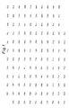
- glycoprotein provided by the present invention is a protein composed of 128 amino acids as shown in Figure 1, which is referred to hereinafter as "1F5 antigen".
- 1F5 antigen a protein composed of 128 amino acids as shown in Figure 1, which is referred to hereinafter as "1F5 antigen”.
- those which are altered by deletion or modification of some of the constituent amino acids, or by addition of amino acids, or other insofar as such alterations cause no impairment of the function of inhibiting complement-mediated cell membrane damage may be included as the glycoprotein of this invention.
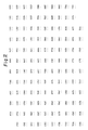
- the gene coding for the above 1F5 antigen may be exemplified by the one possessing the base sequence shown in Figure 2. In the Figure, only one strand of the base sequence is shown with the opposite complementary base sequence being omitted and expressed only as "---".
- DNA fragments of the gene coding for the protein which possesses the function of inhibiting complement-mediated cell membrane damage by the present invention may be obtained by, for example, the following method.
- human peripheral blood lymphocyte cDNA library In the screening for the gene coding for 1F5 antigen, human peripheral blood lymphocyte cDNA library, human T cell cDNA library, K562 cell cDNA library, human monocyte cDNA library, human placental cDNA library, etc. may be utilized, among which the placental cDNA library is particularly preferable. These libraries are purchased from Clonetech Inc.
- the cDNA library may also be prepared in a usual manner from any cell line expressing 1F5 antigen. In such case, MT-2 cDNA library and MOLT2 cDNA library are preferable.
- Escherichia coli is infected with ⁇ phage packaging such cDNA library, and cultured according to the method of Tomizawa et al. ("Experimental Techniques for Bacteriophages", Iwanami Shoten, Tokyo, pp. 99-174, 1970).
- the desired cDNA can be readily selected by screening resultant plaques the plaque hybridization technique ("Molecular Cloning", Cold Spring Harbor Laboratory, pp. 320-328, 1982) using as a probe an oligonucleotide having a base sequence deduced from the amino acid sequence of 1F5 antigen.
- a partial cDNA fragment of the gene which encodes 1F5 antigen may be prepared by polymerase chain reaction (PCR) and used as the probe (Science, 239 , 487-491, 1988).
- PCR is carried out with +strand DNA primer 5′ CA A/G TG C/T TA C/T AA C/T TG C/T CC 3′ (17 nucleotides) corresponding to 27-32 of the amino acid sequence in Figure 1 and -strand primer 5′ CA A/G TG C/T TC A/G AA C/T TTCCA 3′ (17 nucleotides) corresponding to 65-70 of the amino acid sequence in Figure 1, to prepare as the probe a 131 bp-oligonucleotide fragment shown in Figure 3 corresponding to 27-70 of the amino acid sequence in Figure 1.
- a synthetic oligonucleotide may also be prepared according to the DNA base sequence estimated from the amino acid sequence of 1F5 antigen, and used as the probe.
- the base sequence of the desired cDNA fragment may be determined by propagating the phage from a positive plaque on the above-mentioned screening as described by Tomizawa et al., by purifying DNA from the propagated phage according to the method of T. Maniatis et al. ("Molecular Cloning", Cold Spring Harbor Laboratory, 85, (1982)), then by cloning the DNA into a plasmid such as pUC18 or pUC19 after cleaving with a suitable restriction enzyme, e.g. Eco RI, and finally by subjecting it to the dideoxy method of Sanger et al. (Proc. Natl. Acad. Sci. USA, 74 , 5463 (1977)).
- a suitable restriction enzyme e.g. Eco RI
- cDNA fragment consists of a total of 387 base pairs (base sequence in Figure 2), which code for the protein composed of 128 amino acids (the 25 amino acids at N-terminal serve as a signal peptide) containing the partial amino acid sequence (26-70) of 1F5 antigen shown in Figure 1, and contains the entire length of the structural gene of 1F5 antigen.
- the cDNA fragment obtained as described above may be inserted as it is or after modification of its 5′-terminal into a known vector by a conventional method at the downstream of a promoter.
- the expression vector with the inserted cDNA may be then introduced into known host cells such as E. coli , yeasts or animal cells by a conventional method.
- the protein possessing the activity of inhibiting complement-mediated cell membrane damage may be expressed by the above cDNA in the host cells, isolated and purified there from by a conventional method.
- CNBr-activated Sepharose 4B (Pharmacia) was soaked in 1 mM hydrochloric acid for 15-30 minutes. The resulting gel was washed repeatedly by aspiration on a glass filter with subsequent addition of 200 ml of 1 mM hydrochloric acid per g of dry gel.
- the gel was then washed with coupling buffer (0.2M sodium hydrogencarbonate solution containing 0.5M sodium chloride, pH 8.5-8.7), 10 ml per g of dry gel.
- the washed gel was immediately transferred to a flask containing a 1F5 antibody solution in the coupling buffer to initiate a coupling reaction.
- the amount of 1F5 antibody in the reaction mixture was 5-10 mg per ml of swollen gel.
- the coupling reaction was carried out over-night at 4°C with constant and gentle stirring. After the reaction, the reaction mixture was transferred onto a glass funnel to recover unreacted antibody solution. The gel remaining on the funnel was then transferred to a flask containing 1 M ethanolamine solution and allowed to stand for 16 hours at 4°C.
- the resulting gel was transferred onto a glass funnel and aspirated. It was then washed by aspiration ten times with the coupling buffer, and subsequently ten times with 0.1 M acetate buffer (pH 4.0) containing 0.5 M sodium chloride. Finally, the gel was washed by aspiration three times using PBS to obtain 1F5 antibody affinity Sepharose.
- 300 - 400 ml of human blood was centrifuged at 500 ⁇ g for 15 minutes to separate red cells as a precipitate from a supernatant.
- PBS containing 10 mM EDTA and 1 mM PMSF were added and centrifuged at 400 - 500 ⁇ g for 15 minutes.
- the resulting precipitate was further washed with PBS containing 1 mM EDTA and 1 mM PMSF by centrifugation at 400 - 500 ⁇ g for 15 minutes.
- the supernatant was removed to obtain a precipitate. Such washing processes were repeated till the supernatant became clear.
- the washed erythrocytes obtained in (a) above were transferred to a flask containing 3 - 4 l of 10 mM Tris-HCl buffer (pH 7.5) containing 1 mM EDTA and 1 mM PMSF, hereinafter referred to as buffer A, and allowed to hemolyze with stirring overnight at 4°C.
- the resulting mixture was centrifuged at 25,000 ⁇ g for 30 minutes to collect an erythrocyte membrane fraction as a precipitate, which was resuspended in buffer A and centrifuged at 25,000 ⁇ g for 30 minutes. This process was repeated until a faintly pink precipitate of red cell membrane was obtained.
- the red cell membrane fraction obtained in (b) above was suspended in 500 ml of buffer A containing 1% n-octyl- ⁇ -D-glucopyranoside (Sigma; NOG) and stirred overnight at 4°C.
- the eluate was collected in 1-ml fractions and quickly neutralized by adding 1 M glycine-NaOH buffer (pH 9.5).
- the eluate fractions were monitored for protein spectrophotometrically at A280, and those positive for protein were pooled, concentrated by ultrafiltration (exclusion molecular weight; 10,000 or less), and dialyzed against PBS containing 0.1% CHAPS using the above dialysis membrane, to obtain 100 - 150 ⁇ g of the purified glycoprotein of the present invention.
- a control was prepared in the same manner as described above except that endoglycosidase F was not contained in above reaction.
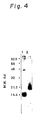
- the result demonstrated on SDS-PAGE showed that the endoglycosidase F-treated sample had migrated as a band shifting by about 0.5 Kd to the lower-molecular-weight side, compared with the control (Fig. 4).
- glycoprotein provided by the present invention contains N-glycoside type carbohydrate chain.
- MT2 cells a human T cell leukemia virus-1 (HTLV-1)-infected human T cell line, possessing the glycoprotein of this invention on their cell surface were used.
- MT2 cells were collected from an MT2 cell suspension in culture medium containing 1 ⁇ 106 cells by centrifugation (100 - 400 ⁇ g for 5 minutes). The cells were resuspended in PBS, gently stirred and centrifuged as above, and the resulting supernatant was discarded.
- HTLV-1 human T cell leukemia virus-1
- the pellet consisting of 1 ⁇ 106 MT2 cells was resupended in 150 ⁇ l of 10 mM Tris-HCl buffer (pH 7.5) containing 15 units of phosphatidyl-inositol-specific phospholipase C (PI-PLC; purified from B. thruringeinsis IAM 12077 according to the method described in J. Biochem. 93 , 1717 (1983)), 0.25 M sucrose, 10 mM EDTA and 0.1% bovine serum albumin, and allowed to stand for 90 minutes at 37°C with a gentle stirring.
- 10 mM Tris-HCl buffer pH 7.5
- PI-PLC phosphatidyl-inositol-specific phospholipase C
- reaction mixture was centrifuged with added 2 ml of PBS at 100 - 400 ⁇ g for 10 minutes, and the resulting supernatant was discarded. This process was further repeated twice.
- Ten ⁇ l of sheep IgG (Cappel.) (10 mg/ml) was added to the cell pellet and stirred. The cell suspension was then allowed to stand for 10 minutes at room temperature, followed by three cycles of washing with PBS by centrifugation as described above. To the resulting precipitate, 20 ⁇ l of 1F5 antibody (concentration, A280 0.1) was added and stirred. This cell suspension was left for 20 minutes at room temperature, and washed three times with PBS.
- the cell pellet was stirred with added 20 ⁇ l of FITC-labeled anti-mouse IgG (Cappel.), then allowed to stand for 20 minutes at room temperature, and washed three times with PBS by centrifugation.
- the cell pellet was dispersed in 200 ⁇ l of Sheath solution (Fujisawa Pharmaceutical Co., Ltd.), and the resulting cell suspension was filtered through a nylon mesh filter. To the filtrate, 500 ⁇ l of Sheath solution was added and the mixture was subjected to flow cytometry using a FACS analyzer .
- a control was prepared with MT2 cells by the same treatment as described above but omitting the use of PI-PLC.
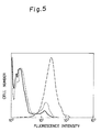
- the PI-PLC-treated MT2 cells were shown to be negative for the glycoprotein of the present invention by 95% of the control cells (Fig. 5).
- glycoprotein of the present invention possesses phosphatidylinositol via which it is bound to the cell membrane.
- the precipitate was resuspended in 2 ml of Mg-EGTA-GVB and centrifuged as above. The supernatant was discarded, and the precipitate was again resuspended in added 100 ⁇ l of Mg-EGTA-GVB and 50 ⁇ l of normal human serum, and gently stirred for 60 minutes at 37°C. To this mixture, 1.0 ml of Veronal buffer containing 40 mM EDTA and 0.1% geletin was added and centrifuged at 100 - 400 x g for 10 minutes. The resulting supernatant was spectrophotometrically analyzed at A414, to determine the percent hemolysis of guinea pig erythrocytes by human complement. Bovine serum albumin at the same concentration was used for control in place of the glycoprotein of the present invention.
- the guinea pig red cell hemolysis was inhibited by 90% in the presence of the glycoprotein of this invention added at a final concentration of 1 ⁇ g/ml.
- the results have indicated that the glycoprotein provided by the present invention is adsorbed onto the erythrocyte membrane and inhibits complement-mediated cell damage (Fig. 6).
- Isoelectric point Analysis was carried out by isoelectric focusing with Ampholyne (Pharmacia) pI 3.5 to 10 in one dimension and by subsequent SDS-PAGE (12.5%) under non-reductive condition in the second dimension and demonstrated the formation of bands at pI values of about 5.0 (main band) and about 6.5.
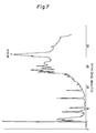
- the one eluted at about 36 minutes was dried in vacuo, then dissolved in 100 ⁇ l of Tris buffer (1M Tris-HCl buffer, pH 8.5, containing 5M guanidine hydrochloride), and reduced by the addition of 0.5 ⁇ l of 2-mercaptoethanol for 2 hours at 40°C in nitrogen gas atmosphere. Subsequently, the resulting mixture was incubated with 1 mg of monoiodoacetic acid in nitrogen gas atmosphere at room temperature for 1 hour with protection from light, to obtain a reductive carboxymethylated protein.
- Tris buffer 1M Tris-HCl buffer, pH 8.5, containing 5M guanidine hydrochloride
- the reductive carboxymethylated protein was subjected to reverse phase high performance liquid chromatography under the same conditions as above with a linear gradient elution of from 30% to 90% acetonitrile/isopropyl alcohol (3/7:v/v) mixture over 30 minutes. Detection made in the same manner as above disclosed approximately 3 protein fractions (Fig. 8).
- the one eluted at about 21 minutes was dried in vacuo, and dissolved in 60 ⁇ l of 50% trifluoroacetic acid.
- the solution was transferred to a polyprene-coated glass filter and subjected to Edman degradation on an Applied Biosystems Model 470A sequencer.
- PTH-amino acid was identified chromatographically on an "MCI gel ODS IHU" column (0.46 x 15 cm) (Mitsubishi Kasei Corporation) with single solvent elution of acetate buffer (10 mM acetate buffer, pH 4.7, containing 0.01% SDS and 38% acetonitrile) at a flow rate of 1.2 ml/min. at 43°C. Detection of PTH-amino acids was carried out by measurement of the absorbance at 269 nm.
- KLH keyhole limpet hemocyanin
- Equal volumes of the two KLH-peptide conjugate preparations obtained above were combined and adjusted to a total protein concentration of about 2 mg/ml using PBS. A portion of this mixture was emulsified with an equal volume of Freund's complete adjuvant (Difco). Rabbits were immunized with the resulting emulsion by subcutaneous injections of 0.1-ml doses at four sites in the dorsal and into each footpad. The animals received additional immunizing doses subcutaneously at four sites in the dorsal 4 weeks afterwards. At 2 weeks after the second injections, 1 ml of the emulsified KLH. peptide conjugate (1 mg/ml) was injected subcutaneously at six sites in the dorsal for further immunization.
- Freund's complete adjuvant Freund's complete adjuvant
- the animals were totally bled 2 - 3 weeks after the last injections and their antisera were obtained.
- the antisera obtained were mixed with solid ammonium sulfate to 40% saturation while stirring, and the resultant mixture was further stirred for 2 - 3 hours at 4°C.
- the mixture was then centrifuged at 10,000 ⁇ g for 15 minutes, and the resulting precipitate was dissolved in a small volume of PBS.
- This solution was applied to a Protein A Cellurofine (Seikagaku Kogyo) column equilibrated with PBS, washed with a sufficient amount of PBS, and eluted with 0.1 M glycine-HCl buffer (pH 2.8).
- Eluted protein fractions were collected, immediately neutralized by the addition of 1 M Tris solution, and concentrated by ultrafiltration (exclusion molecular weight: 30,000). The concentrated antibody solution was dialyzed against PBS at 4°C overnight to obtain a purified polyclonal antibody.
- An affinity Sepharose gel was prepared in the same manner and with the same reagents as in (2)-(d) of Example 1, except for using the polyclonal antibody obtained in (2) above in place of 1F5 antibody.
- the solubilized membrane protein solution obtained in (2)-(a) to (c) of Example 1 was purified by affinity chromatography on a column of this polyclonal antibody Sepharose gel in the same manner as in Example 1.
- the glycoprotein of this invention having properties identical with those of the preparation in Example 1 was obtained.
- the K562 cell cDNA which serves as a substrate may be prepared in an usual manner. That is, RNA possessing poly (A) was prepared from 1 ⁇ 109 K562 cells by the guanidine thiocyanate-cesium chloride method DNA, 2 , 329 (1983)) as follows. A starting material obtained from 1 ⁇ 109 K562 cells was solubilized in 40 ml of a solution consisting of 5M guanidine thiocyanate, 10 mM EDTA, 50 mM Tris-HCl (pH 7.0) and 8% (v/v) ⁇ - mercaptoethanol.
- RNA precipitate was dissolved in 10 ml of a solution consisting of 0.1% sodium laurylsulfate, 1 mM EDTA and 10 mM Tris-HCl (pH 7.5), then extracted with a phenol-chloroform mixture, and precipitated in ethanol. About 3.95 mg of the resulting RNA precipitate was dissolved in 1 ml of a solution consisting of 10 mM Tris-HCl (pH 8.0) and 1 mM EDTA, and incubated at 65°C for 5 minutes, followed by addition of 0.1 ml of 5M sodium chloride.
- the resultant mixture was chromatographed on an Oligo[dT] Cellulose Column (P-L Biochemical) (column volume, 0.5 ml).
- the adsorbed mRNA possessing poly (A) was eluted with an eluant consisting of 10 mM Tris-HCl (pH 7.5) and 1 mM EDTA, and thereby approximately 100 ⁇ g of mRNA possessing poly (A) was obtained.
- T4 DNA ligase buffer 0.1M Tris-HCl (pH 7.5), 0.1M DTT and 60 mM magnesium chloride
- the double strand DNA obtained as above was extracted with an equal volume of water-saturated phenol. After removal of phenol from the aqueous layer with ether, it was precipitated in ethanol.
- the precipitate obtained was dissolved in 50 ⁇ l of water, and 10 ⁇ l of 10 ⁇ conc.
- T4 DNA polymerase buffer (0.33M Tris-acetic acid buffer (pH 7.9), 0.66 M potassium acetate, 0.1M magnesium acetate and 5 mM DTT), 10 ⁇ l of 10 mM 4dNTP and 6 units of T4 DNA polymerase were added to this solution.
- the resulting mixture was made up to a volume of 100 ⁇ l, and incubated for 1 hour at 37°C to obtain a double stranded DNA possessing blunt ends.
- This mixture was subjected to extraction with phenol as above, and after subsequent deproteinization, precipitated in ethanol to purify the DNA. The purified DNA was then allowed to dry in air.
- cDNA substrate Two ⁇ g of cDNA thus obtained was dissolved in 1 ml of H2O to obtain a cDNA substrate.
- PCR was carried out with a Perkin Elmer Cetus DNA Thermal Cyster using Gene AmpTM DNA Amplification Reagent Kit (Takara Shuzo) according to the directions provided by the manufacturer. That is, to 10 ⁇ l of the cDNA substrate (equivalent to 20 ng of the cDNA), 10 ⁇ l of 10 x conc.
- reaction buffer 500 mM potassium chloride, 100 mM Tris-HCl (pH 8.3), 15 mM magnesium chloride and 0.1% (w/v) gelatin
- 16 ⁇ l of 1.25 mM 4d NTP 5 ⁇ l of +strand DNA primer 5′ CA A/G TC C/T TA C/T AA C/T TG C/T CC 3′ (17 nucleotide) corresponding to the amino acid sequence 27-32 in Figure 1 as 20 ⁇ M primer #1
- 5 ⁇ l of 20 ⁇ M -strand DNA primer 5′ CA A/G TG C/T TC A/G AA C/T TTCCA 3′ (17 nucleotide) corresponding to the amino acid sequence 65-70 in Figure 1 as (20 ⁇ M) primer #2 and 0.5 ⁇ l of Taq DNA polymerase were added.
- This mixture was made up to a volume of 100 ⁇ l, pretreated for 10 minutes at 94°C, and subjected to reaction by 35 cycles of successive incubation for 1 minute at 94°C (denaturation step), for 2 minutes at 42°C (annealing step) and for 1.5 minutes at 72°C (elongation step). The reaction mixture was further incubated for 7 minutes at 72°C, and then precipitated in ethanol. The precipitate was dissolved in 50 ⁇ l of water.
- T4 DNA polymerase buffer (0.33M Tris-acetic acid (pH 7.9), 0.66M potassium acetate, 0.1M magnesium acetate and 5 mM DTT), 10 ⁇ l of 10 mM 4d NTP and 6 units of T4 DNA polymerase were added.
- the resultant mixture was made up to a volume of 100 ⁇ l, and incubated for 1 hour at 37°C to obtain double stranded blunt ends.
- the band of about 150 bp was cut out of the polyacrylamide gel strip and cDNA contained therein was inserted into Sma I site of cloning vector pUC12 by the dideoxy method, and its base sequence was determined using commercially available primers 5′ CAGGAAACAGCATGAC 3′ and 5′ AGTCACGACGTTGTA 3′ .
- the base sequence of the said PCR fragment was found to be as shown in Figure 3, and the amino acid sequence estimated by the 131-bp fragment corresponded to the sequence 27-70 in Figure 1 and entirely coincided with the amino acid sequence determined from the purified 1F5 antigen protein.
- the inserted fragment was cleaved at Kpn I and BamH I out of this recombinant plasmid purified and cut out of the acrylamide gel, and radiolabeled by the multiprimer DNA labeling technique (Anal. Biochem., 132 , 6 (1983)) to obtain a probe for cDNA library screening. That is, 100 ng of this DNA fragment was dissolved in water sufficient to make 32 ⁇ l, denatured by heating for 2 minutes at 95°C, and immediately cooled on an ice bath.
- OLB buffer [0.25M Tris-HCl (pH 8.0), 0.025M magnesium chloride, 0.4% ⁇ -mercaptoethanol, 0.1 mM dATP, 0.1 mM dTTP, 0.1 mM dGTP, 1M HEPES (pH 6.6), 10 ⁇ l of oligo primer p dN6 (P-L Biochemical, 15 units)] 2 ⁇ l of 10 mg/ml bovine serum albumin, 5 ⁇ l of ⁇ -32Pd CTP 3000 ci/mmol) and 2 units of DNA polymerase large (Klenow) fragment was added and the mixture was incubated for 30 minutes at 37°. This reaction mixture was used as a probe.
- E. coli strain Y1090 infected with ⁇ gt11 (Proc. Natl. Acad, Sci. USA, 80 , 1194, (1983)) was inoculated together with a soft agar overlay maintained at 42°C in Petri dishes, and incubated at 42°C for 8 hours. The dishes were then cooled for 30 minutes at 4°C, and a nitrocellulose filter (S&S Inc.; BA-83; pore size, 0.2 ⁇ m) was placed on each dish and allowed to stand for 2 minutes. Subsequently, another filter was placed on that filter and allowed to stand for 5 minutes. Both nitrocellulose filters were immersed twice for 20 seconds each in a solution of 0.1N sodium hydroxide and 1.5M sodium chloride.
- the filters were then neutralized by being immersed twice for 20 seconds each in a solution of 2 ⁇ SSCP (30 mM sodium citrate, 240 mM sodium chloride, 26 mM monopotassium phosphate and 2 mM EDTA, pH 7.2) and 0.2M Tris-HCl (pH 7.4), and then dried in air for 2 hours at 80°C. Subsequently, they were soaked in 2 ⁇ SSC (450 mM sodium chloride and 45 mM sodium citrate) for 30 minutes at 65°C.
- 2 ⁇ SSCP 30 mM sodium citrate, 240 mM sodium chloride, 26 mM monopotassium phosphate and 2 mM EDTA, pH 7.2
- Tris-HCl pH 7.4
- the bacterial cells were evenly removed with a sponge and the resulting filters were immersed in a solution consisting of 3 ⁇ SSC and 10 ⁇ Denhardt's mixture (0.2% Ficoll, 0.2% polyvinylpyrrolidone and 0.2% BSA) for 1 hour at 65°C. They were then immersed in a hybridization solution [1 ⁇ Solution A (50 mM Tris-HCl pH 7.8), 10 mM EDTA, 1M sodium chloride and 10 ⁇ Denhardt's mixture), 0.1% SDS and herring testis DNA at 250 ⁇ g/ml] for 30 minutes at 65°C.
- a hybridization solution [1 ⁇ Solution A (50 mM Tris-HCl pH 7.8), 10 mM EDTA, 1M sodium chloride and 10 ⁇ Denhardt's mixture), 0.1% SDS and herring testis DNA at 250 ⁇ g/ml] for 30 minutes at 65°C.
- the filters thus treated were placed in a polyvinyl bag with the hybridization solution containing the PCR probe at 0.1 ng/ml was added, and the bag was sealed.
- the filters in the bag were subjected to hybridization by incubation for 15 hours at 50°C. They were then washed twice with 2 ⁇ SSC-0.1% SDS mixture for 5 minutes each at room temperature, followed by washing for 15 minutes at 60°C, and they were subsequently washed twice with 0.1 ⁇ SSC-0.1% SDS mixture for 15 minutes each at 60°C.
- the filters were wrapped up in polyvinylidene chloride film (Saran Wrap® ; Asahi Chemical Co., Ltd.) and subjected to autoradiography.
- E. coli strain Y1090 was grown overnight in 10 ml of NZ medium prepared by dissolving 10 g of NZ amine, 5 g of sodium chloride and 5 mM of magnesium chloride in 1000 ml of water and by adjusting the solution to pH 7.2.
- One ml of the overnight culture was infected with the phage at a multiplicity of infection (m.o.i.) of 0.1 and incubated for 10 minutes at 37°C, then transferred to a flask containing 1 liter of NZ medium, and incubated for 7-8 hours at 37°C with a constant shaking until the bacteria became lyzed.
- m.o.i. multiplicity of infection
- the reaction mixture was stirred with added 20 ml of chloroform, followed by removal of the chloroform from an aqueous layer by dissolving polyethyleneglycol therein.
- the aqueous layer remained was ultracentrifuged at 28,000 r.p.m. for 60 minutes to collect the pellet of phage particles.
- the fraction was dialyzed against TM buffer overnight, followed by incubation for 1 hour at 37°C with added proteinase K at 100 ⁇ g/ml.
- the mixture was subjected to a gentle phenol extraction by adding an equal volume of water-saturated phenol. It was then centrifuged at 6,500 r.p.m. for 10 minutes, and the aqueous layer was transferred to a dialysis tube and dialyzed against water at 4°C overnight. Approximately 5 mg of DNA was thus obtained.
Landscapes
- Chemical & Material Sciences (AREA)
- Health & Medical Sciences (AREA)
- Organic Chemistry (AREA)
- Life Sciences & Earth Sciences (AREA)
- Medicinal Chemistry (AREA)
- Biophysics (AREA)
- General Health & Medical Sciences (AREA)
- Genetics & Genomics (AREA)
- Biochemistry (AREA)
- Molecular Biology (AREA)
- Proteomics, Peptides & Aminoacids (AREA)
- Gastroenterology & Hepatology (AREA)
- Toxicology (AREA)
- Zoology (AREA)
- Peptides Or Proteins (AREA)
- Preparation Of Compounds By Using Micro-Organisms (AREA)
- Medicines That Contain Protein Lipid Enzymes And Other Medicines (AREA)
Applications Claiming Priority (4)
| Application Number | Priority Date | Filing Date | Title |
|---|---|---|---|
| JP172187/88 | 1988-07-11 | ||
| JP63172187A JP2587863B2 (ja) | 1988-07-11 | 1988-07-11 | 新規な糖蛋白質 |
| JP1129944A JP2847753B2 (ja) | 1989-05-23 | 1989-05-23 | 新規な蛋白質及びそれをコードする遺伝子 |
| JP129944/89 | 1989-05-23 |
Publications (2)
| Publication Number | Publication Date |
|---|---|
| EP0351313A2 true EP0351313A2 (fr) | 1990-01-17 |
| EP0351313A3 EP0351313A3 (fr) | 1991-09-11 |
Family
ID=26465197
Family Applications (1)
| Application Number | Title | Priority Date | Filing Date |
|---|---|---|---|
| EP19890401996 Withdrawn EP0351313A3 (fr) | 1988-07-11 | 1989-07-11 | Glycoprotéine et son gène codant |
Country Status (3)
| Country | Link |
|---|---|
| US (2) | US5179198A (fr) |
| EP (1) | EP0351313A3 (fr) |
| HU (1) | HUT55412A (fr) |
Cited By (12)
| Publication number | Priority date | Publication date | Assignee | Title |
|---|---|---|---|---|
| EP0394035A3 (fr) * | 1989-04-21 | 1991-08-21 | Yamanouchi Pharmaceutical Co. Ltd. | Gènes codant une protéine avec activité MACIF humaine, vecteurs d'expression contenant lesdits gènes, cellules transformantes et protéines avec activité MACIF humaine |
| AU647586B2 (en) * | 1990-12-17 | 1994-03-24 | Texas Tech University Health Sciences Center | Polypeptide fraction affording protection to cells against mechanical damage and assay |
| DE19652815A1 (de) * | 1996-12-18 | 1998-06-25 | Karlsruhe Forschzent | Mittel zur Identifizierung und Therapie metastasierender Tumore |
| WO1998033817A1 (fr) * | 1997-01-31 | 1998-08-06 | Incyte Pharmaceuticals, Inc. | Proteine membranaire humaine |
| US7166568B1 (en) | 1998-02-09 | 2007-01-23 | Oklahoma Medical Research Foundation | Compositions and methods to inhibit formation of the C5b-9 complex of complement |
| US7388081B2 (en) | 1998-12-09 | 2008-06-17 | Dfb Biotech, Inc. | Method for manufacturing glycoproteins having human-type glycosylation |
| US7601891B2 (en) | 2002-03-19 | 2009-10-13 | Plant Research International B.V. | Optimizing glycan processing plants |
| US7781647B2 (en) | 1999-10-26 | 2010-08-24 | Stichting Dienst Landbouwkundig Onderzoek | Mammalian-type glycosylation in transgenic plants expressing mammalian β1,4-galactosyltransferase |
| US7897842B2 (en) | 2002-03-19 | 2011-03-01 | Plant Research International B.V. | GnTIII expression in plants |
| US8106169B2 (en) | 2002-11-27 | 2012-01-31 | Phyton Holdings, Llc | Plant production of immunoglobulins with reduced fucosylation |
| US8309795B2 (en) | 2001-01-19 | 2012-11-13 | Phyton Holdings, Llc | Method for secretory production of glycoprotein having human-type sugar chain using plant cell |
| US8829276B2 (en) | 2007-04-17 | 2014-09-09 | Stichting Dienst Landbouwkundig Onderzoek | Mammalian-type glycosylation in plants by expression of non-mammalian glycosyltransferases |
Families Citing this family (6)
| Publication number | Priority date | Publication date | Assignee | Title |
|---|---|---|---|---|
| US5573940A (en) * | 1989-06-12 | 1996-11-12 | Oklahoma Medical Research Foundation | Cells expressing high levels of CD59 |
| WO1991002002A1 (fr) * | 1989-07-21 | 1991-02-21 | Washington University | Proteine de cofacteur de membrane (pcm) humaine produite par recombinaison |
| US5708141A (en) * | 1992-05-11 | 1998-01-13 | Corvas International, Inc. | Neutrophil inhibitors |
| JPH0954091A (ja) * | 1995-08-09 | 1997-02-25 | Nippon Zoki Pharmaceut Co Ltd | 抗赤血球抗体の測定法 |
| JP2001245698A (ja) * | 1999-11-22 | 2001-09-11 | Xiao Bing Wang | 核酸検出法 |
| US6824980B2 (en) * | 2000-06-08 | 2004-11-30 | Xiao Bing Wang | Isometric primer extension method and kit for detection and quantification of specific nucleic acid |
Family Cites Families (6)
| Publication number | Priority date | Publication date | Assignee | Title |
|---|---|---|---|---|
| US4132769A (en) * | 1974-10-30 | 1979-01-02 | Osther Kurt B | Cancer antigen, cancer therapy, and cancer diagnosis |
| IN150740B (fr) * | 1978-11-24 | 1982-12-04 | Hoffmann La Roche | |
| US4394443A (en) * | 1980-12-18 | 1983-07-19 | Yale University | Method for cloning genes |
| US4675285A (en) * | 1984-09-19 | 1987-06-23 | Genetics Institute, Inc. | Method for identification and isolation of DNA encoding a desired protein |
| US4800159A (en) * | 1986-02-07 | 1989-01-24 | Cetus Corporation | Process for amplifying, detecting, and/or cloning nucleic acid sequences |
| US5135916A (en) * | 1989-06-12 | 1992-08-04 | Oklahoma Medical Research Foundation | Inhibition of complement mediated inflammatory response |
-
1989
- 1989-07-07 US US07/376,828 patent/US5179198A/en not_active Expired - Fee Related
- 1989-07-10 HU HU893452A patent/HUT55412A/hu unknown
- 1989-07-11 EP EP19890401996 patent/EP0351313A3/fr not_active Withdrawn
-
1991
- 1991-08-01 US US07/739,211 patent/US5521296A/en not_active Expired - Fee Related
Non-Patent Citations (3)
| Title |
|---|
| BIOCHEMISTRY, vol. 24, April 1985, pages 2368-2374, American Chemical Society, Washington, US; B. DAHLB[CK et al.: "Characterization of human S protein, an inhibitor of the membrane attack complex of complement. Demonstration of a free reactive thiol group" * |
| PROC. NATL. ACAD. SCI. USA, vol. 81, December 1984, pages 7298-7302, Washington, US; R.G. DiSCIPIO et al.: "Nucleotide sequence of cDNA and derived amino acid sequence of human complement component C9" * |
| PROC. NATL. ACAD. SCI. USA, vol. 83, September 1986, pages 6975-6979, Washington, US; L.S. ZALMAN et al.: "Isolation of a human erythrocyte membrane protein capable of inhibiting expression of homologous complement transmembrane channels" * |
Cited By (25)
| Publication number | Priority date | Publication date | Assignee | Title |
|---|---|---|---|---|
| EP0394035A3 (fr) * | 1989-04-21 | 1991-08-21 | Yamanouchi Pharmaceutical Co. Ltd. | Gènes codant une protéine avec activité MACIF humaine, vecteurs d'expression contenant lesdits gènes, cellules transformantes et protéines avec activité MACIF humaine |
| EP0672683A1 (fr) * | 1989-04-21 | 1995-09-20 | Yamanouchi Pharmaceutical Co. Ltd. | Protéine MACIF, gènes codant pour cette protéine, vecteurs d'expression contenant lesdits gènes, cellules transformées contenant ces protéines |
| AU647586B2 (en) * | 1990-12-17 | 1994-03-24 | Texas Tech University Health Sciences Center | Polypeptide fraction affording protection to cells against mechanical damage and assay |
| DE19652815A1 (de) * | 1996-12-18 | 1998-06-25 | Karlsruhe Forschzent | Mittel zur Identifizierung und Therapie metastasierender Tumore |
| DE19652815C2 (de) * | 1996-12-18 | 1999-11-11 | Karlsruhe Forschzent | Mittel zur Identifizierung und Therapie metastasierender Tumore |
| WO1998033817A1 (fr) * | 1997-01-31 | 1998-08-06 | Incyte Pharmaceuticals, Inc. | Proteine membranaire humaine |
| US7166568B1 (en) | 1998-02-09 | 2007-01-23 | Oklahoma Medical Research Foundation | Compositions and methods to inhibit formation of the C5b-9 complex of complement |
| US7388081B2 (en) | 1998-12-09 | 2008-06-17 | Dfb Biotech, Inc. | Method for manufacturing glycoproteins having human-type glycosylation |
| US8853370B2 (en) | 1998-12-09 | 2014-10-07 | Phyton Holdings, Llc | Plant-produced glycoprotein comprising human-type sugar chain |
| US8241909B2 (en) | 1998-12-09 | 2012-08-14 | Phyton Holdings, Llc | Method for manufacturing glycoproteins having human-type glycosylation |
| US8907163B2 (en) | 1999-10-26 | 2014-12-09 | Stichting Dienst Landbouwkundig Onderzoek | Transgenic plants expressing galactosyltransferase and sialyl transferase |
| US7781647B2 (en) | 1999-10-26 | 2010-08-24 | Stichting Dienst Landbouwkundig Onderzoek | Mammalian-type glycosylation in transgenic plants expressing mammalian β1,4-galactosyltransferase |
| US8193415B2 (en) | 1999-10-26 | 2012-06-05 | Stichting Dienst Landbouwkundig Onderzock | Plant expressing mammalian β1,4-galactosyltransferase and β1,3-glucuronyltransferase |
| US8309795B2 (en) | 2001-01-19 | 2012-11-13 | Phyton Holdings, Llc | Method for secretory production of glycoprotein having human-type sugar chain using plant cell |
| US8735656B2 (en) | 2001-01-19 | 2014-05-27 | Phyton Holdings, Llc | Method of expressing galactosyltransferase and inhibiting xylosyltransferase or fucosyltransferase in a transgenic plant cell for secretory production of glycoproteins having human-type sugar chains |
| US9574218B2 (en) | 2001-01-19 | 2017-02-21 | Phyton Holdings, Llc | Method of co-expressing galactosyltransferase and a glycoprotein in a transgenic plant cell and sialylating the glycoprotein for production of glycoprotein having human-type sugar chain |
| US8058508B2 (en) | 2002-03-19 | 2011-11-15 | Stichting Dienst Landbouwkundig Onderzoek | Optimizing glycan processing in plants |
| US9255277B2 (en) | 2002-03-19 | 2016-02-09 | Stichting Dienst Landbouwkundig Onderzoek | GNTIII expression in plants |
| US8492613B2 (en) | 2002-03-19 | 2013-07-23 | Stichting Dienst Landbouwkundig Onderzoek | GNTIII expression in plants |
| US7897842B2 (en) | 2002-03-19 | 2011-03-01 | Plant Research International B.V. | GnTIII expression in plants |
| US7601891B2 (en) | 2002-03-19 | 2009-10-13 | Plant Research International B.V. | Optimizing glycan processing plants |
| US8927810B2 (en) | 2002-03-19 | 2015-01-06 | Stichting Dienst Landbouwkundig Onderzoek | Optimizing glycan processing in plants |
| US8106169B2 (en) | 2002-11-27 | 2012-01-31 | Phyton Holdings, Llc | Plant production of immunoglobulins with reduced fucosylation |
| US8829276B2 (en) | 2007-04-17 | 2014-09-09 | Stichting Dienst Landbouwkundig Onderzoek | Mammalian-type glycosylation in plants by expression of non-mammalian glycosyltransferases |
| US9745594B2 (en) | 2007-04-17 | 2017-08-29 | Stichting Dienst Landbouwkundig Onderzoek | Mammalian-type glycosylation in plants by expression of a zebrafish glycosyltransferase |
Also Published As
| Publication number | Publication date |
|---|---|
| US5179198A (en) | 1993-01-12 |
| HUT55412A (en) | 1991-05-28 |
| US5521296A (en) | 1996-05-28 |
| EP0351313A3 (fr) | 1991-09-11 |
Similar Documents
| Publication | Publication Date | Title |
|---|---|---|
| US5179198A (en) | Glycoprotein and gene coding therefor | |
| Xia et al. | Characterization of the CAMPATH‐1 (CDw52) antigen: biochemical analysis and cDNA cloning reveal an unusually small peptide backbone | |
| Sewell et al. | The murine homologue of the T lymphocyte CD2 antigen: molecular cloning, chromosome assignment and cell surface expression | |
| JPH04501502A (ja) | ヒトC3b/C4bレセプター (CR1) | |
| KR940000542B1 (ko) | 재조합 dna 기술에 의해 제조된 생리적 활성물질의 정제방법 | |
| Reiser et al. | Cloning and expression of a cDNA for the T-cell-activating protein TAP. | |
| Carne et al. | Amino acid sequences of transport peptides associated with canine exocrine pancreatic proteins. | |
| EP0580672B1 (fr) | ANTIGENE SYNTHETIQUE DE PEPTIDE CDw52(CAMPATH-1) | |
| Hoijer et al. | Purification and characterization of N-acetylmuramyl-L-alanine amidase from human plasma using monoclonal antibodies | |
| KR100465382B1 (ko) | 말라세지아에서 유래된 항원성 단백질 | |
| JP3497504B2 (ja) | 細胞増殖阻害剤 | |
| US5578714A (en) | DNA encoding Duffy 9pd protein | |
| WO1990002207A1 (fr) | Antigene clone de la nephrite | |
| EP0563627A2 (fr) | Méthode antitumorale et agent antitumoral | |
| JP3231262B2 (ja) | ヒトTh1特異的タンパク質及びこれをコードする遺伝子、並びにこれに関連する形質転換体、組換えベクター及び抗体 | |
| US5434247A (en) | Peptides for inducing monocyte cytotoxicity in diagnostics | |
| CA2025577A1 (fr) | Proteines anticoagulantes et antihelminthiques et methode de production et d'utilisation | |
| WO1986007062A1 (fr) | Anticorps monoclonal du facteur d'acceleration de la decomposition (fad), son procede de production et d'utilisation | |
| Matsumoto et al. | Purification and characterization of a human membrane protein that activates the alternative complement pathway and allows the deposition of homologous complement C3. | |
| Higashino et al. | Purification and some properties of the fast migrating alkaline phosphatase in FL-amnion cells (the Kasahara isoenzyme) and its cDNA cloning | |
| JPH06509227A (ja) | リンパ球仲介細胞障害の活性を持つタンパク質をコードするdna | |
| JP2847753B2 (ja) | 新規な蛋白質及びそれをコードする遺伝子 | |
| JP3983246B2 (ja) | マラセチア由来の抗原性蛋白質 | |
| AU697868C (en) | The cloning of Duffy blood group antigen | |
| JP2587863B2 (ja) | 新規な糖蛋白質 |
Legal Events
| Date | Code | Title | Description |
|---|---|---|---|
| PUAI | Public reference made under article 153(3) epc to a published international application that has entered the european phase |
Free format text: ORIGINAL CODE: 0009012 |
|
| AK | Designated contracting states |
Kind code of ref document: A2 Designated state(s): BE CH DE FR GB IT LI NL SE |
|
| 17P | Request for examination filed |
Effective date: 19901227 |
|
| PUAL | Search report despatched |
Free format text: ORIGINAL CODE: 0009013 |
|
| RHK1 | Main classification (correction) |
Ipc: C07K 15/00 |
|
| AK | Designated contracting states |
Kind code of ref document: A3 Designated state(s): BE CH DE FR GB IT LI NL SE |
|
| 17Q | First examination report despatched |
Effective date: 19930921 |
|
| RAP1 | Party data changed (applicant data changed or rights of an application transferred) |
Owner name: MITSUBISHI CHEMICAL CORPORATION Owner name: OKADA, HIDECHIKA |
|
| STAA | Information on the status of an ep patent application or granted ep patent |
Free format text: STATUS: THE APPLICATION IS DEEMED TO BE WITHDRAWN |
|
| 18D | Application deemed to be withdrawn |
Effective date: 19980218 |