EP0355845A2 - Procédé de fabrication de protéoliposomes et procédé pour la fabrication de protéoliposomes géants - Google Patents
Procédé de fabrication de protéoliposomes et procédé pour la fabrication de protéoliposomes géants Download PDFInfo
- Publication number
- EP0355845A2 EP0355845A2 EP89115717A EP89115717A EP0355845A2 EP 0355845 A2 EP0355845 A2 EP 0355845A2 EP 89115717 A EP89115717 A EP 89115717A EP 89115717 A EP89115717 A EP 89115717A EP 0355845 A2 EP0355845 A2 EP 0355845A2
- Authority
- EP
- European Patent Office
- Prior art keywords
- proteoliposome
- carrier
- liposome
- membrane protein
- membrane
- Prior art date
- Legal status (The legal status is an assumption and is not a legal conclusion. Google has not performed a legal analysis and makes no representation as to the accuracy of the status listed.)
- Granted
Links
- 238000000034 method Methods 0.000 title claims abstract description 114
- 239000003725 proteoliposome Substances 0.000 title claims abstract description 106
- 108010052285 Membrane Proteins Proteins 0.000 claims abstract description 102
- 102000018697 Membrane Proteins Human genes 0.000 claims abstract description 97
- 239000002502 liposome Substances 0.000 claims abstract description 83
- 150000002632 lipids Chemical class 0.000 claims abstract description 58
- 239000012266 salt solution Substances 0.000 claims abstract description 53
- 230000008014 freezing Effects 0.000 claims abstract description 27
- 238000007710 freezing Methods 0.000 claims abstract description 27
- 238000010257 thawing Methods 0.000 claims abstract description 27
- 229910052783 alkali metal Inorganic materials 0.000 claims abstract description 25
- 150000001340 alkali metals Chemical class 0.000 claims abstract description 23
- 239000007853 buffer solution Substances 0.000 claims abstract description 13
- 230000003204 osmotic effect Effects 0.000 claims abstract description 13
- 229910052751 metal Inorganic materials 0.000 abstract description 2
- 239000002184 metal Substances 0.000 abstract description 2
- 239000012528 membrane Substances 0.000 description 65
- 102000004169 proteins and genes Human genes 0.000 description 47
- 108090000623 proteins and genes Proteins 0.000 description 47
- FAPWRFPIFSIZLT-UHFFFAOYSA-M Sodium chloride Chemical compound [Na+].[Cl-] FAPWRFPIFSIZLT-UHFFFAOYSA-M 0.000 description 46
- 108010082845 Bacteriorhodopsins Proteins 0.000 description 43
- 239000000243 solution Substances 0.000 description 33
- 239000006185 dispersion Substances 0.000 description 29
- 239000007788 liquid Substances 0.000 description 28
- 238000011282 treatment Methods 0.000 description 27
- 108010030416 proteoliposomes Proteins 0.000 description 25
- 239000011780 sodium chloride Substances 0.000 description 23
- HEMHJVSKTPXQMS-UHFFFAOYSA-M sodium hydroxide Inorganic materials [OH-].[Na+] HEMHJVSKTPXQMS-UHFFFAOYSA-M 0.000 description 22
- 238000002360 preparation method Methods 0.000 description 21
- 125000003178 carboxy group Chemical group [H]OC(*)=O 0.000 description 19
- 239000000725 suspension Substances 0.000 description 19
- NCYCYZXNIZJOKI-IOUUIBBYSA-N 11-cis-retinal Chemical compound O=C/C=C(\C)/C=C\C=C(/C)\C=C\C1=C(C)CCCC1(C)C NCYCYZXNIZJOKI-IOUUIBBYSA-N 0.000 description 18
- 102000004330 Rhodopsin Human genes 0.000 description 18
- 108090000820 Rhodopsin Proteins 0.000 description 18
- 238000000502 dialysis Methods 0.000 description 16
- 230000015572 biosynthetic process Effects 0.000 description 15
- 239000000499 gel Substances 0.000 description 15
- 239000000126 substance Substances 0.000 description 14
- WCUXLLCKKVVCTQ-UHFFFAOYSA-M Potassium chloride Chemical compound [Cl-].[K+] WCUXLLCKKVVCTQ-UHFFFAOYSA-M 0.000 description 12
- 238000010348 incorporation Methods 0.000 description 12
- IJGRMHOSHXDMSA-UHFFFAOYSA-N Atomic nitrogen Chemical compound N#N IJGRMHOSHXDMSA-UHFFFAOYSA-N 0.000 description 10
- 239000000232 Lipid Bilayer Substances 0.000 description 9
- ATDGTVJJHBUTRL-UHFFFAOYSA-N cyanogen bromide Chemical compound BrC#N ATDGTVJJHBUTRL-UHFFFAOYSA-N 0.000 description 9
- 239000000463 material Substances 0.000 description 9
- 238000000527 sonication Methods 0.000 description 9
- 230000008569 process Effects 0.000 description 8
- 238000012360 testing method Methods 0.000 description 8
- 230000008859 change Effects 0.000 description 7
- 238000004587 chromatography analysis Methods 0.000 description 7
- 239000003599 detergent Substances 0.000 description 7
- 239000012634 fragment Substances 0.000 description 7
- 239000002245 particle Substances 0.000 description 7
- HEDRZPFGACZZDS-UHFFFAOYSA-N Chloroform Chemical compound ClC(Cl)Cl HEDRZPFGACZZDS-UHFFFAOYSA-N 0.000 description 6
- RBHJBMIOOPYDBQ-UHFFFAOYSA-N carbon dioxide;propan-2-one Chemical compound O=C=O.CC(C)=O RBHJBMIOOPYDBQ-UHFFFAOYSA-N 0.000 description 6
- 239000000203 mixture Substances 0.000 description 6
- 239000001103 potassium chloride Substances 0.000 description 6
- 235000011164 potassium chloride Nutrition 0.000 description 6
- 108090000765 processed proteins & peptides Proteins 0.000 description 6
- 239000000047 product Substances 0.000 description 6
- 210000004676 purple membrane Anatomy 0.000 description 6
- QGZKDVFQNNGYKY-UHFFFAOYSA-N Ammonia Chemical compound N QGZKDVFQNNGYKY-UHFFFAOYSA-N 0.000 description 5
- UXVMQQNJUSDDNG-UHFFFAOYSA-L Calcium chloride Chemical compound [Cl-].[Cl-].[Ca+2] UXVMQQNJUSDDNG-UHFFFAOYSA-L 0.000 description 5
- 108090001090 Lectins Proteins 0.000 description 5
- 102000004856 Lectins Human genes 0.000 description 5
- 229910021380 Manganese Chloride Inorganic materials 0.000 description 5
- GLFNIEUTAYBVOC-UHFFFAOYSA-L Manganese chloride Chemical compound Cl[Mn]Cl GLFNIEUTAYBVOC-UHFFFAOYSA-L 0.000 description 5
- KWYUFKZDYYNOTN-UHFFFAOYSA-M Potassium hydroxide Chemical compound [OH-].[K+] KWYUFKZDYYNOTN-UHFFFAOYSA-M 0.000 description 5
- -1 alkali metal salt Chemical class 0.000 description 5
- 239000001110 calcium chloride Substances 0.000 description 5
- 235000011148 calcium chloride Nutrition 0.000 description 5
- 229910001628 calcium chloride Inorganic materials 0.000 description 5
- 230000005684 electric field Effects 0.000 description 5
- 230000002255 enzymatic effect Effects 0.000 description 5
- 239000007863 gel particle Substances 0.000 description 5
- 239000011565 manganese chloride Substances 0.000 description 5
- 235000002867 manganese chloride Nutrition 0.000 description 5
- 229910052757 nitrogen Inorganic materials 0.000 description 5
- 230000002441 reversible effect Effects 0.000 description 5
- 239000010409 thin film Substances 0.000 description 5
- JLPULHDHAOZNQI-ZTIMHPMXSA-N 1-hexadecanoyl-2-(9Z,12Z-octadecadienoyl)-sn-glycero-3-phosphocholine Chemical compound CCCCCCCCCCCCCCCC(=O)OC[C@H](COP([O-])(=O)OCC[N+](C)(C)C)OC(=O)CCCCCCC\C=C/C\C=C/CCCCC JLPULHDHAOZNQI-ZTIMHPMXSA-N 0.000 description 4
- 241000204946 Halobacterium salinarum Species 0.000 description 4
- VEXZGXHMUGYJMC-UHFFFAOYSA-N Hydrochloric acid Chemical compound Cl VEXZGXHMUGYJMC-UHFFFAOYSA-N 0.000 description 4
- TWRXJAOTZQYOKJ-UHFFFAOYSA-L Magnesium chloride Chemical compound [Mg+2].[Cl-].[Cl-] TWRXJAOTZQYOKJ-UHFFFAOYSA-L 0.000 description 4
- 229920002684 Sepharose Polymers 0.000 description 4
- UIIMBOGNXHQVGW-UHFFFAOYSA-M Sodium bicarbonate Chemical compound [Na+].OC([O-])=O UIIMBOGNXHQVGW-UHFFFAOYSA-M 0.000 description 4
- 238000001042 affinity chromatography Methods 0.000 description 4
- 239000011543 agarose gel Substances 0.000 description 4
- 239000000969 carrier Substances 0.000 description 4
- 239000001913 cellulose Substances 0.000 description 4
- 229920002678 cellulose Polymers 0.000 description 4
- 238000010494 dissociation reaction Methods 0.000 description 4
- 230000005593 dissociations Effects 0.000 description 4
- 238000001704 evaporation Methods 0.000 description 4
- 125000002887 hydroxy group Chemical group [H]O* 0.000 description 4
- 239000002523 lectin Substances 0.000 description 4
- 230000004048 modification Effects 0.000 description 4
- 238000012986 modification Methods 0.000 description 4
- 230000007935 neutral effect Effects 0.000 description 4
- 239000000523 sample Substances 0.000 description 4
- 239000002904 solvent Substances 0.000 description 4
- 239000008347 soybean phospholipid Substances 0.000 description 4
- VHUUQVKOLVNVRT-UHFFFAOYSA-N Ammonium hydroxide Chemical compound [NH4+].[OH-] VHUUQVKOLVNVRT-UHFFFAOYSA-N 0.000 description 3
- 108010062580 Concanavalin A Proteins 0.000 description 3
- 102000004190 Enzymes Human genes 0.000 description 3
- 108090000790 Enzymes Proteins 0.000 description 3
- 101710169959 Membrane protein 2 Proteins 0.000 description 3
- 108010033276 Peptide Fragments Proteins 0.000 description 3
- 102000007079 Peptide Fragments Human genes 0.000 description 3
- 239000000654 additive Substances 0.000 description 3
- 125000003277 amino group Chemical group 0.000 description 3
- 239000003012 bilayer membrane Substances 0.000 description 3
- 230000005540 biological transmission Effects 0.000 description 3
- 210000004027 cell Anatomy 0.000 description 3
- 238000007796 conventional method Methods 0.000 description 3
- 230000003247 decreasing effect Effects 0.000 description 3
- 239000001307 helium Substances 0.000 description 3
- 229910052734 helium Inorganic materials 0.000 description 3
- SWQJXJOGLNCZEY-UHFFFAOYSA-N helium atom Chemical compound [He] SWQJXJOGLNCZEY-UHFFFAOYSA-N 0.000 description 3
- 230000036571 hydration Effects 0.000 description 3
- 238000006703 hydration reaction Methods 0.000 description 3
- 230000002209 hydrophobic effect Effects 0.000 description 3
- 150000002500 ions Chemical class 0.000 description 3
- 230000014759 maintenance of location Effects 0.000 description 3
- 238000002156 mixing Methods 0.000 description 3
- 238000007500 overflow downdraw method Methods 0.000 description 3
- 239000011148 porous material Substances 0.000 description 3
- 102000005962 receptors Human genes 0.000 description 3
- 108020003175 receptors Proteins 0.000 description 3
- 150000003839 salts Chemical class 0.000 description 3
- 238000012546 transfer Methods 0.000 description 3
- 230000007704 transition Effects 0.000 description 3
- 108010055851 Acetylglucosaminidase Proteins 0.000 description 2
- 229920000936 Agarose Polymers 0.000 description 2
- 241000894006 Bacteria Species 0.000 description 2
- 241000283690 Bos taurus Species 0.000 description 2
- 101001086405 Bos taurus Rhodopsin Proteins 0.000 description 2
- YDNKGFDKKRUKPY-JHOUSYSJSA-N C16 ceramide Natural products CCCCCCCCCCCCCCCC(=O)N[C@@H](CO)[C@H](O)C=CCCCCCCCCCCCCC YDNKGFDKKRUKPY-JHOUSYSJSA-N 0.000 description 2
- RTZKZFJDLAIYFH-UHFFFAOYSA-N Diethyl ether Chemical compound CCOCC RTZKZFJDLAIYFH-UHFFFAOYSA-N 0.000 description 2
- 229920001917 Ficoll Polymers 0.000 description 2
- 102000051325 Glucagon Human genes 0.000 description 2
- 108060003199 Glucagon Proteins 0.000 description 2
- 229930186217 Glycolipid Natural products 0.000 description 2
- OVRNDRQMDRJTHS-PVFLNQBWSA-N N-acetyl-alpha-D-glucosamine Chemical compound CC(=O)N[C@H]1[C@@H](O)O[C@H](CO)[C@@H](O)[C@@H]1O OVRNDRQMDRJTHS-PVFLNQBWSA-N 0.000 description 2
- CRJGESKKUOMBCT-VQTJNVASSA-N N-acetylsphinganine Chemical compound CCCCCCCCCCCCCCC[C@@H](O)[C@H](CO)NC(C)=O CRJGESKKUOMBCT-VQTJNVASSA-N 0.000 description 2
- 239000004743 Polypropylene Substances 0.000 description 2
- 102100030122 Protein O-GlcNAcase Human genes 0.000 description 2
- JLPULHDHAOZNQI-JLOPVYAASA-N [(2r)-3-hexadecanoyloxy-2-[(9e,12e)-octadeca-9,12-dienoyl]oxypropyl] 2-(trimethylazaniumyl)ethyl phosphate Chemical compound CCCCCCCCCCCCCCCC(=O)OC[C@H](COP([O-])(=O)OCC[N+](C)(C)C)OC(=O)CCCCCCC\C=C\C\C=C\CCCCC JLPULHDHAOZNQI-JLOPVYAASA-N 0.000 description 2
- 229910001413 alkali metal ion Inorganic materials 0.000 description 2
- 230000008901 benefit Effects 0.000 description 2
- 239000000872 buffer Substances 0.000 description 2
- 150000001768 cations Chemical class 0.000 description 2
- 230000001413 cellular effect Effects 0.000 description 2
- 238000005119 centrifugation Methods 0.000 description 2
- 229940106189 ceramide Drugs 0.000 description 2
- ZVEQCJWYRWKARO-UHFFFAOYSA-N ceramide Natural products CCCCCCCCCCCCCCC(O)C(=O)NC(CO)C(O)C=CCCC=C(C)CCCCCCCCC ZVEQCJWYRWKARO-UHFFFAOYSA-N 0.000 description 2
- HVYWMOMLDIMFJA-DPAQBDIFSA-N cholesterol Chemical compound C1C=C2C[C@@H](O)CC[C@]2(C)[C@@H]2[C@@H]1[C@@H]1CC[C@H]([C@H](C)CCCC(C)C)[C@@]1(C)CC2 HVYWMOMLDIMFJA-DPAQBDIFSA-N 0.000 description 2
- 230000008878 coupling Effects 0.000 description 2
- 238000010168 coupling process Methods 0.000 description 2
- 238000005859 coupling reaction Methods 0.000 description 2
- 230000009849 deactivation Effects 0.000 description 2
- 230000036425 denaturation Effects 0.000 description 2
- 238000004925 denaturation Methods 0.000 description 2
- 238000011161 development Methods 0.000 description 2
- 238000004090 dissolution Methods 0.000 description 2
- 230000001747 exhibiting effect Effects 0.000 description 2
- 238000000605 extraction Methods 0.000 description 2
- 238000005188 flotation Methods 0.000 description 2
- 230000004927 fusion Effects 0.000 description 2
- 239000011521 glass Substances 0.000 description 2
- MASNOZXLGMXCHN-ZLPAWPGGSA-N glucagon Chemical compound C([C@@H](C(=O)N[C@H](C(=O)N[C@@H](CCC(N)=O)C(=O)N[C@@H](CC=1C2=CC=CC=C2NC=1)C(=O)N[C@@H](CC(C)C)C(=O)N[C@@H](CCSC)C(=O)N[C@@H](CC(N)=O)C(=O)N[C@@H]([C@@H](C)O)C(O)=O)C(C)C)NC(=O)[C@H](CC(O)=O)NC(=O)[C@H](CCC(N)=O)NC(=O)[C@H](C)NC(=O)[C@H](CCCNC(N)=N)NC(=O)[C@H](CCCNC(N)=N)NC(=O)[C@H](CO)NC(=O)[C@H](CC(O)=O)NC(=O)[C@H](CC(C)C)NC(=O)[C@H](CC=1C=CC(O)=CC=1)NC(=O)[C@H](CCCCN)NC(=O)[C@H](CO)NC(=O)[C@H](CC=1C=CC(O)=CC=1)NC(=O)[C@H](CC(O)=O)NC(=O)[C@H](CO)NC(=O)[C@@H](NC(=O)[C@H](CC=1C=CC=CC=1)NC(=O)[C@@H](NC(=O)CNC(=O)[C@H](CCC(N)=O)NC(=O)[C@H](CO)NC(=O)[C@@H](N)CC=1NC=NC=1)[C@@H](C)O)[C@@H](C)O)C1=CC=CC=C1 MASNOZXLGMXCHN-ZLPAWPGGSA-N 0.000 description 2
- 229960004666 glucagon Drugs 0.000 description 2
- 239000001257 hydrogen Substances 0.000 description 2
- 229910052739 hydrogen Inorganic materials 0.000 description 2
- 239000012535 impurity Substances 0.000 description 2
- 229910010272 inorganic material Inorganic materials 0.000 description 2
- 230000003993 interaction Effects 0.000 description 2
- 229910001629 magnesium chloride Inorganic materials 0.000 description 2
- HOVAGTYPODGVJG-ZFYZTMLRSA-N methyl alpha-D-glucopyranoside Chemical compound CO[C@H]1O[C@H](CO)[C@@H](O)[C@H](O)[C@H]1O HOVAGTYPODGVJG-ZFYZTMLRSA-N 0.000 description 2
- HOVAGTYPODGVJG-VEIUFWFVSA-N methyl alpha-D-mannoside Chemical compound CO[C@H]1O[C@H](CO)[C@@H](O)[C@H](O)[C@@H]1O HOVAGTYPODGVJG-VEIUFWFVSA-N 0.000 description 2
- VVGIYYKRAMHVLU-UHFFFAOYSA-N newbouldiamide Natural products CCCCCCCCCCCCCCCCCCCC(O)C(O)C(O)C(CO)NC(=O)CCCCCCCCCCCCCCCCC VVGIYYKRAMHVLU-UHFFFAOYSA-N 0.000 description 2
- HEGSGKPQLMEBJL-RKQHYHRCSA-N octyl beta-D-glucopyranoside Chemical compound CCCCCCCCO[C@@H]1O[C@H](CO)[C@@H](O)[C@H](O)[C@H]1O HEGSGKPQLMEBJL-RKQHYHRCSA-N 0.000 description 2
- 150000003904 phospholipids Chemical class 0.000 description 2
- 229920001155 polypropylene Polymers 0.000 description 2
- 210000001525 retina Anatomy 0.000 description 2
- 229920006395 saturated elastomer Polymers 0.000 description 2
- 235000017557 sodium bicarbonate Nutrition 0.000 description 2
- 229910000030 sodium bicarbonate Inorganic materials 0.000 description 2
- 238000005406 washing Methods 0.000 description 2
- JSPNNZKWADNWHI-PNANGNLXSA-N (2r)-2-hydroxy-n-[(2s,3r,4e,8e)-3-hydroxy-9-methyl-1-[(2r,3r,4s,5s,6r)-3,4,5-trihydroxy-6-(hydroxymethyl)oxan-2-yl]oxyoctadeca-4,8-dien-2-yl]heptadecanamide Chemical compound CCCCCCCCCCCCCCC[C@@H](O)C(=O)N[C@H]([C@H](O)\C=C\CC\C=C(/C)CCCCCCCCC)CO[C@@H]1O[C@H](CO)[C@@H](O)[C@H](O)[C@H]1O JSPNNZKWADNWHI-PNANGNLXSA-N 0.000 description 1
- ZIIUUSVHCHPIQD-UHFFFAOYSA-N 2,4,6-trimethyl-N-[3-(trifluoromethyl)phenyl]benzenesulfonamide Chemical compound CC1=CC(C)=CC(C)=C1S(=O)(=O)NC1=CC=CC(C(F)(F)F)=C1 ZIIUUSVHCHPIQD-UHFFFAOYSA-N 0.000 description 1
- 108091006112 ATPases Proteins 0.000 description 1
- 102000057290 Adenosine Triphosphatases Human genes 0.000 description 1
- 239000000592 Artificial Cell Substances 0.000 description 1
- 108090000312 Calcium Channels Proteins 0.000 description 1
- 102000003922 Calcium Channels Human genes 0.000 description 1
- 108091006146 Channels Proteins 0.000 description 1
- 108010009685 Cholinergic Receptors Proteins 0.000 description 1
- 102000002004 Cytochrome P-450 Enzyme System Human genes 0.000 description 1
- 108010015742 Cytochrome P-450 Enzyme System Proteins 0.000 description 1
- 108050008072 Cytochrome c oxidase subunit IV Proteins 0.000 description 1
- 102000000634 Cytochrome c oxidase subunit IV Human genes 0.000 description 1
- 102000018832 Cytochromes Human genes 0.000 description 1
- 108010052832 Cytochromes Proteins 0.000 description 1
- 102000016354 Glucuronosyltransferase Human genes 0.000 description 1
- 108010092364 Glucuronosyltransferase Proteins 0.000 description 1
- 102000018899 Glutamate Receptors Human genes 0.000 description 1
- 108010027915 Glutamate Receptors Proteins 0.000 description 1
- JZNWSCPGTDBMEW-UHFFFAOYSA-N Glycerophosphorylethanolamin Natural products NCCOP(O)(=O)OCC(O)CO JZNWSCPGTDBMEW-UHFFFAOYSA-N 0.000 description 1
- DGAQECJNVWCQMB-PUAWFVPOSA-M Ilexoside XXIX Chemical compound C[C@@H]1CC[C@@]2(CC[C@@]3(C(=CC[C@H]4[C@]3(CC[C@@H]5[C@@]4(CC[C@@H](C5(C)C)OS(=O)(=O)[O-])C)C)[C@@H]2[C@]1(C)O)C)C(=O)O[C@H]6[C@@H]([C@H]([C@@H]([C@H](O6)CO)O)O)O.[Na+] DGAQECJNVWCQMB-PUAWFVPOSA-M 0.000 description 1
- 241000446313 Lamella Species 0.000 description 1
- 102000004895 Lipoproteins Human genes 0.000 description 1
- 108090001030 Lipoproteins Proteins 0.000 description 1
- WHXSMMKQMYFTQS-UHFFFAOYSA-N Lithium Chemical compound [Li] WHXSMMKQMYFTQS-UHFFFAOYSA-N 0.000 description 1
- 102000004108 Neurotransmitter Receptors Human genes 0.000 description 1
- 108090000590 Neurotransmitter Receptors Proteins 0.000 description 1
- 239000000020 Nitrocellulose Substances 0.000 description 1
- 241000047703 Nonion Species 0.000 description 1
- REYJJPSVUYRZGE-UHFFFAOYSA-N Octadecylamine Chemical compound CCCCCCCCCCCCCCCCCCN REYJJPSVUYRZGE-UHFFFAOYSA-N 0.000 description 1
- 102000015439 Phospholipases Human genes 0.000 description 1
- 108010064785 Phospholipases Proteins 0.000 description 1
- ZLMJMSJWJFRBEC-UHFFFAOYSA-N Potassium Chemical compound [K] ZLMJMSJWJFRBEC-UHFFFAOYSA-N 0.000 description 1
- 102000004257 Potassium Channel Human genes 0.000 description 1
- 108010083204 Proton Pumps Proteins 0.000 description 1
- 102000006270 Proton Pumps Human genes 0.000 description 1
- 108010052164 Sodium Channels Proteins 0.000 description 1
- 102000018674 Sodium Channels Human genes 0.000 description 1
- 102000005393 Sodium-Potassium-Exchanging ATPase Human genes 0.000 description 1
- 108010006431 Sodium-Potassium-Exchanging ATPase Proteins 0.000 description 1
- CZMRCDWAGMRECN-UGDNZRGBSA-N Sucrose Chemical compound O[C@H]1[C@H](O)[C@@H](CO)O[C@@]1(CO)O[C@@H]1[C@H](O)[C@@H](O)[C@H](O)[C@@H](CO)O1 CZMRCDWAGMRECN-UGDNZRGBSA-N 0.000 description 1
- 229930006000 Sucrose Natural products 0.000 description 1
- 241000209140 Triticum Species 0.000 description 1
- 235000021307 Triticum Nutrition 0.000 description 1
- 101100020289 Xenopus laevis koza gene Proteins 0.000 description 1
- 102000034337 acetylcholine receptors Human genes 0.000 description 1
- 230000004913 activation Effects 0.000 description 1
- 230000004931 aggregating effect Effects 0.000 description 1
- 238000013019 agitation Methods 0.000 description 1
- 229910052784 alkaline earth metal Inorganic materials 0.000 description 1
- 150000001342 alkaline earth metals Chemical class 0.000 description 1
- 125000000217 alkyl group Chemical group 0.000 description 1
- 229910021529 ammonia Inorganic materials 0.000 description 1
- 150000001450 anions Chemical class 0.000 description 1
- 239000000427 antigen Substances 0.000 description 1
- 102000036639 antigens Human genes 0.000 description 1
- 108091007433 antigens Proteins 0.000 description 1
- 239000007864 aqueous solution Substances 0.000 description 1
- QVGXLLKOCUKJST-UHFFFAOYSA-N atomic oxygen Chemical compound [O] QVGXLLKOCUKJST-UHFFFAOYSA-N 0.000 description 1
- 150000001720 carbohydrates Chemical class 0.000 description 1
- 235000014633 carbohydrates Nutrition 0.000 description 1
- 239000000919 ceramic Substances 0.000 description 1
- 229930183167 cerebroside Natural products 0.000 description 1
- RIZIAUKTHDLMQX-UHFFFAOYSA-N cerebroside D Natural products CCCCCCCCCCCCCCCCC(O)C(=O)NC(C(O)C=CCCC=C(C)CCCCCCCCC)COC1OC(CO)C(O)C(O)C1O RIZIAUKTHDLMQX-UHFFFAOYSA-N 0.000 description 1
- 230000003196 chaotropic effect Effects 0.000 description 1
- 238000006243 chemical reaction Methods 0.000 description 1
- 235000012000 cholesterol Nutrition 0.000 description 1
- 238000004440 column chromatography Methods 0.000 description 1
- 150000001875 compounds Chemical class 0.000 description 1
- 230000006378 damage Effects 0.000 description 1
- 238000003745 diagnosis Methods 0.000 description 1
- RNPXCFINMKSQPQ-UHFFFAOYSA-N dicetyl hydrogen phosphate Chemical compound CCCCCCCCCCCCCCCCOP(O)(=O)OCCCCCCCCCCCCCCCC RNPXCFINMKSQPQ-UHFFFAOYSA-N 0.000 description 1
- 229940093541 dicetylphosphate Drugs 0.000 description 1
- FRKBLBQTSTUKOV-UHFFFAOYSA-N diphosphatidyl glycerol Natural products OP(O)(=O)OCC(OP(O)(O)=O)COP(O)(O)=O FRKBLBQTSTUKOV-UHFFFAOYSA-N 0.000 description 1
- 229940079593 drug Drugs 0.000 description 1
- 239000003814 drug Substances 0.000 description 1
- 230000000694 effects Effects 0.000 description 1
- 239000000839 emulsion Substances 0.000 description 1
- 125000003700 epoxy group Chemical group 0.000 description 1
- 210000003743 erythrocyte Anatomy 0.000 description 1
- 230000005284 excitation Effects 0.000 description 1
- 238000002474 experimental method Methods 0.000 description 1
- 239000010408 film Substances 0.000 description 1
- 125000000524 functional group Chemical group 0.000 description 1
- 150000002327 glycerophospholipids Chemical class 0.000 description 1
- 229910052736 halogen Inorganic materials 0.000 description 1
- 150000002367 halogens Chemical class 0.000 description 1
- 150000004677 hydrates Chemical class 0.000 description 1
- 150000002484 inorganic compounds Chemical class 0.000 description 1
- 239000011147 inorganic material Substances 0.000 description 1
- 238000003780 insertion Methods 0.000 description 1
- 230000037431 insertion Effects 0.000 description 1
- 238000011835 investigation Methods 0.000 description 1
- 238000004255 ion exchange chromatography Methods 0.000 description 1
- 239000003446 ligand Substances 0.000 description 1
- 229910052744 lithium Inorganic materials 0.000 description 1
- 238000004519 manufacturing process Methods 0.000 description 1
- 229910021645 metal ion Inorganic materials 0.000 description 1
- 229910044991 metal oxide Inorganic materials 0.000 description 1
- 150000004706 metal oxides Chemical class 0.000 description 1
- 239000000693 micelle Substances 0.000 description 1
- 239000003094 microcapsule Substances 0.000 description 1
- 238000000520 microinjection Methods 0.000 description 1
- 230000003278 mimic effect Effects 0.000 description 1
- 239000011259 mixed solution Substances 0.000 description 1
- 229920001220 nitrocellulos Polymers 0.000 description 1
- 230000003287 optical effect Effects 0.000 description 1
- 150000002894 organic compounds Chemical class 0.000 description 1
- 239000011368 organic material Substances 0.000 description 1
- 239000001301 oxygen Substances 0.000 description 1
- 229910052760 oxygen Inorganic materials 0.000 description 1
- 230000036961 partial effect Effects 0.000 description 1
- WTJKGGKOPKCXLL-RRHRGVEJSA-N phosphatidylcholine Chemical compound CCCCCCCCCCCCCCCC(=O)OC[C@H](COP([O-])(=O)OCC[N+](C)(C)C)OC(=O)CCCCCCCC=CCCCCCCCC WTJKGGKOPKCXLL-RRHRGVEJSA-N 0.000 description 1
- 150000008104 phosphatidylethanolamines Chemical class 0.000 description 1
- 239000004033 plastic Substances 0.000 description 1
- 229920003023 plastic Polymers 0.000 description 1
- 239000005373 porous glass Substances 0.000 description 1
- 229910052700 potassium Inorganic materials 0.000 description 1
- 239000011591 potassium Substances 0.000 description 1
- 108020001213 potassium channel Proteins 0.000 description 1
- 230000009145 protein modification Effects 0.000 description 1
- 238000000746 purification Methods 0.000 description 1
- 238000011084 recovery Methods 0.000 description 1
- 230000002829 reductive effect Effects 0.000 description 1
- 230000003014 reinforcing effect Effects 0.000 description 1
- 230000000717 retained effect Effects 0.000 description 1
- 229910052701 rubidium Inorganic materials 0.000 description 1
- IGLNJRXAVVLDKE-UHFFFAOYSA-N rubidium atom Chemical compound [Rb] IGLNJRXAVVLDKE-UHFFFAOYSA-N 0.000 description 1
- 210000002966 serum Anatomy 0.000 description 1
- 229910052708 sodium Inorganic materials 0.000 description 1
- 239000011734 sodium Substances 0.000 description 1
- 230000002269 spontaneous effect Effects 0.000 description 1
- 239000000758 substrate Substances 0.000 description 1
- 239000005720 sucrose Substances 0.000 description 1
- 239000006228 supernatant Substances 0.000 description 1
- 230000000153 supplemental effect Effects 0.000 description 1
- 230000002194 synthesizing effect Effects 0.000 description 1
- 239000002691 unilamellar liposome Substances 0.000 description 1
- 239000003981 vehicle Substances 0.000 description 1
- 239000011800 void material Substances 0.000 description 1
- XLYOFNOQVPJJNP-UHFFFAOYSA-N water Substances O XLYOFNOQVPJJNP-UHFFFAOYSA-N 0.000 description 1
Images
Classifications
-
- A—HUMAN NECESSITIES
- A61—MEDICAL OR VETERINARY SCIENCE; HYGIENE
- A61K—PREPARATIONS FOR MEDICAL, DENTAL OR TOILETRY PURPOSES
- A61K9/00—Medicinal preparations characterised by special physical form
- A61K9/10—Dispersions; Emulsions
- A61K9/127—Synthetic bilayered vehicles, e.g. liposomes or liposomes with cholesterol as the only non-phosphatidyl surfactant
- A61K9/1277—Preparation processes; Proliposomes
-
- A—HUMAN NECESSITIES
- A61—MEDICAL OR VETERINARY SCIENCE; HYGIENE
- A61K—PREPARATIONS FOR MEDICAL, DENTAL OR TOILETRY PURPOSES
- A61K9/00—Medicinal preparations characterised by special physical form
- A61K9/10—Dispersions; Emulsions
- A61K9/127—Synthetic bilayered vehicles, e.g. liposomes or liposomes with cholesterol as the only non-phosphatidyl surfactant
- A61K9/1271—Non-conventional liposomes, e.g. PEGylated liposomes or liposomes coated or grafted with polymers
Definitions
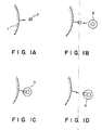
- the inventors of the present invention studying various modification of membrane proteins, found that modification of a membrane protein by an appropriate carrier limits the portion of the membrane protein capable of being inserted into a lipid bilayer membrane and enables the control of the orientation of the membrane protein, and have completed the present invention.
- a salt solution is prepared which contains a membrane protein modified by a carrier at a specific sites and a membrane lipid.
- the concentration of the membrane protein is as such that the ratio of the membrane protein to the lipid is in the range of from 1:1 to 1:200 by weight, preferably 1:10 to 1:120.
- the concentration of the lipid is usually in the range of 1 to 30 mg/ml, preferably 10 to 20 mg/ml.
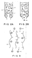
- the above salt solution is subjected to freezing and thawing, and then to dialysis against another salt solution having an osmotic pressure lower than that of the above salt solution, whereby a giant proteoliposome is prepared.
- solubilized disc in an amount of 5 mg as protein, was added to the column to combine the rhodopsin with concanavalin A, and then the column was washed sufficiently with a buffer solution (0.1 M NaCl, 1 mM MnCl2, 1 mM CaCl2, 10 mM Mops-NaOH (pH 7.0)) to eliminate impurities.
- a buffer solution 0.1 M NaCl, 1 mM MnCl2, 1 mM CaCl2, 10 mM Mops-NaOH (pH 7.0)
- a liposome was formed as below by a freeze-thaw method.
- a thin film of 15 mg of asolectin (soy bean phosphatidylcholine, type IV S, supplied by Sigma Co.) was formed on the inside wall of a test tube of 10 x 130 mm.
- the lipid dispersion was transferred into a polypropylene tube (12 x 75 mm: Becton Dickinson Co.: Falkon® 2063) provided with a cap and was saturated with nitrogen.
- This liposome dispersion was subjected to the three cycles of treatments comprising freezing by dry ice-acetone bath, thawing at a room temperature, and treatment by water-bath-type sonication apparatus for 30 seconds (at power level 1), thus giving the intended liposome dispersion (average diameter: ca 1 ⁇ m, volume: 0.5 ⁇ m3).
- a buffer solution (50 mM methyl- ⁇ -D-mannoside, 50 mM methyl- ⁇ -D-glucoside, 0.1 M NaCl, 10 mM Mops-NaOH (pH 7.0)) was made to flow through the column which contains methyl- ⁇ -D-mannoside and methyl- ⁇ -D-glucoside capable of combining with the sugar chain of the rhodopsin antagonistically to concanavalin A.
- the solution leaving the column was collected, and was dialyzed against a buffer solution (0.1 M NaCl, 10 mM Mops-NaOH (pH 8.0)) to eliminate free sugar molecules.
- a monoclonal antibody which will specifically combine with the carboxyl ends of the bacteriorhodopsin was prepared according to the method of K. Kimura (J. Biol. Chem. 257 , 2859-2867 (1980)). This monoclonal antibody specifically combines with fragments comprising 39 residues having carboxyl ends from among the peptide fragments obtained by cyanogen bromide treatment of the bacteriorhodopsin.
- a 0.1 M NaCl solution containing 0.2 mg of purified bacteriorhodopsin was made to flow in the column to combine the carboxyl ends of the bacteriorhodopsin with the CNBr-6 monoclonal antibody immobilized on the gel.
- a liposome liquid dispersion was prepared in the same manner as in Example 1.
- the liposome suspension was added to the column which had 0.2 mg of immobilized bacteriorhodopsin, and was incubated for one hour to incorporate the bacteriorhodopsin into the liposome.
- the proteoliposome prepared by the above procedure incorporated the bacteriorhodopsin with the carboxyl end directing outside, which was confirmed by pH change of the external liquid to alkaline side on light irradiation to the proteoliposome suspension, and formation of peptide having carboxyl ends as the digested product on enzymatic treatment of the proteoliposome.
- Bacteriorhodopsin and CNBr-6 monoclonal antibody were prepared by the methods described in Example 2.
- a liposome liquid dispersion was prepared in the method described in Example 1.
- the membrane filter having the combined monoclonal antibody (dry weight: 10 mg, 0.1 mg as monoclonal antibody) was added to 1 ml of a 0.1 M NaCl solution containing 0.2 mg of the purified bacteriorhodopsin to combine the carboxyl ends of the bacteriorhodopsin with the CNBr-6 monoclonal antibody immobilized on the membrane filter. Then the membrane filter was washed with 0.1 M NaCl to remove free protein, and was added to the liposome dispersion prepared as above, which was incubated for one hour to incorporate the bacteriorhodopsin into the liposome.
- the pH of the dispersion was adjusted to 11 by adding 1 N aqueous ammonia to dissociate the bacteriorhodopsin from the antibody. Thereafter, the dispersion was made neutral by adding 1N hydrochloric acid, and the membrane filter was removed by centrifugation.
- a method for introducing bacteriorhodopsin into a lipid bilayer membrane in a uniform orientation is shown by employing an affinity chromatographic column in which an anti-bacteriorhodopsin antibody is immobilized as a ligand to a cyanogen-bromide-activated agarose carrier.
- a 0.1 M NaCl solution containing 0.2 mg of purified bacteriorhodopsin was made to flow in the column to combine the carboxyl end of the bacteriorhodopsin with the CNBr-6 monoclonal antibody immobilized on the gel.
- the gel was washed again 0.1 M NaCl to remove unreacted bacteriorhodopsin.
- the proteoliposome prepared by the above procedure incorporated the bacteriorhodopsin with the carboxyl ends directing outside, which was confirmed by pH change of the external liquid to alkaline side on light irradiation to the proteoliposome suspension, and formation of peptide having carboxyl ends as the digested product on enzymatic treatment of the proteoliposome.
- Soy bean phospholipid (asolectin) corresponding to 15 mg of lipid dissolved in chloroform was placed in a test tube of 10 mm in diameter and about 130 mm in length.
- the solvent was evaporated off by means of a rotary evaporator, and further the solvent was completely removed in a desiccator in vacuo.
- 1 ml of 3 M aqueous potassium chloride solution which had been adjusted to a certain pH by addition of an aqueous hydrochloric acid or an aqueous potassium hydroxide solution was added. It was treated by a vortex mixer for 30 seconds to disperse the lipid thin membrane, and then was treated by a probe type sonicator for 30 minutes, giving a suspension of unilamellar liposome having a diameter of 100 ⁇ m.
- a membrane protein was added to the liposome suspension.
- Bacteriorhodopsin was used as an example of the membrane protein for forming a proteoliposome.
- the bacteriorhodopsin was prepared by extracting a purple membrane from a hyperhalophilic bacteria, Halobacterium halobium, according to the method of P. Oesterhelt and W. Stoeckenius (Method. Enzymol. 31 , 667-678 (1974)) and delipidating it according to the method of K. S. Huang, et al. (Proc. Natl. Acad. Sci. USA, 77 , 323 (1980)).
- the bacteriorhodopsin was added to the liposome suspension in a ratio of lipid to protein within a range of from 40 to 200 by weight.
- the suspension was subjected to three cycles of the treatments comprising freezing by liquid nitrogen or dry ice-acetone, thawing at room temperature, and vortex mixer treatment for 30 seconds.
- the suspension was then transferred to a dialysis tube, and subjected to dialysis against 10 mM aqueous potassium chloride solution for 2 days.
- a giant proteoliposome having a diameter exceeding 10 ⁇ m was prepared in a large amount.
- the diameter of the resultant proteoliposome depends on the pH during the preparation. In the case where the preparation operation was conducted at the pH within 3 to 11 from beginning to freezing and thawing, the diameter distributed within the range of about 5 to 50 ⁇ m, most of the particles having diameter of about 20 to 30 ⁇ m, while in the case where the operation was performed below pH 2, the particle size distributed non-uniformly over a broad range of from about 5 to 300 ⁇ m.
- the suspension of the giant proteoliposome having the incorporated bacteriorhodopsin was exposed to light of 560 nm, and the change of pH in the external liquid was measured.
- pH in the external liquid changed independently of the pH in the preparation step (in most cases, to alkaline side), from which the bacteriorhodopsin was confirmed to be incorporated with retention of its function.
- a thin film of 15 mg of asolectin (soy bean phosphatidylcholine, type IV S, supplied by Sigma Co.) was formed on the inside wall of a test tube of 10 x 130 mm. Thereto 1 ml of a solution of 3 M KCl, 50 mM MgCl2, 1 M Hepes-KOH (pH 7.5) was added and treated by a vortex mixer for 1 minute (at highest power level) to disperse the lipid thin membrane. The lipid dispersion was transferred into a polypropylene tube (12 x 75 mm; Becton Dickinson Co.: Falkon® 2063) provided with a cap and was saturated with nitrogen.
- asolectin asy bean phosphatidylcholine, type IV S, supplied by Sigma Co.
- a purple membrane, a membrane fragment containing bacteriorhodopsin, was obtained by extraction from a hyperhalophilic Halobacterium halobium according to the method of P. Oesterhelt and W. Stoeckenius (Methods of Enzymology, 31 , 667-678 (1974)).
- the purple membrane was delipidated according to the method of K. S. Huang (Proceedings of National Academy of Science, USA, 77 , 323 (1980)) to purify the bacteriorhodopsin.
- a monoclonal antibody which will specifically combine with the carboxyl ends of the bacteriorhodopsin was prepared according to the method of K. Kimura (J. Biol. Chem., 257 , 2859-2867 (1980)). This monoclonal antibody specifically combines with fragments comprising 39 residues having carboxyl ends from among the peptide fragments obtained by cyanogen bromide treatment of the bacteriorhodopsin.
- soy bean phospholipid dissolved in chloroform was placed in a test tube.
- the solvent was evaporated off by means of a rotary evaporator and a vacuum pump, to form a thin film of the lipid on the wall of the test tube.
- 1 ml of 3 M KCl solution (pH 8.0) was added.
- the lipid was made suspend by a vortex mixer, and the solution was treated by a probe type sonicator, giving a dispersion of liposome having a diameter of 100 nm or less.
- the small pieces of the membrane filter having monoclonal antibody combined were added.
- the suspension was subjected to three cycles of the treatments comprising freezing by liquid nitrogen or dry ice-acetone, thawing at room temperature, and vortex mixer treatment for 30 seconds.
- the suspension was then transferred to a dialysis tube, and subjected to dialysis against 10 mM aqueous potassium chloride solution for 2 days.
- a giant proteoliposome having a diameter exceeding 10 ⁇ m in a large amount.
- the giant proteoliposome prepared by the above procedure incorporated the bacteriorhodopsin with the carboxyl end directing outside, which was confirmed by pH change of the external liquid to alkaline side on light irradiation to the proteoliposome suspension, and formation of peptide having carboxyl ends as the digested product on enzymatic treatment of the proteoliposome.
- a giant proteoliposome was prepared in the same manner as in Example 7.
- the pH of the proteoliposome was made to be 11 by adding 1 N aqueous ammonia to dissociate the bacteriorhodopsin incorporated into the liposome from the CNBr-6 monoclonal antibody, thus giving a giant proteoliposome having a diameter exceeding 5 ⁇ m.
- the giant proteoliposomes prepared by the procedure above incorporated the bacteriorhodopsin with the carboxyl ends directing outside, which was confirmed by pH change of the external liquid to alkaline side on light irradiation to the proteoliposome suspension, and formation of peptide having carboxyl ends as the digested product on enzymatic treatment of the proteoliposome.
- Bovine rhodopsin was isolated and purified by the procedure shown below. The operation was all conducted under dark red light (with Red filter No.1 of Eastman Kodak Co.).
- the purified disc was solubilized by the solution consisting of 50 mM octyl glucoside, 0.1 M NaCl, 1 mM MnCl2, 1 mM CaCl2, and 10 mM Mops-NaOH (pH 7.0), and then the rhodopsin was purified by affinity chromatography employing wheat germ Lectin-Sepharose 6MB; agarose gel, (supplied by Pharmacia Co., average gel particle diameter: ca. 300 ⁇ m) by batchwise treatment (wet volume of 2 ml).
- the gel in an amount of 5 mg as protein, was added to the solution of the solubilized disc, combine the rhodopsin with the lectin, and then the gel was washed sufficiently with a buffer solution (0.1 M NaCl, 1 mM MnCl2, 1 mM CaCl2, 10 mM Mops-NaOH (pH 7.0)) to eliminate impurities.
- a buffer solution 0.1 M NaCl, 1 mM MnCl2, 1 mM CaCl2, 10 mM Mops-NaOH (pH 7.0)
- soy bean phospholipid dissolved in chloroform was placed in a test tube.
- the solvent was evaporated off by means of a rotary evaporator and a vacuum pump to form a thin film of the lipid on the wall of the test tube.
- 1 ml of a solution of 3 M NaCl, 1 mM MnCl2, 1mM CaCl2, and 0.1 M Mops-NaOH (pH 7.0) were added, and made suspend by a vortex mixer, and the suspension was treated by a probe type sonicator, giving a dispersion of liposome having a diameter of 100 nm or less.
- the gel having rhodopsin combined was added to the liposome liquid dispersion and mildly stirred for 30 minutes.
- the suspension was subjected to three cycles of the treatments comprising freezing by liquid nitrogen or dry ice-acetone, thawing at room temperature, and mixing by vortex mixer for 30 seconds.
- the suspension was then transferred to a dialysis tube, and was dialyzed against a solution of 10 mM NaCl, 10 mM Mops-NaOH (pH 7.0) for 2 days.
- a giant proteoliposome having a diameter exceeding 10 ⁇ m in a large amount.
- the incorporation of the rhodopsin into the resultant proteoliposome membrane was confirmed as below.
- the proteoliposome liquid dispersion was freezed by a liquid helium rapid-freeze apparatus (Eiko Enginnering Co.: RF-23), and the replica of the membrane surface prepared by a freeze-fracture replica preparation apparatus (Eiko Engineering Co.: FD-5A) was observed by a transmission type electron microscope (JEOL, Ltd.: JEM 100U). Particle of about 4 nm diameter were observed, which proves the incorporation of rhodopsin.
- a membrane protein having a function of transporting a substance in one direction through the membrane such as a substance transporting ATPase, membrane allows the control of the substance transfer direction from the interior to the exterior of the vesicle or the reverse direction, which raises remarkably the efficiency of the substance transfer since inappropriate protein orientation is decreased and the required amount of the inappropriate protein is decreased.
- a protein such as an enzyme and an antibody which can combine specifically with a substrate or an antigen-antibody and so forth is made possible to be embedded membrane, and the embedding can be made in such an orientation that the combining sites are directed toward the exterior of the liposome, so that the unsuitably orienting protein may be decreased, and the required amount of the protein can be reduced.
- the present invention has made it possible to prepare a giant proteoliposome having a diameter of 5 ⁇ m or larger in a simple procedure under mild conditions in a large amount without deactivation nor denaturation of the protein, which could not be achieved practically by prior art.
- the present invention has achieved, in formation of a giant proteoliposome, the advantage of controlling the orientation of the membrane protein incorporated into the giant proteoliposome.
- a method for forming a proteoliposome comprises incorporating into a liposome a membrane protein combined with a carrier. Further, a method for preparing a giant proteoliposome, comprises freezing and thawing an alkali metal salt solution containing a membrane protein and a lipid, and subsequently dialyzing against a second salt solution or a buffer solution having a lower osmotic pressure than that of said alkali metal salt solution. Further, a metal for forming a proteoliposome, comprises by freezing and thawing an alkali metal salt solution containing a membrane protein combined with a carrier and a lipid.
Landscapes
- Health & Medical Sciences (AREA)
- Chemical & Material Sciences (AREA)
- Life Sciences & Earth Sciences (AREA)
- Medicinal Chemistry (AREA)
- Pharmacology & Pharmacy (AREA)
- Epidemiology (AREA)
- Dispersion Chemistry (AREA)
- Animal Behavior & Ethology (AREA)
- General Health & Medical Sciences (AREA)
- Public Health (AREA)
- Veterinary Medicine (AREA)
- Medicinal Preparation (AREA)
- Manufacturing Of Micro-Capsules (AREA)
- Peptides Or Proteins (AREA)
Applications Claiming Priority (8)
| Application Number | Priority Date | Filing Date | Title |
|---|---|---|---|
| JP211910/88 | 1988-08-26 | ||
| JP63211910A JP2728890B2 (ja) | 1988-08-26 | 1988-08-26 | 巨大プロテオリポソームの調整方法 |
| JP21592488 | 1988-08-29 | ||
| JP215924/88 | 1988-08-29 | ||
| JP19705689 | 1989-07-29 | ||
| JP1197057A JP2727468B2 (ja) | 1989-07-29 | 1989-07-29 | 巨大プロテオリポソームの形成方法 |
| JP197056/89 | 1989-07-29 | ||
| JP197057/89 | 1989-07-29 |
Publications (3)
| Publication Number | Publication Date |
|---|---|
| EP0355845A2 true EP0355845A2 (fr) | 1990-02-28 |
| EP0355845A3 EP0355845A3 (fr) | 1991-07-24 |
| EP0355845B1 EP0355845B1 (fr) | 1993-03-10 |
Family
ID=27475849
Family Applications (1)
| Application Number | Title | Priority Date | Filing Date |
|---|---|---|---|
| EP19890115717 Expired - Lifetime EP0355845B1 (fr) | 1988-08-26 | 1989-08-25 | Procédé de fabrication de protéoliposomes et procédé pour la fabrication de protéoliposomes géants |
Country Status (2)
| Country | Link |
|---|---|
| EP (1) | EP0355845B1 (fr) |
| DE (1) | DE68905264T2 (fr) |
Cited By (2)
| Publication number | Priority date | Publication date | Assignee | Title |
|---|---|---|---|---|
| EP0603671A3 (fr) * | 1992-12-23 | 1995-02-22 | Behringwerke Ag | Méthode de réactivation de protéines membranaires purifiées. |
| WO2008012062A1 (fr) * | 2006-07-24 | 2008-01-31 | Polymun Scientific Immunbiologische Forschung Gmbh | Protéoliposomes antigéniques et procédé de fabrication |
Family Cites Families (2)
| Publication number | Priority date | Publication date | Assignee | Title |
|---|---|---|---|---|
| US4663161A (en) * | 1985-04-22 | 1987-05-05 | Mannino Raphael J | Liposome methods and compositions |
| US4927879A (en) * | 1988-02-25 | 1990-05-22 | Purdue Research Foundation | Method for solid phase membrane mimetics |
-
1989
- 1989-08-25 EP EP19890115717 patent/EP0355845B1/fr not_active Expired - Lifetime
- 1989-08-25 DE DE1989605264 patent/DE68905264T2/de not_active Expired - Fee Related
Cited By (3)
| Publication number | Priority date | Publication date | Assignee | Title |
|---|---|---|---|---|
| EP0603671A3 (fr) * | 1992-12-23 | 1995-02-22 | Behringwerke Ag | Méthode de réactivation de protéines membranaires purifiées. |
| US5599909A (en) * | 1992-12-23 | 1997-02-04 | Behringwerke Aktiengesellschaft | Process for reactivating purified membrane proteins |
| WO2008012062A1 (fr) * | 2006-07-24 | 2008-01-31 | Polymun Scientific Immunbiologische Forschung Gmbh | Protéoliposomes antigéniques et procédé de fabrication |
Also Published As
| Publication number | Publication date |
|---|---|
| DE68905264T2 (de) | 1993-07-01 |
| EP0355845B1 (fr) | 1993-03-10 |
| DE68905264D1 (de) | 1993-04-15 |
| EP0355845A3 (fr) | 1991-07-24 |
Similar Documents
| Publication | Publication Date | Title |
|---|---|---|
| US5374715A (en) | Method for forming proteoliposome and method for forming giant proteoliposome | |
| US4853228A (en) | Method of manufacturing unilamellar lipid vesicles | |
| US5049392A (en) | Osmotically dependent vesicles | |
| Rigaud et al. | Reconstitution of membrane proteins into liposomes: application to energy-transducing membrane proteins | |
| Schubert | Liposome preparation by detergent removal | |
| JP5480764B2 (ja) | 両性リポソーム及びその使用 | |
| AU654120B2 (en) | Solid tumor treatment method and composition | |
| US4241046A (en) | Method of encapsulating biologically active materials in lipid vesicles | |
| Magin et al. | Temperature-dependent drug release from large unilamellar liposomes | |
| US4663161A (en) | Liposome methods and compositions | |
| US6399094B1 (en) | Unilamellar liposomal preparations with high active substance content | |
| AU1543888A (en) | Lipid vesicles formed of surfactants and steroids | |
| US4873089A (en) | Proteoliposomes as drug carriers | |
| EP0355845B1 (fr) | Procédé de fabrication de protéoliposomes et procédé pour la fabrication de protéoliposomes géants | |
| GB2140822A (en) | Transformation of plant cells | |
| Alpes et al. | Reconstitution of Na+/K+-ATPase into phosphatidylcholine vesicles by dialysis of nonionic alkyl maltoside detergents | |
| JP2727469B2 (ja) | プロテオリポソームの形成方法 | |
| JP2727468B2 (ja) | 巨大プロテオリポソームの形成方法 | |
| JPH0564739A (ja) | 巨大リポソームの調製方法 | |
| JP2728890B2 (ja) | 巨大プロテオリポソームの調整方法 | |
| AU646520B2 (en) | Phospholipid analogue vesicle with a succinimidyl moiety | |
| Walker et al. | Controlled multi-stage self-assembly of vesicles | |
| JP2700276B2 (ja) | リポソーム | |
| JPH0773668B2 (ja) | リポソ−ム及びその前駆体の調製法 | |
| Anner et al. | The use of liposomes in the study of Na+, K+-ATPase |
Legal Events
| Date | Code | Title | Description |
|---|---|---|---|
| PUAI | Public reference made under article 153(3) epc to a published international application that has entered the european phase |
Free format text: ORIGINAL CODE: 0009012 |
|
| AK | Designated contracting states |
Kind code of ref document: A2 Designated state(s): CH DE FR GB LI NL |
|
| 17P | Request for examination filed |
Effective date: 19901221 |
|
| PUAL | Search report despatched |
Free format text: ORIGINAL CODE: 0009013 |
|
| AK | Designated contracting states |
Kind code of ref document: A3 Designated state(s): CH DE FR GB LI NL |
|
| 17Q | First examination report despatched |
Effective date: 19920619 |
|
| GRAA | (expected) grant |
Free format text: ORIGINAL CODE: 0009210 |
|
| AK | Designated contracting states |
Kind code of ref document: B1 Designated state(s): CH DE FR GB LI NL |
|
| REF | Corresponds to: |
Ref document number: 68905264 Country of ref document: DE Date of ref document: 19930415 |
|
| ET | Fr: translation filed | ||
| PLBE | No opposition filed within time limit |
Free format text: ORIGINAL CODE: 0009261 |
|
| STAA | Information on the status of an ep patent application or granted ep patent |
Free format text: STATUS: NO OPPOSITION FILED WITHIN TIME LIMIT |
|
| 26N | No opposition filed | ||
| REG | Reference to a national code |
Ref country code: GB Ref legal event code: IF02 |
|
| PGFP | Annual fee paid to national office [announced via postgrant information from national office to epo] |
Ref country code: DE Payment date: 20070823 Year of fee payment: 19 |
|
| PGFP | Annual fee paid to national office [announced via postgrant information from national office to epo] |
Ref country code: CH Payment date: 20070830 Year of fee payment: 19 |
|
| PGFP | Annual fee paid to national office [announced via postgrant information from national office to epo] |
Ref country code: GB Payment date: 20070822 Year of fee payment: 19 |
|
| PGFP | Annual fee paid to national office [announced via postgrant information from national office to epo] |
Ref country code: NL Payment date: 20070815 Year of fee payment: 19 |
|
| PGFP | Annual fee paid to national office [announced via postgrant information from national office to epo] |
Ref country code: FR Payment date: 20070808 Year of fee payment: 19 |
|
| REG | Reference to a national code |
Ref country code: CH Ref legal event code: PL |
|
| GBPC | Gb: european patent ceased through non-payment of renewal fee |
Effective date: 20080825 |
|
| NLV4 | Nl: lapsed or anulled due to non-payment of the annual fee |
Effective date: 20090301 |
|
| PG25 | Lapsed in a contracting state [announced via postgrant information from national office to epo] |
Ref country code: NL Free format text: LAPSE BECAUSE OF NON-PAYMENT OF DUE FEES Effective date: 20090301 |
|
| REG | Reference to a national code |
Ref country code: FR Ref legal event code: ST Effective date: 20090430 |
|
| PG25 | Lapsed in a contracting state [announced via postgrant information from national office to epo] |
Ref country code: LI Free format text: LAPSE BECAUSE OF NON-PAYMENT OF DUE FEES Effective date: 20080831 Ref country code: CH Free format text: LAPSE BECAUSE OF NON-PAYMENT OF DUE FEES Effective date: 20080831 |
|
| PG25 | Lapsed in a contracting state [announced via postgrant information from national office to epo] |
Ref country code: DE Free format text: LAPSE BECAUSE OF NON-PAYMENT OF DUE FEES Effective date: 20090303 Ref country code: FR Free format text: LAPSE BECAUSE OF NON-PAYMENT OF DUE FEES Effective date: 20080901 |
|
| PG25 | Lapsed in a contracting state [announced via postgrant information from national office to epo] |
Ref country code: GB Free format text: LAPSE BECAUSE OF NON-PAYMENT OF DUE FEES Effective date: 20080825 |