EP0361284B1 - T-Zellen-Wachstumsfaktor - Google Patents
T-Zellen-Wachstumsfaktor Download PDFInfo
- Publication number
- EP0361284B1 EP0361284B1 EP89117317A EP89117317A EP0361284B1 EP 0361284 B1 EP0361284 B1 EP 0361284B1 EP 89117317 A EP89117317 A EP 89117317A EP 89117317 A EP89117317 A EP 89117317A EP 0361284 B1 EP0361284 B1 EP 0361284B1
- Authority
- EP
- European Patent Office
- Prior art keywords
- growth factor
- cell growth
- cells
- mammalian
- helper
- Prior art date
- Legal status (The legal status is an assumption and is not a legal conclusion. Google has not performed a legal analysis and makes no representation as to the accuracy of the status listed.)
- Expired - Lifetime
Links
Images


Classifications
-
- C—CHEMISTRY; METALLURGY
- C07—ORGANIC CHEMISTRY
- C07K—PEPTIDES
- C07K14/00—Peptides having more than 20 amino acids; Gastrins; Somatostatins; Melanotropins; Derivatives thereof
- C07K14/435—Peptides having more than 20 amino acids; Gastrins; Somatostatins; Melanotropins; Derivatives thereof from animals; from humans
- C07K14/52—Cytokines; Lymphokines; Interferons
- C07K14/54—Interleukins [IL]
- C07K14/5425—IL-9
-
- A—HUMAN NECESSITIES
- A61—MEDICAL OR VETERINARY SCIENCE; HYGIENE
- A61P—SPECIFIC THERAPEUTIC ACTIVITY OF CHEMICAL COMPOUNDS OR MEDICINAL PREPARATIONS
- A61P37/00—Drugs for immunological or allergic disorders
-
- A—HUMAN NECESSITIES
- A61—MEDICAL OR VETERINARY SCIENCE; HYGIENE
- A61P—SPECIFIC THERAPEUTIC ACTIVITY OF CHEMICAL COMPOUNDS OR MEDICINAL PREPARATIONS
- A61P37/00—Drugs for immunological or allergic disorders
- A61P37/02—Immunomodulators
- A61P37/04—Immunostimulants
-
- C—CHEMISTRY; METALLURGY
- C07—ORGANIC CHEMISTRY
- C07K—PEPTIDES
- C07K16/00—Immunoglobulins [IG], e.g. monoclonal or polyclonal antibodies
- C07K16/18—Immunoglobulins [IG], e.g. monoclonal or polyclonal antibodies against material from animals or humans
- C07K16/24—Immunoglobulins [IG], e.g. monoclonal or polyclonal antibodies against material from animals or humans against cytokines, lymphokines or interferons
- C07K16/244—Interleukins [IL]
-
- A—HUMAN NECESSITIES
- A61—MEDICAL OR VETERINARY SCIENCE; HYGIENE
- A61K—PREPARATIONS FOR MEDICAL, DENTAL OR TOILETRY PURPOSES
- A61K38/00—Medicinal preparations containing peptides
-
- Y—GENERAL TAGGING OF NEW TECHNOLOGICAL DEVELOPMENTS; GENERAL TAGGING OF CROSS-SECTIONAL TECHNOLOGIES SPANNING OVER SEVERAL SECTIONS OF THE IPC; TECHNICAL SUBJECTS COVERED BY FORMER USPC CROSS-REFERENCE ART COLLECTIONS [XRACs] AND DIGESTS
- Y10—TECHNICAL SUBJECTS COVERED BY FORMER USPC
- Y10S—TECHNICAL SUBJECTS COVERED BY FORMER USPC CROSS-REFERENCE ART COLLECTIONS [XRACs] AND DIGESTS
- Y10S530/00—Chemistry: natural resins or derivatives; peptides or proteins; lignins or reaction products thereof
- Y10S530/808—Materials and products related to genetic engineering or hybrid or fused cell technology, e.g. hybridoma, monoclonal products
-
- Y—GENERAL TAGGING OF NEW TECHNOLOGICAL DEVELOPMENTS; GENERAL TAGGING OF CROSS-SECTIONAL TECHNOLOGIES SPANNING OVER SEVERAL SECTIONS OF THE IPC; TECHNICAL SUBJECTS COVERED BY FORMER USPC CROSS-REFERENCE ART COLLECTIONS [XRACs] AND DIGESTS
- Y10—TECHNICAL SUBJECTS COVERED BY FORMER USPC
- Y10S—TECHNICAL SUBJECTS COVERED BY FORMER USPC CROSS-REFERENCE ART COLLECTIONS [XRACs] AND DIGESTS
- Y10S930/00—Peptide or protein sequence
- Y10S930/01—Peptide or protein sequence
- Y10S930/14—Lymphokine; related peptides
- Y10S930/141—Interleukin
Definitions
- the present invention relates generally to a T cell growth factor. More particularly, the present invention relates to a mammalian T cell growth factor which is a glycoprotein capable of supporting interleukin 2- and interleukin 4-independent growth of helper T cells. This factor is further capable of augmenting proliferation if IL3- and IL4-responsive cells. Even more particularly, the present invention relates to the helper T cell growth factor P40, pharmaceutical compositions thereof and antibodies thereto. The present invention also contemplates a method for inducing the proliferation of helper T cells as well as IL3- and IL4-responsive cells.
- the helper T cell growth factor contemplated herein is useful in the stimulation of specific cells in the immune system.
- cytokines are polypeptides which directly or indirectly mediate host defense mechanisms and/or which mediate tissue growth differentiation. Cytokines have been recognized which mediate host defense against cancer and/or infection. Such cytokines include the interferons (IFN- ⁇ , IFN- ⁇ and IFN- ⁇ ), tumor necrosis factor (TNF- ⁇ ), lymphotoxin (TNF- ⁇ ), the interleukins (IL1, 2, 3, 4, 5 and 6), leukoregulin, natural killer cell cytotoxic factor (NKCF), transforming growth factor (TGF), colony stimulating factors (CSF) such as macrophage (M-CSF), granulocyte (G-CSF) and macrophage, granulocyte-CSF (G,M-CSF) and oncostatin M.
- IFN- ⁇ , IFN- ⁇ and IFN- ⁇ tumor necrosis factor
- TNF- ⁇ lymphotoxin
- IL1, 2, 3, 4, 5 and 6 the interleukins
- NKCF natural killer cell cyto
- cytokines are synthesized by leukocytes, commonly in response to stimulation by microorganisms, antigens or mitogens. This has been observed in vitro. Following this stimulation in cell culture, the supernatant fluid is retrieved and cytokine activity identified, isolated and further characterized. In recent years, it has become increasingly clear that IL2 is not the only factor controlling T cell growth. Indeed, several cytokines, including IL4 (Fernandez-Botran et al., Proc. Natl. Acad. Sci. USA , 83 : 9689-9693, 1986; Lichtman et al., Proc. Natl. Acad. Sci.
- T H helper T cell
- T H 1 helps B cells in a linked, antigen-specific manner, and is required early in the response.
- T H 2 helps B cells in a nonlinked manner and is required later in the response.
- helper T cell lines from lymph nodes of antigen-primed mice was obtained using the procedure described by Corradin et al., J. Immunol., 119 : 1048-1053, 1977. These cell lines were initiated by culture in the presence of antigen and were subsequently maintained, without addition of exogenous growth factors, by regular feeding with antigen and irradiated splenic antigen-presenting cells. Most of these cells produce large amounts of IL3, IL4, IL5 and IL6, but no IL2 and, therefore, belong to the T H 2 type defined by Mosmann et al., J. Immunol., 136: 2348-2357, 1986.
- the subject invention relates to a novel T cell growth factor distinct from other known cytokines.
- the new growth factor is useful as a therapeutic compound to stimulate proliferation of helper T cells.
- the present invention is directed to a mammalian T cell growth factor which supports interleukin 2-independent and interleukin 4-independent growth of helper T cells, and is preferably obtained from mouse sources.
- this T cell growth factor is a protein having the identifiable characteristics of P40, derivatives or fragments thereof and the further capability of augmenting proliferation and IL3- and IL4-responsive cells. Methods of isolating P40, its derivatives and fragments are also provided.
- compositions comprising an effective amount of P40, a derivative or fragment thereof and a pharmaceutically acceptable carrier useful in the stimulation of specific cells in the immune system.
- these compositions may also contain IL3 or IL4.
- Still another aspect of the present invention relates to antibodies specific to P40, an antigenic derivative or an antigenic fragment thereof useful in diagnostic assays for P40.
- Yet another aspect of the present invention relates to a recombinant DNA molecule and expression vectors encoding the polypeptide portion of mammalian P40, a derivative or a fragment thereof, thereby providing a convenient source of recombinant P40.
- Still yet another aspect of the present invention contemplates a method of proliferating helper T cells which comprises incubating said cells with a proliferating effective amount of P40 or a derivative thereof for a time and under conditions sufficient for said cells to proliferate.
- a still further aspect of this invention relates to a method of proliferating IL3- or IL4-responsive cells by administering a combination of P40 and IL3 or IL4 to a mammal, especially a human, for a time and under conditions sufficient to stimulate said cells to proliferate.
- Fig. 1 is a graphical representation depicting long-term antigen-independent T cell growth induced by helper T cell supernatant (SN).
- TUC2.15 cells are grown without feeder cells and antigen in normal medium ( ⁇ ), in medium supplemented with IL2 (20 U/ml, ⁇ ) or with TUC2.15 SN (5% v/v, ⁇ ).
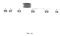
- Fig. 2 is a graphical representation depicting purification of P40.
- TUC7.51 supernatant is fractioned sequentially on an Ultrogel AcA54 gel filtration column (A), a TSK-phenyl hydrophobic interaction column (B), a Mono-Q anion exchange column (C) and Cl-reversed phase column (D).
- the shaded area represents P40 activity.
- Molecular mass standards shown in panel A are bovine serum albumin (BSA, 67 kDa), natural IL5 (45 kDa) and recombinant mouse IL6 (22 kDa).
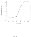
- Fig. 3 is a graphical representation depicting growth factor activity of purified P40.
- TS1 cells (3 x 10 3 cells/well) are cultivated in the presence of increasing doses of purified P40. After 3 days, cells numbers are evaluated by measuring hexosaminidase levels.
- Fig. 4 illustrates the purity of P40 and its extent of glycosylation.
- Panel A is a photograph showing silver-stained NaDodSO 4 /PAGE of purified P40. The sample is run under reducing conditions.
- Panel B is an autoradiograph of 125 I-P40 treated with various glycosylases. Mr of standards is given in kDa.
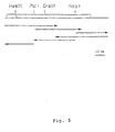
- Fig. 5 is a graphic illustration of the sequencing strategy of the murine P40 gene.
- Fig. 6 is a graphic illustration of the expression of recombinant P40 in fibroblasts.
- Fig. 7 shows the amino acid sequence of murine P40 obtained by chemical sequencing and the various peptides used in obtaining this sequence.
- Fig. 8 is a graphic illustration of the separation of endoproteinase Asp-N peptides of Cm-P40 by RP-HPLC.
- Fig. 9 is a graphic illustration of a multi-wavelength plot of the HPLC profile of Fig. 8 using a hotodiode array detector at wavelengths of (A) 290 nm, (B) 280 nm, (C) 254 nm and (D) 215 nm.
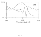
- Fig. 10 is a graphic illustration of a derivative spectral analysis of endoproteinase Asp-N peptide D1 with the zero order spectrum indicated by (---) and the second order spectrum indicated by ( ⁇ ).
- Fig. 11 is a graphic illustration of the microbore RP-HPLC separation of peptides of Cm-P40 derived from digestion with various proteases.
- Panel A shows the peptides from S. aureus V8 protease digestion of Cm-P40 endoproteinase Asp-N peptide D3.
- Panel B shows the peptides from a chymotrypsin digestion of Cm-P40.
- Panel C shows the peptides from a trypsin digestion of Cm-P40.
- Fig. 12 is a graphic illustration of the elution profile of the blocked amino terminal peptide D1 and related synthetic peptides on RP-HPLC.
- Z indicates pyroglutamic acid.
- the present invention relates to a mammalian T cell growth factor which comprises a protein which supports, or is capable of supporting, interleukin 2 (IL2)-independent and interleukin 4 (IL4)-independent growth of helper T cells in the absence of antigen.
- said T cell growth factor is biologically pure.
- biologically pure is meant a composition comprising said T cell growth factor.
- the composition may comprise homogeneous T cell growth factor or may consist essentially of T cell growth factor.
- supporting IL2-independent and IL4-independent growth of helper T cells refers to the ability for said cells to proliferate in the absence of IL2 and/or IL4.
- P40 growth factor
- derivatives of P40 encompass synthetic and naturally occurring amino acid substitutions, deletions and/or insertions as will be apparent to the skilled artisan.
- non-essential amino acid deletions i.e., deletion of amino acids which do not affect the activity of P40 are obtainable by genetic engineering means.
- fragments of P40 are contemplated by the present invention. These fragments are peptides obtained from the P40 protein and may be prepared by proteolysis of purified P40. The peptides are purified by conventional means such as HPLC chromatography and the like, and are useful in determining the P40 amino acid sequence in preparing antibodies to specific domains of P40 and in identifying the P40 domains involved in stimulating T cell growth.
- An antigenic derivative of P40 is defined to be a portion of P40 which is capable of reacting with an antibody specific to P40. All such derivatives are encompassed by the subject invention.
- P40 is a protein, and more particularly, a glycoprotein, capable of supporting long-term IL2-independent and IL4-independent growth of helper T cell lines in the absence of antigen, and is isolated from helper T cell lines, especially mammalian lines like murine helper T cell lines.
- P40 is functionally distinct from all known interleukins and colony-stimulating factors.
- P40 is purified from the supernatant (SN) of lectin-stimulated mouse helper T cell lines to a specific activity of from about 10 U/mg to about 10 10 U/mg, but generally to about 10 8 U/mg and characterized as a basic (pI 10 for murine P40) single chain protein with a Mr of from about 30 to about 40 kDa.
- P40 can be purified from the supernatant fluid of antigen stimulated mouse helper T cell clones (TUC2.15 and TUC7.51). Briefly, the supernatant fluid is concentrated and applied to a TSK-Phenyl chromatography column. Fractions with growth factor activity on factor-dependent TSl cells are pooled, further fractionated on a Mono-Q chromatography column, and the resulting active fractions applied to a C1 reversed-phase HPLC column. Pure murine P40 is eluted at a concentration of about 35% acetonitrile.
- P40 is a glycoprotein: (i) its heterogeneous migration pattern in NaDodSO 4 /PAGE and (ii) its binding to lentil lectin, which points to the presence of N-linked carbohydrate side chains. Consistent with this observation, a number of potential N-glycosylation sites (Asn-X-Thr motif) have been identified in the protein sequence determination. Moreover, additional evidence for extensive glycosylation of the molecule is obtained in experiments with N-glycanase treatment, which reduced the Mr of P40 to about 15 kDa. P40 is a stable molecule whose biological activity is not altered after exposure to NaDodSO 4 , acid pH or acetonitrile.
- P40 is also distinguished from known proteins on the basis of its complete amino acid sequence. The DNA and amino acid sequence of murine P40 is described herein.
- P40 differs functionally from IL2.
- P40 is completely inactive on cytolytic T cell clones under conditions where their response to IL2 is very strong; conversely, IL2 fails to support long-term antigen independent growth of helper T cell lines, whereas P40 is very active in this system.
- long-term growth of helper cells in response to P40 means greater than two months and may be indefinite.
- a correlation is observed between the sensitivity of helper T cell lines to P40 and IL4, indicating that T cell activation by these two molecules is similarly regulated.
- T cell growth factor P40
- P40 is specific for helper T cell lines. This indicates the existence of a growth-stimulatory mechanism restricted to the helper T cell subset. Such a mechanism is important for maintaining the balance between the supply of helper T cell products like IL2 and IL4 and their increased consumption by other lymphocytes activated in the course of the immune response.
- IL3- and IL4-responsive cells are immune system cells which proliferate in response to IL3 or IL4, respectively. These cells may include IL3-dependent cells or IL4-dependent cells, but are not limited thereto.
- IL3-responsive cells include helper T cells, stem cells, mast cells, eosinophils, neutrophils, monocytes, megakaryocytes, basophils and erythropoid cells.
- IL4-responsive cells include helper T cells, activated cytotoxic T cells, macrophages, mast cells and B cells (Smith, K.A., Biotechnology , 7 : 661-667, 1989).
- the combination of cytokines P40 and IL3, or P40 and IL4 can stimulate thymidine uptake by a factor ranging from about 4 to 40 above the stimulatory effect of any one of the cytokines.
- the synergism between P40 and IL3, or P40 and IL4 is dose dependent and cell line dependent.
- suboptimal doses of P40 range from about 1-25% of optimal P40 doses
- suboptimal doses of IL4 range from about 5-30% of optimal IL4 doses
- close to optimal doses of IL3 range from about 70-100% of optimal IL3 doses.
- This synergism provides a further method to stimulate proliferation of IL3- and IL4-responsive cells, especially helper T cells, and is therapeutically useful in treating immune deficiencies, especially those diseases or disease states which benefit from proliferation of specific immune cells such as AIDS, or even from general proliferation of immune cells.
- SN supernatant from a helper T cell line not requiring antigen or feeders
- P40 supernatant
- This SN is able to induce cell proliferation without further requirement for antigen or feeder cells.
- the proliferation activity is not inhibited by either anti-IL4 or anti-IL2 receptor antibodies, indicating that said activity is mediated neither directly nor indirectly by these molecules.
- the active ingredient in the aforementioned SN is shown to be, in accordance with the present invention, P40.
- the SN is active on the test cells, TS1, inducing half-maximal proliferation at dilutions ranging of from about 10 -6 to about 10 -2 (v/v), and generally ranging of from about 10 -5 to about 10 -4 (v/v).
- the novel T cell growth factor P40 is active in biologically pure form and in homogeneous and heterogeneous compositions.
- SN fluid is a form of heterogeneous composition of P40.
- Homogeneous compositions are exemplified herein to include pharmaceutical compositions containing homogeneous preparations of P40, its active derivatives or fragments, and the like.
- the T cell growth factor P40 is contemplated herein to be useful in stimulating the proliferation of T helper cells in mammals.
- P40 is particularly useful in stimulating certain subsets of T helper cells in mammals.
- P40 is a new and useful therapeutic compound capable of stimulating specific cells within the immune cells. For example, this is particularly important for human patients carrying defects in certain subsets of T helper cells as may be the case with various AIDS patients or immune compromised patients.
- the proliferation of helper T cells by P40 will have the additional effect of allowing increased amounts of other cytokines to be produced.
- the present invention also contemplates a method of treatment of immune deficiency comprising the administrtion of a proliferating effective amount of P40, an active derivative, or an active fragment thereof, for a time and under conditions sufficient to effect proliferation of helper T cells.
- the time required for the proliferation of helper T cells ranges from about two days to about seven days.
- the subject invention contemplates a method for inducing and maintaining the proliferation of helper T cells, and preferably, certain subsets thereof, in a mammal which comprises adminstering to said mammal a proliferating-effective amount of a pharmaceutical composition containing P40, an active derivative or fragment thereof, for a time and under conditions sufficient for said cells to proliferate.
- a method for inducing and maintaining the proliferation of helper T cells, and preferably certain subsets thereof, in a mammal is contemplated by this invention in which a nucleic acid molecule encoding P40 is introduced into a T cell in such a manner that said nucleic acid molecule is expressed intracellularly, but extrachromosomally of said cell or following integration into the genome of said cell.
- the nucleic acid molecule is carried to said T cell and transferred into said cell by a second nucleic acid molecule (e.g., various viruses).
- the first nucleic acid molecule is manipulated such that it contains the appropriate signals for expression.
- a method for proliferating T helper cells in a mammal comprising administering a first nucleic acid molecule encoding P40, said nucleic acid molecule being contained in a pharmaceutically acceptable second nucleic acid carrier molecule such that said first nucleic acid molecule enters a T cell and is either maintained extrachromosomally or integrates into the genome of said target all in such a manner that said first nucleic acid molecule is expressed so as to produce an effective amount of P40.
- nucleic acid molecule is meant the nucleotide sequence which encodes, directly or indirectly, P40 or a derivative thereof.
- a nucleic acid molecule is defined herein to mean RNA or DNA.
- the active indredients of a pharmaceutical composition comprising P40 are contemplated to exhibit excellent and effective therapeutic activity, for example, in the treatment of immune compromised diseases in mammals.
- the active ingredients of the therapeutic compositions comprising P40 exhibit helper T cell proliferative activity when administered in therapeutic amounts which depend on the particular disease. For example, from about 0.5 ug to about 2000 mg per kilogram of body weight per day may be administered.
- the dosage regimen may be adjusted to provide the optimum therapeutic response. For example, several divided doses may be administered daily or the dose may be proportionally reduced as indicated by the exigencies of the therapeutic situation.
- the active compound may be administered in a convenient manner such as by the oral, intraveneous (where water soluble), intramuscular, subcutaneous, intranasal, intradermal or suppository routes.
- the active ingredients which comprise P40 may be required to be coated in a material to protect said ingredients from the action of enzymes, acids and other natural conditions which may inactivate said ingredients.
- the low lipophilicity of P40 may allow it to be destroyed in the gastrointestinal tract by enzymes capable of cleaving peptide bonds and in the stomach by acid hydrolysis.
- P40 should be coated by, or administered with, a material to prevent its inactivation.
- P40 may be administered in an adjuvant, co-administered with enzyme inhibitors or in liposomes.
- Adjuvants contemplated herein include resorcinols, non-ionic surfactants such as polyoxyethylene oleyl ether and n-hexadecyl polyethylene ether.
- Enyzme inhibitors include pancreatic trypsin inhibitor, diisopropylfluorophosphate (DFP) and trasylol.
- Liposomes include water-in-oil-in-water P40 emulsions as well as conventional liposomes.
- the active compounds may also be administered parenterally or intraperitoneally.
- Dispersions can also be prepared in glycerol, liquid polyethylene glycols, and mixtures thereof, and in oils. Under ordinary conditions of storage and use, these preparations contain a preservative to prevent the growth of microorganisms.
- the pharmaceutical forms suitable for injectable use include sterile aqueous solutions (where water soluble) or dispersions and sterile powders for the extemporaneous preparation of sterile injectable solutions or dispersion.
- the form must be sterile and must be fluid to the extent that easy syringability exists. It must be stable under the conditions of manufacture and storage and must be preserved against the contaminating action of microorganisms such as bacteria and fungi.
- the carrier can be a solvent or dispersion medium containing, for example, water, ethanol, polyol (for example, glycerol, propylene glycol, liquid polyethylene glycol, and the like), suitable mixtures thereof and vegetable oils.
- the proper fluidity can be maintained, for example, by the use of a coating such as lecithin, by the maintenance of the required particle size in the case of dispersion and by the use of surfactants.
- the preventions of the action of microorganisms can be brought about by various antibacterial and antifungal agents, for example, parabens, chlorobutanol, phenol, sorbic acid, thimerosal, and the like. In many cases, it will be preferable to include isotonic agents, for example, sugars or sodium chloride. Prolonged absorption of the injectable compositions can be brought about by the use in the compositions of agents delaying absorption, for example, aluminum monostearate and gelatin.
- Sterile injectable solutions are prepared by incorporating the active compounds in the required amount in the appropriate solvent with various of the other ingredients enumerated above, as required, followed by filtered sterilization.
- dispersions are prepared by incorporating the various sterilized active ingredient into a sterile vehicle which contains the basic dispersion medium and the required other ingredients from those enumerated above.
- the preferred methods of preparation are vacuum-drying and the freeze-drying technique which yield a powder of the active ingredient plus any additional desired ingredient from previously sterile-filtered solution thereof.
- the active compound may be orally administered, for example, with an inert diluent or with an assimilable edible carrier, or it may be enclosed in hard or soft shell gelatin capsule, or it may be compressed into tablets, or it may be incorporated directly with the food of the diet.
- the active compound may be incorporated with excipients and used in the form of ingestible tablets, buccal tablets, troches, capsules, elixirs, suspensions, syrups, wafers, and the like.
- Such compositions and preparations should contain at least 1% of active compound.
- the percentage of the compositions and preparations may, of course, be varied and may conveniently be between about 5 to about 80% of the weight of the unit.
- the amount of active compound in such therapeutically useful compositions is such that a suitable dosage will be obtained.
- Preferred compositions or preparations according to the present invention are prepared so that an oral dosage unit form contains between about 10 ug and 1000 ug of active compound.
- the tablets, troches, pills, capsules, and the like may also contain the following: a binder such as gum gragacanth, acacia, corn starch or gelatin; excipients such as dicalcium phosphate; a disintegrating agent such as corn starch, potato starch, alginic acid, and the like; a lubricant such as magnesium stearate; and a sweetening agent such as sucrose, lactose or saccharin may be added or a flavoring agent such as peppermint, oil of witnergreen or cherry flavoring.
- a binder such as gum gragacanth, acacia, corn starch or gelatin
- excipients such as dicalcium phosphate
- a disintegrating agent such as corn starch, potato starch, alginic acid, and the like
- a lubricant such as magnesium stearate
- a sweetening agent such as sucrose, lactose or saccharin may be added or a flavoring agent such as
- any material may be present as coatings or to otherwise modify the physical form of the dosage unit.
- tablets, pills or capsules may be coated with shellac, sugar or both.
- a syrup or elixir may contain the active compound,sucrose as a sweetening agent, methyl and propylparabens as preservatives, a dye and flavoring such as cherry or orange flavor.
- any material used in preparing any dosage unit form should be pharmaceutically pure and substantially non-toxic in the amounts employed.
- the active compound may be incorporated into sustained-release preparations and formulations.
- Dosage unit form refers to physically discrete units suited as unitary dosages for the mammalian subjects to be treated; each unit containing a predetermined quantity of active material calculated to produce the desired therapeutic effect in association with the required pharmaceutical carrier.
- the specification for the novel dosage unit forms of the invention are dictated by and directly dependent on (a) the unique characteristics of the active material and the particular therapeutic effect to be achieved, and (b) the limitations inherent in the art of compounding such an active material for the treatment of disease in living subjects having a diseased condition in which bodily health is impaired as herein disclosed in detail.
- the principal active ingredient is compounded for convenient and effective administration in effective amounts with a suitable pharmaceutically acceptable carrier in dosage unit form as hereinbefore disclosed.
- a unit dosage form can, for example, contain the principal active compound in amounts ranging from 0.5 ug to about 2000 mg. Expressed in proportions, the active compound is generally present in from about 10 ug to about 2000 mg/ml of carrier.
- the dosages are determined by reference to the usual dose and manner of administration of the said ingredients.
- pharmaceutically acceptable carrier includes any and all solvents, dispersion media, coatings, antibacterial and antifungal agents, isotonic and absorption delaying agents, and the like.
- the use of such media and agents for pharmaceutical active substances is well known in the art. Except insofar as any conventional media or agent is incompatible with the active ingredient, use thereof in the therapeutic compositions is contemplated. Supplementary active ingredients can also be incorporated into the compositions.
- a further aspect of this invention contemplates the use of P40 with IL3 or IL4 in a method to stimulate proliferation of IL3- or IL4-responsive cells and in a method of treatment of immune deficiency. Such methods are practiced in accordance with the therapeutic methods involving only P40 and as described herein. Likewise, pharmaceutical compositions containing P40 and IL3, or P40 and IL4 are provided in accordance with those which contain P40 alone. Further in this regard, IL3 and IL4 are commercially available and are used in therapeutically effective amounts. Pharmaceutically effective amounts of P40 when used in conjunction with IL3 or IL4 are the same as when P40 is used alone.
- compositions of P40 and IL3 according to the present invention are prepared so that a unit dosage form contains each protein in an amount ranging from about 0.5 ug to about 2000 mg.
- Preferred compositions of P40 and IL4 are likewise prepared so that a unit dosage form contains each protein in an amount ranging from about 0.5 ug to about 2000 mg.
- the relative amount of P40 to IL3 or IL4 can be varied or the same.
- the present invention also relates to antibodies to P40, its derivatives or fragments. Such antibodies are contemplated to be useful in developing detection assays (immunoassays) for P40, especially during the monitoring of a therapeutic regimen and in the purification of P40.
- the antibodies may be monoclonal or polyclonal. Additionally, it is within the scope of this invention to include any second antibodies (monoclonal or polyclonal) directed to the first antibodies discussed above.
- the present invention further contemplates use of these second antibodies in detection assays and, for example, in monitoring the effect of an adminstered pharmaceutical preparation. Furthermore, it is within the scope of the present invention to include antibodies to the glycosylated regions of P40, and to any molecules complexed with said P40. Accordingly, in accordance with this invention, an antibody to P40 encompasses antibodies to P40, or antigenic parts thereof, and to any associated molecules (e.g., glycosylated regions, lipid regions, carrier molecules, and the like).
- the P40, or parts thereof, considered herein are purified, as exemplified in Example 3, then utilized in antibody production. Both polyclonal and monoclonal antibodies are obtainable by immunization with P40, its derivatives, polypeptides or fragments, and either type is utilizable for immunoassays. The methods of obtaining both types of sera are well known in the art. Polyclonal sera are less preferred, but are relatively easily prepared by injection of a suitable laboratory animal with an effective amount of the purified P40, or parts thereof, collecting serum from the animal and isolating specific sera by any of the known immunoadsorbent techniques. Although antibodies produced by this method are utilizable in virtually any type of immunoassay, they are generally less favored because of the potential heterogeneity of the product.
- the use of monoclonal antibodies in the present immunoassay is particularly preferred because of the ability to produce them in large quantities and the homogeneity of the product.
- the preparation of hybridoma cell lines for monoclonal antibody production derived by fusing an immortal cell line and lymphocytes sensitized against the immunogenic preparation can be done by techniques which are well known to those who are skilled in the art. (See, for example, Douillard, J.Y. and Hoffman, T., "Basic Facts About Hybridomas," in Compendium of Immunology, Vol. II, L. Schwartz (Ed.) (1981); Kohler, G.
- the choice of animal for monoclonal antibody production is dependent on the availability of appropriate immortal lines capable of fusing with lymphocytes thereof.
- Mouse and rat have been the animals of choice in hybridoma technology and are preferably used. Humans can also be utilized as sources for sensitized lymphocytes if appropriate immortalized human (or nonhuman) cell lines are available.
- the animal of choice may be injected with from about 1 mg to about 20 mg of the purified P40 or parts thereof. Usually the injecting material is emulsified in Freund's complete adjuvant. Boosting injections may also be required.
- the detection of antibody production can be carried out by testing the antisera with appropriately labeled antigen.
- Lymphocytes can be obtained by removing the spleen or lymph nodes of sensitized animals in a sterile fashion and carrying out fusion. Alternatively, lymphcytes can be stimulated or immunized in vitro, as described, for example, in C. Reading, J. Immunol. Meth., 53 : 261-291, 1982.
- a number of cell lines suitable for fusion have been developed, and the choice of any particular line for hybridization protocols is directed by any one of a number of criteria such as speed, uniformity of growth characteristics, deficiency of its metabolism for a component of the growth medium, and potential for good fusion frequency.
- Intraspecies hybrids particularly between like strains, work better than interspecies fusions.
- Several cell lines are available, including mutants selected for the loss of ability to secrete myeloma immunoglobulin. Included among these are the following mouse myeloma lines: MPC 11 -X45-6TG, P3-NS1-1-Ag4-1. P3-X63-Ag8, or mutants thereof such as X63-Ag8.653, SP2-0-Ag14 (all BALB/C derived), Y3-'Ag1.2.3 (rat) and U266 (human).
- Cell fusion can be induced either by virus, such as Epstein-Barr or Sendai virus, or polyethylene glycol.
- virus such as Epstein-Barr or Sendai virus
- polyethylene glycol Polyethylene glycol (PEG) is the most efficacious agent for the fusion of mammalian somatic cells. PEG itself may be toxic for cells, and various concentrations should be tested for effects on viability before attempting fusion.
- the molecular weight range of PEG may be varied from 1000 to 6000. It gives best results when diluted to about 20% to about 70% (w/w) in saline or serum-free medium. Exposure to PEG at 37°C for about 30 seconds is preferred in the present case, utilizing murine cells.
- the successfully fused cells can be separated from the myeloma line by any technique known by the art.
- the most common and preferred method is to choose a malignant line which is Hypoxanthine Guanine Phosphoribosyl Transferase (HGPRT) deficient, which will not grow in an aminopterin-containing medium used to allow only growth of hybrids and which is generally composed of hypoxanthine 1.10 -4 M, aminopterin 1 x 10 -5 M and thymidien 3 x 10 -5 M, commonly known as the HAT medium.
- the fusion mixture can be grown in the HAT-containing culture medium immediately after the fusion 24 hours later.
- the feeding schedules usually entail maintenance in HAT medium for two weeks and then feeding with either regular culture medium or hypoxanthine, thymidine-containing medium.
- the growing colonies are then tested for the presence of antibodies that recognize the antigenic preparation.
- Detection of hybridoma antibodies can be performed using an assay where the antigen is bound to a solid support and allowed to react to hybridoma supernatants containing putative antibodies.
- the presence of antibodies may be detected by "sandwich” techniques using a variety of indicators. Most of the common methods are sufficiently sensitive for use in the range of antibody concentrations secreted during hybrid growth.
- Cloning of hybrids can be carried out after 21-23 days of cell growth in selected medium. Cloning can be performed by cell limiiting dilution in fluid phase or by directly selecting single cells growing in semi-solid agarose. For limiting dilution, cell suspensions are diluted serially to yield a statistical probability of having only one cell per well. For the agarose technique, hybrids are seeded in a semi-solid upper layer, over a lower layer containing feeder cells. The colonies from the upper layer may be picked up and eventually transferred to wells.
- Antibody-secreting hybrids can be grown in various tissue culture flasks, yielding supernatants with variable concentrations of antibodies. In order to obtain higher concentrations, hybrids may be transferred into animals to obtain inflammatory ascites. Antibody-contaning ascites can be harvested 8-12 days after intraperitoneal injection. The ascites contain a higher concentration of antibodies but include both monoclonals and immunoglobulines from the inflammatory ascites. Antibody purification may then be achieved by, for example, affinity chromatography.
- P40 contemplated herein, or antibodies specific for same, in a patient's serum, tissue or tissue extract can be detected utilizing antibodies prepared as above, either monoclonal or polyclonal, in virtually any type of immunoassay.
- a wide range of immunoassay techniques are available as can be seen by reference to U.S. Patent Nos. 4,016,043; 4,424,279 and 4,018,653. This, of course, includes both single-site and two-site, or "sandwich", assays of the non-competitive types, as well as in the traditional competitive binding assays. Sandwich assays are among the most useful and commonly used assays and are favored for use in the present invention.
- an unlabeled antibody is immobilized in a solid substrate and the sample to be tested brought into contact with the bound molecule.
- a second antibody labeled with a reporter molecule capable of producing a detectable signal is then added and incubated, allowing time sufficient for the formation of a tertiary complex of antibody-antigen-labeled antibody (e.g., antibody-P40-antibody).
- any unreacted material is washed away, and the presence of the antigen is determined by observation of a signal produced by the reporter molecule.
- the results may either be qualitative, by simple observation of the visible signal, or may be quantitated by comparing with a control sample containing known amounts of hapten.
- Variations on the forward assay include a simultaneous assay, in which both sample and labeled antibody are added simultaneously to the bound antibody, or a reverse assay in which the labeled antibody and sample to be tested are first combined incubated and then added to the unlabeled surface bound antibody.
- a first antibody having specificity for P40, or antigenic parts thereof, contemplated in this invention is either covalently or passively bound to a solid surface.
- the solid surface is typically glass or a polymer, the most commonly used polymers being cellulose, polyacrylamide, nylon, polystyrene, polyvinyl chloride or polypropylene.
- the solid supports may be in the form of tubes, beads, discs or microplates, or any other surface suitable for conducting an immunoassay.
- the binding processes are well-known in the art and generally consist of cross-linking, covalently binding or physically absorbing the molecule to the insoluble carrier. Following binding, the polymer-antibody complex is washed in preparation for the test sample.
- reporter molecule a molecule which, by its chemical nature, provides an analytically identifiable signal which allows the detection of antigen-bound antibody. Detection may be either qualitative or quantitative.
- reporter molecules in this type of assay are either enzymers, fluorophores or radionuclide containing molecules (i.e., radioisotopes).
- an enzyme is conjugated to the second antibody, generally by means of glutaraldehyde or periodate.
- Commonly used enzymes include horseradish peroxidase, glucose oxidase, ⁇ -galactosidase and alkaline phosphatase, among other.
- the substrates to be used with the specific enzymes are generally chosed for the production, upon hydrolysis by the corresponding enzyme, of a detectable color change.
- p -nitrophenyl phosphate is suitable for the use with alkaline phosphatase conjugates; for peroxidase conjugates, 1,2-phenylenediamine, 5-aminosalicyclic acid, or tolidine, are commonly used.
- fluorogenic substrates which yield a fluorescent product rather than the chromogenic substrates noted above.
- the enzyme-labeled antibody is added to the first antibody hapten complex, allowed to bind, and then to the first antibody hapten complex, allowed to bind, and then the excess reagent is washed away.
- a solution containing the appropriate substrate is then added to the ternary complex of antibody-antigen-antibody.
- the substrate will react with the enzyme linked to the second antibody, giving a qualitative visual signal, which may be further quantitated, usually spectrophotometrically, to give an indication of the amount of hapten which was present in the sample.
- fluorescent compounds such as fluorescein and rhodamine
- fluorescein and rhodamine may be chemically coupled to antibodies without altering their binding capacity.
- the fluorochrome-labeled antibody When activated by illumination with light of a particular wavelength, the fluorochrome-labeled antibody adsorbs the light energy, inducing a state of excitability in the molecule, followed by emission of the light at a characteristic color visually detectable with a light microscope.
- the fluorescent labeled antibody is allowed to bind to the first antibody-hapten complex. After washing off the unbound reagent, the remaining ternary complex is then exposed to the light of the appropriate wavelength, the fluorescence observed indicates the presence of the hapten of interest.
- Immunofluorescence and EIA techniques are both very well established in the art and are particularly preferred for the present method.
- other reporter molecules such as radioisotope, chemiluminescent or bioluminescent molecules, may also be employed. It will be readily apparent to the skilled technician how to vary the procedure to suit the required purpose. It will also be apparent that the foregoing can be used to detect directly or indirectly (i.e., via antibodies) the P40 of this invention.
- the present invention is also directed to a kit for the rapid and convenient assay of P40 in mammalian body fluids (e.g. serum, tissue extracts, tissue fluids), in vitro cell culture supernatants, and cell lysates.
- the kit is compartmentalized to receive a first container adapted to contain an antibody to P40, or to an antigenic component thereof, and a second container adapted to contain a second antibody to P40, or to an antigenic component thereof, said second antibody being labeled with a reporter molecule capable of giving a detectable signal as hereinbefore described. If the reporter molecule is an enzyme, then a third container adapted to contain a substrate for said enzyme is provided.
- a sample to be tested for P40 is contacted with the contents of the first container for a time and under conditions for P40, if present, to bind to the antibodies contained in said first container.
- the secondary complex is contacted with the contents of the second container. If the antibodies of the first container have bound to P40, then the antibodies of the second container bind to the secondary complex to form a tertiary complex and, since said second antibodies are labeled with a reporter molecule, when subjected to a detecting means, the tertiary complex is detected.
- a recombinant nucleic acid or an isolated nucleic acid molecule said molecule defined herein to be DNA or RNA, encoding P40 or parts thereof.
- the recombinant nucleic acid molecule is complementary DNA (cDNA). It is considered within the scope of the present invention to include the cDNA molecule encoding mammalian P40, preferably murine P40, or to regions or parts thereof including any base deletion, insertion or substitution or any other alteration with respect to nucleotide sequence or chemical composition (e.g. methylation and glycosylation). P40 encoded by cDNA is referred to herein as recombinant P40.
- another embodiment of this invention is directed to the genomic P40 gene, which may include recombinant clones like cosmids encoding the entire gene or subclones encoding exons, introns or any region of the mammalian P40 gene. Recombinant DNA encoding such subregions of the gene are useful as hybridization probes to detect the presence of P40 genes.
- polyadenylated mRNA is obtained from stimulated helper T cells and fractionated on agarose gels.
- aliquots of mRNA can be injected into Xenopus laevis oocytes for translation and assayed for P40 activity using the methods contained herein to enriched fractions of mRNA translating into P40 active molecules.
- mRNA not enriched is used as template for cDNA synthesis.
- Libraries of cDNA clones are constructed in the Pst l site of the vector pBR322 (using homopolymer tailing) or in a variety of other vectors (e.g. the Okayama-Berg cDNA cloning vectors, Messing cDNA cloning vectors and the like).
- Specific cDNA molecules in a vector in said library is then selected by using specific oligonucleotides designed, based on amino acid sequences contained within P40, to encode at least part of said sequence.
- poly(A) + RNA can be prepared from the murine helper T cell line TUC7.51 after 24 hours stimulation with Concanavalin A (Con A) and used as a template for cDNA synthesis.
- the cDNA can be cloned into Bam HI site of a pUC8 vector, transformed into E.
- the murine cDNA clone is used to screen a human genomic library, or other mammalian genomic library, to identify either the entire genomic gene or at least an exon thereof. If only a portion of the gene is isolated by this method, the remainder of the gene can be isolated by "chromosomal walking" with the new clone. Further, a genomic clone is particularly useful to isolate a cDNA clone and vice versa, especially from the same species. Thus, the murine cDNA clone is used to isolate the murine genomic P40 gene.
- the cDNA sequence encoding murine P40 is set forth below with the corresponding amino acid sequence:
- cDNAs or recombinant DNAs encoding all or part of recombinant P40 are ligated into expression vectors. Additional genetic manipulation is routinely carried out to maximize expression of the cDNA in the particular host employed. Accordingly, P40 may be synthesized in vitro by inserting said cDNA sequence into a replicable expression vector, transforming the resulting recombinant molecule into a suitable host and then culturing or growing the transformed host under conditions requisite for the synthesis of the molecule.
- the recombinant molecule defined herein should comprise a nucleic acid sequence encoding a desired polypeptide inserted downstream of a promoter, a eukaryotic or prokaryotic replicon and a selectable marker such as resistance to an antibiotic.
- a promoter consists of a specific nucleic acid sequence that is operably linked to the DNA encoding the desired polypeptide which is capable of effecting expression of said polypeptide.
- the promoter can be replaced or augmented by any other genetic elements capable of effecting gene expression, including such elements as enhancers, transcription terminators, poly(A) signals and the like.
- the latter three elements are not always necessary and their use will depend on both the vector and host system used for gene expression. The need for any of these elements can be easily determined by one skilled in the art. Promoters are DNA sequence elements for controlling gene expression, in particular, they specify transcription initiation sites.
- Prokaryotic promoters that are useful include the lac promoter, the trp promoter, the P L and P R promoters of lambda and the T7 polymerase promoter.
- Eukaryotic promoters are especially useful in the invention and include promoters of viral origin, such as the SV40 late promoter and the Molony Leukemia Virus LTR, yeast promoters and any promoters or variations of promoters designed to control gene expression, including genetically-engineered promoters. Control of gene expression includes the ability to regulate a gene both positively and negatively (i.e., turning gene expression on or off) to obtain the desired level of expression.
- the recombinant molecule may also require a signal sequence to facilitate transport of the synthesized polypeptide to the extracellular environment.
- the polypeptide may be retrieved by first lysing the host cell by a variety of techniques such as sonication, pressure dissintegration or toluene treatment.
- Hosts contemplated in accordance with the present invention can be selected from the group comprising prokaryotes (e.g., Escherichia coli, Bacillus sp., Pseudomonas sp.) and eukaryotes (e.g., mammalian cells, yeast and fungal cultures, insect cells and plant cultures).
- a given amino acid sequence can undergo deletions, substitutions and additions of nucleotides or triplet nucleotides (codons). Such variations are all considered within the scope of the present invention and may be prepared by site-directed mutagenesis technique.
- said P40 may or may not be glycosylated.
- eukaryotic cells for example mammalian T cells and the like, provide glycosylated, recombinant P40.
- Prokaryotic cells for example bacteria such as Escherichia coli and the like, do not glycosylate proteins.
- both glycosylated and non-glycosylated recombinant P40 are encompassed by the present invention.
- Transformant microorganisms and cultured cells are made by introducing the replicable expression vector encoding mammalian P40, a derivative or a fragment thereof, into the desired cell or microorganisms by transformation or transfection or infection of virus or bacteriophage particles.
- Processes for transformation are well-known in the art and include, but are not limited to CaCl 2 treatment and electroporation for bacterial cells and CaPO 4 co-precipitation, protoplast fusion and electroporation for eukaryotic cells.
- Direct infection can be used when the vectors are viruses or bacteriophages. The detailed methods for these techniques can be found in standard laboratory manuals on recombinant DNA technology.
- the invention further contemplates any method for incorporating DNA into a host organism.
- helper T cell lines which produce P40.
- P40 or compositions comprising same stimulate the development of permanent antigen-independent T helper cell lines which are maintained by subcultivation every 3 to 4 days in medium with P40.
- the present invention is directed to TS1, one of the factor-dependent cell lines derived from TUC2.15.
- Dulbecco's modified Eagle's medium supplemented with 10% (v/v) fetal bovine serum (FCS), 50 uM ⁇ -mercaptoethanol, 0.55 mM L-arginine, 0.24 mM L-asparagine and 1.25 mM L-glutamine are used for most cell lines except for 7TDl and BCLl which are grown in Iscove's medium.
- Lines TUC2 and TUC7 are derived from C57BL/6 mice immunized with keyhole limpet hemocyanin.
- Line TUCS is obtained from the same strain of mice but after immunization with human transferrin.
- TUCl3 is an allospecific BALB/c anti-C57B/6 line.
- TUCx.y where x stands for the number of the line and y for the number of the clone.
- the T cells are separated from feeder cells by centrifugation over a layer of Lymphoprep (Nycomed AS, Oslo, Norway) washed and incubated at 5 x 10 4 cells/well. Proliferations are measured on day 3 after a 6 hr pulse with methyl-labeled [ 3 H]-thymidine (0.5 uCi/well).
- TUC2.15 and TUC7.51 cells obtained from cultures stimulated 2 weeks earlier with antigen and feeder cells, are adjusted to 2 x 10 6 cells/ml and incubated for 2-3 days in medium containing 0.5% (v/v) FCS and concanavalin A (ConA, 5 ug/ml).
- Supernatants (SN) are collected by centrifugation at 10,000 g for 20 min. When used for culture, crude SN are supplemented with 0.1 M methyl- ⁇ -D-mannoside.
- Factor-dependent TSl cells are cultured in 1% (v/v) TUC2.15 SN. Before use in the growth factor assay, the cells are washed free of SN and cultured at a density of 3 x 10 3 cells/well in 200 ul with serial dilutions of samples to be tested. After 3 days, cell growth is measured by colorimetric determination of hexosaminidase levels according to Landegren J. Immunol. Methods 67 :379-388, 1984. The dilution giving half-maximal absorbance at 405 nm is arbitrarily assigned one U/ml of activity.
- CTLL-2 (Gillis et al ., J. Immunol. 120 :2027-2032, 1978) is grown with 100 U/ml of human recombinant IL-2 DA-1 (Ihle et al. Adv. Viral Oncol. 4 :95-137, 1984), Ea3.15 (Palacios et al . J. Exp. Med. 152 :1036-1047, 1980) with 10% (v/v) WEHI-3 SN as a source of IL3 and 7TDl with a 1/500 dilution of TUC2.15 SN as a source of IL6 (Van Snick et al. supra ).
- Assays using these cell lines are carried out as described for the TSl line and proliferations are measured either by hexosaminidase determinations or by thymidine incorporation.
- In vivo passaged BCLl cells (Slavin et al . Nature 272 :624-626, 1978) are frozen in aliquots and thawed just before use. Proliferation of BCLl is measured by thymidine incorporation in 7 day-old cultures seeded with 10 4 cells/well.
- ILl ⁇ Purified natural human ILl ⁇ (Van Damme et al., Nature 314 :266-268, 1985), recombinant human IL2 (Devos et al . Nucl. Acids Res. 11 :4307-4323) and purified murine IL3 (Ihle et al . J. Immunol. 129 :2431-2436, 1982) are as described.
- Human recombinant granulocyte colony-stimulating factor (G-CSF) and mouse recombinant granulocyte-macrophage colony stimulating factor (GM-CSF) is described by DeLamarter et al. Embo J. 4 :2575-2581, 1985.
- Platelet-derived growth factor is described by Heldin et al . Proc. Natl. Acad. Sci. USA 76 :3722-3726, 1979.
- Epidermal growth factor is purchased from Boehringer Mannheim (Fed. Rep. Germany).
- Mouse IL4, IL5 and IL6 are purified as described by Van Snick, supra , and Vink et al. Eur. J. Immunol. 18 :607-612, 1988.
- Anti-IL4 antibody llBll (Ohara et al. Nature 315 :333-336, 1985) and anti-IL2 receptor antibody 5A2 (Moreau et al . Eur. J. Immunol. 17 :929-935, 1987) are as described. Purification of TSl Growth Factor
- Adsorption to silicic acid and gel filtration is performed as described (Van Snick supra ). Active fractions from the gel filtration column are pooled, concentrated by ultrafiltration on an Amicon YM-10 membrane in the presence of 10 -4 (v/v) dilution of Tween 20 and transferred to 1 M Na 2 SO 4 buffered to pH 7.0 with 0.1 M sodium phosphate before injection onto a TSK-Phenyl column (LKB, Bromma, Sweden) equilibrated in the same buffer. After a 10 min wash in the starting buffer, elution is carried out at 0.6 ml/min with a linear gradient of a l:l mixture of a sodium phosphate buffer (0.1 M pH 7.0) and ethylene glycol.
- Active fractions are further fractionated on a MonoQ column (Pharmacia Fine Chemicals, Uppsala, Sweden) equilibrated in 20 m ethanolamine-HCl pH 9.5, 20 mM NaCl and 10 -4 (v/v) Tween 20. The column is developed at 0.8 ml/min with a 30 min linear gradient of NaCl (8 mM/min). Pooled active fractions are concentrated and adjusted to contain 0.05% (w/v) trifluoroacetic acid (TFA) before injection on a Cl 25-nm pore-size TSK TMS-250 HPLC column (LKB).
- TFA trifluoroacetic acid
- the column is developed for the first 10 min with a linear gradient from 0 to 35% (W/v) acetonitrile in 0.05% (w/v) TFA, which is followed by a shallow 35-36% gradient for the next 60 min.
- Flow rate is adjusted to 0.8 ml/min; 1 min fractions are collected in Eppendorf tubes containing 10 ul of 1 M NH 4 HCO 3 and 5 ul of Tween 20 (1% (v/v) in water) and lyophilised.
- Total protein is measured fluorometrically with benzoxanthene following Neuhoff et al . Hoppe-Seyler's Z. Physiol. Chem. 360 :1657-1670, 1979.
- the purity of the final product is assessed by NaDodSO 4 /PAGE in 12% (w/v) acrylamide gels. Isoelectric focusing is performed with a LKB (Bromma, Sweden) vertical gel apparatus. Material is recovered from gels by overnight incubation in 130 mM NaCl containing Tween 20 (10 -4 v/v) and 10 mM sodium phosphate pH7.0. Affinity chromatography on lentil lectin-Sepharose is done following the procedure described by the manufacturer (Pharmacia, Uppsala, Sweden).
- TUC2.15 is a C57Bl/6 helper T cell line that requires antigen and antigen-presenting cells for long term growth in vitro.
- this SN is able to induce cell proliferation without further requirement for antigen or feeder cells.
- This growth factor activity is not inhibited by either anti-IL4 or anti-IL2 receptor antibodies (Table 1), indicating that the activity is mediated neither directly nor indirectly by these molecules.
- the SN In addition to its activity in short term proliferations, the SN also readily stimulates the development of permanent antigen-independent cell lines, which are maintained by subcultivation every 3-4 days in medium supplemented with 1% (v/v) SN (Fig. 1). Attempts to derive antigen-independent cells lines with IL2 in this manner are to date unsuccessful.
- a second helper T cell clone, TUC7.51 also gives rise to an antigen-independent cell line upon culture in autologous SN. The factors active on the two cell lines are apparently identical, since TUC7.51 SN supported the growth of TUC2.15 cells and vice versa.
- TSl one of the factor-dependent cell lines derived from TUC2.15 is selected for further identification of the growth factor. This choice is based on the observation that TSl grows quickly, with a doubling time of 11 h, and responds to very small concentrations of SN, half-maximal proliferation being obtained at dilutions between 10 -5 and 10 -4 (v/v).
- TSl grows quickly, with a doubling time of 11 h, and responds to very small concentrations of SN, half-maximal proliferation being obtained at dilutions between 10 -5 and 10 -4 (v/v).
- IL4 and TUC2.15 SN support TSl growth (Table 2). Since anti-IL4 antibodies fail to inhibit the effects of TUC2.15 SN, the aforementioned activity is a new T cell growth factor.
- TUC2.15 Helper T Cells Induced by Autologous Supernatant (SN); Independence from IL2 and IL4 Antibodies Added Proliferation in response to IL2 IL4 TUC2.15 SN (kepm) none 152 18 37 anti-IL2 receptor 4 16 32 anti-IL4 156 1 33 TUC2.15 helper T cells (5 x 10 4 /well) are incubated for 3 days with IL2 (100 U/ml), IL4 (100 U/ml) or TUC2.15 SN (1% v/v) in the presence of anti-IL2 receptor antibody 5A2 (30 ug/ml) or anti-IL4 antibody llBll (10 ug/ml).
- Thymidine incorporation is measured on day 3. Growth of TS1 in Response to Various Cytokines Factors Dose/Dilution Cell Growth (A 405 ) TUC2.15 SN 1/12.500 1.96 IL1 100 U/ml 0 IL2 100 U/ml 0 IL3 100 U/ml 0.01 IL4 100 U/ml 1.36 IL5 100 U/ml 0 IL6 20 ng/ml 0 GM-CSF 10 ng/ml 0 G-CSF 4 ng/ml 0 M-CSF (crude) 1/4 0.02 EGF 50 ng/ml 0 PDGF 4 ug/ml 0.02 TSl cells are incubated for 3 days in the presence of various factors or SN.
- T cell SN Large batches of T cell SN are produced by stimulating TUC2.15 and TUC7.51 cells with ConA as described in Example 1.
- the active material is concentrated by adsorption to silicic acid and applied to an Ultrogel AcA54 gel filtration column.
- the major growth promoting activity which is destroyed by trypsin, elutes as a symmetrical peak in the 30-40 kDa region (Fig. 2A), and is therefore designated P40.
- Subsequent experiments are carried out with TUC7.51 SN because the concentrations of P40 are higher in this material.
- Preliminary characterization of the growth factor indicates that it has a pI of ⁇ 10 and is glycosylated, 60% of the activity being retained on a lentil lectin column. Based in part on this information, the following purification protocol is adopted. Active fractions from the gel filtration step are further separated by hydrophobic interaction chromatography on a TSK-phenyl column (Fig. 2B) followed by passage through a MonoQ anion exchange column equilibrated at pH 9.5. At this elevated pH, most contaminants are retained on the column, whereas P40 elutes mainly in the flow-through fractions, as expected from its high pI (Fig. 2C).
- the purified protein is very heterogeneous with a Mr of about 32 to about 39 kDa in NaDodSO 4 /PAGE both under reducing (Fig. 4) and non-reducing conditions.
- Biological activity is recovered from the corresponding fractions of a non-reduced gel, but exposure to NaDodSO 4 and 2-mercaptoethanol destroys most of the activity.
- Cm-P40 (15 ug) was digested with endoproteinase Asp-N and the digest fractionated by RP-HPLC on a short microbore column (30 x 2.1 mm i.d.), employing a low-pH (F 3 AcOH, pH 2.1) mobile phase and a gradient of acetonitrile.
- Three major peptide-containing peaks were detected: D1, D2 and D3 (Fig. 8).
- Spectral analyses of these peptides were performed using real-time photodiode-array spectroscopy and the absorption spectra of peptides D1, D2 and D3 are shown in Fig. 9.
- the high absorbance at 290 nm of peptide D1 is indicative of the presence of a tryptophan residue.
- the D2 and D3 peptides have high absorbance at 280 nm and low absorbance at 290 nm which is characteristic of tyrosine-containing peptides.
- the presence of tryptophan residue in peptide D1 is supported by the derivative absorbance spectrum shown in Fig. 10. Enhancement of resolution by second-order-derivative spectroscopy reveals extrema at 290; 2 nm and 280 ⁇ 2 nm which are characteristic of tryptophan residues.
- Peptide D3 was subdigested with S. aureus V8 protease and the resultant digest fractionated by RP-HPLC at low pH (F 3 AcOH) (Fig. 11A).
- Cm-P40 (0.5 ug) was iodinated using the iodine monochloride procedure.
- 125 I-Cm-P40 was separated from free 125 I by sequential gel filtration and cation-exchange chromatography.
- the gel was stained with Coomassie Blue R250 using the Phast electrophoresis sytem (Pharmacia, Uppsala, Sweden) according to the manufacturer's instructions.
- 125 I-Cm-P40 was detected by autoradiography using Hyperfilm, MP (Amersham, Buckinghamshire, UK).
- O-glycanase does not contain O-linked carbohydrate chains or that these sites are not accessible in the intact molecule. Since N-glycanase F releases carbohydrate moieties attached to asparagine residues (N-linked) this indicates that P40 consists of a protein core (M r 15,000-16,000) with considerable amounts of N-linked sugars.
- Murine P40 has 126 amino acids.
- the calculated M r from the sequence analysis is 14,150.
- the difference in the calculated M r and the measured M r for native P40 (32-39 kDa) can be attributed to N-glycosylation since upon treatment with N-glycanase F the M r is reduced to 15,000-16,000 (Fig. 4).
- the protein sequence data provides information on the post-translational processing of mature P40. For instance, since no amino acid was identified at positions 32, 60, 83 and 96 (Fig. 10) and since these positions meet the criteria for N-linked glycosylation sites (i.e. Asn-Xaa-Thr/Ser), these data are consistent with asparagines being glycosylated at these four positions. Confirmation of asparagine residues at positions 32, 60, 83 and 96 and the COOH - terminal residue (Pro-126) was provided by sequence analysis of a P40 cDNA clone.
- the exact nature of the blocked N-terminus was determined by a combination of amino acid analysis, fast-atom-bombardment mass spectrometry, and peptide synthesis. These analyses indicated the N-terminus of murine P40 is likely to be pyroglutamic acid.
- Peptides (30 ug in 100 ul 0.1% F 3 AcOH) were acetylated with acetic anhydride by treatment with 6 ul N-ethylmorpholine (Pierce, sequenal grade) followed by 2 ul acetic anhydride (Fluka, puriss grade) for 10 min at 25°C. Formation of pyroglutamyl peptides was accomplished by treating the glutamine peptide at 110°C for 16 h at pH 7.8 under nitrogen.
- D1-D3 Three major Asp-N peptides (D1-D3) and the S. aureus V8 protease subpeptides of D3 provided 65% of the P40 amino acid sequence.
- D1 the single tryptophan-containing peptide was N ⁇ blocked, indicating that this peptide was derived from the N-terminal portion of the polypeptide chain.
- the amino acid composition of the N-blocked Asp-N peptide D1 was consistent with the tryptophan-containing tryptic peptide T1 with the N-terminal addition of two extra residues (Glx and Arg).
- the amino terminus of P40 is likely to be pyroglutamic acid since the amino-terminal endoproteinase Asp-N peptide D1 behaves in exactly the same manner as the synthetic pyroglutamyl peptide before and after acetylation on reversed-phase HPLC.
- FAB-MS of the glutamyl synthetic peptide yielded a molecular mass of-1248 which was in perfect agreement with that obtained for Asp-N peptide D1.
- Double-stranded cDNA was prepared according to Gubler, et al. , Gene 25: 263, 1983, using polyadenylated RNA isolated from P40-producing helper T cells TUC7.51 after a 24 h stimulation with ConA (2.5 ug/ml).
- the cDNA was cloned into the BamHI site of a pUC8 vector and transformed into E. Coli strain DH5. Transformants were screened by in situ hybridization with two end-labeled 20-mer oligonucleotide probes.
- a 64-fold degenerate probe [5'-TGCAT(C+T)TC(X)GT(C+T)TT(C+T)TG(G+A)AA-3'] corresponding to amino acid sequence FQKTEMQ (positions 114-120, see text) was used. Positive clones were subsequently tested with a 129-fold degenerate probe [5'-GG(A+G)TC(A+G)TC(T+C)TT(X)AG(A+G)TT(C+T)TC-3'] corresponding to sequence ENLKDDP (positions 17-23, see text).
- DNA sequencing DNA was sequenced by the dideoxynucleotide procedure after subcloning into a M13 vector. Appropriate fragments were generated by digestion with Pst l and Nco l restriction endonucleases using the sequencing strategy shown in Fig. 5.
- a cDNA library was prepared, in a pUC8 vector, from a helper T cell clone that produces large amounts of P40 after stimulation with ConA. This library was screened with two oligonucleotide probes synthesized on the basis of selected amino acid sequence data obtained by analysis of P40 peptides. Of 20,000 independent transformants, 112 hybridized with the two probes. Most of these clones contained cDNa inserts of about 500bp.
- Insert P40.2B4 was cloned into BamHI site of plasmid pZIPneoSV(X)1 (Cepko, et al ., Cell 37 : 1053, 1984) and transfected into Clone-ld fibroblasts (Kit, et al. , Exp. Cell. Res. 31 : 297, 1963). Cell supernatants collected 48 h after transfection were tested for their growth factor activity on P40-dependent TS1 cells. As shown in Fig.
- the complete nucleotide sequence of the cDNA insert of clone P40.2B4 was determined, and is shown under "Detailed Description of the Invention". It consists of 554 nucleotides with a 5' untranslated sequence of 15 nucleotides, an open reading frame of 432 nucleotides and a 3' untranslated region of 107 nucleotides. The 3'-end terminates with a string of 18 adenine residues located 12 nucleotides downstream from an AATAAA polyadenylation signal consensus sequence.
- the 3'untranslated region contains 3 copies of the sequence ATTTA which is characteristic of transiently expressed genes such as GM-CSF, G-CSF, interferons, several interleukins, tumor necrosis factor, and oncogenes c-fos and c-myc (Shaw et al ., Cell 46: 659, 1986); two of these repeats at nucleotide positions 461-468 and 470-477 are part of an 8 nucleotide motif TATTTATT, which is also present in many of these molecules (Caput et al ., Proc. Natl. Acad. Sci. USA 83: 1670, 1986).
- the predicted polypeptide encoded by the cDNA insert of clone P40.2B4 consists of 144 residues. This size estimation is based on the presumption that the first ATG in the sequence (nucleotide position 16-18) is the initiator codon, a view supported by the efficient expression of the cDNA in fibroblasts and by the presence of an adenine at nucleotide position 13, in concordance with the consensus sequence for an initiator ATG codon; an in-frame TGA translation termination codon occurs as nucleotides 448-450.
- the deducted P40 sequence is characterized by a hydrophobic N-terminal seuqence typical of a signal peptide.
- N-terminal residue Because of the presence of a blocked N-terminus in the native protein, there is some uncertainty concerning the N-terminal residue. Based on the probability weight-matrix described, (Von Heijne, Nucleic Acid Res. 14 : 4683, 1986), the most likely N-terminal sequence of the mature protein would be Gln-Arg-Cys... This is consistent with evidence obtained by biochemical analysis of P40 peptides. Mature P40 would then consist of 126 amino acids with a predicted relative molecular mass of 14,150. The difference with the Mr measured for native P40 appears to be due to glycosylation as suggested by the presence of 4 potential N-linked glycosylation sites and confirmed by the about 15kDa Mr of the native protein after N-glycanase-treatment. The sequence of P40 is further characterized by the presence of 40 cysteines and a strong predominance by cationic residues, which explains the elevated pI(10) of the native protein.
- TUC2.15N and TUC7.41 cells (5x10 4 /well) were cultured with suboptimal amounts of P40 in the presence or absence of a suboptimal dose of IL4 or close to optimal dose of IL3. After 3 days in culture, the cells were pulsed with tritiated thymidine.
- Table 4 indicate that the helper T cells treated with P40 and IL4 or P40 and IL3 incorporated from about 4-40 times more thymidine than those cells treated with any one of these proteins.
- the present invention relates generally to aT cell growth factor. More particularly, the present invention relates to a mammalian T cell growth factor which'is a glycoprotein capable of supporting interleukin 2- and interleukin 4-independent growth of helper T cells. This factor is further capable of augmenting proliferation if IL3- and IL4-responsive cells. Even more particularly, the present invention relates to the helper T cell growth factor P40, pharmaceutical compositions thereof and antibodies thereto. The present invention also contemplates a method for inducing the proliferation of helper T cells as well as IL3- and IL4-responsive cells.
- the helper T cell growth factor contemplated herein is useful in the stimulation of specific cells in the immune system.
- cytokines are polypeptides which directly or indirectly mediate host defense mechanisms and/or which mediate tissue growth differentiation. Cytokines have been recognized which mediate host defense against cancer and/or infection. Such cytokines include the interferons (IFN- ⁇ , IFN- ⁇ and IFN- ⁇ ), tumor necrosis factor (TNF- ⁇ ), lymphotoxin (TNF- ⁇ ), the interleukins (IL1, 2, 3, 4, 5 and 6), leukoregulin, natural killer cell cytotoxic factor (NKCF), transforming growth factor (TGF), colony stimulating factors (CSF) such as macrophage (M-CSF), granulocyte (G-CSF) and macrophage, granulocyte-CSF (G,M-CSF) and oncostatin M.
- IFN- ⁇ , IFN- ⁇ and IFN- ⁇ tumor necrosis factor
- TNF- ⁇ lymphotoxin
- IL1, 2, 3, 4, 5 and 6 the interleukins
- NKCF natural killer cell cyto
- cytokines are synthesized by leukocytes, commonly in response to stimulation by microorganisms, antigens or mitogens. This has been observed in vitro. Following this stimulation in cell culture, the supernatant fluid is retrieved and cytokine activity identified, isolated and further characterized. In recent years, it has become increasingly clear that IL2 is not the only factor controlling T cell growth. Indeed, several cytokines, including IL4 (Fernandez-Botran et al., Proc. Natl. Acad. Sci. USA, 83: 9689-9693, 1986; Lichtman et al., Proc. Natl. Acad. Sci.
- T H helper T cell
- T H 1 helps B cells in a linked, antigen-specific manner, and is required early in the response.
- T H 2 helps B cells in a nonlinked manner and is required later in the response.
- helper T cell lines from lymph nodes of antigen-primed mice was obtained using the procedure described by Corradin et al., J. Immunol., 119 : 1048-1053, 1977. These cell lines were initiated by culture in the presence of antigen and were subsequently maintained, without addition of exogenous growth factors, by regular feeding with antigen and irradiated splenic antigen-presenting cells. Most of these cells produce large amounts of IL3, IL4, IL5 and IL6, but no IL2 and, therefore, belong to the T H 2 type defined by Mosmann et al., J. Immunol., 136: 2348-2357, 1986.
- the subject invention relates to a novel T cell growth factor distinct from other known cytokines.
- the new growth factor is useful as a therapeutic compound to stimulate proliferation of helper T cells.
- the present invention is directed to a process for the preparation of a mammalian T cell growth factor which supports interleukin 2-independent and interleukin 4-independent growth of helper T cells, and is preferably obtained from mouse or human sources.
- this T cell growth factor is a protein having the identifiable characteristics of P40, derivatives or fragments thereof and the further capability of augmenting proliferation and IL3- and IL4-responsive cells. Methods of isolating P40, its derivatives and fragments are also provided.
- compositions comprising an effective amount of P40, a derivative or fragment thereof and a pharmaceutically acceptable carrier useful in the stimulation of specific cells in the immune system.
- these compositions may also contain IL3 or IL4.
- Still another aspect of the present invention relates to antibodies specific to P40, an antigenic derivative or an antigenic fragment thereof useful in diagnostic assays for P40.
- Yet another aspect of the present invention relates to a recombinant DNA molecule and expression vectors encoding the polypeptide portion of mammalian P40, a derivative or a fragment thereof, thereby providing a convenient source of recombinant P40.
- Still yet another aspect of the present invention contemplates a method of proliferating helper T cells which comprises incubating said cells with a proliferating effective amount of P40 or a derivative thereof for a time and under conditions sufficient for said cells to proliferate.
- a still further aspect of this invention relates to a method of proliferating IL3- or IL4-responsive cells by administering a combination of P40 and IL3 or IL4 to a mammal, especially a human, for a time and under conditions sufficient to stimulate said cells to proliferate.
- Fig. 1 is a graphical representation depicting long-term antigen-independent T cell growth induced by helper T cell supernatant (SN).
- TUC2.15 cells are grown without feeder cells and antigen in normal medium ( ⁇ ), in medium supplemented with IL2 (20 U/ml, ⁇ ) or with TUC2.15 SN (5% v/v, ⁇ ).
- Fig. 2 is a graphical representation depicting purification of P40.
- TUC7.51 supernatant is fractioned sequentially on an Ultrogel AcA54 gel filtration column (A), a TSK-phenyl hydrophobic interaction column (B), a Mono-Q anion exchange column (C) and Cl-reversed phase column (D).
- the shaded area represents P40 activity.
- Molecular mass standards shown in panel A are bovine serum albumin (BSA, 67 kDa), natural ILS (45 kDa) and recombinant mouse IL6 (22 kDa).
- Fig. 3 is a graphical representation depicting growth factor activity of purified P40.
- TS1 cells (3 x 10 3 cells/well) are cultivated in the presence of increasing doses of purified P40. After 3 days, cells numbers are evaluated by measuring hexosaminidase levels.
- Fig. 4 illustrates the purity of P40 and its extent of glycosylation.
- Panel A is a photograph showing silver-stained NaDodSO4/PAGE of purified P40. The sample is run under reducing conditions.
- Panel B is an autoradiograph of 125 I-P40 treated with various glycosylases. Mr of standards is given in kDa.
- Fig. 5 is a graphic illustration of the sequencing strategy of the murine P40 gene.
- Fig. 6 is a graphic illustration of the expression of recombinant P40 in fibroblasts.
- Fig. 7 shows the amino acid sequence of murine P40 obtained by chemical sequencing and the various peptides used in obtaining this sequence.
- Fig. 8 is a graphic illustration of the separation of endoproteinase Asp-N peptides of Cm-P40 by RP-HPLC.
- Fig. 9 is a graphic illustration of a multi-wavelength plot of the HPLC profile of Fig. 8 using a hotodiode array detector at wavelengths of (A) 290 nm, (B) 280 nm, (C) 254 nm and (D) 215 nm.
- Fig. 10 is a graphic illustration of a derivative spectral analysis of endoproteinase Asp-N peptide D1 with the zero order spectrum indicated by (---) and the second order spectrum indicated by ( ⁇ ).
- Fig. 11 is a graphic illustration of the microbore RP-HPLC separation of peptides of Cm-P40 derived from digestion with various proteases.
- Panel A shows the peptides from S. aureus V8 protease digestion of Cm-P40 endoproteinase Asp-N peptide D3.
- Panel B shows the peptides from a chymotrypsin digestion of Cm-P40.
- Panel C shows the peptides from a trypsin digestion of Cm-P40.
- Fig. 12 is a graphic illustration of the elution profile of the blocked amino terminal peptide D1 and related synthetic peptides on RP-HPLC.
- Z indicates pyroglutamic acid.
- the present invention relates to a process for the preparation of mammalian T cell growth factor which comprises a protein which supports, or is capable of supporting, interleukin 2 (IL2)-independent and interleukin 4 (IL4)-independent growth of helper T cells in the absence of antigen.
- said T cell growth factor is biologically pure.
- biologically pure is meant a composition comprising said T cell growth factor.
- the composition may comprise homogeneous T cell growth factor or may consist essentially of T cell growth factor.
- supporting IL2-independent and IL4-independent growth of helper T cells refers to the ability for said cells to proliferate in the absence of IL2 and/or IL4.
- This feature distinguishes the subject growth factor from others presently known. In accordance with the present invention, this ability is due to a novel and heretofore unknown T cell growth factor.
- said growth factor is referred to as P40.
- derivatives of P40 encompass synthetic and -naturally occurring amino acid substitutions, deletions and/or insertions as will be apparent to the skilled artisan.
- non-essential amino acid deletions i.e., deletion of amino acids which do not affect the activity of P40 are obtainable by genetic engineering means.
- fragments of P40 are contemplated by the present invention. These fragments are peptides obtained from the P40 protein and may be prepared by proteolysis of purified P40. The peptides are purified by conventional means such as HPLC chromatography and the like, and are useful in determining the P40 amino acid sequence in preparing antibodies to specific domains of P40 and in identifying the P40 domains involved in stimulating T cell growth.
- An antigenic derivative of P40 is defined to be a portion of P40 which is capable of reacting with an antibody specific to P40. All such derivatives are encompassed by the subject invention.
- P40 is a protein, and more particularly, a glycoprotein, capable of supporting long-term IL2-independent and IL4-independent growth of helper T cell lines in the absence of antigen, and is isolated from helper T cell lines, especially mammalian lines like murine and human helper T cell lines. P40 is functionally distinct from all known interleukins and colony-stimulating factors.
- P40 is purified from the supernatant (SN) of lectin-stimulated mouse helper T cell lines to a specific activity of from about 10 U/mg to about 10 10 U/mg, but generally to about 10 8 U/mg and characterized as a basic (pI 10 for murine P40) single chain protein with a Mr of from about 30 to about 40 kDa.
- P40 can be purified from the supernatant fluid of antigen stimulated mouse helper T cell clones (TUC2.15 and TUC7.51). Briefly, the supernatant fluid is concentrated and applied to a TSK-Phenyl chromatography column. Fractions with growth factor activity on factor-dependent TS1 cells are pooled, further fractionated on a Mono-Q chromatography column, and the resulting active fractions applied to a C1 reversed-phase HPLC column. Pure murine P40 is eluted at a concentration of about 35% acetonitrile.
- P40 is a glycoprotein: (i) its heterogeneous migration pattern in NaDodSO 4 /PAGE and (ii) its binding to lentil lectin, which points to the presence of N-linked carbohydrate side chains. Consistent with this observation, a number of potential N-glycosylation sites (Asn-X-Thr motif) have been identified in the protein sequence determination. Moreover, additional evidence for extensive glycosylation of the molecule is obtained in experiments with N-glycanase treatment, which reduced the Mr of P40 to about 15 kDa. P40 is a stable molecule whose biological activity is not altered after exposure to NaDodSO 4 , acid pH or acetonitrile.
- P40 is also distinguished from known proteins on the basis of its complete amino acid sequence.
- the DNA and amino acid sequence of murine P40 and human P40 are described herein and indicate that the two proteins are 55% homologous.
- P40 differs functionally from IL2.
- P40 is completely inactive on cytolytic T cell clones under conditions where their response to IL2 is very strong; conversely, IL2 fails to support long-term antigen independent growth of helper T cell lines, whereas P40 is very active in this system.
- long-term growth of helper cells in response to P40 means greater than two months and may be indefinite.
- a correlation is observed between the sensitivity of helper T cell lines to P40 and IL4, indicating that T cell activation by these two molecules is similarly regulated.
- T cell growth factor P40
- P40 is specific for helper T cell lines. This indicates the existence of a growth-stimulatory mechanism restricted to the helper T cell subset. Such a mechanism is important for maintaining the balance between the supply of helper T cell products like IL2 and IL4 and their increased consumption by other lymphocytes activated in the course of the immune response.
- IL3- and IL4-responsive cells are immune system cells which proliferate in response to IL3 or IL4, respectively. These cells may include IL3-dependent cells or IL4-dependent cells, but are not limited thereto.
- IL3-responsive cells include helper T cells, stem cells, mast cells, eosinophils, neutrophils, monocytes, megakaryocytes, basophils and erythropoid cells.
- IL4-responsive cells include helper T cells, activated cytotoxic T cells, macrophages, mast cells and B cells (Smith, K.A., Biotechnology, 7 : 661-667, 1989).
- the combination of cytokines P40 and IL3, or P40 and IL4 can stimulate thymidine uptake by a factor ranging from about 4 to 40 above the stimulatory effect of any one of the cytokines.
- the synergism between P40 and IL3, or P40 and IL4 is dose dependent and cell line dependent.
- suboptimal doses of P40 range from about 1-25% of optimal P40 doses
- suboptimal doses of IL4 range from about 5-30% of optimal IL4 doses
- close to optimal doses of IL3 range from about 70-100% of optimal IL3 doses.
- This synergism provides a further method to stimulate proliferation of IL3- and IL4-responsive cells, especially helper T cells, and is therapeutically useful in treating immune deficiencies, especially those diseases or disease states which benefit from proliferation of specific immune cells such as AIDS, or even from general proliferation of immune cells.
- SN supernatant from a helper T cell line not requiring antigen or feeders
- P40 supernatant
- This SN is able to induce cell proliferation without further requirement for antigen or feeder cells.
- the proliferation activity is not inhibited by either anti-IL4 or anti-IL2 receptor antibodies, indicating that said activity is mediated neither directly nor indirectly by these molecules.
- the active ingredient in the aforementioned SN is shown to be, in accordance with the present invention, P40.
- the SN is active on the test cells, TS1, inducing half-maximal proliferation at dilutions ranging of from about 10 -6 to about 10 -2 (v/v), and generally ranging of from about 10 -5 to about 10 -4 (v/v).
- the novel T cell growth factor P40 is active in biologically pure form and in homogeneous and heterogeneous compositions.
- SN fluid is a form of heterogeneous composition of P40.
- Homogeneous compositions are exemplified herein to include pharmaceutical compositions containing homogeneous preparations of P40, its active derivatives or fragments, and the like.
- the T cell growth factor P40 is contemplated herein to be useful in stimulating the proliferation of T helper cells in mammals.
- P40 is particularly useful in stimulating certain subsets of T helper cells in mammals.
- P40 is a new and useful therapeutic compound capable of stimulating specific cells within the immune cells. For example, this is particularly important for human patients carrying defects in certain subsets of T helper cells as may be the case with various AIDS patients or immune compromised patients.
- the proliferation of helper T cells by P40 will have the additional effect of allowing increased amounts of other cytokines to be produced.
- the present invention also contemplates a method of treatment of immune deficiency comprising the administrtion of a proliferating effective amount of P40, an active derivative, or an active fragment thereof, for a time and under conditions sufficient to effect proliferation of helper T cells.
- the time required for the proliferation of helper T cells ranges from about two days to about seven days.
- the subject invention contemplates a method for inducing and maintaining the proliferation of helper T cells, and preferably, certain subsets thereof, in a mammal which comprises adminstering to said mammal a proliferating-effective amount of a pharmaceutical composition containing P40, an active derivative or fragment thereof, for a time and under conditions sufficient for said cells to proliferate.
- a method for inducing and maintaining the proliferation of helper T cells, and preferably certain subsets thereof, in a mammal is contemplated by this invention in which a nucleic acid molecule encoding P40 is introduced into a T cell in such a manner that said nucleic acid molecule is expressed intracellularly, but extrachromosomally of said cell or following integration into the genome of said cell.
- the nucleic acid molecule is carried to said T cell and transferred into said cell by a second nucleic acid molecule (e.g., various viruses).
- the first nucleic acid molecule is manipulated such that it contains the appropriate signals for expression.
- a method for proliferating T helper cells in a mammal comprising administering a first nucleic acid molecule encoding P40, said nucleic acid molecule being contained in a pharmaceutically acceptable second nucleic acid carrier molecule such that said first nucleic acid molecule enters a T cell and is either maintained extrachromosomally or integrates into the genome of said target all in such a manner that said first nucleic acid molecule is expressed so as to produce an effective amount of P40.
- nucleic acid molecule is meant the nucleotide sequence which encodes, directly or indirectly, P40 or a derivative thereof.
- a nucleic acid molecule is defined herein to mean RNA or DNA.
- the active indredients of a pharmaceutical composition comprising P40 are contemplated to exhibit excellent and effective therapeutic activity, for example, in the treatment of immune compromised diseases in mammals.
- the active ingredients of the therapeutic compositions comprising P40 exhibit helper T cell proliferative activity when administered in therapeutic amounts which depend on the particular disease. For example, from about 0.5 ug to about 2000 mg per kilogram of body weight per day may be administered.
- the dosage regimen may be adjusted to provide the optimum therapeutic response. For example, several divided doses may be administered daily or the dose may be proportionally reduced as indicated by the exigencies of the therapeutic situation.
- the active compound may be administered in a convenient manner such as by the oral, intraveneous (where water soluble), intramuscular, subcutaneous, intranasal, intradermal or suppository routes.
- the active ingredients which comprise P40 may be required to be coated in a material to protect said ingredients from the action of enzymes, acids and other natural conditions which may inactivate said ingredients.
- the low lipophilicity of P40 may allow it to be destroyed in the gastrointestinal tract by enzymes capable of cleaving peptide bonds and in the stomach by acid hydrolysis.
- P40 should be coated by, or administered with, a material to prevent its inactivation.
- P40 may be administered in an adjuvant, co-administered with enzyme inhibitors or in liposomes.
- Adjuvants contemplated herein include resorcinols, non-ionic surfactants such as polyoxyethylene oleyl ether and n-hexadecyl polyethylene ether.
- Enyzme inhibitors include pancreatic trypsin inhibitor, diisopropylfluorophosphate (DFP) and trasylol.
- Liposomes include water-in-oil-in-water P40 emulsions as well as conventional liposomes.
- the active compounds may also be administered parenterally or intraperitoneally.
- Dispersions can also be prepared in glycerol, liquid polyethylene glycols, and mixtures thereof, and in oils. Under ordinary conditions of storage and use, these preparations contain a preservative to prevent the growth of microorganisms.
- the pharmaceutical forms suitable for injectable use include sterile aqueous solutions (where water soluble) or dispersions and sterile powders for the extemporaneous preparation of sterile injectable solutions or dispersion.
- the form must be sterile and must be fluid to the extent that easy syringability exists. It must be stable under the conditions of manufacture and storage and must be preserved against the contaminating action of microorganisms such as bacteria and fungi.
- the carrier can be a solvent or dispersion medium containing, for example, water, ethanol, polyol (for example, glycerol, propylene glycol, liquid polyethylene glycol, and the like), suitable mixtures thereof and vegetable oils.
- the proper fluidity can be maintained, for example, by the use of a coating such as lecithin, by the maintenance of the required particle size in the case of dispersion and by the use of surfactants.
- the preventions of the action of microorganisms can be brought about by various antibacterial and antifungal agents, for example, parabens, chlorobutanol, phenol, sorbic acid, thimerosal, and the like. In many cases, it will be preferable to include isotonic agents, for example, sugars or sodium chloride. Prolonged absorption of the injectable compositions can be brought about by the use in the compositions of agents delaying absorption, for example, aluminum monostearate and gelatin.
- Sterile injectable solutions are prepared by incorporating the active compounds in the required amount in the appropriate solvent with various of the other ingredients enumerated above, as required, followed by filtered sterilization.
- dispersions are prepared by incorporating the various sterilized active ingredient into a sterile vehicle which contains the basic dispersion medium and the required other ingredients from those enumerated above.
- the preferred methods of preparation are vacuum-drying and the freeze-drying technique which yield a powder of the active ingredient plus any additional desired ingredient from previously sterile-filtered solution thereof.
- the active compound may be orally administered, for example, with an inert diluent or with an assimilable edible carrier, or it may be enclosed in hard or soft shell gelatin capsule, or it may be compressed into tablets, or it may be incorporated directly with the food of the diet.
- the active compound may be incorporated with excipients and used in the form of ingestible tablets, buccal tablets, troches, capsules, elixirs, suspensions, syrups, wafers, and the like.
- Such compositions and preparations should contain at least 1% of active compound.
- the percentage of the compositions and preparations may, of course, be varied and may conveniently be between about 5 to about 80% of the weight of the unit.
- the amount of active compound in such therapeutically useful compositions is such that a suitable dosage will be obtained.
- Preferred compositions or preparations according to the present invention are prepared so that an oral dosage unit form contains between about 10 ug and 1000 ug of active compound.
- the tablets, troches, pills, capsules, and the like may also contain the following: a binder such as gum gragacanth, acacia, corn starch or gelatin; excipients such as dicalcium phosphate; a disintegrating agent such as corn starch, potato starch, alginic acid, and the like; a lubricant such as magnesium stearate; and a sweetening agent such as sucrose, lactose or saccharin may be added or a flavoring agent such as peppermint, oil of witnergreen or cherry flavoring.
- a binder such as gum gragacanth, acacia, corn starch or gelatin
- excipients such as dicalcium phosphate
- a disintegrating agent such as corn starch, potato starch, alginic acid, and the like
- a lubricant such as magnesium stearate
- a sweetening agent such as sucrose, lactose or saccharin may be added or a flavoring agent such as
- any material may be present as coatings or to otherwise modify the physical form of the dosage unit.
- tablets, pills or capsules may be coated with shellac, sugar or both.
- a syrup or elixir may contain the active compound,sucrose as a sweetening agent, methyl and propylparabens as preservatives, a dye and flavoring such as cherry or orange flavor.
- any material used in preparing any dosage unit form should be pharmaceutically pure and substantially non-toxic in the amounts employed.
- the active compound may be incorporated into sustained-release preparations and formulations.
- Dosage unit form refers to physically discrete units suited as unitary dosages for the mammalian subjects to be treated; each unit containing a predetermined quantity of active material calculated to produce the desired therapeutic effect in association with the required pharmaceutical carrier.
- the specification for the novel dosage unit forms of the invention are dictated by and directly dependent on (a) the unique characteristics of the active material and the particular therapeutic effect to be achieved, and (b) the limitations inherent in the art of compounding such an active material for the treatment of disease in living subjects having a diseased condition in which bodily health is impaired as herein disclosed in detail.
- the principal active ingredient is compounded for convenient and effective administration in effective amounts with a suitable pharmaceutically acceptable carrier in dosage unit form as hereinbefore disclosed.
- a unit dosage form can, for example, contain the principal active compound in amounts ranging from 0.5 ug to about 2000 mg. Expressed in proportions, the active compound is generally present in from about 10 ug to about 2000 mg/ml of carrier.
- the dosages are determined by reference to the usual dose and manner of administration of the said ingredients.
- pharmaceutically acceptable carrier includes any and all solvents, dispersion media, coatings, antibacterial and antifungal agents, isotonic and absorption delaying agents, and the like.
- the use of such media and agents for pharmaceutical active substances is well known in the art. Except insofar as any conventional media or agent is incompatible with the active ingredient, use thereof in the therapeutic compositions is contemplated. Supplementary active ingredients can also be incorporated into the compositions.
- a further aspect of this invention contemplates the use of P40 with IL3 or IL4 in a method to stimulate proliferation of IL3- or IL4-responsive cells and in a method of treatment of immune deficiency. Such methods are practiced in accordance with the therapeutic methods involving only P40 and as described herein. Likewise, pharmaceutical compositions containing P40 and IL3, or P40 and IL4 are provided in accordance with those which contain P40 alone. Further in this regard, IL3 and IL4 are commercially available and are used in therapeutically effective amounts. Pharmaceutically effective amounts of P40 when used in conjunction with IL3 or IL4 are the same as when P40 is used alone.
- compositions of P40 and IL3 according to the present invention are prepared so that a unit dosage form contains each protein in an amount ranging from about 0.5 ug to about 2000 mg.
- Preferred compositions of P40 and IL4 are likewise prepared so that a unit dosage form contains each protein in an amount ranging from about 0.5 ug to about 2000 mg.
- the relative amount of P40 to IL3 or IL4 can be varied or the same.
- the present invention also relates to antibodies to P40, its derivatives or fragments. Such antibodies are contemplated to be useful in developing detection assays (immunoassays) for P40, especially during the monitoring of a therapeutic regimen and in the purification of P40.
- the antibodies may be monoclonal or polyclonal. Additionally, it is within the scope of this invention to include any second antibodies (monoclonal or polyclonal) directed to the first antibodies discussed above.
- the present invention further contemplates use of these second antibodies in detection assays and, for example, in monitoring the effect of an adminstered pharmaceutical preparation. Furthermore, it is within the scope of the present invention to include antibodies to the glycosylated regions of P40, and to any molecules complexed with said P40. Accordingly, in accordance with this invention, an antibody to P40 encompasses antibodies to P40, or antigenic parts thereof, and to any associated molecules (e.g., glycosylated regions, lipid regions, carrier molecules, and the like).
- the P40, or parts thereof, considered herein are purified, as exemplified in Example 3, then utilized in antibody production. Both polyclonal and monoclonal antibodies are obtainable by immunization with P40, its derivatives, polypeptides or fragments, and either type is utilizable for immunoassays. The methods of obtaining both types of sera are well known in the art. Polyclonal sera are less preferred, but are relatively easily prepared by injection of a suitable laboratory animal with an effective amount of the purified P40, or parts thereof, collecting serum from the animal and isolating specific sera by any of the known immunoadsorbent techniques. Although antibodies produced by this method are utilizable in virtually any type of immunoassay, they are generally less favored because of the potential heterogeneity of the product.
- the use of monoclonal antibodies in the present immunoassay is particularly preferred because of the ability to produce them in large quantities and the homogeneity of the product.
- the preparation of hybridoma cell lines for monoclonal antibody production derived by fusing an immortal cell line and lymphocytes sensitized against the immunogenic preparation can be done by techniques which are well known to those who are skilled in the art. (See, for example, Douillard, J.Y. and Hoffman, T., "Basic Facts About Hybridomas," in Compendium of Immunology, Vol. II, L. Schwartz (Ed.) (1981); Kohler, G.
- the choice of animal for monoclonal antibody production is dependent on the availability of appropriate immortal lines capable of fusing with lymphocytes thereof.
- Mouse and rat have been the animals of choice in hybridoma technology and are preferably used. Humans can also be utilized as sources for sensitized lymphocytes if appropriate immortalized human (or nonhuman) cell lines are available.
- the animal of choice may be injected with from about 1 mg to about 20 mg of the purified P40 or parts thereof. Usually the injecting material is emulsified in Freund's complete adjuvant. Boosting injections may also be required.
- the detection of antibody production can be carried out by testing the antisera with appropriately labeled antigen.
- Lymphocytes can be obtained by removing the spleen or lymph nodes of sensitized animals in a sterile fashion and carrying out fusion. Alternatively, lymphcytes can be stimulated or immunized in vitro, as described, for example, in C. Reading, J. Immunol. Meth., 53 : 261-291, 1982.
- a number of cell lines suitable for fusion have been developed, and the choice of any particular line for hybridization protocols is directed by any one of a number of criteria such as speed, uniformity of growth characteristics, deficiency of its metabolism for a component of the growth medium, and potential for good fusion frequency.
- Intraspecies hybrids particularly between like strains, work better than interspecies fusions.
- Several cell lines are available, including mutants selected for the loss of ability to secrete myeloma immunoglobulin. Included among these are the following mouse myeloma lines: MPC 11 -X45-6TG, P3-NS1-1-Ag4-1. P3-X63-Ag8, or mutants thereof such as X63-Ag8.653, SP2-0-Ag14 (all BALB/C derived), Y3-'Ag1.2.3 (rat) and U266 (human).
- Cell fusion can be induced either by virus, such as Epstein-Barr or Sendai virus, or polyethylene glycol.
- virus such as Epstein-Barr or Sendai virus
- polyethylene glycol Polyethylene glycol (PEG) is the most efficacious agent for the fusion of mammalian somatic cells. PEG itself may be toxic for cells, and various concentrations should be tested for effects on viability before attempting fusion.
- the molecular weight range of PEG may be varied from 1000 to 6000. It gives best results when diluted to about 20% to about 70% (w/w) in saline or serum-free medium. Exposure to PEG at 37°C for about 30 seconds is preferred in the present case, utilizing murine cells.
- the successfully fused cells can be separated from the myeloma line by any technique known by the art.
- the most common and preferred method is to choose a malignant line which is Hypoxanthine Guanine Phosphoribosyl Transferase (HGPRT) deficient, which will not grow in an aminopterin-containing medium used to allow only growth of hybrids and which is generally composed of hypoxanthine 1.10 -4 M, aminopterin 1 x 10 -5 M and thymidien 3 x 10 -5 M, commonly known as the HAT medium.
- the fusion mixture can be grown in the HAT-containing culture medium immediately after the fusion 24 hours later.
- the feeding schedules usually entail maintenance in HAT medium for two weeks and then feeding with either regular culture medium or hypoxanthine, thymidine-containing medium.
- the growing colonies are then tested for the presence of antibodies that recognize the antigenic preparation.
- Detection of hybridoma antibodies can be performed using an assay where the antigen is bound to a solid support and allowed to react to hybridoma supernatants containing putative antibodies.
- the presence of antibodies may be detected by "sandwich” techniques using a variety of indicators. Most of the common methods are sufficiently sensitive for use in the range of antibody concentrations secreted during hybrid growth.
- Cloning of hybrids can be carried out after 21-23 days of cell growth in selected medium. Cloning can be performed by cell limiiting dilution in fluid phase or by directly selecting single cells growing in semi-solid agarose. For limiting dilution, cell suspensions are diluted serially to yield a statistical probability of having only one cell per well. For the agarose technique, hybrids are seeded in a semi-solid upper layer, over a lower layer containing feeder cells. The colonies from the upper layer may be picked up and eventually transferred to wells.
- Antibody-secreting hybrids can be grown in various tissue culture flasks, yielding supernatants with variable concentrations of antibodies. In order to obtain higher concentrations, hybrids may be transferred into animals to obtain inflammatory ascites. Antibody-contaning ascites can be harvested 8-12 days after intraperitoneal injection. The ascites contain a higher concentration of antibodies but include both monoclonals and immunoglobulines from the inflammatory ascites. Antibody purification may then be achieved by, for example, affinity chromatography.
- P40 contemplated herein, or antibodies specific for same, in a patient's serum, tissue or tissue extract can be detected utilizing antibodies prepared as above, either monoclonal or polyclonal, in virtually any type of immunoassay.
- a wide range of immunoassay techniques are available as can be seen by reference to U.S. Patent Nos. 4,016,043; 4,424,279 and 4,018,653. This, of course, includes both single-site and two-site, or "sandwich", assays of the non-competitive types, as well as in the traditional competitive binding assays. Sandwich assays are among the most useful and commonly used assays and are favored for use in the present invention.
- an unlabeled antibody is immobilized in a solid substrate and the sample to be tested brought into contact with the bound molecule.
- a second antibody labeled with a reporter molecule capable of producing a detectable signal is then added and incubated, allowing time sufficient for the formation of a tertiary complex of antibody-antigen-labeled antibody (e.g., antibody-P40-antibody).
- any unreacted material is washed away, and the presence of the antigen is determined by observation of a signal produced by the reporter molecule.
- the results may either be qualitative, by simple observation of the visible signal, or may be quantitated by comparing with a control sample containing known amounts of hapten.
- Variations on the forward assay include a simultaneous assay, in which both sample and labeled antibody are added simultaneously to the bound antibody, or a reverse assay in which the labeled antibody and sample to be tested are first combined incubated and then added to the unlabeled surface bound antibody.
- a first antibody having specificity for P40, or antigenic parts thereof, contemplated in this invention is either covalently or passively bound to a solid surface.
- the solid surface is typically glass or a polymer, the most commonly used polymers being cellulose, polyacrylamide, nylon, polystyrene, polyvinyl chloride or polypropylene.
- the solid supports may be in the form of tubes, beads, discs or microplates, or any other surface suitable for conducting an immunoassay.
- the binding processes are well-known in the art and generally consist of cross-linking, covalently binding or physically absorbing the molecule to the insoluble carrier. Following binding, the polymer-antibody complex is washed in preparation for the test sample.
- reporter molecule a molecule which, by its chemical nature, provides an analytically identifiable signal which allows the detection of antigen-bound antibody. Detection may be either qualitative or quantitative.
- reporter molecules in this type of assay are either enzymers, fluorophores or radionuclide containing molecules (i.e., radioisotopes).
- an enzyme is conjugated to the second antibody, generally by means of glutaraldehyde or periodate.
- Commonly used enzymes include horseradish peroxidase, glucose oxidase, ⁇ -galactosidase and alkaline phosphatase, among other.
- the substrates to be used with the specific enzymes are generally chosed for the production, upon hydrolysis by the corresponding enzyme, of a detectable color change.
- p -nitrophenyl phosphate is suitable for the use with alkaline phosphatase conjugates; for peroxidase conjugates, 1,2-phenylenediamine, 5-aminosalicyclic acid, or tolidine, are commonly used.
- fluorogenic substrates which yield a fluorescent product rather than the chromogenic substrates noted above.
- the enzyme-labeled antibody is added to the first antibody hapten complex, allowed to bind, and then to the first antibody hapten complex, allowed to bind, and then the excess reagent is washed away.
- a solution containing the appropriate substrate is then added to the ternary complex of antibody-antigen-antibody.
- the substrate will react with the enzyme linked to the second antibody, giving a qualitative visual signal, which may be further quantitated, usually spectrophotometrically, to give an indication of the amount of hapten which was present in the sample.
- fluorescent compounds such as fluorescein and rhodamine
- fluorescein and rhodamine may be chemically coupled to antibodies without altering their binding capacity.
- the fluorochrome-labeled antibody When activated by illumination with light of a particular wavelength, the fluorochrome-labeled antibody adsorbs the light energy, inducing a state of excitability in the molecule, followed by emission of the light at a characteristic color visually detectable with a light microscope.
- the fluorescent labeled antibody is allowed to bind to the first antibody-hapten complex. After washing off the unbound reagent, the remaining ternary complex is then exposed to the light of the appropriate wavelength, the fluorescence observed indicates the presence of the hapten of interest.
- Immunofluorescence and EIA techniques are both very well established in the art and are particularly preferred for the present method.
- other reporter molecules such as radioisotope, chemiluminescent or bioluminescent molecules, may also be employed. It will be readily apparent to the skilled technician how to vary the procedure to suit the required purpose. It will also be apparent that the foregoing can be used to detect directly or indirectly (i.e., via antibodies) the P40 of this invention.
- the present invention is also directed to a kit for the rapid and convenient assay of P40 in mammalian body fluids (e.g. serum, tissue extracts, tissue fluids), in vitro cell culture supernatants, and cell lysates.
- the kit is compartmentalized to receive a first container adapted to contain an antibody to P40, or to an antigenic component thereof, and a second container adapted to contain a second antibody to P40, or to an antigenic component thereof, said second antibody being labeled with a reporter molecule capable of giving a detectable signal as hereinbefore described. If the reporter molecule is an enzyme, then a third container adapted to contain a substrate for said enzyme is provided.
- a sample to be tested for P40 is contacted with the contents of the first container for a time and under conditions for P40, if present, to bind to the antibodies contained in said first container.
- the secondary complex is contacted with the contents of the second container. If the antibodies of the first container have bound to P40, then the antibodies of the second container bind to the secondary complex to form a tertiary complex and, since said second antibodies are labeled with a reporter molecule, when subjected to a detecting means, the tertiary complex is detected.
- a recombinant nucleic acid or an isolated nucleic acid molecule said molecule defined herein to be DNA or RNA, encoding P40 or parts thereof.
- the recombinant nucleic acid molecule is complementary DNA (cDNA). It is considered within the scope of the present invention to include the cDNA molecule encoding mammalian P40, preferably murine and human P40, or to regions or parts thereof including any base deletion, insertion or substitution or any other alteration with respect to nucleotide sequence or chemical composition (e.g. methylation and glycosylation). P40 encoded by cDNA is referred to herein as recombinant P40.
- another embodiment of this invention is directed to the genomic P40 gene, which may include recombinant clones like cosmids encoding the entire gene or subclones encoding exons, introns or any region of the mammalian P40 gene. Recombinant DNA encoding such subregions of the gene are useful as hybridization probes to detect the presence of P40 genes.
- polyadenylated mRNA is obtained from stimulated helper T cells and fractionated on agarose gels.
- aliquots of mRNA can be injected into Xenopus laevis oocytes for translation and assayed for P40 activity using the methods contained herein to enriched fractions of mRNA translating into P40 active molecules.
- mRNA not enriched is used as template for cDNA synthesis.
- Libraries of cDNA clones are constructed in the Pst l site of the vector pBR322 (using homopolymer tailing) or in a variety of other vectors (e.g. the Okayama-Berg cDNA cloning vectors, Messing cDNA cloning vectors and the like).
- Specific cDNA molecules in a vector in said library is then selected by using specific oligonucleotides designed, based on amino acid sequences contained within P40, to encode at least part of said sequence.
- poly(A) + RNA can be prepared from the murine helper T cell line TUC7.51 after 24 hours stimulation with Concanavalin A (Con A) and used as a template for cDNA synthesis.
- the cDNA can be cloned into Bam HI site of a pUC8 vector, transformed into E.
- the murine cDNA clone is used to screen a human genomic library, or other mammalian genomic library, to identify either the entire genomic gene or at least an exon thereof. If only a portion of the gene is isolated by this method, the remainder of the gene can be isolated by "chromosomal walking" with the new clone. Further, a genomic clone is particularly useful to isolate a cDNA clone and vice versa, especially from the same species. Thus, the murine cDNA clone is used to isolate the murine genomic P40 gene.
- the cDNA sequence encoding murine P40 is set forth below with the corresponding amino acid sequence:
- cDNAs or recombinant DNAs encoding all or part of recombinant P40 are ligated into expression vectors. Additional genetic manipulation is routinely carried out to maximize expression of the cDNA in the particular host employed. Accordingly, P40 may be synthesized in vitro by inserting said cDNA sequence into a replicable expression vector, transforming the resulting recombinant molecule into a suitable host and then culturing or growing the transformed host under conditions requisite for the synthesis of the molecule.
- the recombinant molecule defined herein should comprise a nucleic acid sequence encoding a desired polypeptide inserted downstream of a promoter, a eukaryotic or prokaryotic replicon and a selectable marker such as resistance to an antibiotic.
- a promoter consists of a specific nucleic acid sequence that is operably linked to the DNA encoding the desired polypeptide which is capable of effecting expression of said polypeptide.
- the promoter can be replaced or augmented by any other genetic elements capable of effecting gene expression, including such elements as enhancers, transcription terminators, poly(A) signals and the like.
- the latter three elements are not always necessary and their use will depend on both the vector and host system used for gene expression. The need for any of these elements can be easily determined by one skilled in the art. Promoters are DNA sequence elements for controlling gene expression, in particular, they specify transcription initiation sites.
- Prokaryotic promoters that are useful include the lac promoter, the trp promoter, the P L and P R promoters of lambda and the T7 polymerase promoter.
- Eukaryotic promoters are especially useful in the invention and include promoters of viral origin, such as the SV40 late promoter and the Molony Leukemia Virus LTR, yeast promoters and any promoters or variations of promoters designed to control gene expression, including genetically-engineered promoters. Control of gene expression includes the ability to regulate a gene both positively and negatively (i.e., turning gene expression on or off) to obtain the desired level of expression.
- the recombinant molecule may also require a signal sequence to facilitate transport of the synthesized polypeptide to the extracellular environment.
- the polypeptide may be retrieved by first lysing the host cell by a variety of techniques such as sonication, pressure dissintegration or toluene treatment.
- Hosts contemplated in accordance with the present invention can be selected from the group comprising prokaryotes (e.g., Escherichia coli, Bacillus sp., Pseudomonas sp.) and eukaryotes (e.g., mammalian cells, yeast and fungal cultures, insect cells and plant cultures).
- a given amino acid sequence can undergo deletions, substitutions and additions of nucleotides or triplet nucleotides (codons). Such variations are all considered within the scope of the present invention and may be prepared by site-directed mutagenesis technique.
- said P40 may or may not be glycosylated.
- eukaryotic cells for example mammalian T cells and the like, provide glycosylated, recombinant P40.
- Prokaryotic cells for example bacteria such as Escherichia coli and the like, do not glycosylate proteins.
- both glycosylated and non-glycosylated recombinant P40 are encompassed by the present invention.
- Transformant microorganisms and cultured cells are made by introducing the replicable expression vector encoding mammalian P40, a derivative or a fragment thereof, into the desired cell or microorganisms by transformation or transfection or infection of virus or bacteriophage particles.
- Processes for transformation are well-known in the art and include, but are not limited to CaCl 2 treatment and electroporation for bacterial cells and CaPO 4 co-precipitation, protoplast fusion and electroporation for eukaryotic cells.
- Direct infection can be used when the vectors are viruses or bacteriophages. The detailed methods for these techniques can be found in standard laboratory manuals on recombinant DNA technology.
- the invention further contemplates any method for incorporating DNA into a host organism.
- helper T cell lines which produce P40.
- P40 or compositions comprising same stimulate the development of permanent antigen-independent T helper cell lines which are maintained by subcultivation every 3 to 4 days in medium with P40.
- the present invention is directed to TSl, one of the factor-dependent cell lines derived from TUC2.15.
- Dulbecco's modified Eagle's medium supplemented with 10% (v/v) fetal bovine serum (FCS), 50 uM ⁇ -mercaptoethanol, 0.55 mM L-arginine, 0.24 mM L-asparagine and 1.25 mM L-glutamine are used for most cell lines except for 7TDl and BCLl which are grown in Iscove's medium.
- Lines TUC2 and TUC7 are derived from C57BL/6 mice immunized with keyhole limpet hemocyanin.
- Line TUCS is obtained from the same strain of mice but after immunization with human transferrin.
- TUCl3 is an allospecific BALB/c anti-C57B/6 line.
- TUCx.y where x stands for the number of the line and y for the number of the clone.
- the T cells are separated from feeder cells by centrifugation over a layer of Lymphoprep (Nycomed AS, Oslo, Norway) washed and incubated at 5 x 10 4 cells/well. Proliferations are measured on day 3 after a 6 hr pulse with methyl-labeled [ 3 H]-thymidine (0.5 uCi/well).
- TUC2.15 and TUC7.51 cells obtained from cultures stimulated 2 weeks earlier with antigen and feeder cells, are adjusted to 2 x 10 6 cells/ml and incubated for 2-3 days in medium containing 0.5% (v/v) FCS and concanavalin A (ConA, 5 ug/ml).
- Supernatants (SN) are collected by centrifugation at 10,000 g for 20 min. When used for culture, crude SN are supplemented with 0.1 M methyl- ⁇ -D-mannoside.
- Factor-dependent TSl cells are cultured in 1% (v/v) TUC2.15 SN. Before use in the growth factor assay, the cells are washed free of SN and cultured at a density of 3 x 10 3 cells/well in 200 ul with serial dilutions of samples to be tested. After 3 days, cell growth is measured by colorimetric determination of hexosaminidase levels according to Landegren J. Immunol. Methods 67 :379-388, 1984. The dilution giving half-maximal absorbance at 405 nm is arbitrarily assigned one U/ml of activity.
- CTLL-2 (Gillis et al ., J. Immunol. 120 :2027-2032, 1978) is grown with 100 U/ml of human recombinant IL-2 DA-1 (Ihle et al. Adv. Viral Oncol. 4 :95-137, 1984), Ea3.15 (Palacios et al. J.Exp. Med.152 :1036-1047, 1980) with 10% (v/v) WEHI-3 SN as a source of IL3 and 7TDl with a 1/500 dilution of TUC2.15 SN as a source of IL6 (Van Snick et al. supra).
- Assays using these cell lines are carried out as described for the TSl line and proliferations are measured either by hexosaminidase determinations or by thymidine incorporation.
- In vivo passaged BCLl cells (Slavin et al. Nature 272 :624-626, 1978) are frozen in aliquots and thawed just before use. Proliferation of BCLl is measured by thymidine incorporation in 7 day-old cultures seeded with 10 4 cells/well.
- ILl ⁇ Purified natural human ILl ⁇ (Van Damme et al ., Nature 314 :266-268, 1985), recombinant human IL2 (Devos et al. Nucl. Acids Res. 11 :4307-4323) and purified murine IL3 (Ihle et al . J. Immunol. 129 :2431-2436, 1982) are as described.
- Human recombinant granulocyte colony-stimulating factor (G-CSF) and mouse recombinant granulocyte-macrophage colony stimulating factor (GM-CSF) is described by DeLamarter et al. Embo J. 4 :2575-2581, 1985.
- Platelet-derived growth factor is described by Heldin et al. Proc. Natl. Acad. Sci. USA 76 :3722-3726, 1979.
- Epidermal growth factor (EGF) is purchased from Boehringer Mannheim (Fed. Rep. Germany).
- Mouse IL4, IL5 and IL6 are purified as described by Van Snick, supra, and Vink et al. Eur. J. Immunol. 18 :607-612, 1988.
- Anti-IL4 antibody llBll (Ohara et al. Nature 315 :333-336, 1985) and anti-IL2 receptor antibody 5A2 (Moreau et al. Eur. J. Immunol. 17 :929-935, 1987) are as described.
- Adsorption to silicic acid and gel filtration is performed as described (Van Snick supra). Active fractions from the gel filtration column are pooled, concentrated by ultrafiltration on an Amicon YM-10 membrane in the presence of 10 -4 (v/v) dilution of Tween 20 and transferred to 1 M Na 2 SO 4 buffered to pH 7.0 with 0.1 M sodium phosphate before injection onto a TSK-Phenyl column (LKB, Bromma, Sweden) equilibrated in the same buffer. After a 10 min wash in the starting buffer, elution is carried out at 0.6 ml/min with a linear gradient of a 1:1 mixture of a sodium phosphate buffer (0.1 M pH 7.0) and ethylene glycol.
- Active fractions are further fractionated on a MonoQ column (Pharmacia Fine Chemicals, Uppsala, Sweden) equilibrated in 20 m ethanolamine-HCl pH 9.5, 20 mM NaCl and 10 -4 (v/v) Tween 20. The column is developed at 0.8 ml/min with a 30 min linear gradient of NaCl (8 mM/min). Pooled active fractions are concentrated and adjusted to contain 0.05% (w/v) trifluoroacetic acid (TFA) before injection on a Cl 25-nm pore-size TSK TMS-250 HPLC column (LKB).
- TFA trifluoroacetic acid
- the column is developed for the first 10 min with a linear gradient from 0 to 35% (W/v) acetonitrile in 0.05% (w/v) TFA, which is followed by a shallow 35-36% gradient for the next 60 min.
- Flow rate is adjusted to 0.8 ml/min; 1 min fractions are collected in Eppendorf tubes containing 10 ul of 1 M NH 4 HCO 3 and 5 ul of Tween 20 (1% (v/v) in water) and lyophilised.
- Total protein is measured fluorometrically with benzoxanthene following Neuhoff et al. Hoppe-Seyler's Z. Physiol. Chem. 360 :1657-1670, 1979.
- the purity of the final product is assessed by NaDodSO 4 /PAGE in 12% (w/v) acrylamide gels. Isoelectric focusing is performed with a LKB (Bromma, Sweden) vertical gel apparatus. Material is recovered from gels by overnight incubation in 130 mM NaCl containing Tween 20 (10 -4 v/v) and 10 mM sodium phosphate pH7.0. Affinity chromatography on lentil lectin-Sepharose is done following the procedure described by the manufacturer (Pharmacia, Uppsala, Sweden).
- TUC2.15 is a C57B1/6 helper T cell line that requires antigen and antigen-presenting cells for long term growth in vitro.
- this SN is able to induce cell proliferation without further requirement for antigen or feeder cells.
- This growth factor activity is not inhibited by either anti-IL4 or anti-IL2 receptor antibodies (Table 1), indicating that the activity is mediated neither directly nor indirectly by these molecules.
- the SN In addition to its activity in short term proliferations, the SN also readily stimulates the development of permanent antigen-independent cell lines, which are maintained by subcultivation every 3-4 days in medium supplemented with 1% (v/v) SN (Fig. 1). Attempts to derive antigen-independent cells lines with IL2 in this manner are to date unsuccessful.
- a second helper T cell clone, TUC7.51 also gives rise to an antigen-independent cell line upon culture in autologous SN. The factors active on the two cell lines are apparently identical, since TUC7.51 SN supported the growth of TUC2.15 cells and vice versa.
- TSl one of the factor-dependent cell lines derived from TUC2.15 is selected for further identification of the growth factor. This choice is based on the observation that TSl grows quickly, with a doubling time of 11 h, and responds to very small concentrations of SN, half-maximal proliferation being obtained at dilutions between 10 -5 and 10 -4 (v/v).
- TSl grows quickly, with a doubling time of 11 h, and responds to very small concentrations of SN, half-maximal proliferation being obtained at dilutions between 10 -5 and 10 -4 (v/v).
- IL4 and TUC2.15 SN support TSl growth (Table 2). Since anti-IL4 antibodies fail to inhibit the effects of TUC2.15 SN, the aforementioned activity is a new T cell growth factor.
- TUC2.15 Helper T Cells Induced By Autologous Supernatant (SN); Independence from IL2 and IL4 Antibodies Added Proliferation in response to IL2 IL4 TUC2.15 SN (kepm) none 152 18 37 anti-IL2 receptor 4 16 32 anti-IL4 156 1 33 TUC2.15 helper T cells (5 x 10 4 /well) are incubated for 3 days with IL2 (100 U/ml), IL4 (100 U/ml) or TUC2.15 SN (1% v/v) in the presence of anti-IL2 receptor antibody 5A2 (30 ug/ml) or anti-IL4 antibody llBll (10 ug/ml).
- Thymidine incorporation is measured on day 3. Growth of TSl in Response to Various Cytokines Factors Dose/Dilution Cell Growth (A 405 ) TUC2.15 SN 1/12.500 1.96 IL1 100 U/ml 0 IL2 100 U/ml 0 IL3 100 U/ml 0.01 IL4 100 U/ml 1.36 IL5 100 U/ml 0 IL6 20 ng/ml 0 GM-CSF 10 ng/ml 0 G-CSF 4 ng/ml 0 M-CSF (crude) 1/4 0.02 EGF 50 ng/ml 0 PDGF 4 ug/ml 0.02 TSl cells are incubated for 3 days in the presence of various factors or SN.
- T cell SN Large batches of T cell SN are produced by stimulating TUC2.15 and TUC7.51 cells with ConA as described in Example 1.
- the active material is concentrated by adsorption to silicic acid and applied to an Ultrogel AcA54 gel filtration column.
- the major growth promoting activity which is destroyed by trypsin, elutes as a symmetrical peak in the 30-40 kDa region (Fig. 2A), and is therefore designated P40.
- Subsequent experiments are carried out with TUC7.51 SN because the concentrations of P40 are higher in this material.
- Preliminary characterization of the growth factor indicates that it has a pI of ⁇ 10 and is glycosylated, 60% of the activity being retained on a lentil lectin column. Based in part on this information, the following purification protocol is adopted. Active fractions from the gel filtration step are further separated by hydrophobic interaction chromatography on a TSK-phenyl column (Fig. 2B) followed by passage through a MonoQ anion exchange column equilibrated at pH 9.5. At this elevated pH, most contaminants are retained on the column, whereas P40 elutes mainly in the flow-through fractions, as expected from its high pI (Fig. 2C).
- the purified protein is very heterogeneous with a Mr of about 32 to about 39 kDa in NaDodSO 4 /PAGE both under reducing (Fig. 4) and non-reducing conditions.
- Biological activity is recovered from the corresponding fractions of a non-reduced gel, but exposure to NaDodSO 4 and 2-mercaptoethanol destroys most of the activity.
- Cm-P40 (15 ug) was digested with endoproteinase Asp-N and the digest fractionated by RP-HPLC on a short microbore column (30 x 2.1 mm i.d.), employing a low-pH (F 3 AcOH, pH 2.1) mobile phase and a gradient of acetonitrile.
- Three major peptide-containing peaks were detected: D1, D2 and D3 (Fig. 8).
- Spectral analyses of these peptides were performed using real-time photodiode-array spectroscopy and the absorption spectra of peptides D1, D2 and D3 are shown in Fig. 9.
- the high absorbance at 290 nm of peptide D1 is indicative of the presence of a tryptophan residue.
- the D2 and D3 peptides have high absorbance at 280 nm and low absorbance at 290 nm which is characteristic of tyrosine-containing peptides.
- the presence of tryptophan residue in peptide D1 is supported by the derivative absorbance spectrum shown in Fig. 10. Enhancement of resolution by second-order-derivative spectroscopy reveals extrema at 290; 2 nm and 280 ⁇ 2 nm which are characteristic of tryptophan residues.
- Peptide D3 was subdigested with S. aureus V8 protease and the resultant digest fractionated by RP-HPLC at low pH (F 3 AcOH) (Fig. 11A).
- Cm-P40 (0.5 ug) was iodinated using the iodine monochloride procedure. 125 I-Cm-P40 was separated from free 125 I by sequential gel filtration and cation-exchange chromatography. 1 25 I-Cm-P40, untreated, reduced with 2-mercaptoethanol for 5 min at 95°C, or digested with N-glycanase F (Genzyme, Boston, MA, USA) or endo- ⁇ -N-acetylgalactosaminidase (O-glycan-peptide hydrolase, Boehringer Mannheim) for 16 h at 37°C according to the manufacturers' instructions was electrophoresed on a 10-15% gradient polyacrylamide gel in the presence of SDS.
- the gel was stained with Coomassie Blue R250 using the Phast electrophoresis sytem (Pharmacia, Uppsala, Sweden) according to the manufacturer's instructions.
- 125 I-Cm-P40 was detected by autoradiography using Hyperfilm, MP (Amersham, Buckinghamshire, UK).
- O-glycanase does not contain O-linked carbohydrate chains or that these sites are not accessible in the intact molecule. Since N-glycanase F releases carbohydrate moieties attached to asparagine residues (N-linked) this indicates that P40 consists of a protein core (M r 15,000-16,000) with considerable amounts of N-linked sugars.
- Murine P40 has 126 amino acids.
- the calculated M r from the sequence analysis is 14,150.
- the difference in the calculated M r and the measured M r for native P40 (32-39 kDa) can be attributed to N-glycosylation since upon treatment with N-glycanase F the M r is reduced to 15,000-16,000 (Fig. 4).
- the protein sequence data provides information on the post-translational processing of mature P40. For instance, since no amino acid was identified at positions 32, 60, 83 and 96 (Fig. 10) and since these positions meet the criteria for N-linked glycosylation sites (i.e. Asn-Xaa-Thr/Ser), these data are consistent with asparagines being glycosylated at these four positions. Confirmation of asparagine residues at positions 32, 60, 83 and 96 and the COOH - terminal residue (Pro-126) was provided by sequence analysis of a P40 cDNA clone.
- the exact nature of the blocked N-terminus was determined by a combination of amino acid analysis, fast-atom-bombardment mass spectrometry, and peptide synthesis. These analyses indicated the N-terminus of murine P40 is likely to be pyroglutamic acid.
- Peptides (30 ug in 100 ul 0.1% F 3 AcOH) were acetylated with acetic anhydride by treatment with 6 ul N-ethylmorpholine (Pierce, sequenal grade) followed by 2 ul acetic anhydride (Fluka, puriss grade) for 10 min at 25°C. Formation of pyroglutamyl peptides was accomplished by treating the glutamine peptide at 110°C for 16 h at pH 7.8 under nitrogen.
- D1-D3 Three major Asp-N peptides (D1-D3) and the S. aureus V8 protease subpeptides of D3 provided 65% of the P40 amino acid sequence.
- D1 the single tryptophan-containing peptide was N ⁇ blocked, indicating that this peptide was derived from the N-terminal portion of the polypeptide chain.
- the amino acid composition of the N-blocked Asp-N peptide D1 was consistent with the tryptophan-containing tryptic peptide T1 with the N-terminal addition of two extra residues (Glx and Arg).
- the amino terminus of P40 is likely to be pyroglutamic acid since the amino-terminal endoproteinase Asp-N peptide D1 behaves in exactly the same manner as the synthetic pyroglutamyl peptide before and after acetylation on reversed-phase HPLC.
- FAB-MS of the glutamyl synthetic peptide yielded a molecular mass of 1248 which was in perfect agreement with that obtained for Asp-N peptide D1.
- Double-stranded cDNA was prepared according to Gubler, et al ., Gene 25 : 263, 1983, using polyadenylated RNA isolated from P40-producing helper T cells TUC7.51 after a 24 h stimulation with ConA (2.5 ug/ml).
- the cDNA was cloned into the Bam HI site of a pUC8 vector and transformed into E. Coli strain DH5. Transformants were screened by in situ hybridization with two end-labeled 20-mer oligonucleotide probes.
- a 64-fold degenerate probe [5'-TGCAT(C+T)TC(X)GT(C+T)TT(C+T)TG(G+A)AA-3'] corresponding to amino acid sequence FQKTEMQ (positions 114-120, see text) was used. Positive clones were subsequently tested with a 129-fold degenerate probe [5'-GG(A+G)TC(A+G)TC(T+C)TT(X)AG(A+G)TT(C+T)TC-3'] corresponding to sequence ENLKDDP (positions 17-23, see text).
- DNA sequencing DNA was sequenced by the dideoxynucleotide procedure after subcloning into a M13 vector. Appropriate fragments were generated by digestion with Pst l and Nco l restriction endonucleases using the sequencing strategy shown in Fig. 5.
- a cDNA library was prepared, in a pUC8 vector, from a helper T cell clone that produces large amounts of P40 after stimulation with ConA. This library was screened with two oligonucleotide probes synthesized on the basis of selected amino acid sequence data obtained by analysis of P40 peptides. Of 20,000 independent transformants, 112 hybridized with the two probes. Most of these clones contained cDNa inserts of about 500bp.
- Insert P40.2B4 was cloned into BamHI site of plasmid pZIPneoSV(X)1 (Cepko, et al ., Cell 37 : 1053, 1984) and transfected into Clone-ld fibroblasts (Kit, et al. , Exp . Cell. Res. 31 : 297, 1963). Cell supernatants collected 48 h after transfection were tested for their growth factor activity on P40-dependent TSl cells. As shown in Fig.
- the complete nucleotide sequence of the cDNA insert of clone P40.2B4 was determined, and is shown under "Detailed Description of the Invention". It consists of 554 nucleotides with a 5' untranslated sequence of 15 nucleotides, an open reading frame of 432 nucleotides and a 3' untranslated region of 107 nucleotides. The 3'-end terminates with a string of 18 adenine residues located 12 nucleotides downstream from an AATAAA polyadenylation signal consensus sequence.
- the 3'untranslated region contains 3 copies of the sequence ATTTA which is characteristic of transiently expressed genes such as GM-CSF, G-CSF, interferons, several interleukins, tumor necrosis factor, and oncogenes c-fos and c-myc (Shaw et al., Cell 46 : 659, 1986); two of these repeats at nucleotide positions 461-468 and 470-477 are part of an 8 nucleotide motif TATTTATT, which is also present in many of these molecules (Caput et al. , Proc. Natl. Acad. Sci. USA 83 : 1670, 1986).
- the predicted polypeptide encoded by the cDNA insert of clone P40.2B4 consists of 144 residues. This size estimation is based on the presumption that the first ATG in the sequence (nucleotide position 16-18) is the initiator codon, a view supported by the efficient expression of the cDNA in fibroblasts and by the presence of an adenine at nucleotide position 13, in concordance with the consensus sequence for an initiator ATG codon; an in-frame TGA translation termination codon occurs as nucleotides 448-450.
- the deducted P40 sequence is characterized by a hydrophobic N-terminal seuqence typical of a signal peptide.
- N-terminal residue Because of the presence of a blocked N-terminus in the native protein, there is some uncertainty concerning the N-terminal residue. Based on the probability weight-matrix described, (Von Heijne, Nucleic Acid Res. 14 : 4683, 1986), the most likely N-terminal sequence of the mature protein would be Gln-Arg-Cys... This is consistent with evidence obtained by biochemical analysis of P40 peptides. Mature P40 would then consist of 126 amino acids with a predicted relative molecular mass of 14,150. The difference with the Mr measured for native P40 appears to be due to glycosylation as suggested by the presence of 4 potential N-linked glycosylation sites and confirmed by the about 15kDa Mr of the native protein after N-glycanase-treatment. The sequence of P40 is further characterized by the presence of 40 cysteines and a strong predominance by cationic residues, which explains the elevated pI(10) of the native protein.
- the human genomic P40 gene was cloned using the murine a P40 cDNA clone as a probe. Briefly, a human genomic library was constructed in phage ⁇ GEMII with Sau 3A-cut DNA isolated from an EBV-immortalized lymphoblastoid cell line (CESS). A resulting clone ( ⁇ H403A1) was isolated by cross-hybridization with the murine P40 cDNA.
- a cDNA library was constructed with poly-A + RNA prepared from peripheral blood mononuclear cells stimulated for 24 h with phytohemagglutinin and phorbol myristate acetate.
- the library was screened with a restriction fragment of the human genomic clone described in Example 10.
- One of the positive clones was sequenced by the methods described in Example 10, and the DNA sequence with its deduced amino acid sequence is shown under the "Detailed Description of the Invention". Sequence analysis indicated that the murine and human P40 amino acid sequences are 55% homologous.
- TUC2.15N and TUC7.41 cells (5x10 4 /well) were cultured with suboptimal amounts of P40 in the presence or absence of a suboptimal dose of IL4 or close to optimal dose of IL3. After 3 days in culture, the cells were pulsed with tritiated thymidine.
- Table 4 indicate that the helper T cells treated with P40 and IL4 or P40 and IL3 incorporated from about 4-40 times more thymidine than those cells treated with any one of these proteins.
Landscapes
- Health & Medical Sciences (AREA)
- Chemical & Material Sciences (AREA)
- Organic Chemistry (AREA)
- Life Sciences & Earth Sciences (AREA)
- Immunology (AREA)
- Medicinal Chemistry (AREA)
- General Health & Medical Sciences (AREA)
- Biophysics (AREA)
- Proteomics, Peptides & Aminoacids (AREA)
- Genetics & Genomics (AREA)
- Biochemistry (AREA)
- Molecular Biology (AREA)
- General Chemical & Material Sciences (AREA)
- Animal Behavior & Ethology (AREA)
- Toxicology (AREA)
- Engineering & Computer Science (AREA)
- Bioinformatics & Cheminformatics (AREA)
- Chemical Kinetics & Catalysis (AREA)
- Gastroenterology & Hepatology (AREA)
- Nuclear Medicine, Radiotherapy & Molecular Imaging (AREA)
- Pharmacology & Pharmacy (AREA)
- Zoology (AREA)
- Public Health (AREA)
- Veterinary Medicine (AREA)
- Micro-Organisms Or Cultivation Processes Thereof (AREA)
- Preparation Of Compounds By Using Micro-Organisms (AREA)
- Medicines That Contain Protein Lipid Enzymes And Other Medicines (AREA)
- Peptides Or Proteins (AREA)
- Medicines Containing Antibodies Or Antigens For Use As Internal Diagnostic Agents (AREA)
- Measuring Or Testing Involving Enzymes Or Micro-Organisms (AREA)
- Transition And Organic Metals Composition Catalysts For Addition Polymerization (AREA)
Claims (66)
- Isolierter Säugetier-T-Zellen-Wachstumsfaktor mit einem Molekulargewicht von ca. 30 kDa bis 40 kDa wie mit SDS-PAGE ermittelt, wobei der Wachstumsfaktor ein Glykoprotein ist und T-Hilfszellen in Abwesenheit von IL-2 und IL-4 unterstützt.
- T-Zellen-Wachstumsfaktor gemäß Anspruch 1, wobei der genannte Wachstumsfaktor ein Maus-Wachstumsfaktor ist.
- T-Zellen-Wachstumsfaktor gemäß Anspruch 1, wobei das genannte Glykoprotein ein Einzelketten-Glykoprotein ist.
- T-Zellen-Wachstumsfaktor gemäß jedem der Ansprüche 1 bis 3, wobei das genannte Glykoprotein einen Proteinanteil mit einem Molekulargewicht von ca. 14 kDa bis 16 kDa aufweist.
- T-Zellen-Wachstumsfaktor gemäß Anspruch 2 wobei der genannte Maus-Wachstumsfaktor eine Aminosäuresequenz aufweist, die aus Folgendem besteht:
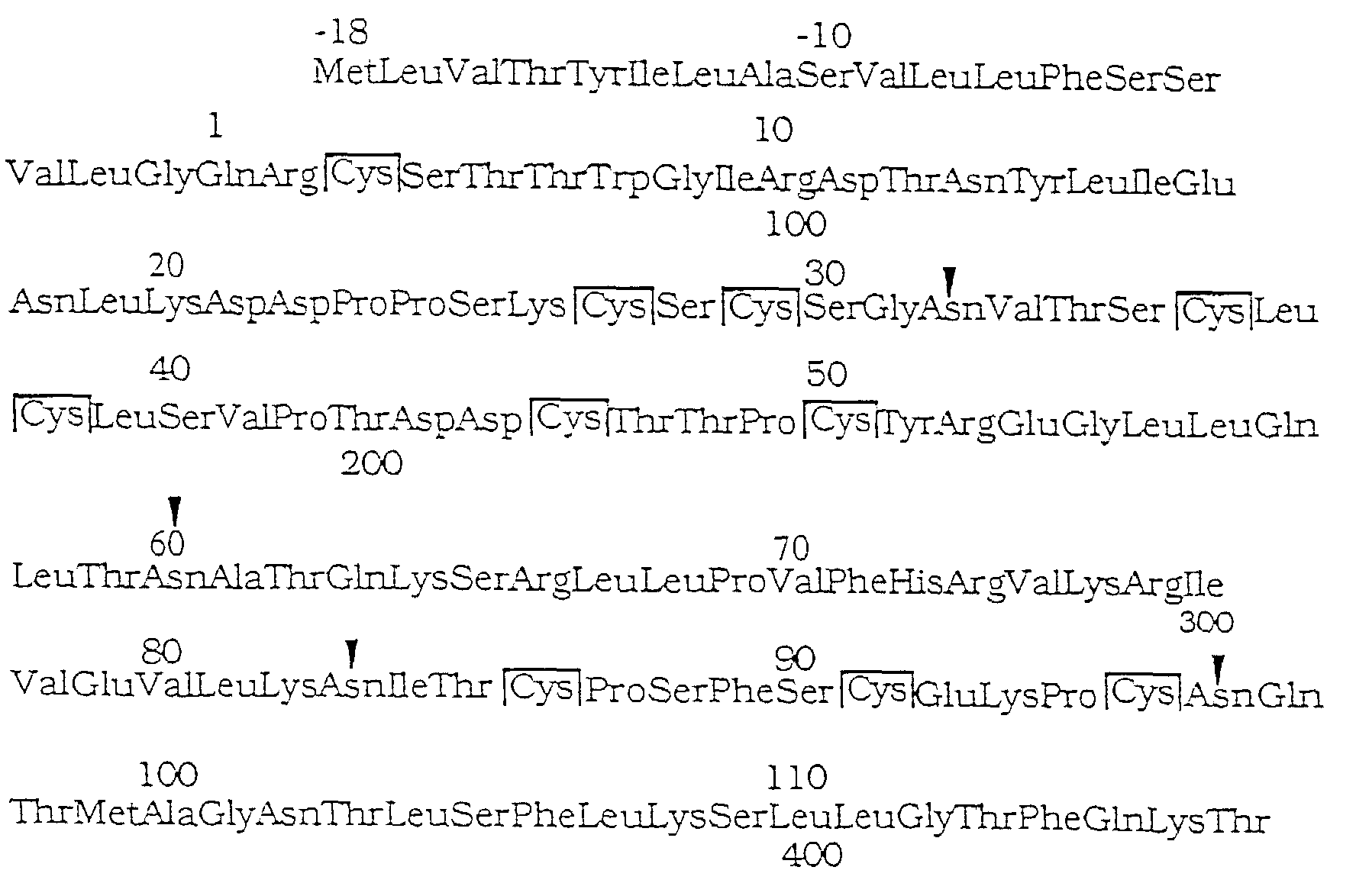

- T-Zellen-Wachstumsfaktor gemäß Anspruch 2, wobei der genannte Maus-Wachstumsfaktor eine Aminosäuresequenz aufweist, die aus Folgendem besteht:

- T-Zellen-Wachstumsfaktor gemäß Anspruch 1, wobei das genannte Glykoprotein in der Lage ist, die Vermehrung der auf IL3- oder IL4- reagierenden Zellen zu verbessern.
- Ein aktives Derivat oder aktives Fragment eines Säugetier-T-Zellen-Wachtumsfaktors gemäß jedem der Ansprüche 1 bis 7, wobei die genannte Aktivität das Interleukin-2- und Interleukin-4-unabhängige Wachstum von T-Hilfszellen stimuliert.
- Ein isoliertes proteolytisches Fragment eines T-Zellen-Wachstumsfaktors gemäß jedem der Ansprüche 1 bis 7.
- Methode zum Präparieren eines Säugetier-T-Zellen-Wachstumsfaktors mit einem Molekulargewicht von ca. 30 kDa bis 40 kDa wie mit SDS-PAGE ermittelt, wobei der genannte Wachstumsfaktor ein Glykoprotein und dazu in der Lage ist, Interleulin-2-und Interleukin-4-unabhängiges Wachstum der T-Hilfszellen zu unterstützen und die Methode vorsieht, eine T-Hilfszellen-Kultur mit einem Antigen oder einem Mitogen für eine bestimmte Zeit in Kontakt zu bringen und unter Bedingungen die ausreichen, um die Produktion des genannten Faktors zu induzieren und den genannten Faktor von der genannten Kultur zurückzugewinnen.
- Methode zum Reinigen eines Säugetier-T-Zellen-Wachstumsfaktors mit einem Molekulargewicht von ca. 30 kDa bis 40 kDa wie mit SDS-PAGE ermittelt, wobei der genannte Wachstumsfaktor ein Glykoprotein ist und die Methode vorsieht, eine überstehende Kulturflüssigkeit einer antigen- oder mitogen-stimulierten T-Hilfszell-Linie mit einem hydrophobischen Chromatografieharz in Kontakt zu bringen, eine erste Fraktion davon mit T-Zellert-Wachstumsaktivität zu identifizieren und zurückzugewinnen, die genannte erste Fraktion mit einem Ionenaustausch-Chromatographieharz in Kontakt zu bringen und eine zweite Fraktion davon mit der genannten Aktivität zu identifizieren und zurückzugewinnen, die genannte zweite Fraktion mit einem Hochdruck-Flüssigkeitschromatografieharz mit umgekehrten Phasen in Kontakt zu bringen und eine dritte Fraktion davon mit der genannten Aktivität zu identifizieren und zurückzugewinnen, wobei die dritte Fraktion ein homogener Säugetier-T-Zellen-Wachstumsfaktor ist.
- Methode gemäß Anspruch 11, wobei das hydrophobische Harz TSK-Phenyl-Chromatografieharz ist, das genannte Ionenaustauschharz Mono-Q-Chromatographie-Harzsand ist und der genannte Harz mit umgekehrten Phasen ein C1-Hochdruck-Flüssigkeitschromatografieharz mit umgekehrten Phasen ist.
- Methode gemäß Anspruch 11, wobei die genannte Aktivität das Interleukin-2- und Interleukin-4-unabhängige Wachstums von T-Hilfszellen stimuliert.
- Einsatz einer wirksamen Menge eines Säugetier-T-Zellen-Wachstumsfaktors, wie in jedem der Ansprüche 1 bis 7 definiert, einem aktiven Derivat davon oder einem aktiven Fragment davon zum Präparieren eines pharmazeutischen Mittels das zur Vermehrung von T-Hilfszellen dient.
- Einsatz einer wirksamen Menge eines Säugetier-T-Zellen-Wachstumsfaktors, wie in jedem der Ansprüche 1 bis 7 definiert und von IL3 zum Präparieren eines pharmazeutischen Mittels, das zur Vermehrung von auf IL3-reagierenden Zellen in einem Säugetier dient.
- Einsatz einer wirksamen Menge eines Säugetier-T-Zellen-Wachstumsfaktors, wie in jedem der Ansprüche 1 bis 7 definiert und von IL4 zum Präparieren eines pharmazeutischen Mittels, das zur Vermehrung von auf IL4-reagierenden Zellen in einem Säugetier dient.
- Einsatz gemäß Anspruch 15 oder 16 wobei die genannten Zellen T-Hilfszellen sind.
- Einsatz gemäß Anspruch 14, 15 oder 16 wobei das genannte Säugetier ein Mensch ist.
- Einsatz von Ansprüchen 14, 15 oder 16, wobei die wirksame Menge des genannten T-Zellen-Wachstumsfaktors, des genannten Derivats davon, des genannten Fragments davon, des genannten IL3 oder des genannten IL4 ca. 10 µg bis 1000 µg pro kg Körpergewicht pro Tag ist.
- Einsatz von Ansprüchen 14, 15 oder 16 wobei das pharmazeutische Mittel intravenös, intramuskulär, intranasal, intradermal, intraperitoneal, in Zäpfenform oder oral verabreicht wird.
- Einsatz einer wirksamen Menge eines Nucleinsäuremoleküls zum Kodieren eines Säugetier-T-Zellen-Wachstumsfaktors, wie in jedem der Ansprüche 1 bis 7 definiert, eines aktiven Derivats davon oder eines aktiven Fragments davon zum Präparieren eines pharmazeutischen Mittels, das zur Vermehrung der T-Hilfszellen in einem Säugetier dient, wobei die genannte Nucleinsäure die Darstellung einer wirksamen Menge des genannten Säugetier-T-Zellen-Wachstumsfaktors, wie in jedem der Ansprüche 1 bis 7 definiert, des genannten Derivats oder des genannten Fragments beeinflußt.
- Einsatz einer wirksamen Menge eines Nucleinsäuremoleküls zum Kodieren von IL3 oder von IL3 und einer wirksamen Menge einer Nucleinsäure zum Kodieren eines Säugetier-T-Zellen-Wachstumsfaktors, wie in jedem der Ansprüche 1 bis 7 definiert oder eines Säugetier-T-Zellen-Wachstumsfaktors, wie in jedem der Ansprüche 1 bis 7 definiert, zum Präparieren eines pharmazeutischen Mittels, das zur Vermehrung der auf IL3-reagierenden Zellen in einem Säugetier dient, wobei die genannte Nucleinsäure die Darstellung einer wirksamen Menge des genannten IL3 oder des genannten Säugetier-T-Zellen-Wachstumsfaktors, wie in jedem der Ansprüche 1 bis 7 definiert, beeinflußt.
- Einsatz einer wirksamen Menge eines Nucleinsäuremoleküls zum Kodieren von IL4 oder von IL4 und einer wirksamen Menge einer Nucleinsäure zum Kodieren eines Säugetier-T-Zellen-Wachstumsfaktors, wie in jedem der Ansprüche 1 bis 7 definiert oder eines Säugetier-T-Zellen-Wachstumsfaktors, wie in jedem der Ansprüche 1 bis 7 definiert, zum Präparieren eines pharmazeutischen Mittels, das zur Vermehrung der auf IL4-reagierendert Zellen in einem Säugetier dient, wobei die genannte Nucleinsäure die Darstellung einer wirksamen Menge IL4 oder des genannten Säugetier-T-Zellen-Wachstumsfaktors beeinflußt.
- Einsatz einer wirksamen Menge eines Säugetier-T-Zellen-Wachstumsfaktors, wie in jedem der Ansprüche 1 bis 7 definiert oder eines aktiven Derivats davon oder eines aktiven Fragments davon zum Präparieren eines pharmazeutischen Mittels das zur Vermehrung der T-Hilfszellen dient, um Immunitätsdefekte zu behandeln.
- Einsatz einer wirksamen Menge von IL3 und eines Säugetier-T-Zellen-Wachstumsfaktors, wie in jedem der Ansprüche 1 bis 7 definiert zum Präparieren eines pharmazeutischen Mittels, das zur Vermehrung der auf IL3-reagierenden Zellen dient, um Immunitätsdefekte zu behandeln.
- Einsatz einer wirksamen Menge von IL4 und eines Säugetier-T-Zellen-Wachstumsfaktors, wie in jedem der Ansprüche 1 bis 7 definiert zum Präparieren eines pharmazeutischen Mittels, das zur Vermehrung der auf IL4-reagierenden Zellen dient, um Immunitätsdefekte zu behandeln.
- Einsatz gemäß Anspruch 25 oder 26 wobei die genannten Zellen T-Hilfszellen sind.
- Einsatz gemäß Anspruch 24, 25 oder 26 wobei das genannte Säugetier ein Mensch ist.
- Einsatz gemäß den Ansprüchen 24, 25 oder 26 wobei die wirksame Menge des genannten Säugetier-T-Zellen-Wachstumsfaktors, des genannten Derivats davon oder des genannten Fragments davon, des genannten IL3 oder des genannten IL4 zwischen ca. 10 µg und 1000 µg pro kg Körpergewicht pro Tag liegt.
- Einsatz gemäß Anspruch 24, 25 oder 26 wobei das pharmazeutische Mittel intravenös, intramuskulär, intranasal, intradermal, intraperitoneal in Zäpfenform oder oral verabreicht wird.
- Pharmazeutische Zusammensetzung, die aus einer wirksamen Menge eines Säugetier-T-Zellen-Wachstumsfaktors, wie in jedem der Ansprüche 1 bis 7 definiert, eines aktiven Derivats davon, eines aktiven Fragments davon oder eines Ausgangsstoffes davon und einem pharmazeutisch akzeptablen Träger besteht.
- Eine pharmazeutische Zusammensetzung, die aus einer wirksamen Menge IL3 und einem Säugetier-T-Zellen-Wachstumsfaktor, wie in jedem der Ansprüche 1 bis 7 definiert oder einem Ausgangsstoff davon und einem pharmazeutisch akzeptablen Träger besteht.
- Eine pharmazeutische Zusammensetzung, die aus einer wirksamen Menge IL4 und einem Säugetier-T-Zellen-Wachstumsfaktor, wie in jedem der Ansprüche 1 bis 7 definiert oder einem Ausgangsstoff davon und einem pharmazeutisch akzeptablen Träger besteht.
- Eine pharmazeutische Zusammensetzung von Anspruch 31, 32 oder 33, wobei der genannte Ausgangsstoff eine Nucleinsäure zum Kodieren des genannten Säugetier-T-Zellen-Wachstumsfaktors, wie in jedem der Ansprüche 1 bis 7 definiert, des genannten Derivats davon, des genannten Fragments davon, des genannten IL3 oder des genannten IL4 ist.
- Verfahren von jedem der Ansprüche 31, 32 oder 33, wobei die wirksame Menge des genannten Säugetier-T-Zellen-Wachstumsfaktors gemäß den Ansprüchen 1 bis 7, des genannten Derivats davon, des genannten Fragments davon, des genannten IL3 oder des genannten IL4 zwischen ca. 10 µg und 1000 µg pro kg Körpergewicht pro Tag liegt.
- Antikörper zum T-Zellen-Wachstumsfaktor von jedem der Ansprüche 1 bis 7, ein antigenes Derivat davon oder ein antigenes Fragment davon.
- Antikörper gemäß Anspruch 36 wobei der Säugetier-T-Zellen-Wachstumsfaktor ein Maus-Wachstumsfaktor ist.
- Antikörper gemäß Anspruch 36 oder 37 wobei der Antikörper polyklonal oder monoklonal ist.
- Methode zum Nachweis des genannten Säugetier-T-Zellen-Wachstumsfaktors, wie in jedem der Ansprüche 1 bis 7 definiert, in Säugetiergewebe, Gewebeextrakt oder Serum, die vorsieht, das genannte Gewebe, Gewebeextrakt oder Serum mit einem Antikörper von Anspruch 36 solange in Kontakt zu bringen, daß sich ein Antikörperkomplex des Säugetier-T-Zellen-Wachstumsfaktors bilden kann und der Antikörperkomplex des Säugetier-T-Zellen-Wachstumsfaktors von einer Nachweisvorrichtung untersucht wird.
- Isolierte Nucleinsäure zum Kodieren eines Säugetier-T-Zellen-Wachstumsfaktors, wie in jedem der Ansprüche 1 bis 7 definiert, eines Derivats davon oder eines Fragments davon.
- Nucleinsäure von Anspruch 40, wobei die genannte Nucleinsäure ein RNS ist, ein mRNS, ein cDNS oder ein rekombiniertes DNS, das ein cDNS oder einen genomischen Klon des genannten Säugetier-T-Zellen-Wachstumsfaktors, wie in jedem der Ansprüche 1 bis 7 definiert oder des genannten Derivats kodiert.
- Nucleinsäure gemäß Anspruch 40, wobei das genannte Säugetier eine Maus ist.
- Nucleinsäure gemäß Anspruch 41, wobei das genannte cDNS eine Nucleotidsequenz zum Kodieren von Säugetier-T-Zellen-Wachstumsfaktor aufweist, die aus Folgendem besteht:
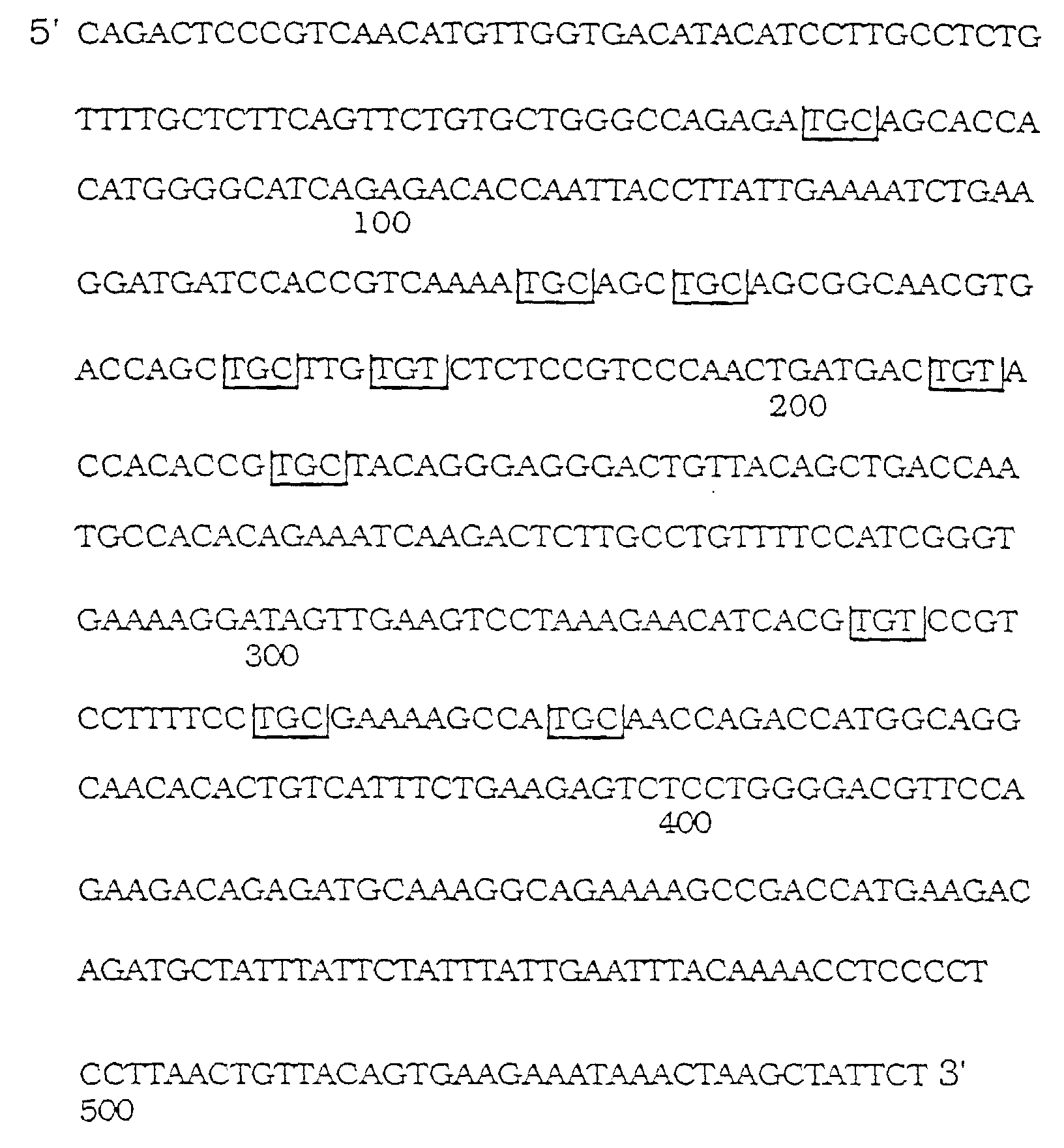
- Nucleinsäure, die eine Nucleotidsequenz zum Kodieren einer Aminosäuresequenz oder eines Maus-T-Zellen-Wachstumsfaktors aufweist, die aus Folgendem besteht:


- Nucleinsäure, die eine Nucleotidsequenz zum Kodieren einer Aminosäuresequenz oder eines Maus-T-Zellen-Wachstumsfaktors aufweist, die aus Folgendem besteht:

- Isolierte Nucleinsäure zum Kodieren des genomischen Gens für den Maus-T-Zellen-Wachstumsfaktor gemäß jedem der Ansprüche 1 bis 7.
- Replizierbarer Darstellungsvektor der aus Nucleinsäure gemäß jedem der Ansprüche 40 bis 46 besteht, wobei die genannte Nucleinsäure funktionell mit einer Nucleotidsequenz verbunden ist, die in der Lage ist die Darstellung der genannten Nucleinsäure zu bewirken.
- Vektor gemäß Anspruch 47, wobei der genannte Vektor in der Lage ist, sich in einem Eukaryot oder Protokaryot zu replizieren.
- Mikroorganismus oder Zelle der/die von der Nucleinsäure gemäß jedem der Ansprüche 40 bis 46 umgewandelt wird.
- Mikroorganismus oder Zelle der/die vom Vektor gemäß Anspruch 47 umgewandelt wird.
- Rekombinierter Säugetier-T-Zellen-Wachstumsfaktor mit einem Molekulargewicht von ca. 30 kDa bis 40 kDa wie mit SDS-PAGE ermittelt, wobei der Wachstumsfaktor ein Glykoprotein ist und den T-Hilfszellen-Wachstumsfaktor in Abwesenheit von IL-2 und IL-4, einem Derivat davon oder einem Fragment davon unterstützt.
- Säugetier-T-Zellen-Wachstumsfaktor von Anspruch 51 wobei das genannte Säugetier eine Maus ist.
- Säugetier-T-Zellen-Wachstumsfaktor der aus dem Vektor von Anspruch 47 erzeugt wird.
- Kompartimentierter Testsatz zum Nachweis des Säugetier-T-Zellen-Wachstumsfaktors, wie in jedem der Ansprüche 1 bis 7 definiert und adaptiert um Folgendes aufzunehmen: einen ersten Behälter der einen Antikörper zu den Säugetier-T-Zellen-Wachstumsfaktor enthält, ein antigenes Derivat davon oder ein antigenisches Fragment davon und einen zweiten Behälter, der einen zweiten Antikörper enthält, wobei der zweite Antikörper dazu in der Lage ist, einen Antikörper-Antigenkomplex des genannten ersten Antikörpers mit dem genannten Säugetier-T-Zellen-Wachstumsfaktor, dem genannten Derivat oder dem genannten Fragment nachzuweisen und der genannte zweite Antikörper mit einem Signalmolekül markiert wird und in der Lage ist ein erkennbares Signal abzugeben.
- Testsatz gemäß Abspruch 54, wobei das Signalmolekül ein Radioisotop, ein Enzym, ein Fluoreszenzmolekül, ein Chemilumineszenzmolekül oder ein Biolumineszenzmolekül ist.
- Testsatz gemäß Anspruch 54, wobei das Signalmolekül ein Enzym ist.
- Testsatz gemäß Anspruch 56, wobei der Testsatz weiterhin einen dritten Behälter aufweist, der ein Substrat für die Enzyme enthält.
- Verfahren gemäß Anspruch 1, wobei der T-Zellen-Wachstumsfaktor eine spezifische Aktivität von ca. 10 U/mg bis ca. 1010 U/mg aufweist.
- Verfahren gemäß Anspruch 1, wobei der T-Zellen-Wachstumsfaktor eine spezifische Aktivität von ca. 108 U/mg aufweist.
Applications Claiming Priority (2)
| Application Number | Priority Date | Filing Date | Title |
|---|---|---|---|
| US246482 | 1988-09-19 | ||
| US07/246,482 US5208218A (en) | 1988-09-19 | 1988-09-19 | T cell growth factor glycoproteins |
Publications (3)
| Publication Number | Publication Date |
|---|---|
| EP0361284A2 EP0361284A2 (de) | 1990-04-04 |
| EP0361284A3 EP0361284A3 (de) | 1990-09-05 |
| EP0361284B1 true EP0361284B1 (de) | 1998-06-17 |
Family
ID=22930867
Family Applications (1)
| Application Number | Title | Priority Date | Filing Date |
|---|---|---|---|
| EP89117317A Expired - Lifetime EP0361284B1 (de) | 1988-09-19 | 1989-09-19 | T-Zellen-Wachstumsfaktor |
Country Status (12)
| Country | Link |
|---|---|
| US (2) | US5208218A (de) |
| EP (1) | EP0361284B1 (de) |
| JP (1) | JP3223260B2 (de) |
| AT (1) | ATE167519T1 (de) |
| AU (1) | AU621661B2 (de) |
| CA (1) | CA1341461C (de) |
| DE (1) | DE68928710T2 (de) |
| DK (1) | DK175845B1 (de) |
| ES (1) | ES2118058T3 (de) |
| IE (1) | IE81152B1 (de) |
| IL (1) | IL91691A (de) |
| PT (1) | PT91750B (de) |
Families Citing this family (18)
| Publication number | Priority date | Publication date | Assignee | Title |
|---|---|---|---|---|
| US5208218A (en) * | 1988-09-19 | 1993-05-04 | Ludwig Institute For Cancer Research | T cell growth factor glycoproteins |
| US5587302A (en) * | 1988-09-19 | 1996-12-24 | Ludwig Institute For Cancer Research | T cell growth factor |
| US5414071A (en) * | 1989-05-23 | 1995-05-09 | Genetics Institute, Inc. | Human cytokine IL-9 |
| US5580753A (en) * | 1989-05-23 | 1996-12-03 | Ludwig Institute For Cancer Research | DNA encoding the human cytokine, interleukin-9 |
| CA2069090A1 (en) * | 1989-11-21 | 1991-05-22 | John A. Hamilton | Anti-inflammatory compositions and methods |
| US5132109A (en) * | 1990-10-05 | 1992-07-21 | Ludwig Institute For Cancer Research | Method for inhibiting production of ige and method for enhancing production of igg using interleukin 9 and inhibitors thereof |
| US5250295A (en) * | 1991-11-04 | 1993-10-05 | The Regents Of The Univ. Of California | Natural killer cell enhancing factor |
| AU5102993A (en) * | 1992-09-18 | 1994-04-12 | Merck & Co., Inc. | Dna encoding murine precursor interleukin 1beta converting enzyme |
| TW257792B (de) * | 1992-10-01 | 1995-09-21 | Lilly Co Eli | |
| US5643881A (en) * | 1994-08-04 | 1997-07-01 | Tampa Bay Research Institute | Protein having anti-HIV activity and method for obtaining it |
| CA2666298A1 (en) * | 1995-08-24 | 1997-03-06 | Genaera Corporation | Asthma associated factors as targets for treating atopic allergies including asthma and related disorders |
| ZA967182B (en) * | 1995-08-24 | 1998-05-25 | Magainin Pharma | Asthma associated factors as targets for treating atopic allergies including asthma and related disorders. |
| AU2007202406B2 (en) * | 1995-08-24 | 2010-08-12 | Ligand Pharmaceuticals Inc. | Asthma associated factors as targets for treating atopic allergies including asthma and related disorders |
| WO1998024904A2 (en) | 1996-12-02 | 1998-06-11 | Magainin Pharmaceuticals Inc. | Il-9 receptor variants, useful in treating and diagnosing atopic allergies including asthma and related disorders |
| WO2002100432A1 (en) | 2001-06-08 | 2002-12-19 | Genaera Corporation | Methods for the modulation of il-13 |
| WO2003086458A1 (en) | 2002-04-12 | 2003-10-23 | Medimmune, Inc. | Recombinant anti-interleukin-9 antibodies |
| DE602004028007D1 (de) * | 2003-05-14 | 2010-08-19 | Bayer Pharmaceuticals Corp | Interleukin-9 antagonist-muteine und ihre pharmakologischen anwendungsverfahren |
| EP2224946B1 (de) * | 2007-12-28 | 2016-11-02 | Kalos Therapeutics, Inc. | Proliferationshemmende verbindungen und ihre verwendung |
Family Cites Families (5)
| Publication number | Priority date | Publication date | Assignee | Title |
|---|---|---|---|---|
| US4636463A (en) * | 1984-04-05 | 1987-01-13 | Scripps Clinic And Research Foundation | Antibodies to human interleukin-2 induced by synthetic polypeptides |
| US5006459A (en) * | 1986-03-31 | 1991-04-09 | T Cell Sciences, Inc. | Therapeutic and diagnostic methods using soluble T cell surface molecules |
| US4935343A (en) * | 1986-08-08 | 1990-06-19 | Syntex (U.S.A.) Inc. | Monoclonal antibodies for interleukin-1β |
| US5208218A (en) * | 1988-09-19 | 1993-05-04 | Ludwig Institute For Cancer Research | T cell growth factor glycoproteins |
| GR1001018B (el) * | 1989-05-24 | 1993-03-31 | Abbott Lab | Μεθοδος παρασκευης προδρομικων πεπτικων παραγωγων. |
-
1988
- 1988-09-19 US US07/246,482 patent/US5208218A/en not_active Expired - Lifetime
-
1989
- 1989-09-15 US US07/408,155 patent/US5157112A/en not_active Expired - Lifetime
- 1989-09-18 DK DK198904588A patent/DK175845B1/da not_active IP Right Cessation
- 1989-09-18 CA CA000611819A patent/CA1341461C/en not_active Expired - Fee Related
- 1989-09-19 EP EP89117317A patent/EP0361284B1/de not_active Expired - Lifetime
- 1989-09-19 IE IE298389A patent/IE81152B1/en not_active IP Right Cessation
- 1989-09-19 ES ES89117317T patent/ES2118058T3/es not_active Expired - Lifetime
- 1989-09-19 DE DE68928710T patent/DE68928710T2/de not_active Expired - Lifetime
- 1989-09-19 AU AU41563/89A patent/AU621661B2/en not_active Expired
- 1989-09-19 AT AT89117317T patent/ATE167519T1/de not_active IP Right Cessation
- 1989-09-19 IL IL91691A patent/IL91691A/xx not_active IP Right Cessation
- 1989-09-19 PT PT91750A patent/PT91750B/pt not_active IP Right Cessation
- 1989-09-19 JP JP24328789A patent/JP3223260B2/ja not_active Expired - Lifetime
Also Published As
| Publication number | Publication date |
|---|---|
| CA1341461C (en) | 2004-10-26 |
| EP0361284A2 (de) | 1990-04-04 |
| DK458889D0 (da) | 1989-09-18 |
| ES2118058T3 (es) | 1998-09-16 |
| EP0361284A3 (de) | 1990-09-05 |
| JP3223260B2 (ja) | 2001-10-29 |
| IE81152B1 (en) | 2000-05-03 |
| US5208218A (en) | 1993-05-04 |
| DK175845B1 (da) | 2005-03-29 |
| PT91750B (pt) | 1995-05-31 |
| IE892983L (en) | 1990-03-19 |
| AU621661B2 (en) | 1992-03-19 |
| DE68928710D1 (de) | 1998-07-23 |
| IL91691A0 (en) | 1990-06-10 |
| IL91691A (en) | 1997-07-13 |
| AU4156389A (en) | 1990-03-22 |
| JPH034796A (ja) | 1991-01-10 |
| PT91750A (pt) | 1990-03-30 |
| ATE167519T1 (de) | 1998-07-15 |
| DE68928710T2 (de) | 1999-02-11 |
| DK458889A (da) | 1990-03-20 |
| US5157112A (en) | 1992-10-20 |
Similar Documents
| Publication | Publication Date | Title |
|---|---|---|
| EP0361284B1 (de) | T-Zellen-Wachstumsfaktor | |
| Uyttenhove et al. | Functional and structural characterization of P40, a mouse glycoprotein with T-cell growth factor activity. | |
| EP0790255B1 (de) | Monoklonale Antikörper spezifisch für den zytotoxischen Lymphozyten-Reifefaktor | |
| Brown et al. | A family of small inducible proteins secreted by leukocytes are members of a new superfamily that includes leukocyte and fibroblast-derived inflammatory agents, growth factors, and indicators of various activation processes. | |
| AU696764C (en) | Vascular endothelial growth factor 2 | |
| EP0712931B2 (de) | Interferon-gamma-induzierendes Polypeptid monoklonal Antikörper, und Zusammensetzung für Interferon-gamma gebunden Krankheiten | |
| EP0871672A1 (de) | Menschliche chemokine beta - 8 beta - 1 und macrophagen - entzündungsprotein -4 | |
| AU7672394A (en) | Human chemokine polypeptides | |
| EP0409091A1 (de) | Vom Carbonylende abgeleitete Antagonisten von GM-CSF | |
| WO1996032481A1 (en) | New chemokine expressed in eosinophils | |
| EP0799311A1 (de) | Menschlicher beta-9 chemokin | |
| WO1996024668A1 (en) | Human chemokine beta-11 and human chemokine alpha-1 | |
| US5587302A (en) | T cell growth factor | |
| EP0832112A1 (de) | Monocytäres chemotaktisches protein-4 | |
| US5962647A (en) | Bovine interleukin-1β protein and amino acid sequence | |
| EP0777494A1 (de) | Menschliche chemokine polypeptide | |
| EP0988319A1 (de) | Humane l105 proteine und dafür kodierende polynukleotide |
Legal Events
| Date | Code | Title | Description |
|---|---|---|---|
| PUAI | Public reference made under article 153(3) epc to a published international application that has entered the european phase |
Free format text: ORIGINAL CODE: 0009012 |
|
| AK | Designated contracting states |
Kind code of ref document: A2 Designated state(s): AT BE CH DE ES FR GB GR IT LI LU NL SE |
|
| PUAL | Search report despatched |
Free format text: ORIGINAL CODE: 0009013 |
|
| AK | Designated contracting states |
Kind code of ref document: A3 Designated state(s): AT BE CH DE ES FR GB GR IT LI LU NL SE |
|
| 17P | Request for examination filed |
Effective date: 19910207 |
|
| 17Q | First examination report despatched |
Effective date: 19930126 |
|
| RAP1 | Party data changed (applicant data changed or rights of an application transferred) |
Owner name: RESEARCH CORPORATION TECHNOLOGIES, INC. |
|
| RAP1 | Party data changed (applicant data changed or rights of an application transferred) |
Owner name: LUDWIG INSTITUTE FOR CANCER RESEARCH |
|
| GRAG | Despatch of communication of intention to grant |
Free format text: ORIGINAL CODE: EPIDOS AGRA |
|
| GRAG | Despatch of communication of intention to grant |
Free format text: ORIGINAL CODE: EPIDOS AGRA |
|
| GRAH | Despatch of communication of intention to grant a patent |
Free format text: ORIGINAL CODE: EPIDOS IGRA |
|
| GRAH | Despatch of communication of intention to grant a patent |
Free format text: ORIGINAL CODE: EPIDOS IGRA |
|
| GRAA | (expected) grant |
Free format text: ORIGINAL CODE: 0009210 |
|
| AK | Designated contracting states |
Kind code of ref document: B1 Designated state(s): AT BE CH DE ES FR GB GR IT LI LU NL SE |
|
| REF | Corresponds to: |
Ref document number: 167519 Country of ref document: AT Date of ref document: 19980715 Kind code of ref document: T |
|
| REG | Reference to a national code |
Ref country code: CH Ref legal event code: EP |
|
| REF | Corresponds to: |
Ref document number: 68928710 Country of ref document: DE Date of ref document: 19980723 |
|
| ITF | It: translation for a ep patent filed | ||
| REG | Reference to a national code |
Ref country code: CH Ref legal event code: NV Representative=s name: BRAUN & PARTNER PATENT-, MARKEN-, RECHTSANWAELTE |
|
| REG | Reference to a national code |
Ref country code: ES Ref legal event code: FG2A Ref document number: 2118058 Country of ref document: ES Kind code of ref document: T3 |
|
| ET | Fr: translation filed | ||
| PLBE | No opposition filed within time limit |
Free format text: ORIGINAL CODE: 0009261 |
|
| STAA | Information on the status of an ep patent application or granted ep patent |
Free format text: STATUS: NO OPPOSITION FILED WITHIN TIME LIMIT |
|
| 26N | No opposition filed | ||
| REG | Reference to a national code |
Ref country code: GB Ref legal event code: IF02 |
|
| PGFP | Annual fee paid to national office [announced via postgrant information from national office to epo] |
Ref country code: ES Payment date: 20080924 Year of fee payment: 20 |
|
| PGFP | Annual fee paid to national office [announced via postgrant information from national office to epo] |
Ref country code: AT Payment date: 20080924 Year of fee payment: 20 Ref country code: NL Payment date: 20080930 Year of fee payment: 20 |
|
| PGFP | Annual fee paid to national office [announced via postgrant information from national office to epo] |
Ref country code: GB Payment date: 20080918 Year of fee payment: 20 |
|
| PGFP | Annual fee paid to national office [announced via postgrant information from national office to epo] |
Ref country code: LU Payment date: 20081003 Year of fee payment: 20 Ref country code: DE Payment date: 20081017 Year of fee payment: 20 Ref country code: CH Payment date: 20081016 Year of fee payment: 20 |
|
| PGFP | Annual fee paid to national office [announced via postgrant information from national office to epo] |
Ref country code: BE Payment date: 20080929 Year of fee payment: 20 Ref country code: SE Payment date: 20080924 Year of fee payment: 20 |
|
| PGFP | Annual fee paid to national office [announced via postgrant information from national office to epo] |
Ref country code: IT Payment date: 20080930 Year of fee payment: 20 |
|
| PGFP | Annual fee paid to national office [announced via postgrant information from national office to epo] |
Ref country code: FR Payment date: 20080930 Year of fee payment: 20 |
|
| PGFP | Annual fee paid to national office [announced via postgrant information from national office to epo] |
Ref country code: GR Payment date: 20080930 Year of fee payment: 20 |
|
| BE20 | Be: patent expired |
Owner name: *LUDWIG INSTITUTE FOR CANCER RESEARCH Effective date: 20090919 |
|
| REG | Reference to a national code |
Ref country code: CH Ref legal event code: PL |
|
| REG | Reference to a national code |
Ref country code: GB Ref legal event code: PE20 Expiry date: 20090918 |
|
| NLV7 | Nl: ceased due to reaching the maximum lifetime of a patent |
Effective date: 20090919 |
|
| EUG | Se: european patent has lapsed | ||
| REG | Reference to a national code |
Ref country code: ES Ref legal event code: FD2A Effective date: 20090921 |
|
| PG25 | Lapsed in a contracting state [announced via postgrant information from national office to epo] |
Ref country code: NL Free format text: LAPSE BECAUSE OF EXPIRATION OF PROTECTION Effective date: 20090919 Ref country code: GB Free format text: LAPSE BECAUSE OF EXPIRATION OF PROTECTION Effective date: 20090918 |
|
| PG25 | Lapsed in a contracting state [announced via postgrant information from national office to epo] |
Ref country code: ES Free format text: LAPSE BECAUSE OF EXPIRATION OF PROTECTION Effective date: 20090921 |