EP0362494A1 - Katheterset für die Spinalanästhesie - Google Patents
Katheterset für die Spinalanästhesie Download PDFInfo
- Publication number
- EP0362494A1 EP0362494A1 EP89112699A EP89112699A EP0362494A1 EP 0362494 A1 EP0362494 A1 EP 0362494A1 EP 89112699 A EP89112699 A EP 89112699A EP 89112699 A EP89112699 A EP 89112699A EP 0362494 A1 EP0362494 A1 EP 0362494A1
- Authority
- EP
- European Patent Office
- Prior art keywords
- cannula
- spinal
- catheter
- epidural
- catheter set
- Prior art date
- Legal status (The legal status is an assumption and is not a legal conclusion. Google has not performed a legal analysis and makes no representation as to the accuracy of the status listed.)
- Ceased
Links
Images

Classifications
-
- A—HUMAN NECESSITIES
- A61—MEDICAL OR VETERINARY SCIENCE; HYGIENE
- A61M—DEVICES FOR INTRODUCING MEDIA INTO, OR ONTO, THE BODY; DEVICES FOR TRANSDUCING BODY MEDIA OR FOR TAKING MEDIA FROM THE BODY; DEVICES FOR PRODUCING OR ENDING SLEEP OR STUPOR
- A61M25/00—Catheters; Hollow probes
- A61M25/01—Introducing, guiding, advancing, emplacing or holding catheters
- A61M25/06—Body-piercing guide needles or the like
- A61M25/065—Guide needles
-
- A—HUMAN NECESSITIES
- A61—MEDICAL OR VETERINARY SCIENCE; HYGIENE
- A61B—DIAGNOSIS; SURGERY; IDENTIFICATION
- A61B17/00—Surgical instruments, devices or methods
- A61B17/34—Trocars; Puncturing needles
- A61B17/3401—Puncturing needles for the peridural or subarachnoid space or the plexus, e.g. for anaesthesia
-
- A—HUMAN NECESSITIES
- A61—MEDICAL OR VETERINARY SCIENCE; HYGIENE
- A61M—DEVICES FOR INTRODUCING MEDIA INTO, OR ONTO, THE BODY; DEVICES FOR TRANSDUCING BODY MEDIA OR FOR TAKING MEDIA FROM THE BODY; DEVICES FOR PRODUCING OR ENDING SLEEP OR STUPOR
- A61M25/00—Catheters; Hollow probes
- A61M25/01—Introducing, guiding, advancing, emplacing or holding catheters
- A61M25/06—Body-piercing guide needles or the like
Definitions
- the invention relates to a catheter set for spinal anesthesia according to the preamble of claim 1.
- spinal catheters have been placed intrathecally through a cannula.
- the spinal cord is punctured with a spinal or epidural cannula.
- a spinal catheter is then inserted intrathecally through the epidural cannula.
- One difficulty is that the spinal or epidural cannula must have a small outer diameter in order to minimize the size of the dura injury and the resulting post-spinal headache.
- the spinal or epidural cannula must have such a large diameter that the spinal catheter can be passed through it.
- spinal or epidural cannulas are currently used in which the outer diameter is larger than 0.65 mm (23 G).
- the invention has for its object to provide a catheter set of the type specified in the preamble of claim 1, with which the smallest possible dura opening is created when puncturing the dura and with which a relatively thick spinal catheter can still be used.
- the epidural cannula is first advanced to the outer wall of the dura.
- the thinner spinal cannula is then inserted into the epidural cannula to perforate the dura.
- the stylet of the spinal cannula is pulled out, the guide wire is inserted into the spinal cannula, the spinal cannula is pulled out over the guide wire and the spinal catheter is pushed over the guide wire.
- the catheter is inserted over the guide wire in a manner similar to that in the known Seldinger technique, a catheter is inserted into a blood vessel. After the tip of the catheter has reached the spinal space, the guidewire and epidural cannula are withdrawn.
- the relatively thick epidural cannula was used to keep the perforation channel open through the muscle tissue and to give the catheter the necessary directional stability when advancing.
- the epidural cannula also surrounds the spinal cannula as it is being inserted. The Advancing the spinal cannula is therefore not affected by the surrounding tissue. Only when the tip of the spinal cannula hits the dura does a clearly noticeable resistance appear. The perforation of the dura can then be carried out carefully by carefully pushing the spinal cannula forward.
- An important advantage of the invention is that the perforation site of the dura is created with the smallest possible diameter, the catheter being inserted into the perforation site only after the spinal cannula with which the perforation was carried out has been withdrawn. This makes it possible to insert a catheter into the perforation opening that completely fills this opening. The dura injury is thus kept as low as possible.
- the spinal cannula preferably contains a pull-out stylet. This stylet is located in the spinal cannula when the spinal space is perforated. After the perforation has taken place, the stylet is pulled out.
- the spinal space can be identified using the refluxing CSF.
- the guide wire used is very thin (about 0.25 mm) and preferably has a flexible tip. It can also be covered with plastic.
- the guide wire is pushed through the spinal cannula and, after the spinal cannula has been pulled out, serves as a guide element for the spinal catheter pushed through the epidural cannula.
- the insertion of the spinal catheter into the cannula tube of the epidural cannula is facilitated.
- the cannula tube is slightly funnel-shaped at the rear end, it fits into the base of a syringe that can be connected to the epidural cannula.
- an epidural cannula 10 which consists of a cannula tube 11 and a handle 12 (or cannula hub). This epidural cannula is also referred to as a Crawford cannula.
- the cannula tube has a cut 13 suitable for tissue puncture at its front tip.
- the epidural cannula 10 can e.g. Dimensions of 0.7 x 0.9 x 80 mm, the first number indicating the inner diameter, the second number the outer diameter and the third number the free length of the cannula tube.
- the handle 12 At the rear end of the handle 12 there is a funnel-shaped trough 14. The rear end of the cannula tube 11 projects through the trough 14 and still extends out of it. At the back A funnel-shaped extension 15 is provided at the end of the cannula tube 11 in order to facilitate the insertion of the spinal catheter. With the trough 14, the handle 12 is designed as a connecting element for attaching a (not shown) syringe.
- the spinal cannula 16 has dimensions of e.g. 0.35 x 0.55 x 120 mm, where the different numbers have the same meanings as in the epidural cannula explained above.
- a handle 18 At the rear end of the cannula tube 17 there is a handle 18.
- a stylet 19 In the spinal cannula 16 there is a stylet 19 which extends through the handle 18 and has a handle 20 at the rear end.
- the catheter set also includes the guide wire 21 with a flexible tip.
- This guide wire has dimensions e.g. of 0.25 x 900 mm, where the first number indicates the diameter and the second number the length. Markings 22 are attached to the guide wire 21 in order to be able to determine the feed length.
- the spinal catheter 23 consists of an elongated flexible catheter tube with dimensions of 0.30 x 0.60 x 600 mm.
- the outer diameter of the spinal catheter is essentially the same as that of the spinal cannula.
- Markings 24 are located in the vicinity of the rear end so that the insertion length can be checked.
- FIGS. 2 to 7 The use of the described catheter set is shown in FIGS. 2 to 7 recognizable.
- the skin 27 and the muscle tissue 26 are perforated with the epidural cannula 10 until the grinding of the epidural cannula 10 lies against the outside of the dura 25.
- the spinal cannula 16 with the stylet 19 located therein is pushed into the epidural cannula until intrathecally.
- the front end of the spinal cannula penetrates the dura 25.
- FIG. 4 shows how the guide wire 21 is pushed through the spinal cannula 16 lying in the epidural cannula after the stylet 19 has been pulled out beforehand.
- the soft end 21a of the guide wire 21 emerges from the front end of the spinal cannula penetrating the dura.
- the catheter 23 is pushed onto the guidewire 21 according to FIG. 5 and inserted into the epidural cannula 10.
- the funnel-shaped widening 15 facilitates the insertion of the catheter into the rear end of the cannula tube 11.
- the guide wire 21 is pulled out of the catheter as shown in FIG. 6. 7, the epidural cannula 10 is then removed above the spinal catheter 23. If necessary, the catheter can be tunneled. Finally, a connector (not shown) is connected to the rear end of the catheter to allow intrathecal injection.
Landscapes
- Health & Medical Sciences (AREA)
- Life Sciences & Earth Sciences (AREA)
- General Health & Medical Sciences (AREA)
- Public Health (AREA)
- Engineering & Computer Science (AREA)
- Anesthesiology (AREA)
- Biomedical Technology (AREA)
- Heart & Thoracic Surgery (AREA)
- Veterinary Medicine (AREA)
- Animal Behavior & Ethology (AREA)
- Biophysics (AREA)
- Pulmonology (AREA)
- Hematology (AREA)
- Surgery (AREA)
- Pathology (AREA)
- Nuclear Medicine, Radiotherapy & Molecular Imaging (AREA)
- Medical Informatics (AREA)
- Molecular Biology (AREA)
- Media Introduction/Drainage Providing Device (AREA)
- Infusion, Injection, And Reservoir Apparatuses (AREA)
Abstract
In eine Epiduralkanüle (10), die bis an die Dura vorgetrieben werden kann, wird eine Spinalkanüle (16) eingeschoben, deren Ende intrathekal placiert wird, wobei die Spinalkanüle (16) die Dura durchstößt. Dann wird ein Führungsdraht (21) durch die Spinalkanüle (16) hindurch nach intrathekal eingeschoben. Nach dem Entfernen der Spinalkanüle (16) wird über dem Führungsdraht (21) und innerhalb der Epiduralkanüle (10) ein Spinalkatheter (23) vorgeschoben. Die Epiduralkanüle (10) verleiht dem Katheter (23) beim Vorschieben die notwendige Richtungsstabilität und erleichtert das Durchdringen der bereits von der Spinalkanüle (16) perforierten Dura.
Description
- Die Erfindung betrifft ein Katheterset für die Spinalanästhesie nach dem Oberbegriff des Anspruchs 1.
- Spinalkatheter werden bisher durch eine Kanüle intrathekal placiert. Dabei erfolgt mit einer Spinal- oder Epiduralkanüle eine Punktion der Dura. Anschließend wird durch die Epiduralkanüle hindurch ein Spinalkatheter intrathekal eingeführt. Eine Schwierigkeit besteht darin, daß die Spinal- oder Epiduralkanüle einen kleinen Außendurchmesser haben muß, um die Größe der Duraverletzung und daraus entstehenden postspinalen Kopfschmerzen so gering wie möglich zu halten. Andererseits muß die Spinal- oder Epiduralkanüle einen so großen Durchmesser haben, daß der Spinalkatheter hindurchgeführt werden kann. Für sehr dünne Spinalkatheter gibt es jedoch keine Konnektoren, die eine dichte Katheter-Konnektorverbindung ermöglichen.
- Aufgrund der genannten Schwierigkeiten werden derzeit Spinal- oder Epiduralkanülen benutzt, bei denen der Außendurchmesser größer ist als 0,65 mm (23 G).
- Der Erfindung liegt die Aufgabe zugrunde, ein Katheterset der im Oberbegriff des Anspruchs 1 angegebenen Art zu schaffen, mit dem bei der Punktion der Dura eine möglichst kleine Duraöffnung erzeugt wird und bei dem dennoch ein verhältnismäßig dicker Spinalkatheter benutzbar ist.
- Die Lösung dieser Aufgabe erfolgt erfindungsgemäß mit den im kennzeichnenden Teil des Anspruchs 1 angegebenen Merkmalen.
- Mit dem erfindungsgemäßen Katheterset wird zunächst die Epiduralkanüle bis zur Außenwand der Dura vorgeschoben. Danach wird in die Epiduralkanüle die dünnere Spinalkanüle eingeführt, um mit dieser die Dura zu perforieren. Wenn die Spinalkanüle den Spinalraum erreicht hat, wird der Mandrin der Spinalkanüle herausgezogen, der Führungsdraht wird in die Spinalkanüle eingeschoben, die Spinalkanüle über den Führungsdraht herausgezogen und über den Führungsdraht wird der Spinalkatheter geschoben. Das Einführen des Katheters über den Führungsdraht erfolgt in ähnlicher Weise wie bei der bekannten Seldinger-Technik ein Katheter in ein Blutgefäß eingeführt wird. Nachdem die Katheterspitze den Spinalraum erreicht hat, werden Führungsdraht und Epiduralkanüle zurückgezogen. Die relativ dicke Epiduralkanüle diente dazu, den Perforationskanal durch das Muskelgewebe hindurch offenzuhalten und dem Katheter beim Vorschieben die notwendige Richtungsstabilität zu geben. Ferner umgibt die Epiduralkanüle die Spinalkanüle, während diese eingeführt wird. Das Vorschieben der Spinalkanüle wird daher nicht durch umgebendes Gewebe beeinträchtigt. Erst wenn die Spitze der Spinalkanüle gegen die Dura stößt, tritt ein deutlich spürbarer Widerstand auf. Die Perforation der Dura kann nachfolgend durch vorsichtiges Weiterschieben der Spinalkanüle behutsam vorgenommen werden.
- Ein wesentlicher Vorteil der Erfindung besteht darin, daß die Perforationsstelle der Dura mit geringstmöglichem Durchmesser erzeugt wird, wobei der Katheter erst in die Perforationsstelle eingeschoben wird, nachdem die Spinalkanüle, mit der die Perforation durchgeführt wurde, zurückgezogen ist. Hierdurch ist es möglich, in die Perforationsöffnung einen Katheter einzuschieben, der diese Öffnung vollständig ausfüllt. Die Duraverletzung wird somit so gering wie möglich gehalten.
- Vorzugsweise enthält die Spinalkanüle einen herausziehbaren Mandrin. Dieser Mandrin befindet sich bei der Perforation des Spinalraums in der Spinalkanüle. Nach erfolgter Perforation wird der Mandrin herausgezogen. Anhand des rückfließenden Liquor kann der Spinalraum identifiziert werden.
- Der verwendete Führungsdraht ist sehr dünn (etwa 0,25 mm) und hat vorzugsweise eine flexible Spitze. Er kann auch mit Kunststoff überzogen sein. Der Führungsdraht wird durch die Spinalkanüle hindurchgeschoben und dient nach dem Herausziehen der Spinalkanüle als Führungselement für den durch die Epiduralkanüle hindurchgeschobenen Spinalkatheter.
- Mit dem Merkmal des Anspruchs 3 wird das Einschieben des Spinalkatheters in das Kanülenrohr der Epiduralkanüle erleichtert. Obwohl das Kanülenrohr am rückwärtigen Ende leicht trichterförmig aufgebogen ist, paßt es in den Ansatz einer Spritze, die an die Epiduralkanüle angeschlossen werden kann.
- Im folgenden wird unter Bezugnahme auf die Zeichnungen ein Ausführungsbeispiel der Erfindung näher erläutert.
- Es zeigen:
- Fig. 1 eine Darstellung der Komponenten des Kathetersets und
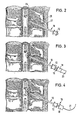
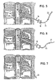
- Fign. 2 bis 7 die verschiedenen Stadien der Benutzung des Katehtersets.
- Gemäß Fig. 1 ist eine Epiduralkanüle 10 vorgesehen, die aus einem Kanülenrohr 11 und einem Griffstück 12 (oder Kanülenansatz) besteht. Diese Epiduralkanüle wird auch als Crawford-Kanüle bezeichnet. Das Kanülenrohr weist an seiner vorderen Spitze einen für die Gewebepunktion geeigneten Schliff 13 auf. Die Epiduralkanüle 10 kann z.B. Abmessungen von 0,7 x 0,9 x 80 mm haben, wobei die erste Zahl den Innendurchmesser, die zweite Zahl den Außendurchmesser und die dritte Zahl die freie länge des Kanülenrohrs angibt.
- Am rückwärtigen Ende des Griffstücks 12 befindet sich eine trichterförmige Mulde 14. Das rückwärtige Ende des Kanülenrohrs 11 ragt durch die Mulde 14 hindurch und erstreckt sich noch aus dieser heraus. Am rückwärtigen Ende des Kanülenrohrs 11 ist eine trichterförmige Erweiterung 15 vorgesehen, um das Einführen des Spinalkatheters zu erleichtern. Mit der Mulde 14 ist das Griffstück 12 als Anschlußelement zum Ansetzen einer (nicht dargestellten) Spritze ausgebildet.
- Die Spinalkanüle 16 hat Abmessungen von z.B. 0,35 x 0,55 x 120 mm, wobei die verschiedenen Zahlen die gleichen Bedeutungen haben wie bei der zuvor erläuterten Epiduralkanüle. Am rückwärtigen Ende des Kanülenrohrs 17 befindet sich ein Griffstück 18. In der Spinalkanüle 16 sitzt ein Mandrin 19, der sich durch das Griffstück 18 hindurch erstreckt und am rückwärtigen Ende einen Handgriff 20 aufweist.
- Zum Katheterset gehört ferner der Führungsdraht 21 mit flexibler Spitze. Dieser Führungsdraht hat Abmessungen z.B. von 0,25 x 900 mm, wobei die erste Zahl den Durchmesser und die zweite Zahl die Länge angibt. Am Führungsdraht 21 sind Markierungen 22 angebracht, um die Vorschublänge feststellen zu können.
- Der Spinalkatheter 23 besteht aus einem langgestreckten flexiblen Katheterschlauch mit Abmessungen von 0,30 x 0,60 x 600 mm. Der Außendurchmesser des Spinalkatheters ist im wesentlichen gleich demjenigen der Spinalkanüle. In der Nähe des rückwärtigen Endes befinden sich Markierungen 24, um die Einschublänge kontrollieren zu können.
- Die Anwendung des beschriebenen Kathetersets ist aus den Fign. 2 bis 7 erkennbar.
- Zunächst werden mit der Epiduralkanüle 10 die Haut 27 und das Muskelgewebe 26 perforiert, bis der Schliff der Epiduralkanüle 10 an der Außenseite der Dura 25 anliegt.
- Gemäß Fig. 3 wird die Spinalkanüle 16 mit dem in ihr befindlichen Mandrin 19 in die Epiduralkanüle bis nach intrathekal eingeschoben. Dabei durchdringt das vordere Ende der Spinalkanüle die Dura 25.
- Fig. 4 zeigt, wie der Führungsdraht 21 durch die in der Epiduralkanüle liegende Spinalkanüle 16 hindurchgeschoben wird, nachdem zuvor der Mandrin 19 herausgezogen worden ist. Das weiche Ende 21a des Führungsdrahtes 21 tritt aus dem vorderen Ende der die Dura durchdringenden Spinalkanüle aus.
- Nachdem die Spinalkanüle 16 über das rückwärtige Ende des Führungsdrahtes 21 hinweg zurückgezogen und aus der Epiduralkanüle 10 entfernt worden ist, wird gemäß Fig. 5 der Katheter 23 auf den Führungsdraht 21 aufgeschoben und über diesem in die Epiduralkanüle 10 eingeschoben. Hierbei erleichtert die trichterförmige Aufweitung 15 das Einführen des Katheters in das rückwärtige Ende des Kanülenrohrs 11.
- Nachdem der Katheter intrathekal liegt, wird der Führungsdraht 21 gemäß Fig. 6 aus dem Katheter herausgezogen. Anschließend wird gemäß Fig. 7 über dem Spinalkatheter 23 die Epiduralkanüle 10 entfernt. Erforderlichenfalls kann der Katheter getunnelt werden. Schließlich wird an das rückwärtige Katheterende ein (nicht dargestellter) Konnektor angeschlossen, um eine intrathekale Injektion zu ermöglichen.
Claims (9)
1. Katheterset für die Spinalanästhesie, mit
einer Epiduralkanüle (10), deren Innendurchmesser kleiner als 1,0 mm ist,
und einem durch die Epiduralkanüle (10) schiebbaren Spinalkatheter (23),
gekennzeichnet durch
eine in die Epiduralkanüle (10) einschiebbare Spinalkanüle (16), die im eingeschobenen Zustand mit ihrem vorderen Ende aus der Epiduralkanüle heraus vorsteht,
und einen durch die Spinalkanüle (16) schiebbaren Führungsdraht (21), dessen Durchmesser kleiner ist als der Innendurchmesser des Spinalkatheters (23).
einer Epiduralkanüle (10), deren Innendurchmesser kleiner als 1,0 mm ist,
und einem durch die Epiduralkanüle (10) schiebbaren Spinalkatheter (23),
gekennzeichnet durch
eine in die Epiduralkanüle (10) einschiebbare Spinalkanüle (16), die im eingeschobenen Zustand mit ihrem vorderen Ende aus der Epiduralkanüle heraus vorsteht,
und einen durch die Spinalkanüle (16) schiebbaren Führungsdraht (21), dessen Durchmesser kleiner ist als der Innendurchmesser des Spinalkatheters (23).
2. Katheterset nach Anspruch 1, dadurch gekennzeichnet, daß die Spinalkanüle (16) einen herausziehbaren Mandrin (19) enthält.
3. Katheterset nach Anspruch 1 oder 2, dadurch gekennzeichnet, daß die Epiduralkanüle (10) ein Griffstück (12) aufweist und daß das Kanülenrohr (11) am rückwärtigen Ende aus dem Griffstück (12) vorsteht und dort mit einer Aufweitung (15) versehen ist.
4. Katheterset nach Anspruch 3, dadurch gekennzeichnet, daß das Kanülenrohr (11) aus einer Mulde (14) am rückwärtigen Ende des Griffstückes (12) vorsteht.
5. Katheterset nach einem der Ansprüche 1 bis 4, dadurch gekennzeichnet, daß der Außendurchmesser der Spinalkanüle (16) kleiner ist als 0,6 mm.
6. Katheterset nach einem der Ansprüche 1 bis 5, dadurch gekennzeichnet, daß der Innendurchmesser der Epiduralkanüle etwa 0,7 mm beträgt.
7. Katheterset nach einem der Ansprüche 1 bis 6, dadurch gekennzeichnet, daß die Länge des Kanülenrohrs (11) der Epiduralkanüle (10) etwa 80 mm beträgt.
8. Katheterset nach einem der Ansprüche 1 bis 7, dadurch gekennzeichnet, daß die Spinalkanüle (16) im eingeschobenen Zustand etwa 10 bis 20 mm aus der Epiduralkanüle heraus vorsteht.
9. Katheterset nach einem der Ansprüche 1 bis 8, dadurch gekennzeichnet, daß das Griffstück (12) der Epiduralkanüle (10) als Anschlußelement zum Ansetzen einer Spritze ausgebildet ist.
Applications Claiming Priority (2)
| Application Number | Priority Date | Filing Date | Title |
|---|---|---|---|
| DE8811408U | 1988-09-09 | ||
| DE8811408U DE8811408U1 (de) | 1988-09-09 | 1988-09-09 | Kathetervorrichtung für die Spinalanästhesie |
Publications (1)
| Publication Number | Publication Date |
|---|---|
| EP0362494A1 true EP0362494A1 (de) | 1990-04-11 |
Family
ID=6827738
Family Applications (1)
| Application Number | Title | Priority Date | Filing Date |
|---|---|---|---|
| EP89112699A Ceased EP0362494A1 (de) | 1988-09-09 | 1989-07-12 | Katheterset für die Spinalanästhesie |
Country Status (3)
| Country | Link |
|---|---|
| US (1) | US4994036A (de) |
| EP (1) | EP0362494A1 (de) |
| DE (1) | DE8811408U1 (de) |
Cited By (4)
| Publication number | Priority date | Publication date | Assignee | Title |
|---|---|---|---|---|
| WO1993007926A1 (en) * | 1991-10-17 | 1993-04-29 | Imagyn Medical, Inc. | Linear eversion catheter system with extension control |
| US5239982A (en) * | 1991-06-07 | 1993-08-31 | Baxter International Inc. | Catheter depth gauge and method of use |
| EP0575878A3 (de) * | 1992-06-24 | 1994-03-16 | Haindl Hans | |
| CN110974365A (zh) * | 2019-11-29 | 2020-04-10 | 浙江量子医疗器械有限公司 | 一种椎间盘穿刺针 |
Families Citing this family (84)
| Publication number | Priority date | Publication date | Assignee | Title |
|---|---|---|---|---|
| US5129889A (en) * | 1987-11-03 | 1992-07-14 | Hahn John L | Synthetic absorbable epidural catheter |
| US4973312A (en) * | 1989-05-26 | 1990-11-27 | Andrew Daniel E | Method and system for inserting spinal catheters |
| DE3918431C1 (de) * | 1989-06-06 | 1990-07-26 | B. Braun Melsungen Ag, 3508 Melsungen, De | |
| DE3922406C1 (de) * | 1989-07-07 | 1990-10-11 | B. Braun Melsungen Ag, 3508 Melsungen, De | |
| US5232442A (en) * | 1989-12-11 | 1993-08-03 | Brigham And Women's Hospital | Method and apparatus for inducing anesthesia |
| US5100390A (en) * | 1990-10-22 | 1992-03-31 | Norma A. Lubeck | Lubeck spinal catheter needle |
| JPH0630205Y2 (ja) * | 1991-01-30 | 1994-08-17 | 方希 百合野 | 持続局所麻酔セット |
| WO1992018174A1 (en) * | 1991-04-19 | 1992-10-29 | Biotime, Inc. | Cannula |
| US5688246A (en) | 1991-04-19 | 1997-11-18 | Biotime, Inc. | Microcannula |
| US5484417A (en) * | 1991-04-19 | 1996-01-16 | Biotime, Inc. | Microcannula |
| US5207647A (en) * | 1991-11-05 | 1993-05-04 | Phelps David Y | Needle device |
| US5344399A (en) * | 1992-05-26 | 1994-09-06 | Dlp, Inc. | Dual flexible introducer and cannula |
| US5306239A (en) * | 1993-07-30 | 1994-04-26 | Simon Gurmarnik | Method of and set for spinal anesthesia |
| DE9405166U1 (de) * | 1994-03-26 | 1994-05-26 | Krebs, Peter, Dr., 78048 Villingen-Schwenningen | Spinalkanüle mit transparentem Griffteil |
| USD371195S (en) | 1994-03-29 | 1996-06-25 | Peter Krebs | Spinal cannula with hollow needle mandrel and holding grip |
| USD379515S (en) * | 1994-06-17 | 1997-05-27 | Baxter International Inc. | Stylet for a bone marrow biopsy needle |
| US5538009A (en) * | 1994-07-21 | 1996-07-23 | Baxter International, Inc. | Biopsy needle assembly |
| GB9501424D0 (en) * | 1995-01-25 | 1995-03-15 | Carrie Len | Epidural catheter |
| US5746720A (en) * | 1995-10-18 | 1998-05-05 | Stouder, Jr.; Albert E. | Method and apparatus for insertion of a cannula and trocar |
| US5669882A (en) * | 1996-04-23 | 1997-09-23 | Pyles; Stephen | Curved epidural needle system |
| US5976110A (en) | 1998-01-14 | 1999-11-02 | Duke University | Catheter system for administration of continuous peripheral nerve anesthetic |
| US6558353B2 (en) * | 2001-01-25 | 2003-05-06 | Walter A. Zohmann | Spinal needle |
| US7022109B1 (en) | 2001-07-09 | 2006-04-04 | Ditto Deborah L | Pain abatement catheter system |
| WO2003089031A1 (en) * | 2002-04-19 | 2003-10-30 | Neuron Therapeutics | Subarachnoid spinal catheter for transporting cerebrospinal fluid |
| US6866648B2 (en) * | 2002-05-28 | 2005-03-15 | Macosta Medical U.S.A., L.L.C. | Method and apparatus to decrease the risk of intraneuronal injection during administration of nerve block anesthesia |
| US7120487B2 (en) * | 2002-08-08 | 2006-10-10 | Nelson David A | Catheter system and method for administering regional anesthesia to a patient |
| US7776042B2 (en) | 2002-12-03 | 2010-08-17 | Trans1 Inc. | Methods and apparatus for provision of therapy to adjacent motion segments |
| US8333734B2 (en) * | 2003-07-03 | 2012-12-18 | Walter A. Zohmann | Fenestrated peripheral nerve block needle and method for using the same |
| EP1691848B1 (de) * | 2003-10-23 | 2012-08-22 | TRANS1, Inc. | Werkzeuge und werkzeugsets zur durchführung von minimalinvasiven verfahren an der wirbelsäule |
| US20050090801A1 (en) * | 2003-10-27 | 2005-04-28 | Racz N. S. | Safety spinal catheter |
| US7824390B2 (en) | 2004-04-16 | 2010-11-02 | Kyphon SÀRL | Spinal diagnostic methods and apparatus |
| US7452351B2 (en) | 2004-04-16 | 2008-11-18 | Kyphon Sarl | Spinal diagnostic methods and apparatus |
| US8062300B2 (en) * | 2006-05-04 | 2011-11-22 | Baxano, Inc. | Tissue removal with at least partially flexible devices |
| US20080312660A1 (en) * | 2007-06-15 | 2008-12-18 | Baxano, Inc. | Devices and methods for measuring the space around a nerve root |
| US7887538B2 (en) | 2005-10-15 | 2011-02-15 | Baxano, Inc. | Methods and apparatus for tissue modification |
| US8617163B2 (en) * | 2004-10-15 | 2013-12-31 | Baxano Surgical, Inc. | Methods, systems and devices for carpal tunnel release |
| US8430881B2 (en) | 2004-10-15 | 2013-04-30 | Baxano, Inc. | Mechanical tissue modification devices and methods |
| US8221397B2 (en) | 2004-10-15 | 2012-07-17 | Baxano, Inc. | Devices and methods for tissue modification |
| US8257356B2 (en) * | 2004-10-15 | 2012-09-04 | Baxano, Inc. | Guidewire exchange systems to treat spinal stenosis |
| US20080161809A1 (en) * | 2006-10-03 | 2008-07-03 | Baxano, Inc. | Articulating Tissue Cutting Device |
| US20110190772A1 (en) | 2004-10-15 | 2011-08-04 | Vahid Saadat | Powered tissue modification devices and methods |
| US8048080B2 (en) | 2004-10-15 | 2011-11-01 | Baxano, Inc. | Flexible tissue rasp |
| AU2005295589B2 (en) * | 2004-10-15 | 2009-12-03 | Baxano, Inc. | Devices and methods for tissue removal |
| US7738969B2 (en) * | 2004-10-15 | 2010-06-15 | Baxano, Inc. | Devices and methods for selective surgical removal of tissue |
| US7857813B2 (en) | 2006-08-29 | 2010-12-28 | Baxano, Inc. | Tissue access guidewire system and method |
| US20090018507A1 (en) * | 2007-07-09 | 2009-01-15 | Baxano, Inc. | Spinal access system and method |
| US20060122458A1 (en) * | 2004-10-15 | 2006-06-08 | Baxano, Inc. | Devices and methods for tissue access |
| US7938830B2 (en) * | 2004-10-15 | 2011-05-10 | Baxano, Inc. | Powered tissue modification devices and methods |
| US9101386B2 (en) | 2004-10-15 | 2015-08-11 | Amendia, Inc. | Devices and methods for treating tissue |
| US20100331883A1 (en) | 2004-10-15 | 2010-12-30 | Schmitz Gregory P | Access and tissue modification systems and methods |
| US7959577B2 (en) | 2007-09-06 | 2011-06-14 | Baxano, Inc. | Method, system, and apparatus for neural localization |
| US7578819B2 (en) * | 2005-05-16 | 2009-08-25 | Baxano, Inc. | Spinal access and neural localization |
| US9247952B2 (en) * | 2004-10-15 | 2016-02-02 | Amendia, Inc. | Devices and methods for tissue access |
| US7738968B2 (en) * | 2004-10-15 | 2010-06-15 | Baxano, Inc. | Devices and methods for selective surgical removal of tissue |
| DE102004057785A1 (de) * | 2004-11-30 | 2006-06-01 | Arno Raths | Piercingnadel |
| US7803142B2 (en) | 2005-02-02 | 2010-09-28 | Summit Access Llc | Microtaper needle and method of use |
| CA2619900A1 (en) * | 2005-08-26 | 2007-03-01 | Novodural Pty Ltd | Improvements relating to epidural administration systems |
| US20080033465A1 (en) * | 2006-08-01 | 2008-02-07 | Baxano, Inc. | Multi-Wire Tissue Cutter |
| US8366712B2 (en) | 2005-10-15 | 2013-02-05 | Baxano, Inc. | Multiple pathways for spinal nerve root decompression from a single access point |
| US8092456B2 (en) * | 2005-10-15 | 2012-01-10 | Baxano, Inc. | Multiple pathways for spinal nerve root decompression from a single access point |
| US8062298B2 (en) | 2005-10-15 | 2011-11-22 | Baxano, Inc. | Flexible tissue removal devices and methods |
| US20080091227A1 (en) * | 2006-08-25 | 2008-04-17 | Baxano, Inc. | Surgical probe and method of making |
| WO2008070867A2 (en) * | 2006-12-07 | 2008-06-12 | Baxano, Inc. | Tissue removal devices and methods |
| US8414587B2 (en) * | 2007-01-26 | 2013-04-09 | Laurimed, Llc | Styli used to position device for carrying out selective discetomy |
| US8088119B2 (en) * | 2007-02-01 | 2012-01-03 | Laurimed, Llc | Methods and devices for treating tissue |
| JP5004771B2 (ja) * | 2007-11-22 | 2012-08-22 | 株式会社リコー | 画像形成装置 |
| US8192436B2 (en) | 2007-12-07 | 2012-06-05 | Baxano, Inc. | Tissue modification devices |
| NZ586548A (en) * | 2008-01-14 | 2012-07-27 | Custom Med Applications Inc | Flexible spinal needle assembly with an internal flow element configured to avoid kinking or restriction of the needle |
| US8287496B2 (en) * | 2008-01-17 | 2012-10-16 | Custom Medical Applications, Inc. | Flow elements for use with flexible spinal needles, needle assemblies and methods therefor |
| USD597667S1 (en) | 2008-03-20 | 2009-08-04 | Custom Medical Applications, Inc. | Flexible spinal needle |
| US8277437B2 (en) * | 2008-04-02 | 2012-10-02 | Laurimed, Llc | Method of accessing two lateral recesses |
| US9314253B2 (en) | 2008-07-01 | 2016-04-19 | Amendia, Inc. | Tissue modification devices and methods |
| US8398641B2 (en) | 2008-07-01 | 2013-03-19 | Baxano, Inc. | Tissue modification devices and methods |
| US8409206B2 (en) | 2008-07-01 | 2013-04-02 | Baxano, Inc. | Tissue modification devices and methods |
| WO2010009093A2 (en) * | 2008-07-14 | 2010-01-21 | Baxano, Inc | Tissue modification devices |
| EP2405823A4 (de) | 2009-03-13 | 2012-07-04 | Baxano Inc | Vorrichtungen und verfahren für flexible nervenortung |
| US8394102B2 (en) * | 2009-06-25 | 2013-03-12 | Baxano, Inc. | Surgical tools for treatment of spinal stenosis |
| US8814807B2 (en) | 2009-08-19 | 2014-08-26 | Mirador Biomedical | Spinal canal access and probe positioning, devices and methods |
| US10463838B2 (en) | 2009-08-19 | 2019-11-05 | Medline Industries, Inc | Vascular access methods and devices |
| US8685052B2 (en) | 2010-06-30 | 2014-04-01 | Laurimed, Llc | Devices and methods for cutting tissue |
| ES2545816T3 (es) | 2010-06-30 | 2015-09-16 | Laurimed, Llc | Dispositivos de corte y evacuación de tejido |
| BR112013004683A2 (pt) * | 2010-09-10 | 2016-05-10 | Sang-Sik Choi | "conjunto de cateter para tratamento de nervo" |
| US9770289B2 (en) | 2012-02-10 | 2017-09-26 | Myromed, Llc | Vacuum powered rotary devices and methods |
| US8815099B1 (en) | 2014-01-21 | 2014-08-26 | Laurimed, Llc | Devices and methods for filtering and/or collecting tissue |
Citations (2)
| Publication number | Priority date | Publication date | Assignee | Title |
|---|---|---|---|---|
| US2922420A (en) * | 1957-11-29 | 1960-01-26 | Sierra Eng Co | Epidural needle |
| DE3327585A1 (de) * | 1982-08-06 | 1984-02-09 | John Martin Oxford Evans | Chirurgisches instrument fuer die epidurale und spinale anaesthesie |
Family Cites Families (6)
| Publication number | Priority date | Publication date | Assignee | Title |
|---|---|---|---|---|
| US4230123A (en) * | 1978-10-31 | 1980-10-28 | Hawkins Jr Irvin F | Needle sheath complex and process for decompression and biopsy |
| US4737146A (en) * | 1979-12-25 | 1988-04-12 | Yoshikiyo Amaki | Multi-lumen epidural catheter |
| US4573448A (en) * | 1983-10-05 | 1986-03-04 | Pilling Co. | Method for decompressing herniated intervertebral discs |
| US4650472A (en) * | 1985-08-30 | 1987-03-17 | Cook, Incorporated | Apparatus and method for effecting percutaneous catheterization of a blood vessel using a small gauge introducer needle |
| US4721506A (en) * | 1986-11-07 | 1988-01-26 | Teves Leonides Y | Flat-inclined tip needle |
| US4808157A (en) * | 1987-07-13 | 1989-02-28 | Neuro Delivery Technology, Inc. | Multi-lumen epidural-spinal needle |
-
1988
- 1988-09-09 DE DE8811408U patent/DE8811408U1/de not_active Expired
-
1989
- 1989-07-12 EP EP89112699A patent/EP0362494A1/de not_active Ceased
- 1989-08-07 US US07/390,608 patent/US4994036A/en not_active Expired - Fee Related
Patent Citations (2)
| Publication number | Priority date | Publication date | Assignee | Title |
|---|---|---|---|---|
| US2922420A (en) * | 1957-11-29 | 1960-01-26 | Sierra Eng Co | Epidural needle |
| DE3327585A1 (de) * | 1982-08-06 | 1984-02-09 | John Martin Oxford Evans | Chirurgisches instrument fuer die epidurale und spinale anaesthesie |
Non-Patent Citations (1)
| Title |
|---|
| ACTA RADIOLOGICA, Band 39, JÛnner-Juni 1953, Stockholm S.I.SELDINGER "Catheter replacement of the needle in percutaneous arteriography" Seiten 368-376 * |
Cited By (4)
| Publication number | Priority date | Publication date | Assignee | Title |
|---|---|---|---|---|
| US5239982A (en) * | 1991-06-07 | 1993-08-31 | Baxter International Inc. | Catheter depth gauge and method of use |
| WO1993007926A1 (en) * | 1991-10-17 | 1993-04-29 | Imagyn Medical, Inc. | Linear eversion catheter system with extension control |
| EP0575878A3 (de) * | 1992-06-24 | 1994-03-16 | Haindl Hans | |
| CN110974365A (zh) * | 2019-11-29 | 2020-04-10 | 浙江量子医疗器械有限公司 | 一种椎间盘穿刺针 |
Also Published As
| Publication number | Publication date |
|---|---|
| US4994036A (en) | 1991-02-19 |
| DE8811408U1 (de) | 1988-12-01 |
Similar Documents
| Publication | Publication Date | Title |
|---|---|---|
| EP0362494A1 (de) | Katheterset für die Spinalanästhesie | |
| DE69013532T2 (de) | Aus einem abgestuften Dilator und einer Hülle bestehende Einheit und Verfahren zu deren Herstellung. | |
| DE3918431C1 (de) | ||
| EP0406586B1 (de) | Anästhesiebesteck | |
| DE3327585C2 (de) | ||
| DE69225977T2 (de) | Arterienkatheter | |
| EP0679408B1 (de) | Vorrichtung zum Einführen eines Katheters in einen Körperhohlraum | |
| DE69819207T2 (de) | Vorrichtung zum einführen einer kanüle | |
| DE3420455C1 (de) | Trenneinrichtung fuer eine Einfuehrungshuelse | |
| EP0575878B1 (de) | Anästhesiebesteck | |
| DE102013101538B3 (de) | Set für die periphere Nervenblockade | |
| DE2941333A1 (de) | Geraet zur intravenoesen katheterisierung | |
| DE2238722B2 (de) | Katheter | |
| EP1270039A1 (de) | Katheter | |
| DE4128530C2 (de) | ||
| DE8033392U1 (de) | Vorrichtung zur einfuehrung eines katheters oder einer sonde in ein blutgefaess | |
| DE3218242C2 (de) | Doppelkanüle für die Peridural-Anästhesie | |
| DE2933687A1 (de) | Anordnung zum einlegen von kathetern, mit einer einrichtung zum axialen und winkelmaessigen ausrichten | |
| DE2453840A1 (de) | Herzschrittmacherelektrode | |
| EP0359987A2 (de) | Stahlkanüle für die Spinal- und Periduralanästhesie | |
| WO1996018429A1 (de) | Kanüle für die gefässpunktion | |
| DE10244118B4 (de) | Kanüle für die kontinuierliche Epiduralanästhesie | |
| DE3712869C2 (de) | ||
| DE2204664A1 (de) | Trokar | |
| DE3543799A1 (de) | Katheter |
Legal Events
| Date | Code | Title | Description |
|---|---|---|---|
| PUAI | Public reference made under article 153(3) epc to a published international application that has entered the european phase |
Free format text: ORIGINAL CODE: 0009012 |
|
| AK | Designated contracting states |
Kind code of ref document: A1 Designated state(s): AT BE CH DE ES FR GB IT LI LU NL SE |
|
| 17P | Request for examination filed |
Effective date: 19900526 |
|
| 17Q | First examination report despatched |
Effective date: 19910712 |
|
| STAA | Information on the status of an ep patent application or granted ep patent |
Free format text: STATUS: THE APPLICATION HAS BEEN REFUSED |
|
| 18R | Application refused |
Effective date: 19920921 |