EP0365928B1 - Procédé pour l'exploitation d'images de cellules - Google Patents
Procédé pour l'exploitation d'images de cellules Download PDFInfo
- Publication number
- EP0365928B1 EP0365928B1 EP89118940A EP89118940A EP0365928B1 EP 0365928 B1 EP0365928 B1 EP 0365928B1 EP 89118940 A EP89118940 A EP 89118940A EP 89118940 A EP89118940 A EP 89118940A EP 0365928 B1 EP0365928 B1 EP 0365928B1
- Authority
- EP
- European Patent Office
- Prior art keywords
- image
- cell
- cells
- marker symbols
- images
- Prior art date
- Legal status (The legal status is an assumption and is not a legal conclusion. Google has not performed a legal analysis and makes no representation as to the accuracy of the status listed.)
- Expired - Lifetime
Links
Images




Classifications
-
- G—PHYSICS
- G01—MEASURING; TESTING
- G01N—INVESTIGATING OR ANALYSING MATERIALS BY DETERMINING THEIR CHEMICAL OR PHYSICAL PROPERTIES
- G01N15/00—Investigating characteristics of particles; Investigating permeability, pore-volume or surface-area of porous materials
- G01N15/10—Investigating individual particles
- G01N15/14—Optical investigation techniques, e.g. flow cytometry
- G01N15/1468—Optical investigation techniques, e.g. flow cytometry with spatial resolution of the texture or inner structure of the particle
Definitions
- a purely visual tracking of individual cells is associated with an immense amount of work due to the large number.
- J MICROBIOLOGY, Vol. 31, No. 1, pages 35-44, (1985) is a visual and quantitative method Measurement of microbial growth known.
- a temporal sequence of microscope images of the culture is recorded with a video camera and subsequently evaluated in a computer, the growth averaged over several cells being calculated on the basis of the difference images of successive digital images and represented either graphically or in the form of the difference images.
- Histochemistry, Vol. 84, pages 525-529, (1986) describes a system for determining the movement behavior of leukocytes.
- a chronological sequence of microscope images of the culture is recorded with a video camera.
- the positions of the leukocytes in an initial image are determined, stored in the computer and the result is displayed on a monitor in the form of an image in which the image areas outside of leukocytes are darkened.
- the observer can select the leukocytes to be tracked in the following images.
- the next adjacent leukocyte to each selected leukocyte is then searched by image analysis, its center is determined and the distance between the centers is determined.
- the positions of the center points in the image sequence can subsequently be displayed on a monitor by illuminating the center points.
- the results of the method are particularly meaningful. In this way it is possible to eliminate the falsifying influence of dead cells or cells that have migrated into or out of the image field on the result.
- the statistical parameters can be, for example, the cell division rate for individual groups of cells, the mobility of the different cells or the maximum number of generations until the cell culture dies.
- the method also allows cell lineages to be formed for individual cells and displayed on the screen.
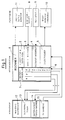
- the workstation shown in the block diagram in FIG. 1 for carrying out the method according to the invention is divided into three areas: the actual microscope, the computer system connected to the microscope and the peripheral devices which enable interactive operation with the user.
- a conventional inverted microscope (12) with a scanning table (13) movable in two coordinates is selected as the microscope.
- the microscope is equipped with a motorized focus drive (14) and has a TV camera (15) at an image output.
- the scanning table (13) serves to close the cell cultures to be examined over the objective of the microscope position. It has an extra large driving range so that a large number of sample vessels can be picked up and processed one after the other.
- a microscope suitable for carrying out the method is sold, for example, by the applicant under the name "Axiovert".
- the computer (1) can be, for example, a personal computer of the type IBM PC / AT, which is equipped as standard with a mass storage device (2) in the form of a floppy disk drive and a hard disk drive.
- the computer (1) also has a tablet interface (3) for connecting the digitizing tablet (8) and a keyboard interface (4) to which the input keyboard (9) is connected.
- the computer (1) is also provided with an image memory (5).
- This image memory also contains the necessary components for digitizing the image taken by the camera (15) and additional image processing hardware.
- the module offered by Matrox under the type designation MVP-AT can be used as image memory.
- a monitor (10) is connected to the image memory (5), on which the cell images can be displayed and the graphics generated by the computer (1) for dialogue with the user can be superimposed.
- a separate data dialog monitor (11) such as, for example, the computer screen (13) already present, can be used.
- the motor controller (7) is also integrated into the computer (1), from which the drive motors of the scanning table (13) and the drive motor for the focus drive (14) of the microscope are controlled.
- the motor control also includes an autofocus device (6) to which the signal from the camera (15) is fed and which moves the focus drive (Z) in the direction of maximum image contrast.
- the Cell cultures positioned on the scanning table (13) are examined at regular intervals of, for example, two hours under the microscope, and images from adjacent image fields of each culture are taken with the TV camera (15) and transferred to the image memory (5).
- the digitized images are stored separately on the hard disk of the mass storage device (2) according to image fields. From there, after all image acquisition sequences have been completed, the cell images in each image field can be called up for the different observation times.
- the cell image sequences are evaluated interactively.
- the cell images are displayed on the monitor (10).
- the cells in the image are marked by the user by means of a cursor mark that can be positioned on the screen (10), the movement of which is controlled with the aid of the digitizing tablet (8).
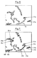
- the interactive evaluation process is described in more detail below with reference to FIGS. 2-7, in which the image sequence of an arbitrarily selected image field is shown as an example:
- the next image of the sequence shown in FIG. 3 is first adjusted in relation to the original (FIG. 2).
- the computer (1) performs a real-time subtraction of the signals of both images belonging to certain image pixels and thereby corrects a shift in the center of gravity of the cells due to drifting of the apparatus.
- the cell marks already set in FIG. 2 are adopted in the new image (FIG. 3). The user therefore only has to correct the changes that occur. These are movements individual cells, cell division and death, as well as the emigration and immigration of cells from or into the image field.
- FIG. 3 for example, in comparison to FIG. 2, divisions have been made at the locations of the cell group (20) designated (20a and 20b) and at the location designated (21a) in the cell group (21).
- the daughter cells are given a different brand here. These can be differentiated by the color.
- a double symbol is chosen for better visibility, namely the square with the diagonal cross for the daughter cells of the first generation in the cell group (20) and the cross with a circle for the daughter cells of the cell group (21).
- this variant of the method can be easily automated further, for example, using the known means of image analysis from a corresponding processor to examine each new image in a sequence, taking into account the knowledge of the previous image, for cell division, cell migration, etc., and for the results to be entered, ie the corresponding new brands have already been registered by the image analysis device. The user then only has to take corrective action and correct the image-analytically determined result if necessary.
- the computer (1) stores the number and the positions of all different types of marks stored in the images of a sequence. From this stored information, statements about the observed cell culture are then derived according to various criteria.
- FIG. 8 shows a type of evaluation which is based on a result representation, which corresponds to the currently customary evaluation of cell images by simply counting the total number of cells.
- the curves labeled (30, 31 and 32) indicate the cell growth, i.e. the absolute number of cells in all image fields of a culture for three different cell cultures in successive observation periods.
- the cell cultures to which the graphs (31 and 32) belong were exposed to different concentrations of a toxic substance, while the graph (30) represents the development of an untreated control culture.
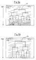
- FIG. 9a shows the cell lineage for a cell of the untreated control culture in a timely representation.
- the horizontal stripes that are placed over the cell family tree (33) each represent constant time periods. From this it can be seen very nicely that the cycle time between successive generations is approximately constant and its value can easily be determined roughly.
- Figure 9b shows the pedigree of a cell of the treated culture. From the family tree (34) it can be seen that, compared to FIG. 9a, fewer division processes have taken place and the cycle time is longer for the individual generations.
- FIG 10a the average values for the cycle time and the mitotic index of all cells of the control population are shown in their temporal behavior, i.e. shown in successive time intervals.
- the mitosis index is the division rate of a cell culture, normalized to the total number of cells that are present in the culture in the relevant time period.
Landscapes
- Chemical & Material Sciences (AREA)
- General Physics & Mathematics (AREA)
- Dispersion Chemistry (AREA)
- Health & Medical Sciences (AREA)
- Life Sciences & Earth Sciences (AREA)
- Analytical Chemistry (AREA)
- Biochemistry (AREA)
- Physics & Mathematics (AREA)
- Immunology (AREA)
- General Health & Medical Sciences (AREA)
- Pathology (AREA)
- Image Processing (AREA)
- Investigating Or Analysing Biological Materials (AREA)
- Apparatus Associated With Microorganisms And Enzymes (AREA)
- Image Analysis (AREA)
- Investigating Or Analysing Materials By Optical Means (AREA)
Claims (11)
- Procédé pour analyse d'images de cellules qui permet- de réaliser plusieurs images successives d'une culture cellulaire donnée et d'enregistrer ces images sous forme numérique, en choisissant l'étendue du champ d'image de manière à ce que celui-ci contienne plusieurs cellules individuelles,- de visualiser lesdites images numériques sur l'écran de manière consécutive,- de marquer les différentes cellules individuelles reproduites sur l'écran de symboles, les différentes cellules ou groupes de cellules (20-23) étant dotés de symboles différents et les positions des différents symboles étant mises en mémoire,- et de déterminer ensuite des caractéristiques statistiques de la culture cellulaire en fonction des variations relevées dans les images numériques consécutives,caractérisé en ce que- les images consécutives sont réalisées parallèlement pour une multitude de champs d'image différents, puis enregistrées sous forme numérique,- les cellules individuelles doivent être marquées au moins sur la première image numérique respectivement (fig. 2) d'une succession de prises de vues,- les mêmes symboles de marquage sont apposés automatiquement aux cellules correspondantes respectivement dans les images numériques suivantes et que les symboles attribués par voie automatique sont ensuite corrigés par l'utilisateur,- les caractéristiques statistiques des différents champs d'image ou des différents symboles des successions de prises de vues sont déterminées et mises en mémoire séparément.
- Procédé selon la revendication 1, caractérisé en ce que les cellules sont classées par générations et que le nombre de cellules compté pour chaque génération est déterminé et mis en mémoire.
- Procédé selon la revendication 1, caractérisé en ce que le taux de division cellulaire est déterminé et mis en mémoire pour différents champs d'image ou pour différentes cellules ou différents groupes de cellules, identifiés par leurs symboles.
- Procédé selon la revendication 1, caractérisé en ce que la mobilité des cellules ou groupes de cellules ainsi marqués est déterminée et mise en mémoire.
- Procédé selon la revendication 1, caractérisé en ce que des arbres généalogiques (33, 34) de cellules individuelles ou de lignées de cellules sont formés et visualisés sur l'écran.
- Procédé selon la revendication 1, caractérisé en ce que les prises de vues des différents champs d'image sont réalisées au moyen d'une platine motorisée à mouvements croisés (13) qui positionne différentes zones adjacentes d'une culture cellulaire sous un microscope (12) respectivement en plusieurs séquences consécutives.
- Procédé selon la revendication 1, caractérisé en ce que les symboles employés dans une image de départ (fig. 2) sont repris dans l'image numérique suivante (fig. 3) et que les changements qui surviennent sont corrigés par déplacement ou par attribution de symboles.
- Procédé selon la revendication 7, caractérisé en ce que les cellules sont localisées à l'aide d'un système d'analyse d'images.
- Procédé selon la revendication 8, caractérisé en ce qu'une image numérique, avant d'être analysée, est décalée par rapport à l'image précédente de manière à ce que les décalages des positions de cellules dans leur ensemble soient réduits à un minimum.
- Dispositif pour la mise en oeuvre du procédé selon les revendications 1 à 9, comprenant- un microscope (12) équipé d'une platine porte-objet (13) qui peut être positionnée selon au moins deux axes (x, y),- des moyens pour réaliser un grand nombre d'images vidéo de l'échantillon à examiner,- un ordinateur à fonctions graphiques (1) muni d'un générateur de marques servant à créer les différents types de symboles,- une mémoire d'images (5) destinée au stockage, sous forme numérique, des images vidéo enregistrées dans des intervalles réguliers,- un écran (10) pouvant visualiser les images numériques stockées dans la mémoire d'images avec superposition des symboles générés par l'ordinateur,- un dispositif (8) servant à positionner les symboles dans le champ d'image et à mettre en mémoire les coordonnées des symboles dans les différentes images,caractérisé en ce que- les moyens d'enregistrement d'images vidéo sont constitués par un équipement TV,- la mémoire d'images est conçue de façon à permettre le stockage d'une multitude de séquences d'images vidéo prises dans des champs d'images attenants l'un à l'autre,- un programme d'analyse est prévu qui déduit de la somme des différents symboles dans les images consécutives et des coordonnées desdits symboles les signes caractéristiques de la culture cellulaire observée,- un autre programme est prévu pour assurer l'insertion de symboles dans une image numérique en fonction des symboles mis en place dans l'image numérique précédente, les symboles ainsi insérés étant susceptibles d'être corrigés par l'utilisateur.
- Dispositif selon la revendication 10, caractérisé en ce que les symboles mis en place peuvent être corrigés à l'aide du dispositif (8) servant à positionner les symboles.
Applications Claiming Priority (2)
| Application Number | Priority Date | Filing Date | Title |
|---|---|---|---|
| DE3836716A DE3836716A1 (de) | 1988-10-28 | 1988-10-28 | Verfahren zur auswertung von zellbildern |
| DE3836716 | 1988-10-28 |
Publications (3)
| Publication Number | Publication Date |
|---|---|
| EP0365928A2 EP0365928A2 (fr) | 1990-05-02 |
| EP0365928A3 EP0365928A3 (fr) | 1991-07-10 |
| EP0365928B1 true EP0365928B1 (fr) | 1995-05-03 |
Family
ID=6366086
Family Applications (1)
| Application Number | Title | Priority Date | Filing Date |
|---|---|---|---|
| EP89118940A Expired - Lifetime EP0365928B1 (fr) | 1988-10-28 | 1989-10-12 | Procédé pour l'exploitation d'images de cellules |
Country Status (5)
| Country | Link |
|---|---|
| US (1) | US5031099A (fr) |
| EP (1) | EP0365928B1 (fr) |
| JP (1) | JP2930618B2 (fr) |
| AT (1) | ATE122145T1 (fr) |
| DE (2) | DE3836716A1 (fr) |
Cited By (1)
| Publication number | Priority date | Publication date | Assignee | Title |
|---|---|---|---|---|
| US7817841B2 (en) | 2005-11-12 | 2010-10-19 | General Electric Company | Time-lapse cell cycle analysis of unstained nuclei |
Families Citing this family (67)
| Publication number | Priority date | Publication date | Assignee | Title |
|---|---|---|---|---|
| DE10136481A1 (de) * | 2001-07-27 | 2003-02-20 | Leica Microsystems | Anordnung zum Mikromanipulieren von biologischen Objekten |
| DE4110217A1 (de) * | 1991-03-28 | 1992-10-01 | Ullrich Juergen Heinz | Verfahren zur herstellung von kristallisaten zur fruehdiagnose oder zur praevention von erkrankungen und zur herstellung von arzneimitteln |
| US5218645A (en) * | 1991-03-29 | 1993-06-08 | Cell Analysis Systems, Inc. | Method and apparatus for separating cell objects for analysis |
| DE4211904C2 (de) * | 1991-04-09 | 1994-03-17 | Werner Maier | Automatisches Verfahren zum Erstellen einer Liste unterschiedlicher Arten für eine flüssige Probe |
| CA2077781A1 (fr) * | 1991-09-23 | 1993-03-24 | James W. Bacus | Methode et appareil de dosage automatise de prelevements bilologiques |
| US5889881A (en) * | 1992-10-14 | 1999-03-30 | Oncometrics Imaging Corp. | Method and apparatus for automatically detecting malignancy-associated changes |
| US6026174A (en) * | 1992-10-14 | 2000-02-15 | Accumed International, Inc. | System and method for automatically detecting malignant cells and cells having malignancy-associated changes |
| JP2675962B2 (ja) * | 1993-04-28 | 1997-11-12 | 株式会社トプコン | 眼科装置 |
| US5692220A (en) * | 1993-09-02 | 1997-11-25 | Coulter Corporation | Decision support system and method for diagnosis consultation in laboratory hematopathology |
| CA2132269C (fr) * | 1993-10-12 | 2000-02-01 | Rainer Hermann Doerrer | Methode cytologique automatisee interactive et systeme |
| CH690384A5 (de) | 1995-03-24 | 2000-08-15 | Zeiss Carl Fa | Computerunterstütztes Video-Mikroskop. |
| US5889880A (en) * | 1995-06-07 | 1999-03-30 | Autocyte, Inc. | Interactive automated cytology method incorporating both manual and automatic determinations |
| DE19709348C2 (de) * | 1996-05-29 | 1999-07-01 | Schubert Walter Dr Md | Automatisches Multi-Epitop-Ligand-Kartierungsverfahren |
| DE19635666C1 (de) * | 1996-09-03 | 1997-12-18 | Kapitza Hans Georg Dr | Integriertes Mikroskop |
| US6008010A (en) * | 1996-11-01 | 1999-12-28 | University Of Pittsburgh | Method and apparatus for holding cells |
| GB2322269B (en) * | 1997-02-13 | 2001-08-22 | Quantel Ltd | A video image processing apparatus and method |
| AU773088B2 (en) * | 1997-02-27 | 2004-05-13 | Cellomics, Inc. | A system for cell-based screening |
| US6753161B2 (en) * | 1997-03-27 | 2004-06-22 | Oncosis Llc | Optoinjection methods |
| DE19726226C2 (de) * | 1997-06-22 | 2001-07-26 | Zentrum Fuer Neuroinformatik G | Verfahren zum automatisierten Erkennen von Strukturen in Schnitten durch biologische Zellen oder biologisches Gewebe |
| DE19845883B4 (de) * | 1997-10-15 | 2007-06-06 | LemnaTec GmbH Labor für elektronische und maschinelle Naturanalytik | Verfahren zur Bestimmung der Phytotoxizität einer Testsubstanz |
| DE19758533B4 (de) * | 1997-12-04 | 2005-09-29 | Micronas Gmbh | Verfahren zum Strukturieren einer Oberflächenschicht |
| DE19753790C2 (de) * | 1997-12-04 | 2001-07-19 | Micronas Gmbh | Verfahren zur Untersuchung einer Oberflächenschicht |
| US6181811B1 (en) | 1998-01-13 | 2001-01-30 | Neopath, Inc. | Method and apparatus for optimizing biological and cytological specimen screening and diagnosis |
| US6834238B1 (en) * | 1998-06-08 | 2004-12-21 | Cytoscan Sciences Llc | Method for identifying optical contrast enhancing agents |
| DE19913922C2 (de) * | 1998-06-15 | 2002-10-24 | Steag Rtp Systems Gmbh | Verfahren zum Bestimmen von Parameterverteilungen von Substrateigenschaften |
| JP3582390B2 (ja) * | 1999-01-13 | 2004-10-27 | 松下電器産業株式会社 | 微細物体の自動探索装置 |
| DE19916749B4 (de) * | 1999-04-14 | 2004-02-12 | Carl Zeiss Jena Gmbh | Verfahren zur Untersuchung von Proben |
| DE19919539C5 (de) * | 1999-04-29 | 2004-12-09 | Gerhard Lewandovski | Verfahren zur Messung der Aktivität einer biologisch wirksamen Substanz in einem histologischen Präparat |
| DE19930607C2 (de) * | 1999-07-02 | 2002-08-01 | Max Planck Gesellschaft | Verfahren zur Gewinnung von Daten, die Aufschluß geben über die Kinetik der Reaktionen von Reaktanten in einer Vielzahl von Proben und Vorrichtung zur Durchführung des Verfahrens |
| CA2392534A1 (fr) * | 1999-11-30 | 2001-06-07 | Oncosis | Procede et dispositif permettant de cibler de maniere selective des cellules specifiques dans une population de cellules |
| EP2363828A1 (fr) | 2000-10-24 | 2011-09-07 | Cyntellect, Inc. | Appareil pour le ciblage sélectif de cellules au sein d'un échantillon à trois dimensions |
| EP1339017A4 (fr) * | 2000-12-01 | 2007-08-29 | Japan Science & Tech Corp | Procede de determination des zones nucleaires et procede d'etablissement de genealogie nucleaire |
| DE10128552B4 (de) * | 2001-06-13 | 2015-01-22 | Jens Stockhausen | Verfahren zur Zellanalyse und Zellanalyseeinrichtung |
| DE10152404C5 (de) | 2001-10-24 | 2017-06-08 | Carl Zeiss Microscopy Gmbh | Laser-Mikrodissektionssystem |
| DE10158964A1 (de) * | 2001-11-30 | 2003-08-07 | Digi Plan Gmbh Planungsgesells | Verfahren und Vorrichtung zur Untersuchung von Medien auf das Vorhandensein von Lebendmaterie |
| DE10221124A1 (de) * | 2002-05-13 | 2003-11-27 | Bayer Cropscience Ag | Verfahren und Vorrichtung zur automatischen Auszählung und Klassifizierung von tierischen Schädlingen |
| DE10229880A1 (de) * | 2002-07-03 | 2004-01-29 | Siemens Ag | Bildanalyseverfahren und Vorrichtung zur Bildauswertung für die in vivo Kleintierbildgebung |
| CA2492416A1 (fr) * | 2002-07-15 | 2004-01-22 | Baylor College Of Medicine | Interface-utilisateur facilitant l'acquisition et l'analyse de caracteristiques de specimens biologiques |
| DE10306970B4 (de) * | 2003-02-19 | 2017-02-09 | Carl Zeiss Microscopy Gmbh | Elektronisches Mikroskop |
| GB0317679D0 (en) * | 2003-07-29 | 2003-09-03 | Amersham Biosciences Uk Ltd | Analysing biological entities |
| JP2007509334A (ja) | 2003-10-21 | 2007-04-12 | ライカ マイクロシステムス ツェーエムエス ゲーエムベーハー | レーザマイクロ切開におけるレーザ切断線の自動生成方法 |
| DE102004051508B4 (de) * | 2003-10-21 | 2006-09-21 | Leica Microsystems Cms Gmbh | Verfahren zur automatischen Erzeugung von Laser-Schnittlinien in der Laser-Mikrodissektion |
| US20050095578A1 (en) * | 2003-10-31 | 2005-05-05 | Koller Manfred R. | Method and apparatus for cell permeabilization |
| GB0403611D0 (en) * | 2004-02-18 | 2004-03-24 | Univ Glasgow | Analysis of cell morphology and motility |
| US7425426B2 (en) * | 2004-03-15 | 2008-09-16 | Cyntellect, Inc. | Methods for purification of cells based on product secretion |
| DE102004022484B4 (de) | 2004-05-07 | 2007-12-20 | P.A.L.M. Microlaser Technologies Ag | Mikroskoptisch |
| DE102004023262B8 (de) * | 2004-05-11 | 2013-01-17 | Carl Zeiss Microimaging Gmbh | Verfahren zur Bearbeitung einer Masse mittels Laserbestrahlung und Steuersystem |
| KR20070086024A (ko) * | 2004-12-08 | 2007-08-27 | 고꾸리쯔 다이가꾸 호우징 오사까 다이가꾸 | 세포 평가방법, 세포 측정용 시스템 및 세포 측정용프로그램 |
| DE102004062218A1 (de) * | 2004-12-23 | 2006-07-13 | P.A.L.M. Microlaser Technologies Ag | Verfahren und Vorrichtung zum Erstellen von Aufnahmeserien von biologischen Objekten und zur Laser-Mikrodissektion |
| JP2006238802A (ja) * | 2005-03-03 | 2006-09-14 | Olympus Corp | 細胞観察装置、細胞観察方法、顕微鏡システム、及び細胞観察プログラム |
| JP4690119B2 (ja) * | 2005-06-16 | 2011-06-01 | オリンパス株式会社 | 画像処理装置および画像処理プログラム |
| US8265357B2 (en) * | 2005-10-14 | 2012-09-11 | Unisense Fertilitech A/S | Determination of a change in a cell population |
| US20070250548A1 (en) * | 2006-04-21 | 2007-10-25 | Beckman Coulter, Inc. | Systems and methods for displaying a cellular abnormality |
| JP4921858B2 (ja) * | 2006-06-07 | 2012-04-25 | オリンパス株式会社 | 画像処理装置および画像処理プログラム |
| JP5143227B2 (ja) | 2007-06-29 | 2013-02-13 | ウニセンス フェルティリテック アー/エス | 顕微鏡対象物の監視および/または培養用の機器、システムおよび方法 |
| KR20110108390A (ko) | 2009-01-12 | 2011-10-05 | 신텔렉트 인코포레이티드 | 레이저로 매개된 박막 절개 및 세포 군체의 이동 |
| DE102010035908A1 (de) | 2009-08-28 | 2011-03-10 | Perner, Petra, Dr.-Ing. | Einrichtung und Verfahren zur automatischen Erfassung der dynamischen Prozesse von Zellen von Zellproben |
| JP5854418B2 (ja) * | 2011-02-28 | 2016-02-09 | 国立大学法人名古屋大学 | 細胞培養環境評価方法及びその装置 |
| KR102179088B1 (ko) * | 2013-12-12 | 2020-11-18 | 메스 메디컬 일렉트로닉 시스템즈 리미티드 | 홈 테스팅 장치 |
| WO2016139925A1 (fr) * | 2015-03-04 | 2016-09-09 | Sony Corporation | Appareil, procédé et système de traitement d'image |
| JP6759550B2 (ja) * | 2015-03-04 | 2020-09-23 | ソニー株式会社 | 情報処理装置、プログラム、情報処理方法及び観察システム |
| JP6851635B2 (ja) | 2015-03-31 | 2021-03-31 | スライブ バイオサイエンス, インコーポレイテッド | 自動細胞培養インキュベータ |
| US10696937B2 (en) | 2015-03-31 | 2020-06-30 | Thrive Bioscience, Inc. | Cell culture incubators with integrated cell manipulation systems |
| EP3308281A4 (fr) * | 2015-03-31 | 2019-12-04 | Thrive Bioscience, Inc. | Incubateurs de culture cellulaire comportant des systèmes d'imagerie intégrés |
| JPWO2019021472A1 (ja) * | 2017-07-28 | 2020-07-30 | オリンパス株式会社 | 細胞特定方法、細胞集団の製造方法及び細胞特定システム |
| JP6960053B2 (ja) * | 2018-06-07 | 2021-11-05 | オリンパス株式会社 | 画像処理装置および培養評価システム |
| WO2020218393A1 (fr) * | 2019-04-26 | 2020-10-29 | 株式会社ニコン | Procédé de suivi de cellule, dispositif de traitement d'image et programme |
Family Cites Families (5)
| Publication number | Priority date | Publication date | Assignee | Title |
|---|---|---|---|---|
| US4320415A (en) * | 1979-06-14 | 1982-03-16 | National Research Development Corporation | Method of and apparatus for measuring electrophoretic mobility of cells |
| US4513438A (en) * | 1982-04-15 | 1985-04-23 | Coulter Electronics, Inc. | Automated microscopy system and method for locating and re-locating objects in an image |
| US4700298A (en) * | 1984-09-14 | 1987-10-13 | Branko Palcic | Dynamic microscope image processing scanner |
| US4724543A (en) * | 1985-09-10 | 1988-02-09 | Beckman Research Institute, City Of Hope | Method and apparatus for automatic digital image analysis |
| DE3718066A1 (de) * | 1987-05-29 | 1988-12-08 | Zeiss Carl Fa | Verfahren zur mikroinjektion in zellen bzw. zum absaugen aus einzelnen zellen oder ganzer zellen aus zellkulturen |
-
1988
- 1988-10-28 DE DE3836716A patent/DE3836716A1/de not_active Withdrawn
-
1989
- 1989-10-12 DE DE58909215T patent/DE58909215D1/de not_active Expired - Lifetime
- 1989-10-12 EP EP89118940A patent/EP0365928B1/fr not_active Expired - Lifetime
- 1989-10-12 AT AT89118940T patent/ATE122145T1/de not_active IP Right Cessation
- 1989-10-27 JP JP1278796A patent/JP2930618B2/ja not_active Expired - Lifetime
- 1989-10-27 US US07/428,217 patent/US5031099A/en not_active Expired - Lifetime
Cited By (1)
| Publication number | Priority date | Publication date | Assignee | Title |
|---|---|---|---|---|
| US7817841B2 (en) | 2005-11-12 | 2010-10-19 | General Electric Company | Time-lapse cell cycle analysis of unstained nuclei |
Also Published As
| Publication number | Publication date |
|---|---|
| EP0365928A3 (fr) | 1991-07-10 |
| US5031099A (en) | 1991-07-09 |
| DE3836716A1 (de) | 1990-05-03 |
| ATE122145T1 (de) | 1995-05-15 |
| JP2930618B2 (ja) | 1999-08-03 |
| EP0365928A2 (fr) | 1990-05-02 |
| JPH02171866A (ja) | 1990-07-03 |
| DE58909215D1 (de) | 1995-06-08 |
Similar Documents
| Publication | Publication Date | Title |
|---|---|---|
| EP0365928B1 (fr) | Procédé pour l'exploitation d'images de cellules | |
| DE69520004T2 (de) | Verfahren zum feststellen und/oder zum identifizieren von mikrobenkolonien | |
| DE112004002219B4 (de) | Verfahren zum Antreiben eines Roboters | |
| DE102008059788B4 (de) | Analyse und Klassifizierung insbesondere biologischer oder biochemischer Objekte auf Basis von Zeitreihen-Bildern, anwendbar bei der zytometrischen Time-Lapse-Zellanalyse in der bildbasierten Zytometrie | |
| EP0425595B1 (fr) | Procede et dispositif pour la classification optique automatique de plantes | |
| DE69411268T2 (de) | Verfahren zur schnellen quantifizierung von microorganismenwachstum | |
| EP3948688B1 (fr) | Entraînement pour réseaux neuronaux artificiels avec une meilleure utilisation des jeux de données d'apprentissage | |
| EP2656277B1 (fr) | Dispositif et procédé de détermination d'objets dans une image en couleurs | |
| EP0752101B1 (fr) | Procede et dispositif pour determiner la toxicite, et application | |
| DE102007046704B4 (de) | Verfahren und Vorrichtung zur Unterstützung der Auswertung von medizinischen Bilddaten | |
| DE3146423T1 (de) | Flow-through optical analyzer | |
| DE10108295A1 (de) | Zahnidentifikation auf digitalen Röntgenaufnahmen und Zuordnung von Informationen zu digitalen Röntgenaufnahmen | |
| EP3168783A1 (fr) | Procédé et dispositif d'évaluation des résultats d'un enregistrement de vue | |
| DE4211904C2 (de) | Automatisches Verfahren zum Erstellen einer Liste unterschiedlicher Arten für eine flüssige Probe | |
| EP2920577A1 (fr) | Dispositif et procédé pour la microscopie d'une pluralité d'échantillons | |
| WO2008014960A1 (fr) | Procédé de reconnaissance automatisée des objets 3d et détermination de position | |
| DE69116543T2 (de) | Anordnung zur intelligenz-prüfung | |
| DE3543515C2 (fr) | ||
| DE3622222A1 (de) | Zeichenerkennung in zweidimensionalen signalen | |
| EP3958036B1 (fr) | Procédé et dispositif de génération d'une image à contraste de synthèse d'un porte-échantillon dans un microscope | |
| DE10317142A1 (de) | Automatische Trefferzählung bei der numerischen Röntgenologie, insbesondere bei der Mammografie | |
| EP1556476A2 (fr) | Procede et dispositif pour manipuler des echantillons | |
| DE4244708C2 (de) | Verfahren und Vorrichtung zum Erstellen einer Artenliste für eine flüssige Probe | |
| DE10221124A1 (de) | Verfahren und Vorrichtung zur automatischen Auszählung und Klassifizierung von tierischen Schädlingen | |
| DE102020106857A1 (de) | Mikroskopiesystem und verfahren zum verarbeiten von mikroskopbildern |
Legal Events
| Date | Code | Title | Description |
|---|---|---|---|
| PUAI | Public reference made under article 153(3) epc to a published international application that has entered the european phase |
Free format text: ORIGINAL CODE: 0009012 |
|
| AK | Designated contracting states |
Kind code of ref document: A2 Designated state(s): AT CH DE LI |
|
| PUAL | Search report despatched |
Free format text: ORIGINAL CODE: 0009013 |
|
| AK | Designated contracting states |
Kind code of ref document: A3 Designated state(s): AT CH DE LI |
|
| 17P | Request for examination filed |
Effective date: 19911214 |
|
| 17Q | First examination report despatched |
Effective date: 19930802 |
|
| GRAA | (expected) grant |
Free format text: ORIGINAL CODE: 0009210 |
|
| AK | Designated contracting states |
Kind code of ref document: B1 Designated state(s): AT CH DE LI |
|
| REF | Corresponds to: |
Ref document number: 122145 Country of ref document: AT Date of ref document: 19950515 Kind code of ref document: T |
|
| REF | Corresponds to: |
Ref document number: 58909215 Country of ref document: DE Date of ref document: 19950608 |
|
| PG25 | Lapsed in a contracting state [announced via postgrant information from national office to epo] |
Ref country code: AT Effective date: 19951012 |
|
| PG25 | Lapsed in a contracting state [announced via postgrant information from national office to epo] |
Ref country code: LI Effective date: 19951031 Ref country code: CH Effective date: 19951031 |
|
| PLBE | No opposition filed within time limit |
Free format text: ORIGINAL CODE: 0009261 |
|
| STAA | Information on the status of an ep patent application or granted ep patent |
Free format text: STATUS: NO OPPOSITION FILED WITHIN TIME LIMIT |
|
| 26N | No opposition filed | ||
| REG | Reference to a national code |
Ref country code: CH Ref legal event code: PL |
|
| PGFP | Annual fee paid to national office [announced via postgrant information from national office to epo] |
Ref country code: DE Payment date: 20081022 Year of fee payment: 20 |