EP0369566A2 - Bifunktionelle chimäre Antikörper - Google Patents
Bifunktionelle chimäre Antikörper Download PDFInfo
- Publication number
- EP0369566A2 EP0369566A2 EP89302313A EP89302313A EP0369566A2 EP 0369566 A2 EP0369566 A2 EP 0369566A2 EP 89302313 A EP89302313 A EP 89302313A EP 89302313 A EP89302313 A EP 89302313A EP 0369566 A2 EP0369566 A2 EP 0369566A2
- Authority
- EP
- European Patent Office
- Prior art keywords
- thr
- ser
- gly
- ala
- leu
- Prior art date
- Legal status (The legal status is an assumption and is not a legal conclusion. Google has not performed a legal analysis and makes no representation as to the accuracy of the status listed.)
- Withdrawn
Links
Images




Classifications
-
- C—CHEMISTRY; METALLURGY
- C12—BIOCHEMISTRY; BEER; SPIRITS; WINE; VINEGAR; MICROBIOLOGY; ENZYMOLOGY; MUTATION OR GENETIC ENGINEERING
- C12N—MICROORGANISMS OR ENZYMES; COMPOSITIONS THEREOF; PROPAGATING, PRESERVING, OR MAINTAINING MICROORGANISMS; MUTATION OR GENETIC ENGINEERING; CULTURE MEDIA
- C12N15/00—Mutation or genetic engineering; DNA or RNA concerning genetic engineering, vectors, e.g. plasmids, or their isolation, preparation or purification; Use of hosts therefor
- C12N15/09—Recombinant DNA-technology
- C12N15/63—Introduction of foreign genetic material using vectors; Vectors; Use of hosts therefor; Regulation of expression
- C12N15/79—Vectors or expression systems specially adapted for eukaryotic hosts
-
- C—CHEMISTRY; METALLURGY
- C07—ORGANIC CHEMISTRY
- C07K—PEPTIDES
- C07K16/00—Immunoglobulins [IG], e.g. monoclonal or polyclonal antibodies
- C07K16/46—Hybrid immunoglobulins
- C07K16/468—Immunoglobulins having two or more different antigen binding sites, e.g. multifunctional antibodies
Definitions
- the present invention relates to monoclonal bifunctional antibodies directed against human carcinoembryonic antigen and metal chelates. More particularly, it relates to novel bifunctional chimeric monoclonal antibodies directed against these antigens, and DNA constructs encoding for such antibodies, for in vitro and in vivo application.
- Monoclonal antibodies are becoming increasingly important for both in vitro application in immunoassays and for the in vivo diagnosis and treatment of disease.
- Monoclonal antibodies which are directed against human carcinoembryonic antigen ("CEA") are particularly useful for the in vivo imaging and treatment of tumors associated with certain carcinomas, including colorectal and breast carcinomas.
- CCA human carcinoembryonic antigen
- these monoclonal antibodies are generally conjugated with radionuclides, drugs or toxins.
- murine antibodies are derived from murine, i.e., mouse, hybridomas.
- the in vitro application of murine antibodies in immunoassays presents potential problems associated with false positive results which are attributable to the reaction of serum components with murine immunoglobulins.
- the in vivo application of murine antibodies in human medicine is often limited due to their inherent immunogenicity.
- the administration of murine antibodies will, in many patients, induce an immune response which results in a gradual decline in the efficacy of the antibodies during multiple dose regimens. This decrease in efficacy is attributable, at least in part, to the rapid clearance from circulation or alteration of pharmacokinetic properties of murine antibodies by the patient's immune response.
- Chimeric antibodies in which the binding or variable regions of antibodies derived from one species are combined with the constant regions of antibodies derived from a different species, have been constructed by recombinant DNA methodology. Chimeric antibodies are described, for example, in European Patent Publication 173494; Shaw, et al ., J. Immun. , 138:4534 (1987), Sun, L.K. et al ., Proc. Natl. Acad. Sci. USA , 84:214-218 (1987); Neuberger, M.S. et al ., Nature , 314:268 (1985), Boulianne, G.L. et al ., Nature , 312:643-646 (1984); and Morrison, S.L.
- variable region of a murine antibody is joined with the constant region of a human antibody. It is expected that as such chimeric antibodies are largely human in composition, they will be substantially less immunogenic than murine antibodies. Accordingly, chimeric monoclonal antibodies are highly desirable for in vivo application.
- bifunctional antibodies are those antibodies which comprise a light chain and a heavy chain from one antibody which recognizes a first specific antigen, in association with a light chain and a heavy chain from a second antibody which recognizes a second specific antigen.
- Such antibodies with dual specificities are quite useful in the prognosis, diagnosis and treatment of disease states. For example, if one set of light and heavy chains of a bifunctional antibody recognize a tumor associated antigen, while the corresponding light and heavy chains recognize a metal chelate, the bifunctional antibody can be used for tumor imaging and therapy.
- the light and heavy chains of one part of the antibody Upon introduction of the bifunctional antibody into the system of a patient, the light and heavy chains of one part of the antibody will specifically bind to the tumor associated antigen.
- Introduction of a metal chelate into the patient's system will provide a known quantity of antigen with which the metal chelate-binding arms of the bifunctional antibody can react.
- the free metal chelates being very small molecules, will quickly pass from the patients system, thereby allowing enhanced imaging of any tumor site where the bifunctional antibodies have bound.
- the present invention specifically provides chimeric bifunctional antibodies wherein one light and heavy chain recognize CEA, while the second light and heavy chain recognize metal chelates.
- the specific metal chelate-binding light and heavy chain variable regions of the present bifunctional chimeric antibody were derived from monoclonal antibody CHA255, which was disclosed in Reardon et al ., (1985) Nature 316:265-268.
- the CHA255 antibody most efficiently binds to the EDTA-chelate of Indium (III), but also recognizes the EDTA chelates of Iron (II) and Cadmium (II).
- bifunctional and chimeric antibodies While the general concepts of bifunctional and chimeric antibodies have been described, there exists a need for the development of novel bifunctional chimeric antibodies having specificity for predetermined antigens of interest, particularly human carcinoembryonic antigen and metal chelates, and variable regions of defined amino acid sequences. Further, there exists a need for the development of DNA constructs having defined DNA coding sequences for light and heavy chain variable regions which comprise novel bifunctional chimeric antibodies, and which are capable of expressing these novel bifunctional chimeric proteins in eukaryotic cells. The present invention meets these needs.
- a - deoxyadenosine Ala - an alanine residue.
- Ap R the ampicillin-resistant phenotype or gene conferring same.
- Asn - an asparagine residue.
- Asp - an aspartic acid residue.
- Bifunctional antibody an antibody comprising a light chain variable region and a heavy chain variable region which specifically react with a first antigen in conjunction with a light chain variable region and a heavy chain variable region which specifically react with a second and different antigen.
- C deoxycytosine.
- Chimeric antibody - an antibody comprising a variable region from one species, typically mouse, joined to a constant region from a second and different species, typically human.
- Ile - an isoleucine residue.
- MoAB monoclonal antibody.
- Nascent protein the polypeptide produced upon translation of a mRNA transcript, prior to any post-translational modifications.
- pA a DNA sequence encoding a polyadenylation signal.
- Phe a phenylalanine residue.
- Pro a proline residue.
- Promoter a DNA sequence that directs transcription of DNA into RNA.
- Recombinant DNA Cloning Vector any autonomously replicating agent, including, but not limited to, plasmids and phages, comprising a DNA molecule to which one or more additional DNA segments can be or have been added.
- Recombinant DNA Expression Vector any recombinant DNA cloning vector into which a promoter has been incorporated.
- Replicon A DNA sequence that controls and allows for autonomous replication of a plasmid or other vector.
- Restriction Fragment any linear DNA sequence generated by the action of one or more restriction endonuclease enzymes.
- Sensitive Host Cell - a host cell that cannot grow in the presence of a given antibiotic or other toxic compound without a DNA segment that confers resistance thereto.
- Structural Gene any DNA sequence that encodes a functional polypeptide, inclusive of translational start and stop signals.
- T deoxythymidine.
- Tc R the tetracycline-resistant phenotype or gene conferring same. Thr - a threonine residue. Trp - a tryptophane residue. Tyr - a tyrosine residue. Val - a valine residue.
- the present invention is a bifunctional chimeric monoclonal antibody (and DNA compounds encoding said antibody) wherein one light chain variable region and one corresponding heavy chain variable region comprise light and heavy chain variable regions which specifically recognize human carcinoembryonic antigen and the second light chain variable region and corresponding second heavy chain variable region comprise light and heavy chain variable regions which specifically recognize metal chelates and the constant regions of all light and heavy chains comprise human constant regions.
- the bifunctional chimeric monoclonal antibody light chain variable region which recognizes human carsinoembryonic antigen has an amino acid sequence substantially the same as: Asp - Ile - Val - Met - Thr - Gln - Ser - Gln - Lys Phe - Met - Ser - Thr - Ser - Val - Gly - Asp - Arg Val - Ser - Ile - Thr - Cys - Lys - Ala - Ser - Gln Asn - Val - Arg - Thr - Ala - Val - Ala - Trp - Tyr Gln - Gln - Lys - Pro - Gly - Gln - Ser - Pro - Lys Ala - Leu - Ile - Tyr - Leu - Ala - Ser - Asn - Arg Tyr - Thr - Gly - Val - Pro - As
- the bifunctional chimeric monoclonal antibody heavy chain variable region which recognizes human carcinoembryonic antigen has an amino acid sequence substantially the same as: Asp - Val - Gln - Leu - Val - Glu - Ser - Gly - Gly Gly - Leu - Val - Gln - Pro - Gly - Gly - Ser - Arg Lys - Leu - Ser - Cys - Ala - Ala - Ser - Gly - Phe Thr - Phe - Ser - Asn - Phe - Gly - Met - His - Trp Ile - Arg - Gln - Als - Pro - Glu - Lys - Gly - Leu Gly - Trp - Val - Ala - Tyr - Ile - Ser - Gly - Gly Ser - Ser - Thr - Ile - Tyr - Tyr - Ala - Asp
- the bifunctional chimeric monoclonal antibody light chain variable region which recognizes metal chelates has an amino acid sequence substantially the same as: Gln Ala Val Val Thr Gln Glu Ser Ala Leu Thr Thr Ser Pro Gly Glu Thr Val Thr Leu Thr Cys Arg Ser Ser Thr Gly Ala Val Thr Thr Ser Asn Tyr Ala Asn Trp Val Gln Glu Lys Pro Asp His Leu Phe Thr Gly Leu Ile Gly Gly Thr Asn Asn Arg Ala Pro Gly Val Pro Ala Arg Phe Ser Gly Ser Leu Ile Gly Asp Lys Ala Ala Leu Thr Ile Thr Gly Ala Gln Thr Glu Asp Glu Ala Arg Tyr Phe Cys Ala Leu Trp Tyr Ser Asn Leu Trp Val Phe Gly Gly Gly Thr Lys Leu Thr Val Leu Gly.
- the bifunctional chimeric monoclonal antibody heavy chain variable region which recognizes metal chelates has an amino acid sequence substantially the same as: Glu Val Thr Leu Val Glu Ser Gly Gly Asp Ser Val Lys Pro Gly Gly Ser Leu Lys Leu Ser Cys Ala Ala Ser Gly Phe Thr Leu Ser Gly Glu Thr Met Ser Trp Val Arg Gln Thr Pro Glu Lys Arg Leu Glu Trp Val Ala Thr Thr Leu Ser Gly Gly Gly Phe Thr Phe Tyr Ser Ala Ser Val Lys Gly Arg Phe Thr Ile Ser Arg Asp Asn Ala Gln Asn Asn Leu Tyr Leu Gln Leu Asn Ser Leu Arg Ser Glu Asp Thr Ala Leu Tyr Phe Cys Ala Ser His Arg Phe Val His Trp Gly His Gly Thr Leu Val Thr Val Ser Ala Ala.
- the compounds of the present invention repreversiblysent recombinant bifunctional chimeric monoclonal antibody CEM/CHA. Due to the complementary nature of DNA base pairing, the sequence of one strand of a double-stranded DNA molecule is sufficient to determine the sequence of the opposing strand.
- the nucleotide sequence of the gene encoding the bifunctional chimeric monoclonal antibody light chain variable region which recognizes human carcinoembryonic antigen is: GAC - ATT - GTG - ATG - ACC - CAG - TCT - CAA - AAA TTC - ATG - TCC - ACA - TCA - GTA - GGA - GAC - AGG GTC - AGC - ATC - ACC - TGC - AAG - GCC - AGT - CAG AAT - GTT - CGT - ACT - GCT - GTT - GCC - TGG - TAT CAA - CAG - AAA - CCA - GGG - CAG - TCT - CCT - AAA GCA - CTG - ATT - TAC - TTG - GCA - TCC - AAC - CGG TAC - ACT - GGA
- the nucleotide sequence of the gene encoding the bifunctional chimeric monoclonal antibody heavy chain variable region which recognizes human carcinoembryonic antigen is: GAT - GTG - CAG - CTG - GTG - GAG - TCT - GGG - GGA GGC - TTA - GTG - CAG - CCT - GGA - GGG - TTC - CGG AAA - CTC - TCC - TGT - GCA - GCC - TCT - GGA - TTC ACT - TTC - AGT - AAC - TTT - GGA - ATG - CAC - TGG ATT - CGT - CAG - GCT - CCA - GAG - AAG - GGA - CTG GAG - TGG - GTC - GCA - TAC - ATT - AGT - GGT - GGC AGT - AGT -
- the nucleotide sequence of the gene encoding the bifunctional chimeric monoclonal antibody light chain variable region which recognizes metal chelates is: CAG GCT GTT GTG ACT CAG GAA TCT GCA CTC ACC ACA TCA CCT GGT GAA ACA GTC ACA CTC ACT TGT CGC TCA AGT ACT GGG GCT GTT ACA ACT AGT AAC TAT GCC AAC TGG GTC CAA GAA AAA CCA GAT CAT TTA TTC ACT GGT CTA ATA GGT GGT ACC AAT AAC CGG GCT CCA GGT GTT CCT GCC AGA TTC TCA GGC TCC CTG ATT GGA GAC AAG GCT GCC CTC ACC ATC ACA GGG GCA CAG ACT GAA GAT GAG GCA AGA TAT TTC TGT GCT CTA TGG TAC AGC AAC CTC TGG GTA TTC GGT GGA GGA ACC AAA CTG ACT GTC CTA G
- the nucleotide sequence of the gene encoding the bifunctional chimeric monoclonal antibody heavy chain variable region which recognizes metal chelates is: GAA GTG ACG CTG GTG GAG TCT GGG GGA GAC TCA GTG AAG CCT GGA GGG TCC CTG AAA CTC TCC TGT GCA GCC TCT GGA TTC ACT TTA AGT GGT GAA ACC ATG TCT TGG GTT CGC CAG ACT CCG GAG AAG AGG CTG GAG TGG GTC GCA ACC ACT CTT AGT GGT GGT GGT TTC ACC TTC TAT TCA GCC AGT GTG AAG GGT CGT TTC ACC ATC TCC AGA GAC AAT GCC CAG AAC AAC CTC TAT CTA CAA CTG AAT AGT CTG AGG TCT GAG GAC ACG GCC TTG TAT TTC TGT GCA AGT CAT CGG TTT GTT CAC TGG GGC CAC GGG ACT CTG
- the novel DNA compounds of the present invention are derived from genomic DNA clones prepared from two separate hybridoma cell lines.
- the light and heavy chain variable region genes which express immunoglobulin chains which recognize human carcinoembryonic antigen, were cloned from a genomic DNA library of hybridoma CEM 231.6.7.
- Murine hybridoma CEM 231.6.7 is part of the permanent stock culture collection of the American Type Culture Collection, Rockville, MD, and is available to the public under the accession number ATCC HB 9620.
- the light and heavy chain variable region genes which express immunoglobulin chains which recognize metal chelates, were cloned from a genomic DNA library of hybridoma CHA255.5.
- Murine hybridoma CHA255.5 is disclosed in Reardon et al ., (1985) Nature 316:265-268.
- Murine hybridoma CHA255.5 is also disclosed in Meares and David, U.S. Patent No. 4,722,892, issued February 2, 1988, the entire disclosure of which is herein incorporated by reference.
- the genes which encode the human constant regions of all light and heavy chain constructions were cloned from a gene library derived from human blood cells.
- the present invention provides a needed benefit to medical science in that, for the first time, large amounts of bifunctional chimeric antibodies can now be biologically produced.
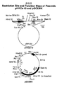
- Plasmid pMLCE-10 comprises the genomic sequence of the light chain variable region of monoclonal antibody CEM, which recognizes human carcinoembryonic antigen. Plasmid pMLCE-10 is part of the permanent collection of the American Type Culture Collection (deposited March 1, 1988) and is available under accession number ATCC 67639. Plasmid pHKF-1 comprises the genomic sequence of the light chain constant region of human antibody. Plasmid pHKF-1 is part of the ATCC permanent collection (deposited March 1, 1988) under accession number ATCC 67637. Restriction site and function map of plasmids pMLCE-10 ad pHKF-1 are presented in Figure 1 of the accompanying drawings.
- Plasmid pHKCE-10 was constructed by isolating the approximately 3.8 kb Hin dIII fragment containing the CEM light chain variable region gene from plasmid pMLCE-10 and ligating this fragment into HIn dIII digested plasmid pHKF-1. Plasmid pSV2gpt (available from the ATCC under accession number ATCC 37145) was digested with restriction enzyme Eco RI and Cla I linkers (sequence dCATCCGATG) were ligated into the Eco RI site to form plasmid pSV2gpt Cla.
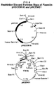
- Plasmid pHKCE-10 was next digested with restriction enzymes Cla I and Bam HI and the approximately 9.0 kb Cla I/ Bam HI restriction fragment, which comprises the CEM light chain variable region gene linked to the human light chain constant region gene, was isolated. Plasmid pSV2gpt-Cla was also digested with restriction enzymes Cla I and Bam HI and the approximately 4.5 kb Cla I/ Bam HI restriction fragment was isolated. The about 9.0 kb fragment from plasmid pHKCE-10 was ligated into the about 4.5 kb vector fragment of plasmid pSV2gpt-Cla to form expression plasmid pGCEMK. Restriction site and function maps of plasmids pHKCE-10 and pGCEMK are presented in Figure 2 of the accompanying drawings.
- Plasmid pMHCE-30 comprises the genomic sequence of the heavy chain variable region of monoclonal antibody CEM, which recognizes human carcinoembryonic antigen. Plasmid pMHCE-30 is part of the ATCC collection (deposited March 1, 1988) and is available under accession number ATCC 67640. Plasmid pHG1Z comprises the genomic sequence of the heavy chain variable region of human antibody. Plasmid pHG1Z is deposited with the ATCC (deposited March 1, 1988) under accession number ATCC 67638. Restriction site and function maps of plasmid pMHCE-30 and pHG1Z are presented in Figure 3 of the accompanying drawings.
- Plasmid pHGCEM-30 was constructed by isolating the approximately 5.3 kb Cla I/ Hin dIII fragment containing the CEM heavy chain variable region gene from plasmid pMHCE-30 and ligating this fragment into Cla I/ Hin dIII digested vector pHG1Z. Inasmuch as plasmid pMHCE-30 contains more than one Bam HI site, the 5.3 kb Cla I/ Hin dIII restriction fragment of plasmid pMHCE-30 is most easily isolated following a total Cla I digested and a subsequent partial Bam HI digestion.
- Plasmid pSV2neo (ATCC 37149) was digested with restriction enzyme Eco RI and Cla I linkers (dCATCCGATG) were ligated into the Eco RI site to form plasmid pSV2neo-Cla. Plasmid pSV2neo-Cla was then totally digested with restriction enzymes Bam HI and Cla I and the about 4.5 kb vector fragment was isolated. Plasmid pHGCEM-30 was totally digested with restriction enzyme Cla I, then partially digested with restriction enzyme Bam HI and the about 12.7 kb restriction fragment, comprising the gene encoding the CEM heavy chain variable region linked to the gene encoding the human heavy chain constant region, was isolated.
- Plasmid PGCEMK was digested with restriction enzymes Cla I and Bam HI and the approximately 8.9 kb Cla I/ Bam HI restriction fragment was isolated.
- Plasmid pNCEMG1 was also digested with restriction enzymes Cla I and Bam HI and the approximately 17.6 kb Cla I/ Bam HI restriction fragment was isolated.
- the about 8.9 kb Cla I/ Bam HI restriction fragment of plasmid pGCEMK was then ligated into the about 17.6 kb Cla I/ Bam HI restriction fragment of plasmid pNCEMG1 to form expression vector pNCEMKG1.
- Plasmid pNCEMKG1 comprises the gene encoding the CEM light chain variable region linked to the gene encoding the human light chain constant region as well as the gene encoding the CEM heavy chain variable region linked to the gene encoding the human heavy chain variable region. Plasmid pNCEMKG1 also contains the neomycin resistance-conferring gene. A restriction site and function map of plasmid pNCEMKG1 is presented in Figure 5 of the accompanying drawings.
- Plasmid pMLCH1 comprises the genomic sequence encoding the light chain variable region of monoclonal antibody CHA, which recognizes metal chelates.
- Plasmid pMLCH1 can be conventionally isolated from E . coli K12 HB101/pMLCH1, a strain deposited and made part of the permanent stock culture collection of the Northern Regional Research Laboratory (NRRL), Peoria, Illinois.
- a culture of E . coli K12 HB101/pMLCH1 (deposited November 14, 1988) can be obtained from the NRRL under the accession number NRRL B-18432.
- Plasmid pMLCH1 was digested with restriction enzyme Bam HI, then treated with Klenow enzyme to remove the Bam HI site which occurs 5′ to the structural gene in the plasmid. Religation of this vector fragment created plasmid pMLCH1dB. Restriction site and function maps of plasmids pMLCH1 and pMLCH1dB are presented in Figure 6 of the accompanying drawings.
- Plasmid pMLCH1dB was digested with restriction enzymes Hin dIII and Bam HI and the approximately 5.75 kb Hin dIII/ Bam HI restriction fragment, which comprises the gene encoding the CHA heavy chain variable region, was isolated. Plasmid pBR322 was also digested with restriction enzymes Hin dIII and Bam HI and the large vector fragment was purified. The about 5.75 kb Hin dIII/ Bam HI restriction fragment of plasmid pMLCH1dB was then ligated into the Hin dIII/ Bam HI-digested vector fragment of plasmid pBR322 to form plasmid pMLCH2.
- Plasmid pMLCH2 was then digested with restriction enzymes Cla I and Bam HI and the approximately 5.75 kb Cla I/ Bam HI restriction fragment was isolated and ligated into Cla I/ Bam HI digested plasmid pSV2gpt-Cla to form plasmid pGCHA.
- Plasmid pGCHA was digested with restriction enzyme Bam HI and the 11.2 kb fragment was purified.
- Plasmid pHKF1 which comprises the gene encoding the human light chain constant region, was digested with restriction enzyme Hin dIII, filled in with Klenow, phosphorylated Bam HI (NEB) linkers were added, then the vector was cut with Bam HI, and the approximately 5.2 kb Bam HI restriction fragment was isolated.
- Plasmid pUCVHInc-1A comprises the genomic sequence encoding the heavy chain variable region of monoclonal antibody CHA, which recognizes metal chelates. Plasmid pUCVHInc-1A can be conventionally isolated from E . coli K12 HB101/pUCVHInc-1A, a strain deposited (November 14, 1988) and made part of the permanent stock culture collection of the NRRL. A culture of E . coli K12 HB101/pUCVHInc-1A can be obtained under the accession number NRRL B-18433.
- Plasmid pUCVHInc-1A was digested with restriction enzyme Hin dIII and the approximately 3.4 kb Hin dIII restriction fragment, which comprises the gene encoding the CHA heavy chain variable region, was isolated.
- Plasmid pHG1Z (ATCC 67638) which comprises the gene encoding the human heavy chain constant region, was also digested with restriction enzyme Hin dIII and then treated with Calf Intestine Alkaline Phosphatase to create blunt ends.
- Plasmid pHG1-CHA which comprises the gene encoding the CHA heavy chain variable region linked to the gene encoding the human heavy chain constant region, was digested with restriction enzymes Cla I and Bam HI and the approximately 10.5 kb Cla I/ Bam HI restriction fragment was isolated. Plasmid pSV2neo-Cla was next digested with restriction enzymes Bam HI and Cla I and the approximately 5.0 kb Cla I/ Bam HI vector fragment was purified.
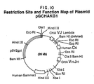
- Plasmid pGCHAG1 was digested with restriction enzyme Cla I and the approximately 15.05 kb Cla I restriction fragment was isolated. Plasmid pGCHAK was digested with restriction enzyme Bam HI and Cla I linkers were ligated into the Bam HI site to form plasmid pGCHAK-Cla. Plasmid pGCHAK-Cla was then digested with restriction enzyme Cla I and the approximately 10.85 kb Cla I restriction fragment was isolated. The about 10.85 kb Cla I restriction fragment of plasmid pGCHAK-Cla was then ligated into the about 15.05 kb Cla I fragment of vector pGCHAG1 to form expression plasmid pGCHAKG1.
- Plasmid pGCHAKG1 comprises the gene encoding the CHA light chain variable region joined to the gene encoding the human light chain constant region as well as the gene encoding the CHA heavy chain variable region joined to the gene encoding the human heavy chain constant region. Plasmid pGCHAKG1 also comprises the gene which confers resistance to the mycophenolic acid. A restriction site and function maps of plasmid pGCHAKG1 is presented in Figure 10 of the accompanying drawings.
- the present DNA compounds which encode recombinant CEM and CHA immunoglobulins and derivatives are especially preferred for the construction of vectors for transformation and expression of the various antibody chains in mammalian and other eukaryotic cells.
- Many mammalian host cells possess the necessary cellular machinery for the recognition and proper processing of the signal peptides present on the amino-terminus of the various antibody chains embodied in the present invention. Some mammalian host cells also provide the post-translational modifications, such as glycosylation, that are observed in antibody molecules.
- a wide variety of vectors exist for the transformation of eukaryotic host cells, and the specific vectors exemplified below are in no way intended to limit the scope of the present invention.
- the various expression vectors of the present invention can be transformed into and expressed in a variety of eukaryotic, especially mammalian, host cells.
- the expression vectors also comprise sequences that allow for replication in E . coli .
- Expression of antibodies occurs in host cells in which the particular promoter associated with the antibody's structural gene functions. Skilled artisans will understand that a variety of eukaryotic host cells can be used to express the various antibody chains of the present invention.
- the SP2/0-Ag14 cell line is a myeloma cell line which ordinarily does not secrete antibody. Following transfection of cell line SP2/0 with plasmids pGCHAKG1 and pNCEMKG1, the transfected cell line secretes bifunctional chimeric CEM/CHA antibody into the culture fluid.
- the host cells used in the invention may be transformed in a variety of ways by standard transfection procedures well known in the art.
- standard transfection procedures which may be used are electroporation techniques, protoplast fusion and calcium-phosphate precipitation techniques.
- electroporation techniques protoplast fusion and calcium-phosphate precipitation techniques.
- Such techniques are generally described by Toneguzzo, F. et al ., Mol. and Cell. Biol. , 6:703-706 (1986); Chu, G., et al ., Nucleic Acid Res. , 15:1311-1325 (1987); Rice, D., et al ., Proc. Natl. Acad. Sci. USA , 79:7862-7865 (1979) and; Oi, V., et al , Proc. Natl. Acad. Sci. USA , 80:825-829 (1983).
- the recombinant expression vectors comprising the CEM and CHA constructs of the invention are transfected sequentially into host cells.
- the expression vectors comprising the CHA constructs are first transfected into the host cells and transformed host cells expressing the CHA immunoglobulin chains are selected by standard procedures known in the art. See, e.g. Engvall, E. and Perlmann, P., Immunochemistry , 8:871-874 (1971) or Meares and David, U.S. Patent No. 4,722,892, issued February 2, 1988, the entire teaching of which is herein incorporated by reference.
- the expression vectors comprising the CEM immunoglobulin gene constructs are, thereafter, transfected into the selected host cells.
- both the CHA and CEM constructs can be simultaneously introduced into the host cells or introduced in inverse order.
- both the CEM and CHA gene constructs can be combined on a single expression vector, or the two DNA's could be linearized and ligated together prior to transformation into cells.
- standard assays are performed for the detection of antibodies directed against CEA and metal chelates for the identification of transformed cells expressing the bifunctional chimeric antibodies of the present invention.
- heterologous immunoglobulin chains are defined as any immunoglobulin chain other than the murine kappa light chain encoded on plasmid pGCEMK.
- Plasmid pGCHAK-2 which comprises the murine lambda CHA variable region and human constant region genes driven by the CEM kappa promoter on an SV40 enhancer-containing vector is constructed according to the teaching of Example 7.
- Plasmid pGCHAK-3 which comprises the murine lambda CHA variable region and human constant region genes driven by the CEM kappa promoter on an SV40 enhancerless vector is also constructed according to the teaching of Example 7.
- plasmid pGCHAK By replacing plasmid pGCHAK with plasmid pGCHAK-2 in the scheme for building plasmid pGCHAKG1, one can easily construct plasmid pGCHAKG1-2.
- plasmid pGCHAKG1-3 can be constructed by replacing plasmid pGCHAG1 with plasmid pGCHAGI(E-), and replacing pGCHAK with pGCHAK-2 in the scheme for the construction of plasmid pGCHAKG1.
- SP2/0 cells transfected with either of plasmids pGCHAKG1-2 or pGCHAKG1-3, then subsequently transfected with vector pNCEMKG1, will demonstrate higher levels of expression of bifunctional chimeric CEM/CHA antibody than do cells which are transfected with vectors pGCHAKG1 and pNCEMKG1.
- the mature bifunctional chimeric CEM/CHA antibodies are secreted into the supernatant.
- the culture fluid is generally concentrated and treated with a solution of carboxypeptidase after culture collection.
- the bifunctional chimeric CEM/CHA antibodies can then be purified according to techniques well known in the art. (See Catherine B. Beidler, M.J. Johnson, and James R. Ludwig, Chimeric Antibodies Directed against Human Carcinoembryonic Antigen , Attorney Docket No. H- 7587A, U.S. Application No.
- bifunctional chimeric CEM/CHA antibodies can be detected in a wide variety of ways.
- One method is to coat polystyrene beads with monoclonal antibody CEV124, which recognizes CEA.
- CEA is allowed to bind to the polystyrene/CEV124 complex.
- CEA can be allowed to bind directly to microtiter plate wells, rather than via an antibody linker.
- Supernatant containing bifunctional chimeric CEM/CHA antibodies is then incubated with the beads, thereby allowing the CEA-recognizing arms of CEM/CHA to bind to the CEA which is bound to the beads. Radiolabeled Indium-EDTA chelates are then added to the beads.
- the CHA arms of the bifunction chimeric CEM/CHA antibody will bind the Indium-EDTA chelates, thereby allowing for antibody titer determination using a gamma counter.
- Indium-EDTA chelates are disclosed in U.S. Patent No. 4,722,892, issued February 2, 1988, the disclosure of which is herein incorporated by reference. Skilled artisans recognize the virtually limitless methods which are available for the testing and assaying of the secreted CEM/CHA antibodies of the present invention.
- the antibodies of the present invention while having great utility for the in vitro detection of CEA in serum or tissue samples, are also quite useful for the detection of CEA in vivo .
- the chimeric portions of the antibody reduce the possibility of cross-reactivity and serum sickness, and therefore allow multiple dose regimens for both detection and treatment of disease states.
- Bifunctional chimeric CEM/CHA is infused and allowed to localize at the tumor site where CEA is located.
- the radionuclide Indium-EDTA chelate can be infused later, and inasmuch as the metal chelates are small molecules, the unbound metal chelates are rapidly cleared from the body, thereby reducing the possibility of damage to non-tumor tissue.
- the radionuclide is selected on the basis of its emission of radiation, typically a gamma-photon, which can be detected by photoscanning techniques. If it is desired to treat the tumor to reduce its size, preferably the radionuclide emits an electron or an alpha-particle.
- the radionuclide emits an electron or an alpha-particle.
- 111In is preferred.
- Y90 is preferred.
- the bifunctional chimeric CEM/CHA antibody is infused and allowed to localize at the site of the tumor associated CEA.
- a toxin or drug to which is conjugated the metal chelate can be subsequently infused to be bound by the antibody at the tumor site.
- the metal is a radionuclide that, upon decay, changes atomic number to a metal whose chelate is not bound tightly to an antibody, the antibody will release the chelate and the associated drug or toxin in the vicinity of the tumorous cell to facilitate its entry into the cell.
- the radionuclide is selected to have a half-life which insures that release does not occur prematurely or over too long a period.
- Murine hybridoma cells designated as CEM 231.6.7, were used in the examples to derive and clone genomic DNA for variable light and heavy chain variable regions.
- Murine hybridoma CEM 231.6.7 was deposited with the American Type Culture Collection, Rockville, Maryland, under the accession number ATCC HB 9620.
- Transfection of chimeric genes was accomplished by electroporation techniques, essentially as described by Toneguzzo, F., et al ., Molecular and Cell. Biol. , 6:703-706 (1986); and Chu, G. et al ., Nucleic Acid Res. , 15:1311-1325 (1987).
- the host cells SP2/0-Ag14 hybridoma cells were the recipients of the chimeric genes.
- the SP2/0-Ag14 hybridoma cells used are available from the American Type Culture Collection, Rockville, Maryland under the accession number ATCC CRL 1581.
- Genomic DNA from CEM 231.6.7 was isolated by procedures essentially as described by Pellecer et al ., Cell , 41:133 (1978). Approximately 1 x 10⁸ cells were collected by centrifugation (10′, 800 rpm IEC clinical centifuge). Cells were then washed 2x in Phosphate Buffered Saline (PBS). Cells were then resuspended in 4 ml of 10 mM Tris-HCl (pH 8.0) 2 mM EDTA, 40 mM NaCl (TEN), and lysed by the addition of 200 ⁇ l of 10% SDS and 42 ⁇ l of Protease K at 20 mg/ml (Sigma Chemicals, St. Louis, Missouri.
- the DNA was then treated for 2 hours with RNAse A at 50 ⁇ g/ml (Sigma Chemicals, St. Louis, Missouri) followed by Protease K at 200 ⁇ g/ml for one hour at 37°C. DNA was extracted again as detailed above and dialyzed overnight.
- DNA was ethanol precipitated by adding 1/10th volume of 2 M Na Acetate and 2 volumes of 95% ethanol. After 30 minutes at -20°C, the DNA was centrifuged at 8000 RPM for 30 minutes. The pellet was resuspended in TE buffer at a final concentration of 955 ⁇ g/ml.
- Partial DNA digests of the CEM 231 genomic DNA were done by taking 10 ⁇ g of DNA in 11 ⁇ l TE buffer, 20 ⁇ l of H2O, 15 ⁇ l of 10x Core Restriction Digest Buffer (500 mM pH8.0, 100 mM MgCl2, 500 mM NaCl) and then aliquoted into tubes. Restriction enzyme Mbo I was added to each tube in units increasing from .0038 to .5 units/ ⁇ l and placed at 37°C for one hour. Aliquots of the samples were then run on a 0.7% TBE (89 mM Tris 89 mM Borate 2 mM EDTA) agarose gel and electrophoresed overnight at 40 volts.
- TBE 89 mM Tris 89 mM Borate 2 mM EDTA
- CEM 231.6.7 DNA (12-24 kb) was ligated into the EMBL-3 phage arms using 100 ng of DNA, 100 ng of pre-isolated EMBL-3 phage arms, .5 ⁇ l of 10x Ligase Buffer (500 mM Tris-HCl pH8; 70 mM MgCl2; 10 mM dithiothreitol (dtt) and T4 DNA ligase (2 units) at 4°C, overnight.
- Packaging was done using Stratagene's commercially available Gigapack-Gold In Vitro packaging system in accordance with their product protocols and using the host E . coli strain P2.392 supplied by Stratagene, Inc.
- phage dilution buffer per liter 5.8 g NaCl, 2 g MgSO4-6H2O, 50 ml 1M Tris-HCl pH 7.5, 5 ml 2% gelatin
- Phage were then titered using standard protocols as described in Molecular Cloning , edited by T. Maniatis, E.F. Fritsch and J. Sambrook, Cold Spring Harbor Laboratories, Cold Spring Harbor, New York (1982). Following titration, the phage library was plated at a density of 20,000 plaques/100 mM plate using P2.392 cells and incubated at 37°C overnight.
- the kappa oligonucleotide probe used was comprised of the sequence 5′-AGA-TGG-ATA-CAG-TTG-GTG-CAG-CAT-CAG-CCC-3′ and was synthesized by Molecular Biosystems, Inc. (San Diego, CA).
- Duplicate filter lifts were made and phage hybridizing to both probes were analyzed by double southern blots as described in Molecular Cloning , supra, probed with the murine kappa and J L probes which were radiolabeled with 32P. Hybridizations were done as described in Molecular Cloning , supra.
- a single CEM 231.6.7 phage clone was selected on the basis of its hybridizing to both probes, indicating a light chain variable region gene rearrangement to a position directly contiguous to the C kappa gene region. This recombinant phage was designated ⁇ MLCE-10.
- ⁇ MLCE-10 DNA was isolated and 20 ⁇ g were digested with Bam HI (1 unit/ ⁇ g) using React Buffer #3 from Gibco-BRL (Gaithersburg, Maryland) in a total of 220 ⁇ l.
- a 10 kb Bam HI fragment was isolated by electrophoresis of the Bam HI digested DNA in a .75% TBE agarose gel containing .5 ⁇ g/ml of ethidium bromide at 40 V overnight. Following visualization on a UV transparent light box the 10 kb fragment was electrophoresed onto DEAE 81 (Schleicher and Schuell, Keene, New Hampshire) paper followed by elution in 1 M NaCl and ethanol precipitation.
- the eluted fragment was then resuspended in 6 ⁇ l of TE buffer.
- the fragment was ligated to 50 ng of Bam HI digested pBR322, available from the American Type Culture Collection (ATCC Designation 31344) by using 1 ⁇ l (50 ng) pf pBR322 DNA, 6 ⁇ l (300 ng) of Bam HI 10 kb recombinant phage DNA ligase as described in Example 1B.
- E . coli HB101 competent cells available from Gibco-BRL (Gaithersburg, Maryland), were transformed using standard procedures.
- HB101 cells were first thawed on ice; then 10 ⁇ l of the ligation reaction were mixed with 200 ⁇ l of cells and incubated on ice for 30 minutes. Following this, cells were heat shocked for 90 seconds at 42°C, then returned to ice for 2 minutes.
- One ml of LB broth per liter 10 g NaCl, 5 g yeast extrct 10 g tryptone was added and the cells incubated in a New Brunswick air shaker for one hour at 225 rpm, 37°C. Two hundred ⁇ l were then plated on LB-agarose with ampicillin (50 ⁇ g/ml) plated and incubated overnight at 37°C.
- the DNA was extracted twice with an equal volume of phenol:chloroform:isoamyl alcohol (25:24:1), twice more with chloroform:isoamyl alcohol (24:1) and dialyzed against 10 mM Tris-HCl (pH 8.0) 1 mM EDTA.
- the DNA was then treated for two hours with 50 ⁇ g/ml RNAse A (Sigma Chemicals) followed by redigestion with 200 ⁇ g/ml Protease K for 1 hour.
- the DNA was extracted and dialyzed as described above and the concentration determined by OD260 to range from 100-200 mg/ml.
- Human DNA of the fbn haplotype (210 ⁇ g as isolated in Example 1E) was partially digested with MboI, DNA fragments in the molecular weight range 12-24 kb isolated, and cloned into EMBL-3 phage as described in Example 1B.
- the EMBL-3 library was screened with a human kappa probe supplied by Dr. P. Hieter, Johns Hopkins University, Baltimore, Maryland, the sequence of which is available from the NIH Data Base, accession number J00241.
- a total of 5 x 10⁵ recombinant phage were screened as described in Example 1C.
- a single clone was isolated and designated 0 ⁇ HKF-1. Fifteen ⁇ l of 0 ⁇ HKF-1 DNA were digested with Bam HI and Hin dIII in React Buffer #3 (Gibco-BRL, Gaithersburg, Maryland).
- pHKF-1 was deposited under the Budapest Treaty with the American Type Culture Collection on March 1, 1988 (ATCC Deposit #67637. A restriction site and function map of plasmid pHKF-1 is presented in Figure 1 of the accompanying drawings.
- a 3.8 kb Hin dII fragment from pMLCE-10 containing the entire CEM 231.6.7 variable kappa region was further subcloned into the Hin dIII site of plasmid pHKF-1, described in Example 1E, using the methods described in Example 1D to form a chimeric plasmid having the murine CEM 231.6.7 variable light region fused to a human constant kappa region gene.
- One ⁇ g of pMLCE-10 DNA was digested with Hin dIII 1 unit/ ⁇ g using React Buffer #2 (Gibco-BRL, Gaithersburg, Maryland).
- One ⁇ g of pHKF1 was digested in a similar manner.
- the pMLCE-10 digested DNA was electrophoresed as above and the 3.8 kb Hin dIII fragment was isolated onto DEAE 81 paper and eluted in 5 ⁇ l of TE.
- One ⁇ g (2 ⁇ l) of Hin dIII digested pMLCE-10 were ligated to 600 ng of Hin dIII digested pHKF-1 in the presence of 10x Ligation Buffer, 10 mM ATP and 2 units of T4 DNA ligase of total volume of 10 ⁇ l. Ligation was done at 12°C overnight and again HB101 competent cells from BRL were used in the plasmid transformation, described in Example 10.
- the plasmid designated as pHKCE-10 was identified by restriction mapping of the plasmid DNA. A restriction site and function map of plasmid pHKCE-10 is presented in Figure 2 of the accompanying drawings.
- the eukaryotic expression vector containing the murine V L region fused to the human kappa gene was constructed using the vector pSV2gpt, available from the American type Culture Collection, (ATCC Designation #37145).
- pSV2gpt DNA was digested with the restriction enzyme Eco R1 using 1 unit/ ⁇ g of DNA in Reaction Buffer #3 (Gibco-BRL, Gaithersburg, Maryland).
- the Eco R1 ends were then made blunt by adding 10 ⁇ l of 5 mM each of the 4 deoxyribonucleotides dTTP, dGTP, dCTP and dATP, 2 units of Klenow enzyme and a 10x buffer (.5M Tris HCl pH 7.5; .1 M MgCl2; 10 mM diothiothreitol) in a total 50 ⁇ l as described in Molecular Cloning , supra.
- the resulting vector, pSV2gpt-Cla was then digested with Cla I and Bam HI restriction enzymes (1 unit/ ⁇ g of DNA).
- the chimeric vector pHKCE-10 was also digested with these two enzymes.
- the 4.5 kb pSV2gpt Cla - Bam fragment and the 9 kb Cla - Bam pHKCE-10 fragment were isolated on DEAE 81 paper as described in Example 1D.
- a standard ligation reaction as described above was done using 375 ng of the 9 kb fragment insert DNA and 200 ng of the 4.5 kb vector DNA.
- pGCEMK a recombinant plasmid, designated as pGCEMK, was identified by restriction mapping of the plasmid DNA.
- This plasmid which is the chimeric expression vector used to produce the CEM chimeric light chain polypeptide, is shown with restriction sites in Figure 2.
- the EMBL-3 library described in Example 1B was screened with two murine heavy chain probes, one representing the J h 3-4 regions was obtained from Dr. Phil Tucker, University of Texas, Dallas, Texas, and one representing sequences in the murine gamma-1 gene.
- This latter probe was comprised of the sequences 5′-CTG-TAC-ATA-TGC-AAG-GCT-TAC-AAC-CEC-AAT-3′ as determined from the General Bank Data Base (NIH Accession #J00453) and was synthesized by Molecular Biosystems, Inc. (San Diego, CA).
- a total of 4.8 x 10⁵ recombinant phage plaques were screened in duplicate using these two probes in order to identify the clone containing both the J h region and the gamma region. Again, as with the kappa clone, a phage containing sequences for these two regions indicates that that DNA has been rearranged, thus identifying the expressed immunoglobulin gene in the cell line CEM 231.6.7.
- a single CEM 231.6.7 clone was selected on the basis of its being rearranged and was designated 0 ⁇ MHCE-30.
- the vector and isolated 5.6 kb insert DNA was ligated at a ratio of 1:10 in a total of 10 ⁇ l as detailed in Example 1B.
- the correct recombinant was identified by restriction digest mapping, and designated as pMHCE-30.
- pMHCE-30 was deposited under the Budapest Treaty with the American Type Culture Collection on March 1, 1988 (ATCC Deposit #67640).
- a restriction site and function map of plasmid pMHCE-30 is presented in Figure 3 of the accompanying drawings.
- Human DNA of the azg haplotype was digested as 10 ⁇ g aliquots using 30 units each of the restriction endonucleases Bam HI and Hin dIII in Reaction Buffer #3 (Gibco-BRL, Gaithersburg, Maryland) (50 mM Tris-HCl pH 8.0, 10 mM MgCl2, 100 mM NaCl) in a total volume of 200 ⁇ l.
- the digested fragments were concentrated to 20 ⁇ l by ethanol precipitation and separated on a 0.6% low gelling temperature agarose (FMC) gel run for 15 hours at 50 mAmps. DNA fragments in the size range 6-7 kb were excised from the gel.
- the cloning vector pUC 18 (described by Yanisch-Perron C., et al ., Gene , 33:103 (1985)) was also digested with Bam HI and Hin dIII as described above.
- the human DNA fragments (150 ng) were ligated into the pUC 18 vector (50 ng) (New England BioLabs, Beverly, Massachusetts) in a total reaction volume of 400 ⁇ l containing 30 mM Tris-HCl (pH7.6), 10 mM MgCl2, 5 mM dithiothreitol, 1 mM ATP, and 1 ⁇ l T4 DNA ligase (2 units, for 72 hours at 15°C.
- Half (100 ng) of the ligated DNA sample was used to transform 500 ⁇ l of freshly prepared competent E . coli M15 cells and the resulting transformants were plated onto X-gal, IPTG, AMP plates (4 ⁇ g/ml X-gal, 2 ⁇ g/ml IPTG, 100 ⁇ g/ml Ampicillin).
- Ampicillin resistant pUC18 colonies containing recombinant human DNA were screened by the method described in Example 1 using a human Gamma 2 probe supplied by T. Honjo, University of Osaka, Japan and described by Takahashi, N. et al ., Cell. , 29:671-679 (1982).
- a clone containing a 7.5 kb insert corresponding to the human Gamma 1 gene was identified and designated HyHG1.
- This same 7.5 kb Hin dIII- Bam HI fragment containing the human gamma constant region gene was subsequently re-cloned into pBR322 using the same methodology as described in Example 1G.
- the pBR322 vector containing the human gamma 1 gene was designated pHG1Z and was deposited under the Budapest Treaty with the American type Culture Collection on March 1, 1988 (ATCC Deposit #67638).
- a restriction site and function map of plasmid pHG1Z is presented in Figure 3 of the accompanying drawings.
- the murine variable heavy chain region was fused to the human gamma-1 gene in the following manner. Ten ⁇ g of pMHCE-30 was digested with Cla I (1 unit/ ⁇ g) and then partially digested with Hin dIII to give a 5.3 kb fragment containing V h and the major intron. Partial digests were performed by using only .1 unit/ ⁇ g of DNA and a digestion time of 1 hour at 37°C. One ⁇ g of the plasmid pHGZ-1, described in Example 1N containing the human gamma 1 gene was also digested with Cla I and Hin dIII.
- the 5.3 kb fragment from pMHCE-30 was isolated from a TBE gel using the DEAE 81 protocol as described in Example 1D. This fragment was ligated into the Cla - Hin d site of pHGZ-1 by using 500 ng of the insert and 200 ng of the vector DNA in a ligation mixture of 10 ⁇ l total volume. Ligation reactions were carried out as detailed in Example 1B.
- the recombinant plasmids resulting from transformation of HB101 were analyzed by restriction digest mapping in order toidentify a plasmid containing the murine V h region fused to a human gamma 1 gene, and was designated pHGCEM-30.
- a restriction site and function map of plasmid pHGCEM-30 is presented in Figure 4 of the accompanying drawings.
- the chimeric Ig gene was inserted into the eukaryotic expression vector essentially as detailed in Example 1H.
- the vector used was pSV2neo, available from The American Type Culture Collection (ATCC Designation #37149).
- a Cla I site was added to this vector exactly as described for the pSV2gpt vector.
- 1 ⁇ g of pSV2neo-Cla DNA was digested with the enzymes Cla I and Bam HI using 1 unit/ ⁇ g of DNA.
- the plasmid pHGCEM-30 was digested with Cla I and then partially digested with Bam HI (.1 unit/ ⁇ g) in order to obtain a fragment of 12.7 kb which contained the chimeric V h and gamma 1 regions.
- This fragment was isolated on DEAE 81 paper and eluted in 10 ⁇ l of TE buffer.
- the ligation was done using 50 ng of vector DNA, 400 ng of the 12.7 kb insert DNA, 10x ligation buffer, 10 mM ATP and T4 DNA ligase, as in Example 1B at 12°C overnight.
- HB101 cells were transformed and the correct recombinant plasmid constituting the chimeric expression vector pNCEMG1 was identified.
- pNCEMG1 which constitutes the recombinant expression vector used to express chimeric heavy chain immunoglobulin genes and is shown with restriction sites, in Figure 4.
- Plasmid pGCEMK was digested with restriction enzymes Cla I and Bam HI and the approximately 8.9 kb Cla I/ Bam HI restriction fragment was isolated substantially as hereinbefore described.
- plasmid pNCEMG1 was also digested with restriction enzymes Cla I and Bam HI and the approximately 17.6 kb Cla I/ Bam HI restriction fragment was purified.
- the about 8.9 kb Cla I/ Bam HI restriction fragment of plasmid pGCEMK was then ligated into the about 17.6 kb Cla / Bam HI restriction fragment of plasmid pNCEMG1 in substantial accordance with the teaching of Example 1B. After transformation into E .
- Plasmid pNCEMKG1 A restriction site and function map of plasmid pNCEMKG1 is presented in Figure 5 of the accompanying drawings.
- Sequencing of the cloned CEM variable light and heavy chain genes was accomplished by standard procedures for both double and single stranded templates using the protocols provided by the sequencing kit Sequenase, commercially available from U.S. Biochemicals (Cleveland, Ohio), and the Bluescript/DNA Sequencing System, commercially available from Stratagene, Inc. (La Jolla, CA). From the DNA sequences obtained for the cloned CEM variable light and heavy region genes, the amino acid sequences of the polypeptides encoded for were deduced by a computer software program, MAPSEQ, commercially available from DNAStar, (Madison, Wisconsin).
- Plasmid pUCVHInc-1A comprises the gene which encodes the murine variable region of heavy metal-binding monoclonal antibody CHA255.5.
- Plasmid pUCVHInc-1A can be conventionally isolated from E . coli K12 HB101/pUCVHInc-1A, a strain made part of the permanent stock culture collection of the NRRL under accession number NRRL B-18433.
- Plasmid pHG1Z comprises the human gamma gene and is available from the ATCC under the accession number ATCC 67638. About 1 ⁇ g of plasmid pUCVHInc-1A was digested with restriction enzyme Hin dIII and the approximately 3.4 kb Hin dIII restriction fragment was isolated.
- plasmid pHG1Z About 1 ⁇ g of plasmid pHG1Z was also digested with restriction enzyme Hin dIII, then treated with Bacterial Alkaline Phosphatase according to procedures wellknown in the art. The about 3.4 kb Hin dIII fragment of plasmid pUCVHInc-1A was then ligated into the Hin dIII-digested, phosphatased plasmid pHG1Z to form plasmid pHG1-CHA. Restriction site and function maps of plasmids pUCVHInc-1A and pHG1-CHA are presented in Figure 8 of the accompanying drawings.
- Plasmid pSV2gpt-Cla (constructed in Example 1) was digested with restriction enzymes Cla I and Bam HI and then about 5.0 kb Cla I/ Bam HI restriction fragment was isolated.
- plasmid pHG1-CHA was also digested with restriction enzymes Cla I and Bam HI and the approximately 10.5 kb restriction fragment was isolated.
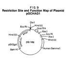
- the about 5.0 kb Cla I/ Bam HI restriction fragment of plasmid pSV2gpt-Cla was ligated to the about 10.5 kb Cla I/ Bam HI restriction fragment of plasmid pHG1-CHA to form plasmid pGCHAG1.
- a restriction site and function map of plasmid pGCHAG1 is presented in Figure 9 of the accompanying drawings.
- plasmid pMLCH-1 from E . coli K12 HB101/pMLCH-1 NRRL B-18432 was digested for three minutes with restriction enzyme Bam HI. After an ethanol precipitation the Bam HI ends were made blunt by adding 10 ⁇ l of 5 mM each of the four deoxyribonucleotides dTTP, dGTP, dATP and dCTP, two units of Klenow enzyme and 5 ⁇ l of 10x Buffer (.5 M Tris-HC1 (pH 7.5), .1 M MgCl2 and 10 mM DTT) in a total of 50 ⁇ l reaction volume.
- plasmid pMLCH1dB Restriction site and function maps of plasmids pMLCH1 and pMLCH1dB are presented in Figure 6 of the accompanying drawings.
- Plasmid pMLCH2 was next constructed by digesting plasmid pMLCH1dB with restriction enzymes Bam HI and Hin dIII and isolating the about 5.75 kb CHA lambda gene region. This fragment was then ligated into Bam HI/ Hin dIII digested plasmid pBR322 to form plasmid pMLCH2. Plasmid pMLCH2 was then digested with restriction enzymes Cla I and Bam HI and the approximately 5.75 kb restriction fragment was isolated and ligated into the Cla I/ Bam HI digested vector fragment of plasmid pSV2gpt-Cla (constructed in Example 1) to form plasmid pGCHA.
- Plasmid pGCHA was digested with restriction enzyme Bam HI and the approximately 11.2 kb restriction fragment was isolated.
- Plasmid pHKF1 (available from the ATCC under the accession number 67637) was digested with restriction enzyme Hin dIII, filled in with Klenow, phosphorylated Bam HI (NEB) linkers were added, then the vector was cut with Bam HI, and the about 5.2 kb restriction fragment was isolated.
- Plasmid pGCHAK The about 11.2 kb Bam HI fragment of plasmid pGCHA was then ligated with the about 5.2 kb Bam HI restriction fragment of plasmid pHKF1 to form plasmid pGCHAK, which comprises the gene which encodes the murine lambda CHA variable region joined to the gene which encode the human gamma region.
- a restriction site and function map of plasmid pCHAK is presented in Figure 7 of the accompanying drawings.
- Plasmid pGCHAK-Cla was digested with restriction enzyme Cla I and the approximately 10.85 kb restriction fragment was isolated in accordance with prior teachings. Plasmid pGCHAG1 was digested with restriction enzyme Cla I and the approximately 15.05 kb restriction fragment was isolated.
- the CHA immunoglobulin plasmid used for transfection was pGCHAKG1 as described in the Example 3 above.
- the pGCHAKG1 plasmid is first transfected into SP2/0 hybridoma cells by the electroporation techniques referenced above.
- the SP2/0-Ag14 cells are grown in media containing 5% FCS and maintained in a log phase of growth for the three days preceeding electroporation.
- Twenty ⁇ g of the plasmid vector pGCHAKG1 is linearized using the restriction enzyme Pvu I (1 u/ ⁇ g) and the Reaction Buffer #7 (Gibco-BRL, Gaithersburg, Maryland).
- the SP2/0 cells are collected by centrifugation in an IEC clinical centrifuge - 800 rpm 10′ room temperature. Cells are then washed 3x in Hanks Buffered Saline Solution (Gibco Laboratories, Grand Island, New York) with 6 mM Dextrose and resuspended at a final concentration of 1.5 x 10⁷ cells/ml. 0.3 mls of cells are aliquoted into cuvettes at a density of 0.5 x 10⁶/.3 ml and the linearized DNA is added. The mixture is maintained on ice 10 minutes.
- Electroporation is done using the .8 mm gap electrode (P/N 472) and the BTX 100 Transfector (BTX, Inc. San Diego, CA.). Conditions are 1 pulse, 5 m seconds each at 250 volts. The electroporated cells are then resuspended in media at a density of 2 x 10⁵/ml (in T75 flasks) for 72 hours. (37°C 5% CO2).
- HMAX 1.0 Media 50 ng/ml Hypoxanthine, 250 ng/ml Mycophenolic Acid and 50 ⁇ g/ml Xanthine
- HMAX resistant colonies Two hundred ⁇ l of supernatant is collected from each well which contained HMAX resistant colonies. This supernatant is then assayed for the presence of a human kappa constant region and a human gamma constant region which would indicate expression of the chimeric immunoglobulin genes of pGCHAKG1.
- Transfected SP2/0 cells expressing the chimeric CHA-human Ig genes were identified by a standard enzyme-linked immunosorbent assay (ELISA), as described by ENgvall, E. and Perlmann, P., Immunochemistry , 8:871-874 (1971), for human Ig.
- ELISA enzyme-linked immunosorbent assay
- this assay is to identify those cells secreting the chimeric kappa and gamma chain polypeptides coded for by the pGCHAKG1 plasmid vector which was constructed from murine variable regions isolated from the murine hybridoma CHA 255 and fused to the human constant region genes.
- a 5 ⁇ g/ml solution of goat anti-human gamma chain (Tago #4100) in 10 mM sodium phosphate pH 7-8 is prepared. Each well of a 96 well plate is coated with 50 ⁇ l of this solution. The plates are then incubated overnight at 37°C. Plates are then rinsed thoroughly in H2O and PBS + 0.1% Tween (w/v).
- a goat anti-human kappa chain alkaline phosphatase conjugate (Tago #2496) is diluted 1:1000 in the same medium as the supernatant material. 100 ⁇ l are added per well and allowed to incubate for 1 hour at room temperature. Plates are rinsed as above.
- the alkaline phosphatase substrate is prepared as per package instruction, one tablet per 3 ml of distilled H2O and 150 ⁇ l of this substrate is added to each well and allowed to incubate 30 minutes at 37°C.
- the reaction is quenched with 50 ⁇ l of 500 mM EDTA and then the absorbance is read at 405 nM. Those supernatants showing the highest levels of Ig expression are identified and the cells from the corresponding wells are pooled and expanded for introduction of the bifunctional chimeric construct pNCEMKG1.
- Cells which were expressing high levels of CHA Ig chain are further assayed for production of immunoglobulin which specifically reacts with metal chelates.
- the assay procedures used for the detection of antibodies directed against metal chelates are standard solid phase radioimmunoassays, essentially as described by Wang, R., et al ., Immunol. Methods , 18:157-164 (1977); Reardon et al ., Nature 316:265-268 (1985); and Meares and David, U.S. Patent No. 4,722,892, issued February 2, 1988.
- the following reagents are added to wells of a microtiter plate and incubated overnight at room temperature with mixing: 25 ⁇ l cell culture supernatent, 50 ⁇ l 111In-EDTA, 20 ⁇ l of Sepharose, bound goat anti-human IgG and 25 ⁇ l cell culture media. Immune complexes bound to the Sepharose-anti-human IgG are collected onto paper filters. Filters are counted in a gamma counter. The radioimmunoassays resulted in the identification of clones which secreted chimeric CHA antibodies. These clones are selected for transfection with plasmid pNCEMKG1.
- the CEM immunoglobulin plasmid used for transfection into SP2/0 cells was pNCEMKG1, derived from constructs as detailed in Example 1.
- the pooled populations of cells expressing the chimeric CHA genes are next electroporated with the plasmid constructs containing the chimeric CEM genes.
- the SP2/0 chimeric CHA producing cells SP2/0-K
- Twenty micrograms of the plasmid DNA pNCEMKG1 are linearized with the enzyme Pvu I in React buffer #6 (Gibco-BRL, Gaithersburg, Maryland).
- Cells are collected, washed and resuspended at a density of 1.5 x 10⁶ cells/ml as detailed in Example 2A.
- the DNA is added and the mixture held on ice for 10 minutes preceeding the electroporation.
- Conditions used are 1 pulse at 5 m seconds, 250 volts.
- Cells were plated at 2.5 x 10⁵/ml in HH2 (or any other commercially available media) 5% FCS plus HMAX 1.0 for 72 hours at 37°C, 5% CO2. Following this, these cells were plated at 5 x 10⁴/ml in 24 well plates in medium containing HMAX 1.0 and G418 antibiotic (Geneticin, Gibco-BRL, Gaithersburg, Maryland) at an active concentration of 500 ⁇ g/ml. Selection was maintained for 14 days at which time those wells with HMAX/G418 resistant colonies are identified for further analysis.
- the following reagents were added to wells of a microtiter plate and incubated overnight at room temperature with mixing: 25 ⁇ l cell culture supernatant, 50 ⁇ l 125I-CEA (affinity purified), 20 ⁇ l of Sepharose bound goat anti-human IgG and 25 ⁇ l cell culture media. Immune complexes bound to the Sepharose-anti-human IgG were collected onto paper filters. Filters were counted in a gamma counter. The radioimmunoassays resulted in the identification clones which were expressing anti-bodies which specifically reacted with CEA.
- Affinity assays were performed to determine the Ka of bifunctional chimeric antibody CEM/CHA for CEA.
- the affinity of bifunctional chimeric antibody CEM/CHA for CEA was determined by standard radioimmunoassay procedures, as described in Example 4A, and Scatchard analysis as described by Scatchard, G., Ann. New York Acad. Sci. , 51:660 (1949).
- Antigen binding assays were done as described in Example 5A. To generate the inhibition curve, 25 ⁇ l volumes of CEA were added to each reaction, substituting for the 25 ⁇ l of cell culture media. The mass of competitor added ranged from 1-100 ng. For the Scatchard analysis, a plot of bound/free vs bound antigen permitted calculation of the affinity constant as defined by the megative of the slope of the line. Affinities of the chimeric antibodies were at least comparable to those of the murine antibody counterparts from which the chimeric antibodies of the invention were derived.
- EOTUBE is a derivative of p-(aminobenzyl) EDTA and is described in L.D. Anderson, J.M. Frincke, and D.L. Meyer, U.S. Patent No. 151855, filed February 3, 1988, (European Patent Application No. 89301012.4) herein incorporated by reference.
- EOTUBE is EDTA substituted at one of the internal ethylene carbons (S stereochemistry at that substituted carbon) through the benzylic carbon of an N-(2-hydroxyethyl)-N′-(p-benzyl)thiourea moiety.
- the synthesis of EOTUBE was performed excluding metal ions as much as possible. (For example, all glassware was washed with 6M HCl, and only dionized water was used).
- (S)- p -nitrobenzyl EDTA was prepared and converted to (S)-4-isothiocyanatobenzyl EDTA (hereinafter abbreviated as ITCBE) as described on U.S. Patent No. 4,622,420, and Meares, C.F., Anal. Biochem 142 : 68-75 (1984).
- the lyophilized ITCBE was resuspended in 0.3M HCl (Ultrex, J.T. Laker, Phillipsburg, N.J.) to a final concentration of roughly 50 mM. This solution was stored at -70°C. Unless indicated otherwise, all reactions were done in aqueous solution as 40°C.
- the product was purified by anion exchange chromatography on an 11 ml column of DEAD Sephadex A-25, eluted with a 110 ml gradient of 0.1 to 1 M ammonium formate, pH 6. (The column was monitored at 280 nm.) Fractions containing the product were pooled and lyophilized. The product has an absorbance maximum at 246 nm with an extinction coefficient of 18000.
- the structure was purified by carbon 13 NMR spectroscopy (on a Varian Instruments Model XL-300 300 Mhz instrument. The spectrum was run deuterated water.)
- the peak corresponding to the carbon in the isothiocyanate moiety in ITGBE was at 139 ppm, and was replaced by a peak at 182 ppm in EOTUBE. This peak corresponds to the carbon in the thiourea linkage.
- the aromatic region (128-138 ppm) of the spectrum of ITCBE shows four peaks, while that of EOTUBE shows three. In the aliphatic regions, there are five peaks in common for ITCBE and EOTUBE, and an additional two peaks at 64 and 49 ppm in the EOTUBE spectrum. The latter peaks correspond to the carbons adjacent to the hydroxyl and thiourea moieties, respectively.
- Polystyrene beads were coated with monoclonal antibody CEV124, which specifically binds CEA.
- CEA was then added to the coated beads and allowed to incubate for one hour at 37°C, then the beads were rinsed with PBS.
- Supernatent containing bifunctional chimeric CEM/CHA was then added to the beads, and the reaction was allowed to proceed for one hour at 37°C. After a rinse with PBS, the beads were mixed with 111IN-EDTA for one hour at 37°C. After another rinse, immune complexes comprising antibody CEM/CHA were collected onto paper filters which were counted in a gamma counter.
- Transfected SP2/0 cells which produced bifunctional chimeric CEM/CHA, wherein one arm binds to CEA and the other arm binds to metal chelates, were identified and subcloned. Following adaptation of the cells to adapt them to serum-free media (HH2), levels of total Ig expression as high as 60 ⁇ g/ml/106 cells were attained.
- HH2 serum-free media
- DNA was resuspended in 5 ⁇ l of TE buffer. About 1 ⁇ l (about 500 ng) of this DNA fragment was mixed with 10 ⁇ l of water, 5 ⁇ l of 5x ligation buffer, 2 ⁇ l of 100 ⁇ MATP and 2 ⁇ l of T4 ligase. After an incubation of 2 hours at room temperature, the ligation mixture was transformed into E . coli HB101 cells in substantial accordance with the teaching of Example 1P. Plasmid DNA was isolated from the trans formants and those plasmids which displayed the proper ⁇ 0.3 kb deletion of the SV40 enhancer region were designated plasmid pSV2gpt(E-).
- SV40 enhancerless pSV3neo was also constructed.
- About 20 ⁇ l (10 ⁇ g) of plasmid pSV2neo-Cla (constructed in Example 1P) was digested with restriction enzymes Bam HI and Hin dIII in a reaction mixture using Gibco Raction Buffer #3, substantially as described above.
- the approximately 2.3 kb Hin dIII/ Bam HI neomycin resistance-conferring gene containing fragment was isolated and purified from DEAE 81 paper following electroelution.
- plasmid pSV2gpt(E-) was also digested with restriction enzymes Bam HI and Hin dIII, and the large vector fragment was purified after electrophoresis.
- the about 2.3 kb Hin dIII/ Bam HI neo containing restriction fragment of plasmid pSV2neo-Cla was then ligated into the Hin dIII/ Bam HI-digested vector fragment of plasmid pSV2gpt(E-) in substantial accordance with the teaching of the previous paragraph. After transformation into E .
- plasmid pSV2neo(E-) those plasmids which the pSV2gpt(E-) vector backbone ligated with the about 2.3 kb BamHI/HindIII neo fragment of plasmid pSV2neo-Cla are designated plasmid pSV2neo(E-).
- Example 5C About 10 ⁇ g (100 ⁇ l) of plasmid pSV2neo(E-) DNA were digested with restriction enzymes Cla I and Bam HI, then the vector fragments were isolated and purified in substantial accordance with the teaching of Example 5C.
- Plasmid pNCEMG(E-) therefore comprises the murine variable, human constant gamma encoding genes on an SV40 enhancerless expression vector.
- Plasmid pGCHAK(E-) is constructed in substantial accordance with the teaching for the construction of plasmid pGCHAK (Example 3B), except plasmid pSV2gpt(E-) is used rather than plasmid pSV2gpt.
- Plasmid pGCHAG1(E-) is constructed in substantial accordance with the teaching for the construction of plasmid pGCHAG1 (Example 3A), except plasmid pSV2gpt(E-) is used rather than plasmid pSV2gpt.
- Plasmid pNCEMKG1(E-) is constructed in substantial accordance with the teaching for the construction of plasmid pNCEMKG1 (Example 1Q), except plasmid pNCEMG1(E-) is used rather than plasmid pNCEMG1.
- Plasmid pGCHAKG1(E-) is constructed in substantial accordance with the teaching for the construction of plasmid pGCHAKG1 (Example 3C), except plasmid pGCHAG1(E-) is used rather than plasmid pGCHAG1.
- Plasmid pGCHAKG1(E-) is first transfected into SP2/0 cells in substantial accordance with the teaching of Example 4. After transfection SP2/0 cells which express the active CHA chimeric are identified by the techniques of Example 5B, these clones are transfected with plasmid pNCEMKG1(E-) to produce cells which will demonstrate high levels of expression of bifunctional chimeric antibody CEM/CHA.
- Intermediate plasmid p19HANCH was first constructed by preparing an oligonucleotide polylinker with the DNA sequence comprising:
- the linker depicted above was synthesized from single-stranded deoxyoligonucleotides by procedures well known in the art.
- the single-stranded deoxyoligonucloeitdes can be synthesized with commercially available instruments, such as the Biosearch 8700 DNA Synthesizer marketed by Biosearch, Inc. (San Raphael, CA), which utilizes phosphoramidite Chemistry. Other procedures for synthesizing DNA are also known in the art.
- the synthesized polylinker was ligated into the EcoRI/HindIII digested plasmid pUC19. After transformation into E . coli and re-isolation of plasmid DNA, those plasmids which demonstrated the proper Hin dIII, Ssp I, Pst I, Sst II and EcoRI sites within the linker region were designated plasmid p19HAN.
- Plasmid S(-)CHAVL was constructed by first digesting the Bluescript vector, M13(-)SK (Stratagene) with restriction enzymes Bam HI and Sst I, then isolating the large vector fragment in substantial accordance with earlier teaching. Plasmid pMLCH-1 was next digested with restriction enzymes Bam HI and Sst I, and the approximately 1100 bp fragment which comprises the gene encoding the murine CHA variable region was isolated. Plasmid pMLCH-1 can be conventionally isolated from E . coli K12 HB101/pMLCH-1, a strain deposited and made part of the permanent stock culture collection of the Northern Regional Research Laboratory, 1815 North University Street, Peoria, IL 61604. E .
- coli K12 HB101/pMLCH-1 is available under the accession number NRRL B-18432.
- the approximately 1100 bp Bam HI/ Sst I restriction fragment of plasmid pMLCH-1 was then ligated into the Bam HI/ Sst I digested vector M13(-)SK and the plasmid was transformed into E . coli in substantial accordance with prior examples.
- those plasmids which contained the proper approximately 1100 bp Bam HI/ Sst I fragment were designated plasmid S(-)CHAVL.
- plasmid S(-)CHAVL was next digested with restriction enzyme Pst I in substantial accordance with prior teachings.
- An approximately 900 bp Pst I restriction fragment which comprises the gene encoding the murine CHA variable region was isolated and ligated into plasmid p19HAN, which had first been cut in the polylinker region by restriction enzyme Pst I.
- those plasmids which comprise the J region of the CHA gene closest to the Hin dIII site of the polylinker are designated plasmid p19HANCH.
- Plasmid pGCEMK (constructed in Example 1) was digested with restriction enzymes Cla I and Ssp I in substantial accordance with earlier teachings, then the approximately 2.2 kb Cla I/ Ssp I restriction fragment, which comprise the CEM kappa promoter, was purified.
- plasmid pGCEMK was digested with restriction enzymes Cla I and Sst II, and the approximately 9.4 kb vector fragment was isolated.
- plasmid p19HANCH was digested with restriction enzymes Ssp I and Sst II and the approximately 931 base pair restriction fragment which comprises the gene encoding the murine kappa CHA variable region was isolated.
- Plasmid pGCHAK-3 differs from plasmid pGCHAK-2 only in the fact that plasmid pGCHAK-3 lacks the SV40 enhancer.
- Plasmid pGCHAKG1-2 is constructed in substantial accordance with the teaching for the construction of plasmid pGCHAKG1 (Example 3C), except plasmid pGCHAK-2 is used, rather than plasmid pGCHAK.
- plasmid pGCHAKG1-3 is constructed in substantial accordance with the teaching for the construction of plasmid pGCHAK (Example 3C), except plasmids pGCHAK-2 is used, rather than plasmid pGCHAK and plasmid pGCHAG1(E-) is used rather than pGCHAG1.
- Plasmid pGCHAKG1-2 comprises the genes encoding the CHA chimeric light chain driven by the CEM kappa promoter as well as the chimeric heavy chain CHA-specific gene.
- Plasmid pGCHAKG1-3 comprises the CHA chimeric light chain driven by the CEM kappa promoter, as well as the chimeric heavy chain CHA-specific gene, on an SV40 enhancerless vector.
Landscapes
- Health & Medical Sciences (AREA)
- Chemical & Material Sciences (AREA)
- Genetics & Genomics (AREA)
- Life Sciences & Earth Sciences (AREA)
- Organic Chemistry (AREA)
- Immunology (AREA)
- Engineering & Computer Science (AREA)
- Biophysics (AREA)
- General Health & Medical Sciences (AREA)
- Molecular Biology (AREA)
- Biomedical Technology (AREA)
- Bioinformatics & Cheminformatics (AREA)
- General Engineering & Computer Science (AREA)
- Wood Science & Technology (AREA)
- Zoology (AREA)
- Biotechnology (AREA)
- Biochemistry (AREA)
- Microbiology (AREA)
- Plant Pathology (AREA)
- Physics & Mathematics (AREA)
- Medicinal Chemistry (AREA)
- Proteomics, Peptides & Aminoacids (AREA)
- Preparation Of Compounds By Using Micro-Organisms (AREA)
- Peptides Or Proteins (AREA)
- Micro-Organisms Or Cultivation Processes Thereof (AREA)
Applications Claiming Priority (2)
| Application Number | Priority Date | Filing Date | Title |
|---|---|---|---|
| US27410588A | 1988-11-17 | 1988-11-17 | |
| US274105 | 1988-11-17 |
Publications (2)
| Publication Number | Publication Date |
|---|---|
| EP0369566A2 true EP0369566A2 (de) | 1990-05-23 |
| EP0369566A3 EP0369566A3 (de) | 1991-10-16 |
Family
ID=23046793
Family Applications (1)
| Application Number | Title | Priority Date | Filing Date |
|---|---|---|---|
| EP19890302313 Withdrawn EP0369566A3 (de) | 1988-11-17 | 1989-03-08 | Bifunktionelle chimäre Antikörper |
Country Status (5)
| Country | Link |
|---|---|
| EP (1) | EP0369566A3 (de) |
| JP (1) | JPH02145187A (de) |
| AU (1) | AU618324B2 (de) |
| DK (1) | DK108189A (de) |
| IL (1) | IL89491A0 (de) |
Cited By (26)
| Publication number | Priority date | Publication date | Assignee | Title |
|---|---|---|---|---|
| EP0404097A3 (de) * | 1989-06-22 | 1991-10-23 | BEHRINGWERKE Aktiengesellschaft | Bispezifische und oligospezifische, mono- und oligovalente Rezeptoren, ihre Herstellung und Verwendung |
| WO1992010209A1 (en) * | 1990-12-04 | 1992-06-25 | The Wistar Institute Of Anatomy And Biology | Bifunctional antibodies and method of preparing same |
| EP0491351A3 (en) * | 1990-12-18 | 1993-03-17 | Takeda Chemical Industries, Ltd. | Chimeric antibodies and their use |
| EP0623675A1 (de) * | 1992-11-12 | 1994-11-09 | Hybritech Incorporated | Polypeptide mit geänderter Affinität von metallchelatbindenden Antikörpern |
| US5532136A (en) * | 1993-06-22 | 1996-07-02 | Bionebraska, Inc. | Monoclonal antibody assay and kit for detecting metal cations in body fluids |
| US5582996A (en) * | 1990-12-04 | 1996-12-10 | The Wistar Institute Of Anatomy & Biology | Bifunctional antibodies and method of preparing same |
| WO2000014119A3 (en) * | 1998-09-04 | 2000-11-30 | Ks Biomedix Ltd | Bifunctional antibodies and their use in targeting anti-tumour agents |
| US8062635B2 (en) | 2003-10-10 | 2011-11-22 | Chugai Seiyaku Kabushiki Kaisha | Bispecific antibody substituting for functional proteins |
| US9334331B2 (en) | 2010-11-17 | 2016-05-10 | Chugai Seiyaku Kabushiki Kaisha | Bispecific antibodies |
| US9670269B2 (en) | 2006-03-31 | 2017-06-06 | Chugai Seiyaku Kabushiki Kaisha | Methods of modifying antibodies for purification of bispecific antibodies |
| US9828429B2 (en) | 2007-09-26 | 2017-11-28 | Chugai Seiyaku Kabushiki Kaisha | Method of modifying isoelectric point of antibody via amino acid substitution in CDR |
| US9975966B2 (en) | 2014-09-26 | 2018-05-22 | Chugai Seiyaku Kabushiki Kaisha | Cytotoxicity-inducing theraputic agent |
| US10011858B2 (en) | 2005-03-31 | 2018-07-03 | Chugai Seiyaku Kabushiki Kaisha | Methods for producing polypeptides by regulating polypeptide association |
| US10759870B2 (en) | 2017-09-29 | 2020-09-01 | Chugai Seiyaku Kabushiki Kaisha | Multispecific antigen-binding molecules having blood coagulation factor VIII (FVIII) cofactor function-substituting activity and pharmaceutical formulations containing such a molecule as an active ingredient |
| US11046784B2 (en) | 2006-03-31 | 2021-06-29 | Chugai Seiyaku Kabushiki Kaisha | Methods for controlling blood pharmacokinetics of antibodies |
| US11124576B2 (en) | 2013-09-27 | 2021-09-21 | Chungai Seiyaku Kabushiki Kaisha | Method for producing polypeptide heteromultimer |
| US11142587B2 (en) | 2015-04-01 | 2021-10-12 | Chugai Seiyaku Kabushiki Kaisha | Method for producing polypeptide hetero-oligomer |
| US11150254B2 (en) | 2014-09-26 | 2021-10-19 | Chugai Seiyaku Kabushiki Kaisha | Method for measuring reactivity of FVIII |
| US11214623B2 (en) | 2014-09-26 | 2022-01-04 | Chugai Seiyaku Kabushiki Kaisha | Antibody capable of neutralizing substance having activity alternative to function of coagulation factor VIII (FVIII) |
| US11352438B2 (en) | 2016-09-06 | 2022-06-07 | Chugai Seiyaku Kabushiki Kaisha | Methods of using a bispecific antibody that recognizes coagulation factor IX and/or activated coagulation factor IX and coagulation factor X and/or activated coagulation factor X |
| US11649262B2 (en) | 2015-12-28 | 2023-05-16 | Chugai Seiyaku Kabushiki Kaisha | Method for promoting efficiency of purification of Fc region-containing polypeptide |
| US12168697B2 (en) | 2015-12-25 | 2024-12-17 | Chugai Seiyaku Kabushiki Kaisha | Bispecific antibodies which bind blood coagulation factor VIII and have enhanced activity, and methods of use thereof for treating bleeding and associated conditions |
| US12421322B2 (en) | 2017-11-01 | 2025-09-23 | Chugai Seiyaku Kabushiki Kaisha | Antibody variant and isoform with lowered biological activity |
| US12460014B2 (en) | 2016-04-28 | 2025-11-04 | Chugai Seiyaku Kabushiki Kaisha | Antibody-containing preparation |
| US12545743B2 (en) | 2016-07-29 | 2026-02-10 | Chugai Seiyaku Kabushiki Kaisha | Bispecific antibody exhibiting increased alternative FVIII-cofactor-function activity |
| US12600798B2 (en) | 2017-03-31 | 2026-04-14 | Public University Corporation Nara Medical University | Medicinal composition usable for preventing and/or treating blood coagulation factor IX abnormality, comprising multispecific antigen binding molecule replacing function of blood coagulation factor VIII |
Families Citing this family (5)
| Publication number | Priority date | Publication date | Assignee | Title |
|---|---|---|---|---|
| IL89489A0 (en) * | 1988-03-09 | 1989-09-10 | Hybritech Inc | Chimeric antibodies directed against human carcinoembryonic antigen |
| IL89490A0 (en) * | 1988-11-17 | 1989-09-10 | Hybritech Inc | Chimeric antibodies directed against metal chelates |
| IL89491A0 (en) * | 1988-11-17 | 1989-09-10 | Hybritech Inc | Bifunctional chimeric antibodies |
| WO2005035754A1 (ja) | 2003-10-14 | 2005-04-21 | Chugai Seiyaku Kabushiki Kaisha | 機能蛋白質を代替する二重特異性抗体 |
| CN108220281A (zh) * | 2017-09-25 | 2018-06-29 | 天津强微特生物科技有限公司 | 一种含有结合核酸的磁珠在生物样品保存液的制备和运用 |
Family Cites Families (6)
| Publication number | Priority date | Publication date | Assignee | Title |
|---|---|---|---|---|
| CA1339445C (en) * | 1986-11-12 | 1997-09-09 | The General Hospital Corporation | Recombinant hybrid immunoglobulin molecules and method of use |
| ATE122237T1 (de) * | 1987-05-29 | 1995-05-15 | Tanox Biosystems Inc | Hiv-1 neutralisierende monoklonale antikörper. |
| AU625613B2 (en) * | 1988-01-05 | 1992-07-16 | Novartis Ag | Novel chimeric antibodies |
| IL89489A0 (en) * | 1988-03-09 | 1989-09-10 | Hybritech Inc | Chimeric antibodies directed against human carcinoembryonic antigen |
| IL89491A0 (en) * | 1988-11-17 | 1989-09-10 | Hybritech Inc | Bifunctional chimeric antibodies |
| IL89490A0 (en) * | 1988-11-17 | 1989-09-10 | Hybritech Inc | Chimeric antibodies directed against metal chelates |
-
1989
- 1989-03-06 IL IL89491A patent/IL89491A0/xx unknown
- 1989-03-07 DK DK108189A patent/DK108189A/da not_active Application Discontinuation
- 1989-03-07 AU AU31074/89A patent/AU618324B2/en not_active Ceased
- 1989-03-08 EP EP19890302313 patent/EP0369566A3/de not_active Withdrawn
- 1989-03-08 JP JP1057675A patent/JPH02145187A/ja active Pending
Cited By (38)
| Publication number | Priority date | Publication date | Assignee | Title |
|---|---|---|---|---|
| EP0404097A3 (de) * | 1989-06-22 | 1991-10-23 | BEHRINGWERKE Aktiengesellschaft | Bispezifische und oligospezifische, mono- und oligovalente Rezeptoren, ihre Herstellung und Verwendung |
| US5591828A (en) * | 1989-06-22 | 1997-01-07 | Behringwerke Aktiengesellschaft | Bispecific and oligospecific mono-and oligovalent receptors, the preparation and use thereof |
| WO1992010209A1 (en) * | 1990-12-04 | 1992-06-25 | The Wistar Institute Of Anatomy And Biology | Bifunctional antibodies and method of preparing same |
| US5582996A (en) * | 1990-12-04 | 1996-12-10 | The Wistar Institute Of Anatomy & Biology | Bifunctional antibodies and method of preparing same |
| EP0491351A3 (en) * | 1990-12-18 | 1993-03-17 | Takeda Chemical Industries, Ltd. | Chimeric antibodies and their use |
| EP0623675A1 (de) * | 1992-11-12 | 1994-11-09 | Hybritech Incorporated | Polypeptide mit geänderter Affinität von metallchelatbindenden Antikörpern |
| US5532136A (en) * | 1993-06-22 | 1996-07-02 | Bionebraska, Inc. | Monoclonal antibody assay and kit for detecting metal cations in body fluids |
| US5620856A (en) * | 1993-06-22 | 1997-04-15 | Bionebraska, Inc. | Monoclonal antibody assay and kit for detecting metal cations in body fluids |
| WO2000014119A3 (en) * | 1998-09-04 | 2000-11-30 | Ks Biomedix Ltd | Bifunctional antibodies and their use in targeting anti-tumour agents |
| US8062635B2 (en) | 2003-10-10 | 2011-11-22 | Chugai Seiyaku Kabushiki Kaisha | Bispecific antibody substituting for functional proteins |
| US11168344B2 (en) | 2005-03-31 | 2021-11-09 | Chugai Seiyaku Kabushiki Kaisha | Methods for producing polypeptides by regulating polypeptide association |
| US10011858B2 (en) | 2005-03-31 | 2018-07-03 | Chugai Seiyaku Kabushiki Kaisha | Methods for producing polypeptides by regulating polypeptide association |
| US9670269B2 (en) | 2006-03-31 | 2017-06-06 | Chugai Seiyaku Kabushiki Kaisha | Methods of modifying antibodies for purification of bispecific antibodies |
| US12473375B2 (en) | 2006-03-31 | 2025-11-18 | Chugai Seiyaku Kabushiki Kaisha | Methods for controlling blood pharmacokinetics of antibodies |
| US10934344B2 (en) | 2006-03-31 | 2021-03-02 | Chugai Seiyaku Kabushiki Kaisha | Methods of modifying antibodies for purification of bispecific antibodies |
| US11046784B2 (en) | 2006-03-31 | 2021-06-29 | Chugai Seiyaku Kabushiki Kaisha | Methods for controlling blood pharmacokinetics of antibodies |
| US9828429B2 (en) | 2007-09-26 | 2017-11-28 | Chugai Seiyaku Kabushiki Kaisha | Method of modifying isoelectric point of antibody via amino acid substitution in CDR |
| US11248053B2 (en) | 2007-09-26 | 2022-02-15 | Chugai Seiyaku Kabushiki Kaisha | Method of modifying isoelectric point of antibody via amino acid substitution in CDR |
| US12122840B2 (en) | 2007-09-26 | 2024-10-22 | Chugai Seiyaku Kabushiki Kaisha | Method of modifying isoelectric point of antibody via amino acid substitution in CDR |
| US12116414B2 (en) | 2007-09-26 | 2024-10-15 | Chugai Seiyaku Kabushiki Kaisha | Method of modifying isoelectric point of antibody via amino acid substitution in CDR |
| US9334331B2 (en) | 2010-11-17 | 2016-05-10 | Chugai Seiyaku Kabushiki Kaisha | Bispecific antibodies |
| US10450381B2 (en) | 2010-11-17 | 2019-10-22 | Chugai Seiyaku Kabushiki Kaisha | Methods of treatment that include the administration of bispecific antibodies |
| US11124576B2 (en) | 2013-09-27 | 2021-09-21 | Chungai Seiyaku Kabushiki Kaisha | Method for producing polypeptide heteromultimer |
| US11001643B2 (en) | 2014-09-26 | 2021-05-11 | Chugai Seiyaku Kabushiki Kaisha | Cytotoxicity-inducing therapeutic agent |
| US11150254B2 (en) | 2014-09-26 | 2021-10-19 | Chugai Seiyaku Kabushiki Kaisha | Method for measuring reactivity of FVIII |
| US9975966B2 (en) | 2014-09-26 | 2018-05-22 | Chugai Seiyaku Kabushiki Kaisha | Cytotoxicity-inducing theraputic agent |
| US11214623B2 (en) | 2014-09-26 | 2022-01-04 | Chugai Seiyaku Kabushiki Kaisha | Antibody capable of neutralizing substance having activity alternative to function of coagulation factor VIII (FVIII) |
| US11142587B2 (en) | 2015-04-01 | 2021-10-12 | Chugai Seiyaku Kabushiki Kaisha | Method for producing polypeptide hetero-oligomer |
| US12359001B2 (en) | 2015-04-01 | 2025-07-15 | Chugai Seiyaku Kabushiki Kaisha | Method for producing polypeptide hetero-oligomer |
| US12168697B2 (en) | 2015-12-25 | 2024-12-17 | Chugai Seiyaku Kabushiki Kaisha | Bispecific antibodies which bind blood coagulation factor VIII and have enhanced activity, and methods of use thereof for treating bleeding and associated conditions |
| US11649262B2 (en) | 2015-12-28 | 2023-05-16 | Chugai Seiyaku Kabushiki Kaisha | Method for promoting efficiency of purification of Fc region-containing polypeptide |
| US12460014B2 (en) | 2016-04-28 | 2025-11-04 | Chugai Seiyaku Kabushiki Kaisha | Antibody-containing preparation |
| US12545743B2 (en) | 2016-07-29 | 2026-02-10 | Chugai Seiyaku Kabushiki Kaisha | Bispecific antibody exhibiting increased alternative FVIII-cofactor-function activity |
| US11352438B2 (en) | 2016-09-06 | 2022-06-07 | Chugai Seiyaku Kabushiki Kaisha | Methods of using a bispecific antibody that recognizes coagulation factor IX and/or activated coagulation factor IX and coagulation factor X and/or activated coagulation factor X |
| US12600798B2 (en) | 2017-03-31 | 2026-04-14 | Public University Corporation Nara Medical University | Medicinal composition usable for preventing and/or treating blood coagulation factor IX abnormality, comprising multispecific antigen binding molecule replacing function of blood coagulation factor VIII |
| US10759870B2 (en) | 2017-09-29 | 2020-09-01 | Chugai Seiyaku Kabushiki Kaisha | Multispecific antigen-binding molecules having blood coagulation factor VIII (FVIII) cofactor function-substituting activity and pharmaceutical formulations containing such a molecule as an active ingredient |
| US12522668B2 (en) | 2017-09-29 | 2026-01-13 | Chugai Seiyaku Kabushiki Kaisha | Multispecific antigen-binding molecules having blood coagulation factor VIII (FVIII) cofactor function-substituting activity and pharmaceutical formulations containing such a molecule as an active ingredient |
| US12421322B2 (en) | 2017-11-01 | 2025-09-23 | Chugai Seiyaku Kabushiki Kaisha | Antibody variant and isoform with lowered biological activity |
Also Published As
| Publication number | Publication date |
|---|---|
| IL89491A0 (en) | 1989-09-10 |
| AU3107489A (en) | 1990-05-24 |
| DK108189D0 (da) | 1989-03-07 |
| AU618324B2 (en) | 1991-12-19 |
| EP0369566A3 (de) | 1991-10-16 |
| JPH02145187A (ja) | 1990-06-04 |
| DK108189A (da) | 1990-05-18 |
Similar Documents
| Publication | Publication Date | Title |
|---|---|---|
| AU618324B2 (en) | Bifunctional chimeric antibodies | |
| EP0332424A2 (de) | Gegen menschliches carcino-embryonisches Antigen gerichteter chimärer Antikörper | |
| CA2035899C (en) | Chimeric anti-cea antibody | |
| Daugherty et al. | Polymerase chain reaction facilitates the cloning, CDRgrafting, and rapid expression of a murine monoclonal antibody directed against the CD18 component of leukocyte integrins | |
| Whittle et al. | Expression in COS cells of a mouse—human chimaeric B72. 3 antibody | |
| US5891996A (en) | Humanized and chimeric monoclonal antibodies that recognize epidermal growth factor receptor (EGF-R); diagnostic and therapeutic use | |
| US5582996A (en) | Bifunctional antibodies and method of preparing same | |
| US5591593A (en) | Minimum recognition unit of a PEM mucin tandem repeat specific monoclonal antibody and detection method using same | |
| AU633336B2 (en) | Chimeric antibodies directed against a human glycoprotein antigen | |
| King et al. | Expression, purification and characterization of a mouse-human chimeric antibody and chimeric Fab'fragment | |
| WO1989004872A1 (en) | Immunoreactive heterochain antibodies | |
| EP0419858A1 (de) | Hund-Maus-Heterohybridoma und für die konstante Region von Hunde-Immunglobulinen kodierendes Genfragment | |
| EP1658095A1 (de) | Deimmunisierter anti-cd3 antikörper | |
| JP2935520B2 (ja) | ガン治療に用いられる新規高親和性改変抗体系統群 | |
| US5075431A (en) | Chimeric anti-CEA antibody | |
| Iwahashi et al. | CDR substitutions of a humanized monoclonal antibody (CC49): contributions of individual CDRs to antigen binding and immunogenicity | |
| Hardman et al. | Generation of a recombinant mouse‐human chimaeric monoclonal antibody directed against human carcinoembryonic antigen | |
| KR970007783B1 (ko) | 특이적 결합제 | |
| US5645817A (en) | Granulocyte-binding antibody constructs, their preparation and use | |
| US5639621A (en) | Monoclonal antibodies against tumor-associated antigens | |
| Hutzell et al. | Generation and characterization of a recombinant/chimeric B72. 3 (human γ1) | |
| EP0369567A2 (de) | Chimäre Antikörper, die gegen Metallchelate gerichtet sind | |
| EP0328578B1 (de) | Von mca 16-88 erkanntes antigen | |
| WO1996009324A1 (en) | Ecdn protein and dna coding for the same | |
| RU2102479C1 (ru) | Химерное моноклональное антитело, взаимодействующее с человеческой плацентарной щелочной фосфатазой, вариабельная область тяжелой гамма-цепи химерного моноклонального антитела, вариабельная область легкой k-цепи химерного моноклонального антитела, фрагмент кднк, кодирующий вариабельную область тяжелой гамма-цепи химерного моноклонального антитела и фрагмент кднк, кодирующий вариабельную область легкой k-цепи химерного моноклонального антитела |
Legal Events
| Date | Code | Title | Description |
|---|---|---|---|
| PUAI | Public reference made under article 153(3) epc to a published international application that has entered the european phase |
Free format text: ORIGINAL CODE: 0009012 |
|
| 17P | Request for examination filed |
Effective date: 19890321 |
|
| AK | Designated contracting states |
Kind code of ref document: A2 Designated state(s): AT BE CH DE ES FR GB GR IT LI LU NL SE |
|
| PUAL | Search report despatched |
Free format text: ORIGINAL CODE: 0009013 |
|
| AK | Designated contracting states |
Kind code of ref document: A3 Designated state(s): AT BE CH DE ES FR GB GR IT LI LU NL SE |
|
| 17Q | First examination report despatched |
Effective date: 19930921 |
|
| STAA | Information on the status of an ep patent application or granted ep patent |
Free format text: STATUS: THE APPLICATION IS DEEMED TO BE WITHDRAWN |
|
| 18D | Application deemed to be withdrawn |
Effective date: 19940202 |