EP0369877B1 - Clonage et expression d'une protéine, qui module la réponse cellulaire à l'interféron du type I - Google Patents
Clonage et expression d'une protéine, qui module la réponse cellulaire à l'interféron du type I Download PDFInfo
- Publication number
- EP0369877B1 EP0369877B1 EP89403131A EP89403131A EP0369877B1 EP 0369877 B1 EP0369877 B1 EP 0369877B1 EP 89403131 A EP89403131 A EP 89403131A EP 89403131 A EP89403131 A EP 89403131A EP 0369877 B1 EP0369877 B1 EP 0369877B1
- Authority
- EP
- European Patent Office
- Prior art keywords
- protein
- cells
- dna
- ifn
- interferon
- Prior art date
- Legal status (The legal status is an assumption and is not a legal conclusion. Google has not performed a legal analysis and makes no representation as to the accuracy of the status listed.)
- Expired - Lifetime
Links
Images




Classifications
-
- C—CHEMISTRY; METALLURGY
- C07—ORGANIC CHEMISTRY
- C07K—PEPTIDES
- C07K14/00—Peptides having more than 20 amino acids; Gastrins; Somatostatins; Melanotropins; Derivatives thereof
- C07K14/435—Peptides having more than 20 amino acids; Gastrins; Somatostatins; Melanotropins; Derivatives thereof from animals; from humans
- C07K14/705—Receptors; Cell surface antigens; Cell surface determinants
- C07K14/715—Receptors; Cell surface antigens; Cell surface determinants for cytokines; for lymphokines; for interferons
-
- C—CHEMISTRY; METALLURGY
- C07—ORGANIC CHEMISTRY
- C07K—PEPTIDES
- C07K16/00—Immunoglobulins [IG], e.g. monoclonal or polyclonal antibodies
- C07K16/18—Immunoglobulins [IG], e.g. monoclonal or polyclonal antibodies against material from animals or humans
- C07K16/28—Immunoglobulins [IG], e.g. monoclonal or polyclonal antibodies against material from animals or humans against receptors, cell surface antigens or cell surface determinants
- C07K16/2866—Immunoglobulins [IG], e.g. monoclonal or polyclonal antibodies against material from animals or humans against receptors, cell surface antigens or cell surface determinants against receptors for cytokines, lymphokines, interferons
-
- A—HUMAN NECESSITIES
- A61—MEDICAL OR VETERINARY SCIENCE; HYGIENE
- A61K—PREPARATIONS FOR MEDICAL, DENTAL OR TOILETRY PURPOSES
- A61K38/00—Medicinal preparations containing peptides
Definitions
- the present invention relates to the molecular cloning of a gene encoding a polypeptide which modulates cellular response to type I interferon, such polypeptide being a putative component of the interferon type I receptor system, and expression of the gene in a suitable host.
- the present invention relates to the polypeptide itself, active analogs thereof and processes for obtaining polypeptides with such properties.
- Human and murine cells can be induced to make three classes of interferon (IFN) designated alpha, beta, and gamma on their basis of their antigenic properties and on the type of cell producing them. These IFN's in turn induce a number of changes in human cells which result in establishment of an antiviral, antitumor, and/or anti-cellular state, and cause a number of alterations in the cell membrane including induction and/or increased expression of major histocompatibility complex (MHC) antigens.
- MHC major histocompatibility complex
- Type I IFN's All human IFN-alpha's and human IFN-,8 (Type I IFN's) bind to a chromosome 21-encoded Type I receptor, while human IFN-gamma (Type II IFN) binds to a chromosome 6-encoded Type II receptor which requires a chromosome 21-encoded gene product in order to confer sensitivity.
- Type II IFN human IFN-gamma
- the human type I IFN receptor has been estimated to have a molecular weight of 95-140 kDa, based on experiments in which cell membrane having bound 125 1-IFN are cross-linked and run on SDS-PAGE (Razziudin, A. et al. (1984), Proc. Natl. Acad. Sci. (USA), 81, pp. 5504-5508 and Thompson, M.R. et al. (1985), J. Biol. Chem., 260, pp. 563-571). There is some evidence that the receptor is a glycoprotein and the lower values may be closer to the size of the polypeptide itself.
- the Type I receptor has neither been isolated from nature and purified, nor produced by recombinant DNA techniques.
- IFN-a,,8 receptors by direct protein purification because of their scarcity (a few thousand molecules per cell) and of the absence of cell lines with an amplified number of receptors.
- the present inventors approached the problem by DNA-mediated gene transfer. By transforming mouse cells with human genomic DNA and selecting cell clones which have acquired increased ability to respond to human alpha and beta IFN, the gene for a protein component of the Type I IFN receptor system was successfully isolated. The present inventors chose to use the increase in H2 antigens of the cell surface as a means to isolate human IFN responder cells.
- the present invention relates to a DNA sequence comprising the DNA sequence encoding a protein which modulates cellular response to Type I interferon and is thus a putative component of the human interferon Type I receptor system.
- it relates to the cDNA of Fig. 1 comprising the coding region 899-3253.
- the present invention also relates to a DNA vector including the DNA of the invention, and preferably to an expression vector consisting essentially of the plasmid pSVE3-L7 containing the cDNA of Fig. 1 fused to the Simian Virus (SV) 40 early gene promoter.
- SV Simian Virus
- host cells preferably eukaryotic, e.g. mammalian cells, transformed with a DNA of the invention in a manner allowing the host cell to express said human IFN Type I receptor-related polypeptide, and to the polypeptide thus expressed.
- Receptors for cytokines or other protein hormones are protein molecules on the surface of cells which interact with specificity with a ligand (i.e., the cytokine or hormone) present in the extracellular environment and transfer a signal into the intracellular compartment in order to effectuate the biological action of the ligand.
- a ligand i.e., the cytokine or hormone
- Known receptors for cytokines e.g., interleukin-2 (IL-2), interferon-y (IFN-y)
- IL-2 interleukin-2
- IFN-y interferon-y
- the function of these protein chains are 1) to bind the ligand specifically at high affinity, 2) to interact with the active site of the ligand which often produces a conformational change, and 3) to transduce a signal into the intracellular compartment.
- the type II interferon receptor system is composed of at least two components: a binding unit and a transducing unit (Langer J.A. and Pestka S., (1988) "Interferon receptors", Immunology Today, 9, 393-400). It is likely that the Type I IFN receptor system is similarly composed of several proteins involved in species-specific binding of IFN, interaction with IFN active site and transduction of the signal which activates transcription of specific genes.
- the present invention relates to a method of identifying a gene encoding a receptor component for a cytokine, particularly an interferon, and more especially a Type I interferon, which avoids the need to attempt to cross-link a labeled cytokine to a surface receptor expressed by the transformed cells and than, through detection of the bound label, distinguish cells expressing such a receptor from those which do not. Instead, the present invention relies on the ability of the cytokine to induce a detectable change in a cell surface characteristic. In a preferred embodiment, this change is an antigenic change. The change may be an increase or a decrease in one or more antigens.
- one such change is an increase in MHC antigens; it is preferred to detect the increased expression of the H2 antigen, but other MHC antigens may similarly be detected, as, for example, ;82 microglobulin or class II MHC antigens.
- This procedure has several advantages over the method discussed above. First, it is capable of identifying cytoplasmic receptors, not merely cell surface receptors. Second, it is capable of identifying receptors which interact only indirectly with the corresponding ligand, not merely those which bind the ligand directly. Finally, it ensures that the selected clones are competent to respond to the ligand. The simplistic method can select clones expressing a molecule which binds interferon but does not, as a result of that interaction, trigger any further cellular activity.
- the cell surface antigen is recognized by an antibody, especially a monoclonal antibody.
- a different class of binding molecule such as a lectin or an enzyme, may be suitable.
- the binding molecule must be labeled. Any label known in the assay art, such as a radiolabel, enzyme label or fluorescent label, may be employed; the fluorescent label is preferred.
- the label may be attached directly or indirectly, covalently or noncovalently.
- the binding molecule may be attached to an affinity chromatography support and cells bearing the receptor can then be affinity separated from those which do not.
- FACS fluorescence-activated cell sorting
- the receptor component gene must be isolated from DNA containing unrelated genes as well as sequences without coding function.
- the DNA to be screened may be genomic DNA or cDNA. It may be derived from the entire genome of the donor, or from a selected chromosome. It may be enriched for DNA of a particular size.
- the DNA may be introduced into the host cell by simple transfection of DNA fragments, or the DNA fragments may be subcloned into a suitable viral or plasmid vector which is then used to transform the host cell.
- the host cell used for screening is preferably one which, prior to transformation with a functional receptor gene, is incapable of responding to the interferon (or other cytokine of interest).
- a host cell with a low level of response can be used if the genetic modification substantially enhances the response to be detected. Since interferons are species-specific, in searching for the human IFN receptor component gene, the use of a non-human host cell is preferred.
- non-naturally occurring analogues of the receptor component are readily prepared by site-specific mutagenesis of the gene, or by chemical treatment (e.g., with proteolytic enzymes).
- Analogues likely to be functional can be identified by careful study of the sequence. For example, potential phosphorylation and transmembrane regions are more likely to be essential to activity. It is better to replace an amino acid with one of similar size and charge than with a radically different amino acid.
- the N- and C-termini may be truncated to determine which is necessary for activity.
- the receptor component gene may be linked to a non-natively associated promoter, which may be constitutive or inducible.
- the preferred promoter is the SV40 early promoter.
- the codon selection of the gene may be modified to match the codon preferences of the host, to eliminate undesirable secondary structure formation, or to facilitate further modifications of the gene by creating or eliminating restriction sites.
- the gene may be introduced into any suitable host cell.
- sheared human DNA was introduced into mouse LTK + cells together with a Herpes virus TK gene and LTK + transfected mouse cells which respond to human Type I IFN's were selected by fluorescence activated cell sorting (FACS).
- FACS fluorescence activated cell sorting
- Indirect, rather than direct, FACS selection for receptor component was used because the number of Type I IFN receptors/cell is below that which can be detected by FACS analysis.
- the IFN induced increase in MHC antigens on the cell surface was chosen for use as a marker to separate responder cells.
- Genomic DNA libraries were prepared from DNA extracted from the secondary transfectants using bacteriophage EMBL3. Human DNA restriction enzyme fragments from these phages were analyzed by Southern blot analysis to obtain unique sequences expressed in cells containing human chromosome 21. One phage isolate contained a unique 2.1 kb EcoR1 restriction fragment which appeared to be from human chromosome 21 on the basis of differential hybridization to genomic DNA from mouse cells vs. human placental DNA and DNA from a human x mouse somatic cell line in which human chromosome 21 was the only human chromosome retained.
- Mouse LM TK- cells (lacking the enzyme thymidylate kinase, TK) were transfected by total human DNA from the T cell leukemia Molt-4 cells using a plasmid pAGO harboring the Herpes Simplex virus TK- cDNA as the selectable marker in Hypoxanthine-Aminopterin-Thymidine (HAT) selective medium.
- LM TK + cells were exposed to 200 U/ml human IFN-,81 and stained with monoclonal antibody anti-H2K 12-41 (see Rosa, et al. (1986), Interferon, 7, 47-87, Academic Press, incorporated by reference) and FITC-(Fab') rabbit antimouse IgG.
- cytofluorograph 50HH Orthodiagnostics
- the 10% most fluorescent cells were sterilely sorted, regrown and subjected to a second round of selection.
- Preliminary experiments established that the twice sorted population was enriched in cells responding to human IFN-alpha or beta (but not gamma) in multiple tests such as induction of the (2'-5') oligo A synthetase, resistance to Vesicular Stomatitis Virus, and 125 l-IFN-alpha binding. Clones with this phenotype were derived by dilution.
- DNA was prepared from subclone cultures which were simultaneously tested for (2'-5') oligo A synthetase induction by human IFN. Subcloning or repeated FACS selection was found to be needed to maintain consistent levels of the phenotypic response. One clone, named ,811-19-26 was selected for further analysis.
- Southern blot analysis with a total human DNA probe showed that the content of human DNA in some of the clones was low enough to warrant attempts at molecular cloning of the transforming DNA.
- a genomic library was prepared from clone ,811-19-26 using the EMBL-3 vector, and screened with total human DNA as probe. About 20 EMBL-3 phages containing human repetitive DNA were purified. From two of the phages giving the strongest signal (15-4 and 6-40), BamHl restriction fragments of 2.1 and 1 kb containing no repetitive DNA were isolated. Southern blot analysis of genomic DNA confirmed that these fragments hybridized with human but not mouse DNA.
- the chromosomal origin of the 6-40-3 probe was determined by hybridization to Southern DNA blots from mouse-human and hamster-human hybrids, which confirmed that the 6-40-3 probe identifies a 300 kb Notl fragment originating from chromosome 21.
- DNA from transformant subclones which either respond to human IFN or lack the phenotype were probed with 6-40-3; only responders showed specific hybridization to the probe.
- T7 RNA polymerase transcripts translated in reticulocyte lysates produced a 100 kd protein in one orientation but not the other.
- a 62 kD protein was made by the same transcripts producing the 100 kD protein, and may be translated from an internal ATG codon.
- the nucleotide sequence of the L7 cDNA was established by sequencing nested deletions from both strands in the Bluescript vector.
- the sequence shown in Fig. 1 indicates an RNA of 3870 nucleotides followed by a poly A tail.
- An open reading frame starts at nucleotide 899 and ends at 3253 encoding a protein of 785 amino acids.
- the location of the coding region 899-3253 was confirmed by translation of deletion transcripts in reticulocyte lysates.
- the length of the protein predicts a molecular weight of 86,000 which is close to the lower values calculated by gel electrophoresis. From the electrophoresis of the translation product of the L7 cDNA in reticulocyte lysate, it appears that the protein migrates more slowly than theoretically expected, with an apparent molecular weight of 100,000. This can be explained by the abundance of acidic amino acids in the sequence.
- L7 protein An interesting feature of the L7 protein is a region of 12 contiguous serines flanked by 6 glutamic acids (bases 1710-1760 in Fig. 1). This region could be phosphorylated. A potential transmembrane region with 15 uncharged amino acids is present from nucleotides 3056 to 3100 in Fig. 1. A hydrophobic signal peptide is observed at the N-terminus of the protein. These features are in line with the location of this protein in the cell membrane as expected for the IFN Type I receptor. There is no significant homology between the L7 protein sequence and the IFN-y receptor or any other known gene.
- an expression vector pSVE3-L7 was constructed containing the entire L7 cDNA fused to the Simian Virus 40 early gene promoter.
- this plasmid was transfected into mouse cells and 24 hours later the cells were treated by 500 U/ml human IFN-,81 or left untreated (Benech, P., at al. (1987), Mol. Cell. Biol., 7, pp. 4498-4504). After another 24 hours cell extracts were prepared and (2'-5') oligo A synthetase was measured as described (Revel M. et al. (1981), Meth. Enzymol., 79, p. 143).
- Stable transformants of Chinese Hamster Ovary cells were prepared by cotransfection of the cDNA fused to the SV40 early promoter and vectors containing the DHFR gene. Clonal cell populations were isolated from transfection after appropriate selection and these clonal lines were screened for the presence of the cDNA and for response to human IFN-,8 as shown by induction of (2'-5') A synthetase. Clones containing L7 DNA by Southern Blot analysis and responding to 100 U/ml human IFN- ⁇ 1 by (2'-5')-A synthetase induction higher than CHO-DHFR + cells, were isolated and subjected to amplification by stepwise selection with methotrexate (MTX).
- MTX methotrexate
- RNA expression becomes detectable by Northern blots of total RNA.
- the response to 100 U/ml human IFN- ⁇ 1 is 4-5 fold higher in the 50 nM MTX clones than in CHO cells containing pSVDHFR only, which were similarly amplified (Table 2).
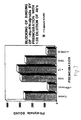
- the CHO-DHFR + L7 clones showed an increase in the response to human IFN-,8 as compared to the highest response in CHO-DHFR control clones which were similarly treated by MTX (Fig. 4).
- the amplitude of the response to human IFN- ⁇ increases, therefore, with gene amplification by a factor of 8 fold.
- the response of such amplified clones to 100 U/ml human IFN was about 15% of that to 20 U/ml hamster IFN.
- the present inventors have concluded that, although expression of the L7 cDNA under SV40 promoter control leads to increased response of the cells to human IFN, the amplitude of the response remains low. This may be due either to problems in expression of the protein or to the fact that the L7 protein is only one part of the IFN receptor system. The latter possibility seems more likely as significant high affinity binding of human 125 1-IFN-a to the CHO transformants was not able to be demonstrated. If the receptor is composed of more than one chain, binding affinity may be very low in the presence of only one of the protein chains.
- Fig. 5 shows that at low concentration of hamster or human IFN, which produce low stimulation of (2'-5') A synthetase activity in the control CHO-DHFR clone (abscissa), a CHO-pSVL7 clone (300 nM MTX) gave a 5 fold higher response with either of the two IFN species (the straight line in Fig. 5 is the expected line if both clones respond equally).
- Example 3 Antibodies to L7 protein show the protein on the surface of cells
- a 27 amino acid long peptide near the amino-terminus of the L7 protein (Fig. 3), was synthesized, conjugated to KLH and used to raise rabbit antisera.
- the antibodies were purified on a column of immobilized peptide and used in Western blots.
- a protein of 130 kDa is detected in Triton X-100-deoxycholate lysates and in membrane preparations. This is probably a glycosylated form of L7 which has an N-glycosylation site near the N-terminal region.
- immunoprecipitation was not possible with the peptide antibodies.
- the L7 cDNA was fused through codon 113 to the protein A gene in vector pRIT-2 (Pharmacia Fine Chemicals) and expressed in E.coli.
- the fused protein PSN and PN2 (Fig. 3), purified on an immunoglobulin column, were used for immunization to obtain rabbit polyclonal antibodies to the L7 protein.
- These antibodies immunoprecipitated from human Daudi cells labelled for 7 hours with 35- methionine, with major bands of 80-85 kDa and minor bands of about 130 kDa.
- the 80-85 kDa bands may be forms of the protein which have lost part off the N-terminal region recognized by the above anti-peptide antibody.
- Fig. 6 shows a fluorescence activated cell sorter (FACS) analysis demonstrating the presence of L7 on the surface of Daudi cells.
- FACS fluorescence activated cell sorter
- the anti-L7 antibodies did not inhibit the binding of 125 l-IFN- ⁇ 2 to human lymphoblastoid Daudi cells.
- Fig. 7 shows that the binding was inhibited by cold IFN-,8, by antibodies against mouse-human hybrid cells containing only chromosome 21 ( ⁇ chr.21), but not by anti-L7 ( ⁇ L7), nor by normal rabbit serum (NRS) or normal mouse serum (NMS). Therefore, the L7 protein seems not to be directly involved in the binding of IFN-a to these human cells. This would distinguish the L7 protein from the protein cross-linked to IFN-a on human cells (Razziudin et al, locus cited).
- the protein which binds IFN-a may be different from that which binds IFN-,8. Since the L7 protein was identified by its action on the response of cells to IFN- ⁇ , there has been an attempt to see if L7 could be cross-linked to IFN-,8. Since radio-iodination of active IFN- ⁇ was not successful, cold IFN- ⁇ was cross-linked to Daudi cells by suberimidate treatment and the electrophoretic mobility of the L7 protein (detected by immunoblots with anti-peptide) was analyzed to determine if it decreased by this treatment. In one experiment, a slower 150 kDa band was observed in addition to the 130 kDa L7 protein, but two other experiments failed to show this phenomenon.
- Identification of the L7 protein as a protein which modulates cellular response to Type I interferon and a component of the type I IFN receptor system is based on the following:
- the type I IFN receptor is probably formed by several different protein chains.
- the L7 protein is probably one component of the type I IFN receptor system and involved in the response of cells to IFN. It is probably not the chain of the receptor system which binds human IFN in a species specific way. It is more likely to be a chain of the receptor increasing the signal produced by the binding of IFN.
- the identification of the L7 protein allows one to study its structure-function relationship, to produce a modified L7 protein with the aim off increasing or decreasing the response of cell's, tissues and whole organisms to interferon of the a or type, and to produce by recombinant DNA technology soluble or lipid- bound L7 protein which can act as a competitor against IFN.
- These inventions can be applied to pathological situations in which one wishes to either enhance or to reduce the response to these types of interferons (e.g., autoimmune processes, chronic and acute infections, tumors and leukemias, etc.).
- L7 protein fragments or analogues which interact with IFN, or antidiotypic antibodies raised against antibodies to the L7 protein may be injected into a patient, acting as "decoys" for any interferon molecules present.
- Gene therapy with functional receptor genes can be used to provide more receptors and thereby increase the response to the IFN.
- the process of the present invention can also be repeated using other markers of interferon activity, e.g., other changes of cell surface characteristics caused by interferon, for detection of the interferon receptor system components.
- Such characteristics may include increased ;82 microglobulin or class II MHC antigens.
- any protein obtained in accordance with the present invention modulates cellular response to interferon. This is true by definition as it is that property which guides the selection of the genetic material. While it is thought that all such proteins are part of the interferon receptor system, applicant does not wish to be limited to such theory. It is known, however, that such proteins are not merely interferon inducers in view of their activity in responding to interferon, rather than creating it.
Landscapes
- Health & Medical Sciences (AREA)
- Chemical & Material Sciences (AREA)
- Life Sciences & Earth Sciences (AREA)
- Organic Chemistry (AREA)
- Immunology (AREA)
- General Health & Medical Sciences (AREA)
- Medicinal Chemistry (AREA)
- Proteomics, Peptides & Aminoacids (AREA)
- Biochemistry (AREA)
- Biophysics (AREA)
- Molecular Biology (AREA)
- Genetics & Genomics (AREA)
- Gastroenterology & Hepatology (AREA)
- Toxicology (AREA)
- Zoology (AREA)
- Cell Biology (AREA)
- Peptides Or Proteins (AREA)
- Preparation Of Compounds By Using Micro-Organisms (AREA)
- Medicines That Contain Protein Lipid Enzymes And Other Medicines (AREA)
- Micro-Organisms Or Cultivation Processes Thereof (AREA)
Claims (15)
Applications Claiming Priority (2)
| Application Number | Priority Date | Filing Date | Title |
|---|---|---|---|
| IL88377 | 1988-11-14 | ||
| IL8837788A IL88377A (en) | 1988-11-14 | 1988-11-14 | Cloning and expression of a protein which modulates cellular response to type I interferon |
Publications (2)
| Publication Number | Publication Date |
|---|---|
| EP0369877A1 EP0369877A1 (fr) | 1990-05-23 |
| EP0369877B1 true EP0369877B1 (fr) | 1995-05-10 |
Family
ID=11059417
Family Applications (1)
| Application Number | Title | Priority Date | Filing Date |
|---|---|---|---|
| EP89403131A Expired - Lifetime EP0369877B1 (fr) | 1988-11-14 | 1989-11-14 | Clonage et expression d'une protéine, qui module la réponse cellulaire à l'interféron du type I |
Country Status (14)
| Country | Link |
|---|---|
| US (1) | US5358867A (fr) |
| EP (1) | EP0369877B1 (fr) |
| JP (1) | JP3156724B2 (fr) |
| AT (1) | ATE122393T1 (fr) |
| AU (1) | AU628895B2 (fr) |
| CA (1) | CA2002862C (fr) |
| DE (1) | DE68922582T2 (fr) |
| DK (1) | DK169890A (fr) |
| ES (1) | ES2071675T3 (fr) |
| FI (1) | FI104495B (fr) |
| IL (1) | IL88377A (fr) |
| NO (1) | NO311181B1 (fr) |
| WO (1) | WO1990005737A1 (fr) |
| ZA (1) | ZA898680B (fr) |
Cited By (2)
| Publication number | Priority date | Publication date | Assignee | Title |
|---|---|---|---|---|
| US7179465B2 (en) | 1992-03-31 | 2007-02-20 | Medarex, Inc. | Monoclonal antibodies against the interferon receptor, with neutralizing activity against type 1 interferon |
| US10947295B2 (en) | 2017-08-22 | 2021-03-16 | Sanabio, Llc | Heterodimers of soluble interferon receptors and uses thereof |
Families Citing this family (8)
| Publication number | Priority date | Publication date | Assignee | Title |
|---|---|---|---|---|
| JPH06510061A (ja) * | 1991-08-30 | 1994-11-10 | ジェネンテク,インコーポレイテッド | Iddmの治療法 |
| IL106591A (en) * | 1992-09-03 | 2008-03-20 | Yeda Res & Dev | Interferon alpha/beta binding protein, its preparation and pharmaceutical compositions containing it |
| US5821078A (en) | 1992-09-03 | 1998-10-13 | Yeda Research And Development Co. Ltd. | Nucleic acid encoding interferon-α/β binding protein |
| WO1995007716A1 (fr) * | 1993-09-17 | 1995-03-23 | Laboratoire Europeen De Biotechnologie S.A. | Composition pharmaceutique comprenant des anticorps monoclonaux diriges contre le recepteur d'interferon et dotes d'une activite neutralisante a l'egard de l'interferon de type i |
| US6740519B1 (en) | 1997-11-03 | 2004-05-25 | Mount Sinai School Of Medicine | Immortalized, homozygous stat1-deficient mammalian cell lines and their uses |
| DK1631312T3 (da) | 2003-04-23 | 2009-01-19 | Medarex Inc | Sammensætning og fremstillingsmåde til behandling af inflammatorisk tarmsygdom |
| KR20150041193A (ko) | 2004-06-21 | 2015-04-15 | 메다렉스, 엘.엘.시. | 인터페론 알파 수용체 1 항체 및 그의 용도 |
| AU2017290389B2 (en) | 2016-07-01 | 2024-09-26 | Resolve Therapeutics, Llc | Optimized binuclease fusions and methods |
Family Cites Families (1)
| Publication number | Priority date | Publication date | Assignee | Title |
|---|---|---|---|---|
| US4808523A (en) * | 1984-11-07 | 1989-02-28 | Yeda Research And Development Co., Ltd. | Constitutive production of human IFN-β1 by mammalian cells transformed by the IFN-β1 gene fused to an SV40 early promoter |
-
1988
- 1988-11-14 IL IL8837788A patent/IL88377A/en not_active IP Right Cessation
-
1989
- 1989-11-14 WO PCT/US1989/005197 patent/WO1990005737A1/fr not_active Ceased
- 1989-11-14 CA CA002002862A patent/CA2002862C/fr not_active Expired - Fee Related
- 1989-11-14 AT AT89403131T patent/ATE122393T1/de not_active IP Right Cessation
- 1989-11-14 ES ES89403131T patent/ES2071675T3/es not_active Expired - Lifetime
- 1989-11-14 ZA ZA898680A patent/ZA898680B/xx unknown
- 1989-11-14 JP JP50126490A patent/JP3156724B2/ja not_active Expired - Fee Related
- 1989-11-14 EP EP89403131A patent/EP0369877B1/fr not_active Expired - Lifetime
- 1989-11-14 AU AU46661/89A patent/AU628895B2/en not_active Ceased
- 1989-11-14 DE DE68922582T patent/DE68922582T2/de not_active Expired - Fee Related
-
1990
- 1990-07-12 NO NO19903118A patent/NO311181B1/no not_active IP Right Cessation
- 1990-07-13 FI FI903577A patent/FI104495B/fi not_active IP Right Cessation
- 1990-07-13 DK DK169890A patent/DK169890A/da not_active Application Discontinuation
-
1993
- 1993-09-13 US US08/120,030 patent/US5358867A/en not_active Expired - Fee Related
Cited By (3)
| Publication number | Priority date | Publication date | Assignee | Title |
|---|---|---|---|---|
| US7179465B2 (en) | 1992-03-31 | 2007-02-20 | Medarex, Inc. | Monoclonal antibodies against the interferon receptor, with neutralizing activity against type 1 interferon |
| US10947295B2 (en) | 2017-08-22 | 2021-03-16 | Sanabio, Llc | Heterodimers of soluble interferon receptors and uses thereof |
| US12129288B2 (en) | 2017-08-22 | 2024-10-29 | Sanabio, Llc | Polynucleotides heterodimers of soluble interferon receptors and uses thereof |
Also Published As
| Publication number | Publication date |
|---|---|
| FI104495B (fi) | 2000-02-15 |
| AU628895B2 (en) | 1992-09-24 |
| ES2071675T3 (es) | 1995-07-01 |
| DK169890D0 (da) | 1990-07-13 |
| WO1990005737A1 (fr) | 1990-05-31 |
| CA2002862C (fr) | 1999-01-26 |
| NO311181B1 (no) | 2001-10-22 |
| IL88377A (en) | 1996-09-12 |
| NO903118D0 (no) | 1990-07-12 |
| DK169890A (da) | 1990-09-14 |
| CA2002862A1 (fr) | 1990-05-14 |
| JP3156724B2 (ja) | 2001-04-16 |
| JPH03503366A (ja) | 1991-08-01 |
| NO903118L (no) | 1990-08-27 |
| DE68922582D1 (de) | 1995-06-14 |
| FI903577A0 (fi) | 1990-07-13 |
| ZA898680B (en) | 1991-04-24 |
| ATE122393T1 (de) | 1995-05-15 |
| EP0369877A1 (fr) | 1990-05-23 |
| US5358867A (en) | 1994-10-25 |
| DE68922582T2 (de) | 1995-09-07 |
| IL88377A0 (en) | 1989-06-30 |
| AU4666189A (en) | 1990-06-12 |
Similar Documents
| Publication | Publication Date | Title |
|---|---|---|
| EP0411946B1 (fr) | DNA codant pour la protéine GP130 humaine | |
| RU2135584C1 (ru) | Гомодимер р40 интерлейкина-12 | |
| US5453491A (en) | Murine interleukin-5 receptor | |
| US6294347B1 (en) | Human FCγ receptor III | |
| JP3255699B2 (ja) | ヒトIL−2レセプターγ鎖分子 | |
| FI104188B (fi) | Rekombinantti interleukiini-2-reseptori | |
| AU628895B2 (en) | Cloning and expression of a protein which modulates cellular response to type i interferon | |
| EP1243597A2 (fr) | Récepteurs pour l'interleukine-12 humaine | |
| KR100382628B1 (ko) | 인터페론α/β결합단백질과이의제조및용도 | |
| JPH06220100A (ja) | インターフェロン−α/β結合タンパク質、その製造法およびそれを含有する医薬組成物 | |
| US5223611A (en) | DNA encoding for human GP130 protein | |
| CA2710760C (fr) | Recepteur de type toll 3 de cynomolgus | |
| Yamaguchi et al. | Structure of the functional interleukin-2 receptor: Evidence for the association of human p55 and murine p75 molecules in a mouse T cell line | |
| EP4538285A1 (fr) | Variant de pd-1 et son utilisation | |
| EP2536435B1 (fr) | Homologue de singe d'interféron oméga humain | |
| Schreiber et al. | The Molecular and Cellular Biology of the IFNγ Receptor | |
| CA2113076A1 (fr) | Interleukine 6 humaine recombinante a extremite n-terminale homogene et production de celle-ci | |
| US20110052499A1 (en) | Baboon homolog of human cd147 | |
| IE902930A1 (en) | Human gp130 protein |
Legal Events
| Date | Code | Title | Description |
|---|---|---|---|
| PUAI | Public reference made under article 153(3) epc to a published international application that has entered the european phase |
Free format text: ORIGINAL CODE: 0009012 |
|
| AK | Designated contracting states |
Kind code of ref document: A1 Designated state(s): AT BE CH DE ES FR GB GR IT LI LU NL SE |
|
| RIN1 | Information on inventor provided before grant (corrected) |
Inventor name: FELLOUS, MARC Inventor name: SHULMAN, LESTER Inventor name: REVEL, MICHEL |
|
| 17P | Request for examination filed |
Effective date: 19901027 |
|
| 17Q | First examination report despatched |
Effective date: 19930211 |
|
| GRAA | (expected) grant |
Free format text: ORIGINAL CODE: 0009210 |
|
| AK | Designated contracting states |
Kind code of ref document: B1 Designated state(s): AT BE CH DE ES FR GB GR IT LI LU NL SE |
|
| REF | Corresponds to: |
Ref document number: 122393 Country of ref document: AT Date of ref document: 19950515 Kind code of ref document: T |
|
| REF | Corresponds to: |
Ref document number: 68922582 Country of ref document: DE Date of ref document: 19950614 |
|
| ET | Fr: translation filed | ||
| REG | Reference to a national code |
Ref country code: ES Ref legal event code: FG2A Ref document number: 2071675 Country of ref document: ES Kind code of ref document: T3 |
|
| ITF | It: translation for a ep patent filed | ||
| REG | Reference to a national code |
Ref country code: GR Ref legal event code: FG4A Free format text: 3016729 |
|
| PLBE | No opposition filed within time limit |
Free format text: ORIGINAL CODE: 0009261 |
|
| STAA | Information on the status of an ep patent application or granted ep patent |
Free format text: STATUS: NO OPPOSITION FILED WITHIN TIME LIMIT |
|
| 26N | No opposition filed | ||
| REG | Reference to a national code |
Ref country code: GB Ref legal event code: IF02 |
|
| PGFP | Annual fee paid to national office [announced via postgrant information from national office to epo] |
Ref country code: SE Payment date: 20021106 Year of fee payment: 14 |
|
| PGFP | Annual fee paid to national office [announced via postgrant information from national office to epo] |
Ref country code: FR Payment date: 20021108 Year of fee payment: 14 |
|
| PGFP | Annual fee paid to national office [announced via postgrant information from national office to epo] |
Ref country code: LU Payment date: 20021113 Year of fee payment: 14 Ref country code: GB Payment date: 20021113 Year of fee payment: 14 Ref country code: AT Payment date: 20021113 Year of fee payment: 14 |
|
| PGFP | Annual fee paid to national office [announced via postgrant information from national office to epo] |
Ref country code: DE Payment date: 20021114 Year of fee payment: 14 |
|
| PGFP | Annual fee paid to national office [announced via postgrant information from national office to epo] |
Ref country code: CH Payment date: 20021115 Year of fee payment: 14 |
|
| PGFP | Annual fee paid to national office [announced via postgrant information from national office to epo] |
Ref country code: ES Payment date: 20021127 Year of fee payment: 14 |
|
| PGFP | Annual fee paid to national office [announced via postgrant information from national office to epo] |
Ref country code: GR Payment date: 20021128 Year of fee payment: 14 |
|
| PGFP | Annual fee paid to national office [announced via postgrant information from national office to epo] |
Ref country code: NL Payment date: 20021129 Year of fee payment: 14 |
|
| PGFP | Annual fee paid to national office [announced via postgrant information from national office to epo] |
Ref country code: BE Payment date: 20030117 Year of fee payment: 14 |
|
| PG25 | Lapsed in a contracting state [announced via postgrant information from national office to epo] |
Ref country code: LU Free format text: LAPSE BECAUSE OF NON-PAYMENT OF DUE FEES Effective date: 20031114 Ref country code: GB Free format text: LAPSE BECAUSE OF NON-PAYMENT OF DUE FEES Effective date: 20031114 Ref country code: AT Free format text: LAPSE BECAUSE OF NON-PAYMENT OF DUE FEES Effective date: 20031114 |
|
| PG25 | Lapsed in a contracting state [announced via postgrant information from national office to epo] |
Ref country code: SE Free format text: LAPSE BECAUSE OF NON-PAYMENT OF DUE FEES Effective date: 20031115 Ref country code: ES Free format text: LAPSE BECAUSE OF NON-PAYMENT OF DUE FEES Effective date: 20031115 |
|
| PG25 | Lapsed in a contracting state [announced via postgrant information from national office to epo] |
Ref country code: LI Free format text: LAPSE BECAUSE OF NON-PAYMENT OF DUE FEES Effective date: 20031130 Ref country code: CH Free format text: LAPSE BECAUSE OF NON-PAYMENT OF DUE FEES Effective date: 20031130 Ref country code: BE Free format text: LAPSE BECAUSE OF NON-PAYMENT OF DUE FEES Effective date: 20031130 |
|
| BERE | Be: lapsed |
Owner name: *YEDA RESEARCH AND DEVELOPMENT CO. LTD Effective date: 20031130 |
|
| PG25 | Lapsed in a contracting state [announced via postgrant information from national office to epo] |
Ref country code: NL Free format text: LAPSE BECAUSE OF NON-PAYMENT OF DUE FEES Effective date: 20040601 |
|
| PG25 | Lapsed in a contracting state [announced via postgrant information from national office to epo] |
Ref country code: DE Free format text: LAPSE BECAUSE OF NON-PAYMENT OF DUE FEES Effective date: 20040602 |
|
| PG25 | Lapsed in a contracting state [announced via postgrant information from national office to epo] |
Ref country code: GR Free format text: LAPSE BECAUSE OF NON-PAYMENT OF DUE FEES Effective date: 20040603 |
|
| EUG | Se: european patent has lapsed | ||
| GBPC | Gb: european patent ceased through non-payment of renewal fee |
Effective date: 20031114 |
|
| REG | Reference to a national code |
Ref country code: CH Ref legal event code: PL |
|
| PG25 | Lapsed in a contracting state [announced via postgrant information from national office to epo] |
Ref country code: FR Free format text: LAPSE BECAUSE OF NON-PAYMENT OF DUE FEES Effective date: 20040730 |
|
| NLV4 | Nl: lapsed or anulled due to non-payment of the annual fee |
Effective date: 20040601 |
|
| REG | Reference to a national code |
Ref country code: FR Ref legal event code: ST |
|
| REG | Reference to a national code |
Ref country code: ES Ref legal event code: FD2A Effective date: 20031115 |
|
| PG25 | Lapsed in a contracting state [announced via postgrant information from national office to epo] |
Ref country code: IT Free format text: LAPSE BECAUSE OF NON-PAYMENT OF DUE FEES;WARNING: LAPSES OF ITALIAN PATENTS WITH EFFECTIVE DATE BEFORE 2007 MAY HAVE OCCURRED AT ANY TIME BEFORE 2007. THE CORRECT EFFECTIVE DATE MAY BE DIFFERENT FROM THE ONE RECORDED. Effective date: 20051114 |